Structure-Based Drug Design: From Foundational Principles to AI-Driven Discovery
This article provides a comprehensive overview of Structure-Based Drug Design (SBDD), a cornerstone of modern rational drug discovery.
Structure-Based Drug Design: From Foundational Principles to AI-Driven Discovery
Abstract
This article provides a comprehensive overview of Structure-Based Drug Design (SBDD), a cornerstone of modern rational drug discovery. Tailored for researchers, scientists, and drug development professionals, it explores the foundational principles of SBDD, from obtaining 3D protein structures via X-ray crystallography, cryo-EM, and computational prediction. It delves into core methodological applications like molecular docking and virtual screening, and examines cutting-edge advances, including equivariant diffusion and multi-modal AI models that generate novel drug candidates. The content also addresses persistent challenges such as scoring function accuracy and protein flexibility, offering troubleshooting and optimization strategies. Finally, it evaluates validation frameworks and comparative performance of various SBDD approaches, synthesizing key takeaways to illuminate future directions for accelerating therapeutic development.
The Bedrock of SBDD: Core Principles and Structural Techniques
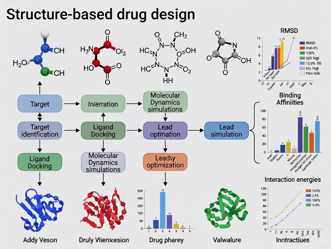
Structure-Based Drug Design (SBDD) represents a paradigm shift in pharmaceutical development, utilizing the three-dimensional structural information of biological targets to guide the discovery and optimization of novel therapeutics. This approach has evolved from a largely experimental technique to a sophisticated computational discipline, fundamentally transforming the drug discovery workflow [1]. By leveraging detailed insights into atomic-level interactions between a drug candidate and its target, SBDD facilitates a more rational and efficient path to identifying lead compounds, optimizing their potency and selectivity, and overcoming challenges such as drug resistance [2]. This article delineates the core principles of SBDD, provides a detailed protocol for a key experimental process, and synthesizes current computational advances that are propelling the field forward, including the integration of machine learning and high-throughput molecular simulations.
At its core, SBDD is an approach to drug discovery that relies on the knowledge of the three-dimensional structure of a biological target, typically a protein or nucleic acid, to design molecules that can interact with it in a specific and therapeutically beneficial manner [1]. This methodology stands in contrast to traditional empirical methods, offering a rational framework that reduces reliance on serendipity and high-volume screening alone.
The strategic value of SBDD is profoundly amplified by treating the underlying structural and chemical data as a high-value product in its own right. High-quality SBDD data products are characterized by rigorous validation, standardized formats, comprehensive metadata, and intuitive interfaces that democratize access across multidisciplinary teams, from structural biologists to medicinal chemists [1]. The process generally follows a cyclical workflow: Target Selection and Validation → Structure Determination → Ligand Docking and Design → Compound Synthesis → Experimental Assay → Lead Optimization, with insights from each stage feeding back into the next design cycle. The subsequent sections will unpack the specific methodologies and tools that make this cycle possible.
Core Methodologies and Data
SBDD integrates a suite of computational and experimental techniques. The table below summarizes the primary computational methods used for identifying and optimizing lead compounds.
Table 1: Key Computational Methods in Structure-Based Drug Design
| Method | Primary Function | Common Tools/Approaches |
|---|---|---|
| Homology Modeling | Constructs a 3D model of a target protein when an experimental structure is unavailable, using a related protein with a known structure as a template [2]. | MODELLER [2] |
| Molecular Docking | Predicts the preferred orientation and binding affinity of a small molecule (ligand) when bound to a target protein [2]. | AutoDock Vina, InstaDock [2] |
| Structure-Based Virtual Screening (SBVS) | Automatically evaluates large libraries of compounds (e.g., 89,399 in a recent study) through docking to identify potential hits for further experimental testing [2]. | AutoDock Vina [2] |
| Molecular Dynamics (MD) Simulations | Models the physical movements of atoms and molecules over time, providing insights into protein-ligand complex stability, conformational changes, and binding dynamics [1] [2]. | GROMACS [1] |
| Machine Learning (ML) Classification | Employs algorithms to distinguish between active and inactive compounds based on chemical descriptor properties, refining hit lists from virtual screening [2]. | PaDEL-Descriptor for feature generation [2] |
The integration of these methods was exemplified in a recent study aiming to identify natural inhibitors of the human αβIII tubulin isotype, a cancer-relevant target. The workflow, summarized in the diagram below, involved homology modeling, virtual screening of a 89,399-compound library, machine learning to narrow 1,000 hits to 20 active compounds, and finally, molecular dynamics simulations to validate the stability of the top four candidates [2].
Diagram 1: SBDD workflow for identifying tubulin inhibitors.
Experimental Protocol: Protein Production for SBDD
A critical bottleneck in SBDD is the production of sufficient quantities of high-quality, pure protein for structural studies. The following protocol details the manufacture and setup of a cost-effective, single-use bubble column reactor (suBCR) array for litre-scale expression of recombinant proteins in E. coli, designed to overcome the limitations of traditional shake-flasks [3].
Materials and Reagents
Table 2: Essential Research Reagents and Materials for suBCR Setup
| Item | Specification/Example | Function |
|---|---|---|
| Layflat Tubing (LFT) | Heavy-duty (125-250 micron) Polyethylene (PE) or autoclaveable Polypropylene (PP) [3]. | Forms the single-use bioreactor bag. |
| Air Pump | Aquarium diaphragm air pump (e.g., Tetra brand) [3]. | Supplies oxygen to the bacterial culture. |
| Airline | Semi-rigid food/lab grade tubing, 4-4.5mm internal diameter (e.g., Legris PUR pipe) [3]. | Transports air from the pump to the bioreactor. |
| Airstones | Cylindrical, 25-30mm (e.g., Tetra air stones) [3]. | Diffuses air into fine bubbles for efficient oxygen transfer. |
| Foam Stopper | Indenti-Plug L800-E, for 46-65mm openings [3]. | Seals the bag while holding the airline; allows gas exchange. |
| Temperature Control | Submersible aquarium heater (200-300W) and/or recirculating lab water chiller [3]. | Maintains optimal culture temperature. |
| Injection Ports | Self-healing, adhesive ports (e.g., 3M) [3]. | Allows for sterile inoculation and sampling. |
| Impulse Sealer | Standard commercial heat sealer. | Creates airtight seals at the ends of the LFT bags. |
Step-by-Step Procedure
Preparing the Airline Assembly:
- Cut a 1.5-1.6m length of airline tubing.
- Insert an airstone into one end.
- Thread the opposite end through a foam stopper, sliding the stopper to approximately 70cm above the airstone. The foam should grip the tubing to hold it in place [3].
Manufacturing the Single-Use Bioreactor (Bag):
- Measure and cut a ~2.8m length of layflat tubing for a 1.2m tall rail system.
- Use an impulse sealer to create a heat seal at one end of the tubing, allowing a 20-30mm seam allowance.
- On the outer face of the bag, below the point where it will hang freely, make a ~100mm vertical slit. This provides access for the airline and for filling the bag [3].
- Affix a self-healing injection port above the intended liquid fill line.
System Setup and Operation:
- Suspend the manufactured bags from a rail system over a water bath.
- Fill the water bath and activate the temperature control system (heater and recirculating pump).
- Connect the airline from the pump to the top of the airline assembly using a flow control valve.
- Fill the bags with sterile culture media through the slit or injection port.
- Insert the airline assembly into the bag through the slit, ensuring the airstone is at the bottom. The foam stopper should form a seal inside the neck of the bag.
- Inoculate the culture through the self-healing injection port.
- Turn on the air supply and adjust the flow rate to achieve adequate aeration and mixing via bubble formation [3].
Current Advances and Future Outlook
The field of SBDD is being rapidly transformed by new computational technologies. A prominent trend is the deep integration of artificial intelligence and machine learning. The quality and organization of training data are now recognized as paramount, with organizations that maintain pristine structural data products gaining a competitive edge in developing next-generation AI tools for predicting protein-ligand interactions [1] [2].
Furthermore, federated data ecosystems are emerging, allowing organizations to collaboratively share structural information while preserving proprietary interests, thus accelerating discovery across the entire industry [1]. Conferences like the SBDD 2025 Congress highlight cutting-edge research in AI-driven approaches, molecular modeling, and advanced simulations, underscoring the dynamic evolution of the field [4]. The industry is also moving towards more integrated enterprise software solutions, such as the Proasis platform, which are designed to translate 3D structural data into a powerful, actionable strategic asset for drug discovery teams [1].
Structure-Based Drug Design has firmly established itself as a rational and indispensable approach in modern drug discovery. By moving beyond pure empiricism to a detailed, structure-guided process, SBDD significantly increases the efficiency and success rate of developing new therapeutics. The continued advancement of the field—through improvements in high-throughput protein production, more sophisticated and integrated computational workflows, and the powerful application of AI—promises to further accelerate the delivery of novel treatments for diseases ranging from cancer to antibiotic resistance. As these tools become more accessible and data ecosystems more collaborative, SBDD will continue to be a cornerstone of innovative drug development.
The Critical Role of 3D Protein Structures
Structure-Based Drug Design (SBDD) is a foundational paradigm in modern drug discovery, focused on the development and interpretation of three-dimensional (3D) models of protein-ligand interactions [5]. This rational approach uses the 3D structure of a biological target, typically a protein, to design and optimize novel drug candidates, thereby streamlining the discovery process [6]. The central premise of SBDD is that knowledge of the target's atomic structure enables researchers to rationally design molecules that bind with high affinity and selectivity, which has become an integral part of most industrial drug discovery programs [5]. The value of SBDD is significantly enhanced by treating the underlying structural and experimental data not as a mere byproduct of research, but as a high-value product in its own right, characterized by rigorous validation, standardized formats, and comprehensive metadata [1].
The Centrality of Accurate 3D Protein Structures
The accuracy of the initial 3D structural model is a critical determinant of success in any SBDD campaign. Inaccurate structures can misdirect design efforts, leading to costly delays and failures. The field relies on both experimental and computational techniques to obtain these essential models, each with distinct advantages and limitations [5].
Experimental Structure Determination Methods
- X-ray Crystallography: This traditional workhorse of structural biology involves crystallizing the target protein, often with a bound ligand, and determining its structure by analyzing the diffraction pattern of X-rays passed through the crystal. While powerful, it can be challenging for certain protein classes (e.g., membrane proteins), is time-consuming, and requires high-resolution data for accurate SBDD, as minute differences in side-chain conformation can be crucial for analyzing binding interactions [5].
- Cryo-Electron Microscopy (Cryo-EM): This emerging alternative to crystallography addresses many of its challenges, particularly for large protein complexes that are difficult to crystallize. Although access to cryo-EM facilities can be limited, its use is expected to grow significantly in the coming decades [5].
- Nuclear Magnetic Resonance (NMR): NMR spectroscopy can be used to determine protein structures in solution, providing insights into dynamic behavior. However, it is generally limited to smaller proteins [5].
Computational Structure Prediction Methods
Computational methods have emerged as powerful alternatives or complements to experimental techniques.
- Machine Learning-Based Prediction: Advances in machine learning, exemplified by models like AlphaFold2, have revolutionized the field by enabling accurate protein structure prediction from amino acid sequence data alone [5]. These models have dramatically expanded the structural coverage of the proteome.
- Docking and Co-folding Algorithms: Docking algorithms (e.g., AutoDock Vina) can predict how a small molecule binds to a protein target. A newer generation of models, including AlphaFold3 and HelixFold3, perform protein-ligand co-folding, simultaneously predicting the protein structure and its binding mode with a ligand [5]. While their accuracy may be lower than high-resolution crystallography, their speed promises to accelerate SBDD, especially for intractable targets.
Table 1: Comparison of Protein Structure Determination and Modeling Techniques
| Method | Key Principle | Typical Resolution/Accuracy | Primary Advantages | Primary Limitations |
|---|---|---|---|---|
| X-ray Crystallography | X-ray diffraction from protein crystals | Atomic resolution (dependent on crystal quality) | High accuracy for well-diffracting crystals; direct experimental data | Difficult for membrane proteins; time-consuming crystallization |
| Cryo-EM | Electron microscopy of frozen-hydrated samples | Near-atomic to atomic resolution | Suitable for large complexes; no crystallization needed | Limited access to facilities; can be resource-intensive |
| AlphaFold2/3 | Deep learning on evolutionary data | High accuracy (varies by protein) [7] | Fast; based on sequence alone; covers many proteins | Can underestimate binding pocket volumes [7] |
| DeepSCFold | Deep learning on sequence-derived complementarity | 11.6% higher TM-score than AlphaFold-Multimer [8] | Excels in protein complex & antibody-antigen modeling [8] | Newer method; requires further community adoption |
A critical evaluation of computational models against experimental structures is essential. For instance, a 2025 comprehensive analysis of nuclear receptor structures revealed that while AlphaFold2 achieves high accuracy in predicting stable conformations with proper stereochemistry, it systematically underestimates ligand-binding pocket volumes by 8.4% on average and captures only single conformational states, missing functionally important asymmetry observed in experimental structures [7]. This highlights the importance of understanding the limitations of predictive models in SBDD.
Application Notes: SBDD in Action
Protocol 1: Structure-Based Virtual Screening (SBVS) for Hit Identification
This protocol details the use of a target protein's 3D structure to computationally screen large libraries of small molecules for potential hits.
1. Target Preparation
- Obtain the 3D structure of the target protein from the PDB, or via prediction tools like AlphaFold or DeepSCFold for complexes [8].
- Prepare the protein structure by adding hydrogen atoms, assigning protonation states, and optimizing side-chain orientations for residues in the binding site using molecular modeling software.
- Define the binding site coordinates, typically centered on a known ligand or a key residue in the active site.
2. Ligand Library Preparation
- Select a compound library (e.g., ZINC natural compounds, in-house corporate library) [2].
- Prepare ligands by generating 3D structures, enumerating plausible tautomers and protonation states at biological pH, and minimizing their energy to achieve a low-energy conformation.
3. Molecular Docking
- Perform high-throughput virtual screening (HTVS) using docking software such as AutoDock Vina or a platform like InstaDock [2].
- Key Parameters: The docking search space should be defined by a grid box centered on the binding site. The exhaustiveness of the global search should be set sufficiently high (e.g., 32-128) to ensure adequate sampling of ligand poses. Each compound is typically docked in multiple flexible conformations.
- The output is a ranked list of compounds based on the computed binding affinity (e.g., Vina score) [2].
4. Post-Docking Analysis
- Visually inspect the top-ranking poses to confirm they form sensible interactions (e.g., hydrogen bonds, hydrophobic contacts) with the protein target.
- Cluster results based on chemical structure and binding mode to prioritize diverse chemotypes for further experimental testing.
Diagram Title: Structure-Based Virtual Screening Workflow
Protocol 2: Hit-to-Lead Optimization Using Molecular Dynamics
After confirming hits, this protocol uses molecular dynamics (MD) to understand and optimize the binding interaction, moving from a static view to a dynamic one.
1. System Setup
- Build the simulation system by placing the protein-ligand complex in a simulation box (e.g., a cubic or rhombic dodecahedron box) filled with water molecules (e.g., TIP3P water model).
- Add ions (e.g., Na⁺, Cl⁻) to neutralize the system's charge and mimic a physiological salt concentration (e.g., 150 mM NaCl).
2. Energy Minimization and Equilibration
- Energy Minimization: Run a steepest descent or conjugate gradient algorithm to remove any steric clashes introduced during system setup.
- Equilibration: Perform a two-step equilibration in the NVT (constant Number of particles, Volume, and Temperature) and NPT (constant Number of particles, Pressure, and Temperature) ensembles to stabilize the temperature and pressure of the system. This is typically done for 100-500 ps.
3. Production MD Simulation
- Run an unbiased MD simulation for a timescale relevant to the biological process (typically 100 ns to 1 µs). Use a integration time step of 2 fs.
- Key Analyses:
- Root-mean-square deviation (RMSD): Measure the stability of the protein and ligand backbone over time.
- Root-mean-square fluctuation (RMSF): Identify flexible regions of the protein, particularly in the binding site.
- Ligand-protein interactions: Calculate the occupancy of specific interactions (hydrogen bonds, hydrophobic contacts, salt bridges) throughout the simulation to identify key binding motifs.
- Binding pocket analysis: Use tools like WaterMap to analyze solvation effects and identify displaceable water molecules for potential affinity gains [9].
4. Insight-Driven Design
- Use the dynamic interaction fingerprints from the MD simulation to guide rational compound design. For example, adding an electron-withdrawing group to a phenol can improve its hydrogen-bond donor capacity, while strategic conformational restriction (e.g., macrocyclization) can minimize the energetic penalty paid upon binding [5].
Table 2: Key Analyses in Molecular Dynamics Simulations for SBDD
| Analysis Metric | Description | Application in SBDD |
|---|---|---|
| RMSD (Root-Mean-Square Deviation) | Measures the average distance between atoms of superimposed structures over time. | Assesses the overall stability of the protein-ligand complex during simulation. |
| RMSF (Root-Mean-Square Fluctuation) | Measures the deviation of a particle/atom from its average position. | Identifies flexible regions in the protein, especially in binding sites and loops. |
| H-Bond Occupancy | The percentage of simulation time a specific hydrogen bond exists. | Quantifies the strength and persistence of critical polar interactions. |
| Rg (Radius of Gyration) | Measures the compactness of the protein structure. | Monitors large-scale conformational changes or folding/unfolding events. |
| SASA (Solvent Accessible Surface Area) | Measures the surface area of a molecule accessible to a solvent. | Evaluates changes in protein folding and ligand burial upon binding. |
Advanced Topics and Future Directions
Generative AI for 3D Molecular Design
A frontier in SBDD is the use of generative artificial intelligence to create novel drug molecules directly within the context of a 3D protein binding pocket. These models aim to generate molecules with high binding affinity, but the field is evolving to incorporate other critical drug-like properties, such as synthetic feasibility and selectivity, which are essential for practical drug discovery [10]. New frameworks like CByG (Controllable Bayesian Flow Network with Integrated Guidance) extend beyond conventional diffusion models to more robustly integrate property-specific guidance during the generation process, addressing limitations in handling the hybrid nature of 3D molecular data (continuous coordinates and categorical atom types) [10]. This highlights a shift from mere generation to controllable generation of viable drug candidates.
The Critical Role of Selectivity and Specificity
Beyond simple binding affinity, a successful drug must be selective for its intended target to minimize off-target side effects. This necessitates evaluating generated or designed molecules against off-target proteins. However, widely used public datasets like CrossDocked2020 were not originally designed for rigorous selectivity assessment, creating a need for new, biologically relevant benchmarks and guidance strategies specifically for selectivity [10]. SBDD protocols must therefore evolve to include multi-target docking and simulation studies to proactively address potential selectivity issues.
The Scientist's Toolkit: Essential Research Reagents and Solutions
Table 3: Key Research Reagent Solutions for SBDD
| Tool/Resource | Type | Primary Function in SBDD |
|---|---|---|
| RCSB Protein Data Bank (PDB) | Data Repository | Primary archive for experimentally determined 3D structures of proteins, nucleic acids, and complexes. |
| AlphaFold Protein Structure Database | Data Repository | Provides access to millions of predicted protein structures generated by the AlphaFold AI system. |
| AutoDock Vina | Software | Widely used open-source molecular docking tool for predicting small molecule binding modes and affinities. |
| ZINC Database | Compound Library | A curated collection of commercially available chemical compounds for virtual screening. |
| DesertSci Proasis / Rowan Platform | Enterprise Software | Integrated platforms that manage 3D structural data, streamline SBDD workflows, and facilitate collaboration. [5] [1] |
| GROMACS | Software | A package for performing molecular dynamics simulations, used to study protein-ligand interactions over time. |
| Schrödinger Suite | Software Suite | A comprehensive commercial software platform for drug discovery, including tools for molecular modeling, simulation, and design. |
Diagram Title: The SBDD Ecosystem Data Flow
Structure-based drug design (SBDD) has become a cornerstone of modern pharmaceutical research, offering a rational framework for transforming initial hits into optimized drug candidates [11]. By leveraging detailed three-dimensional structural information, SBDD enables the design of compounds with enhanced potency, selectivity, and improved pharmacological profiles [12]. The success of SBDD relies heavily on high-resolution structural data of biological targets, primarily obtained through three principal experimental techniques: X-ray crystallography, cryo-electron microscopy (cryo-EM), and nuclear magnetic resonance (NMR) spectroscopy [12] [13]. This article provides a detailed comparison of these techniques, their specific applications in drug discovery, and standardized protocols for their implementation in SBDD workflows.
Technique Comparison and Applications
The selection of an appropriate structure determination technique depends on the target biomolecule's properties, the required resolution, and the specific stage of the drug discovery process. Each method offers distinct advantages and limitations, summarized in the table below.
Table 1: Comparative Analysis of Structural Biology Techniques in Drug Discovery
| Parameter | X-ray Crystallography | Cryo-Electron Microscopy | NMR Spectroscopy |
|---|---|---|---|
| Typical Resolution | Routinely < 2.5 Å, often sub-1 Å possible [14] | Typically 2.5-4.0 Å, with <2 Å possible [13] [14] | Atomic-level for proteins < 30 kDa [15] |
| Optimal Target Size | Best for proteins < 100 kDa [14] | Ideal for complexes > 100 kDa [14] | Suitable for proteins up to ~50 kDa [11] [16] |
| Sample State | Crystalline solid state | Vitrified solution (near-native) [14] | Solution state (physiological conditions) [11] |
| Key Advantage | Atomic precision; well-established pipelines [14] | No crystallization needed; captures conformational states [16] [14] | Studies dynamics & weak interactions; no crystallization [11] [15] |
| Primary Limitation | Requires high-quality crystals; static snapshot [11] [5] | High equipment cost; intensive computation [16] [14] | Low sensitivity; molecular weight constraints [11] |
| Throughput | Medium to High (after crystal optimization) [11] | Medium (data collection: hours to days) [14] | Low to Medium (data acquisition can be time-consuming) |
| Ideal for SBDD | High-throughput ligand screening, fragment growing [11] [14] | Membrane proteins, large complexes, flexible systems [13] [14] | Fragment-based discovery, studying protein dynamics & weak binding [11] [15] |
Table 2: Application-Based Selection Guide for SBDD
| SBDD Application | Recommended Technique | Rationale |
|---|---|---|
| High-Throughput Fragment Screening | X-ray Crystallography (if crystals available) [14] | Established soaking pipelines provide rapid structural data for many compounds. |
| Membrane Protein Target (e.g., GPCR) | Cryo-EM [13] [14] | Eliminates crystallization hurdle and preserves near-native lipid environment. |
| Target with Inherent Flexibility/Disorder | NMR or Cryo-EM [11] [16] | NMR probes dynamics in solution; Cryo-EM can capture multiple conformations. |
| Optimizing Weak Fragment Binders | NMR [11] [15] | Detects and characterizes weak, transient interactions critical for early FBDD. |
| Structure of a Large Viral Complex | Cryo-EM [16] [14] | No size limitations; can resolve large assemblies without crystal packing constraints. |
| Characterizing H-bonding & Protonation States | NMR [11] | Directly probes hydrogen atoms and their interactions, invisible to X-rays. |
Experimental Protocols
Protein Crystallography for Ligand Binding Studies
Objective: To determine the high-resolution structure of a target protein in complex with a small-molecule ligand to guide rational drug design [5].
Workflow Overview:
Protocol Details:
Protein Production and Crystallization:
- Express and purify the target protein to high homogeneity (>95% purity) [14]. Typical yields of >2 mg are required [14].
- Use high-throughput vapor diffusion screens to identify initial crystallization conditions.
- Optimize conditions to grow large, single, and well-ordered crystals. This process can take weeks to months [5] [14].
Ligand Soaking and Harvesting:
- For pre-formed crystals, soak the crystal in a cryoprotectant solution containing the ligand of interest. Ligand concentration should be high enough to ensure saturation, but mindful of DMSO tolerance [11].
- Alternatively, co-crystallize the protein with the ligand.
- Flash-cool the crystal in liquid nitrogen for data collection [14].
Data Collection and Processing:
- Collect X-ray diffraction data at a synchrotron source. Data collection typically takes minutes to hours [14].
- Index, integrate, and scale the diffraction data using established software (e.g., XDS, HKL-3000) [14].
- Solve the phase problem, often by molecular replacement using a known related structure as a search model.
Model Building and Refinement:
- Fit the protein sequence into the electron density map and build the atomic model.
- Identify positive difference density (F~o~ - F~c~) in the binding pocket to place and refine the ligand geometry.
- Iteratively refine the model (coordinates and B-factors) against the diffraction data to achieve the best agreement (low R~work~/R~free~).
Key Reagents: Table 3: Key Research Reagents for Protein Crystallography
| Reagent/Material | Function | Example/Notes |
|---|---|---|
| Highly Pure Protein | The target for crystallization. | Requires high homogeneity; typical concentration 5-20 mg/mL. |
| Crystallization Screen Kits | To identify initial crystallization conditions. | Commercial sparse matrix screens (e.g., from Hampton Research). |
| Ligand Compound | The small molecule for binding studies. | Dissolved in DMSO; final DMSO concentration in soak should be <5%. |
| Cryoprotectant | Prevents ice crystal formation during vitrification. | e.g., Glycerol, ethylene glycol, or various cryoprotectant cocktails. |
Single-Particle Cryo-EM for Complex Structures
Objective: To determine the structure of a large protein or complex, particularly targets resistant to crystallization, in complex with a drug candidate [13].
Workflow Overview:
Protocol Details:
Sample Preparation and Vitrification:
- Prepare a purified sample of the protein-ligand complex. Sample amount required is minimal (0.1-0.2 mg) compared to crystallography [14].
- Incubate the protein with the ligand to form the complex prior to grid preparation.
- Apply 3-4 µL of sample to a freshly plasma-cleaned EM grid. Blot away excess liquid and rapidly plunge-freeze the grid in liquid ethane. Optimize blotting time and humidity to achieve a thin layer of vitreous ice [14].
Data Collection:
- Load the grid into a high-end cryo-electron microscope equipped with a direct electron detector.
- Collect thousands of micrograph movies at a calibrated defocus under low-electron-dose conditions to minimize beam-induced damage. Data collection can take hours to days [14].
Image Processing and 3D Reconstruction:
- Perform motion correction and estimate the contrast transfer function (CTF) for each micrograph [14].
- Autopick or manually pick particles from the micrographs.
- Perform multiple rounds of 2D classification to select a homogeneous set of particles.
- Generate an initial 3D model ab initio or by using a low-resolution structure as a reference, followed by high-resolution 3D refinement. This step is computationally intensive and requires high-performance computing [14].
Model Building and Validation:
- Fit an existing atomic model or de novo build a model into the reconstructed EM density map using software like Coot or Phenix.
- Refine the model against the map and validate using metrics such as Fourier Shell Correlation (FSC).
Key Reagents: Table 4: Key Research Reagents for Single-Particle Cryo-EM
| Reagent/Material | Function | Example/Notes |
|---|---|---|
| Purified Macromolecular Complex | The target for structure determination. | Tolerates some heterogeneity; ideal for complexes >100 kDa. |
| EM Grids | Support for the vitrified sample. | e.g., Quantifoil or C-flat grids with holy carbon film. |
| Ligand Compound | The drug candidate for complex formation. | Pre-incubate with protein to ensure binding. |
| Plasma Cleaner | Makes the grid hydrophilic for even ice distribution. | Critical for achieving thin, homogenous vitreous ice. |
NMR Spectroscopy for Fragment-Based Drug Design
Objective: To identify and characterize the binding of small molecule fragments to a target protein and determine the structure of the complex in solution [11] [15].
Workflow Overview:
Protocol Details:
Sample Preparation:
- Produce uniformly ^15^N- and/or ^13^C-labeled protein by expressing it in bacterial culture media containing these isotopes as the sole nitrogen and carbon sources [11]. For larger proteins, selective labeling strategies can be employed [11].
- The protein must be soluble and stable at concentrations of 20-200 µM for protein-observed experiments [15].
Ligand Binding Experiments:
- Ligand-Observed NMR: Techniques like Saturation Transfer Difference (STD) NMR or WaterLOGSY are used to screen libraries of fragments (at ~100 µM concentration) against unlabeled protein (at ~5-50 µM) to identify binders [15].
- Protein-Observed NMR: For validated hits, record 2D ^1^H-^15^N Heteronuclear Single Quantum Coherence (HSQC) spectra of the labeled protein in the absence and presence of the ligand. Chemical Shift Perturbations (CSPs) of backbone amide resonances indicate binding and map the interaction site [11] [15].
Structure Calculation:
- Assign the protein's NMR resonances (backbone and side-chain) using triple-resonance experiments.
- Collect structural restraints: distance restraints from Nuclear Overhauser Effect (NOE) spectroscopy, dihedral angle restraints from chemical shifts, and orientational restraints from Residual Dipolar Couplings (RDCs).
- Use computational tools and simulated annealing to calculate an ensemble of structures that satisfy all experimental restraints.
Key Reagents: Table 5: Key Research Reagents for NMR in SBDD
| Reagent/Material | Function | Example/Notes |
|---|---|---|
| Isotope-Labeled Protein | Enables detection of protein signals in NMR. | ¹⁵N-labeled for HSQC; ¹³C/¹⁵N-labeled for full structure. |
| NMR Screening Library | A collection of low MW fragments for FBDD. | Typically 500-1000 compounds; solubility is critical. |
| Deuterated Solvent | Reduces background signal from solvent protons. | D₂O or deuterated buffers (e.g., in d³-DMSO for ligands). |
| NMR Tubes | Holds the sample within the NMR magnet. | High-quality Shigemi tubes are used for precious samples. |
X-ray crystallography, cryo-EM, and NMR spectroscopy provide a powerful, complementary toolkit for structure-based drug design. The choice of technique is strategic, depending on the target's properties, the desired information, and the project stage. An integrative approach, combining data from multiple techniques, is increasingly becoming the gold standard for tackling challenging drug targets and accelerating the discovery of novel therapeutics.
Structure-based drug design (SBDD) relies on detailed three-dimensional structural information of biological targets to guide the discovery and optimization of therapeutic compounds [17]. The central challenge has historically been obtaining accurate protein structures, which through experimental methods like X-ray crystallography can take years and considerable resources for a single structure [18]. The emergence of advanced computational predictors, most notably AlphaFold, has fundamentally transformed this landscape by providing rapid, accurate protein structure predictions at an unprecedented scale.
AlphaFold, developed by Google DeepMind, represents a revolutionary artificial intelligence (AI) system that can predict protein structures with atomic accuracy from amino acid sequences alone [19]. Its performance in the 14th Critical Assessment of protein Structure Prediction (CASP14) demonstrated accuracy competitive with experimental structures in most cases, marking a solution to the 50-year-old protein folding problem [20] [19]. This breakthrough has created new paradigms for SBDD, enabling researchers to access structural information for targets previously considered intractable due to lack of experimental data.
The AlphaFold Protein Structure Database, developed in partnership with EMBL-EBI, now provides open access to over 200 million protein structure predictions, dramatically expanding the structural coverage of the proteome [21]. This vast resource offers particular promise for expanding the pool of druggable targets beyond the approximately 3,500 targets currently pursued in drug discovery to potentially include more of the estimated 50,000 unique proteins in the human proteome [17].
Technical Specifications and Performance Metrics
AlphaFold Architecture and Methodological Innovations
The exceptional performance of AlphaFold stems from its novel neural network architecture that integrates evolutionary, physical, and geometric constraints of protein structures [19]. Unlike conventional approaches, AlphaFold employs an end-to-end deep learning model that directly predicts the 3D coordinates of all heavy atoms for a given protein using primary amino acid sequence and aligned sequences of homologs as inputs.
The network architecture consists of two primary components: the Evoformer module and the structure module. The Evoformer, a novel neural network block, processes inputs through repeated layers that operate on both a multiple sequence alignment (MSA) representation and a pair representation [19]. This design enables continuous information exchange between evolving MSA representations and residue-pair relationships, allowing the network to reason about spatial and evolutionary constraints simultaneously. The structure module then generates an explicit 3D structure through a series of rotations and translations for each residue, with key innovations including breaking chain structure to allow simultaneous local refinement and using an equivariant transformer to implicitly reason about side-chain atoms [19].
A critical feature of AlphaFold is its iterative refinement process, where the network repeatedly applies the final loss to outputs and feeds them recursively into the same modules. This recycling process significantly enhances accuracy with minimal extra computational cost during training [19]. The system also provides per-residue confidence estimates through predicted local-distance difference test (pLDDT) scores, enabling researchers to assess the reliability of different regions within a predicted structure [17] [19].
Quantitative Accuracy Assessment
AlphaFold's remarkable accuracy has been rigorously validated through independent assessments. In CASP14, AlphaFold demonstrated median backbone accuracy of 0.96 Å (Cα root-mean-square deviation at 95% residue coverage), dramatically outperforming other methods which achieved median backbone accuracy of 2.8 Å [19]. For context, the width of a carbon atom is approximately 1.4 Å, highlighting the atomic-level precision achieved.
Table 1: AlphaFold Accuracy Metrics from CASP14 Assessment
| Metric | AlphaFold Performance | Next Best Method Performance | Measurement Context |
|---|---|---|---|
| Backbone Accuracy | 0.96 Å RMSD95 | 2.8 Å RMSD95 | Cα atoms at 95% residue coverage |
| All-Atom Accuracy | 1.5 Å RMSD95 | 3.5 Å RMSD95 | All heavy atoms at 95% residue coverage |
| Side-Chain Accuracy | High accuracy when backbone is correct | Substantially less accurate | Precise side-chain positioning |
For drug discovery applications, side-chain positioning is particularly critical for defining binding pockets and modeling ligand interactions [17]. While AlphaFold achieves high overall accuracy, assessment of its all-atom accuracy (including side chains) reveals that for proteins without good templates in the Protein Data Bank, it achieves within 2 Å and 1 Å in 52% and 17% of cases, respectively [17]. This level of precision enables many SBDD applications, though particularly challenging targets may require additional refinement.
Table 2: AlphaFold Performance in Structure-Based Drug Design Context
| Application Parameter | Performance Metric | Implications for SBDD |
|---|---|---|
| Backbone Accuracy (template-free) | Median RMSD95 of 1.46 Å | Suitable for binding site identification |
| First Quartile Backbone Accuracy | RMSD95 of 0.79 Å | High accuracy for many targets |
| All-Atom Accuracy (<2Å) | 52% of template-free cases | Enables many virtual screening applications |
| All-Atom Accuracy (<1Å) | 17% of template-free cases | Suitable for precise binding pocket definition |
| Confidence Estimation | Strong correlation with actual accuracy | Guides appropriate use in SBDD pipelines |
Experimental Protocols and Applications
Protocol: Utilizing AlphaFold Predictions for Druggability Assessment
Purpose: To evaluate the potential of a novel protein target for small-molecule drug development using AlphaFold-predicted structures.
Materials and Reagents:
- Target protein sequence in FASTA format
- AlphaFold Protein Structure Database access or AlphaFold Server for custom predictions
- Molecular visualization software (e.g., PyMOL, ChimeraX)
- Binding site detection tools (e.g., FPOCKET, DeepSite)
- Structural alignment software (if known binding sites from homologs are available)
Procedure:
- Structure Acquisition: Query the AlphaFold Protein Structure Database using the target protein's UniProt identifier. If no prediction exists, submit the amino acid sequence to the AlphaFold Server for prediction [21].
- Quality Assessment: Examine the per-residue pLDDT scores throughout the structure. Regions with scores >90 are considered high confidence, 70-90 as confident, 50-70 as low confidence, and <50 as very low confidence [19].
- Binding Pocket Identification: Use computational tools to detect and characterize potential binding pockets, prioritizing cavities in high-confidence regions with appropriate physicochemical properties for ligand binding [17].
- Conservation Analysis: If multiple sequence alignments are available, assess evolutionary conservation of residues lining the potential binding pocket.
- Structural Comparison: If structures of homologous proteins with known ligands exist, perform structural alignment to assess similarity in binding site architecture.
- Druggability Scoring: Apply quantitative druggability assessment algorithms (e.g., DrugScore, PocketDepth) to estimate the likelihood of successful small-molecule targeting.
Interpretation: Targets with well-defined, conserved binding pockets in high-confidence regions of the AlphaFold model represent promising candidates for further SBDD efforts. Targets with poorly defined or shallow binding surfaces may require experimental structure determination or be less suitable for small-molecule approaches.
Protocol: Integration of AlphaFold Structures with Molecular Dynamics for Binding Site Refinement
Purpose: To improve the accuracy of AlphaFold-predicted binding sites for ligand docking through molecular dynamics simulations.
Materials and Reagents:
- AlphaFold-predicted structure in PDB format
- Molecular dynamics software (e.g., GROMACS, AMBER)
- Force field parameters (e.g., CHARMM36, AMBER ff19SB)
- High-performance computing resources
- Solvation box (e.g., TIP3P water model)
- Ion parameters for physiological concentration
Procedure:
- System Preparation: Import the AlphaFold-predicted structure into the molecular dynamics environment. Add missing hydrogen atoms and assign appropriate protonation states for ionizable residues based on physiological pH.
- Solvation and Ionization: Place the protein in an appropriate water box, ensuring sufficient margin (typically ≥10 Å) from protein atoms to box edges. Add ions to achieve physiological concentration and neutralize system charge.
- Energy Minimization: Perform steepest descent or conjugate gradient minimization to remove steric clashes and optimize the initial structure.
- Equilibration: Conduct gradual heating from 0K to 310K over 100ps with position restraints on protein heavy atoms, followed by equilibrium runs without restraints to stabilize system density and temperature.
- Production Simulation: Run unrestrained molecular dynamics for a time scale sufficient to capture binding site flexibility (typically 100ns-1μs depending on system size and complexity).
- Cluster Analysis: Identify representative conformations of the binding site through cluster analysis of trajectory frames based on binding site residue root-mean-square deviation.
- Ensemble Selection: Select dominant cluster centroids as representative structures for docking studies.
Interpretation: Molecular dynamics simulations can address limitations in static AlphaFold models by sampling flexible regions and providing conformational ensembles that more accurately represent the dynamic nature of binding sites [22]. This is particularly valuable for regions with moderate pLDDT scores (70-90) where some flexibility is expected.
Research Reagent Solutions for Computational SBDD
Table 3: Essential Computational Tools and Resources for AlphaFold-Enabled SBDD
| Resource Name | Type | Primary Function | Access Method |
|---|---|---|---|
| AlphaFold Protein Structure Database | Database | Provides pre-computed structures for over 200 million proteins | Public access via web interface [21] |
| AlphaFold Server | Prediction tool | Generates protein structure predictions from amino acid sequences | Web interface with submission queue [18] |
| GROMACS | Molecular dynamics software | Performs high-performance molecular dynamics simulations for structure refinement | Open-source download [22] |
| PyMOL/ChimeraX | Visualization software | Enables 3D visualization and analysis of predicted structures | Open-source or commercial licenses |
| FPOCKET | Binding site detection | Identifies and characterizes potential small-molecule binding pockets | Open-source download |
| OpenFold | Training framework | Enables retraining of AlphaFold-like models on custom datasets | Open-source implementation [23] |
Advanced Applications and Future Directions
Beyond Monomeric Proteins: Complex Prediction and State-Specific Modeling
While initial AlphaFold implementations focused on single-chain proteins, recent advancements have expanded capabilities to model protein-protein complexes and conformational states highly relevant to drug discovery. RoseTTAFold, developed by David Baker's laboratory, incorporates approaches similar to AlphaFold while supporting protein-protein complexes [17]. This capability is particularly valuable for understanding signaling complexes and allosteric regulatory mechanisms.
For G protein-coupled receptors (GPCRs) - a prominent class of drug targets - specialized implementations like AlphaFold-MultiState have been developed to generate state-specific models [23]. By using activation state-annotated template databases, this approach can produce models representative of active, inactive, or intermediate states critical for understanding ligand efficacy and designing selective compounds [23].
The accurate prediction of GPCR-ligand complex geometries remains challenging. Benchmark studies demonstrate that despite improved binding pocket accuracy with AlphaFold, successful prediction of ligand binding poses (defined as ≤2.0 Å RMSD from experimental structures) does not automatically follow [23]. Integration with molecular dynamics and advanced docking protocols that account for pocket flexibility remains essential for reliable complex prediction.
Protocol: Generation of State-Specific GPCR Models for SBDD
Purpose: To create conformational state-specific models of GPCR targets for structure-based discovery of selective modulators.
Materials and Reagents:
- Target GPCR sequence in FASTA format
- State-annotated GPCR structure database (e.g., GPCRdb)
- AlphaFold-MultiState implementation or template-guided sampling
- Molecular dynamics simulation package
Procedure:
- Template Curation: Collect experimental GPCR structures annotated by activation state (active, inactive, intermediate) and transducer coupling (G-protein, arrestin).
- Sequence Alignment: Generate accurate sequence alignment between target GPCR and state-annotated templates.
- State-Specific Prediction: Utilize AlphaFold-MultiState or modify AlphaFold input to bias toward specific conformational states through template selection and weighting.
- Model Validation: Assess conserved activation motifs (e.g., TXP, DRY, NPxxY) for conformation consistent with target state.
- Molecular Dynamics Validation: Run limited molecular dynamics simulations (50-100ns) to assess model stability and state-specific features.
- Ensemble Generation: If targeting multiple states, repeat process for each relevant conformational state.
Interpretation: State-specific models enable structure-based design of biased agonists or selective antagonists by revealing structural features unique to particular functional states. This approach is particularly valuable for GPCRs with no experimental structures in desired conformational states.
The rise of computational predictors, particularly AlphaFold, represents a paradigm shift in structure-based drug design. By providing rapid access to accurate protein structures at proteome scale, these tools have dramatically expanded the universe of druggable targets and accelerated early drug discovery workflows. The integration of AI-predicted structures with traditional experimental methods and computational techniques like molecular dynamics creates a powerful framework for rational drug design.
While limitations remain - particularly regarding modeling of protein complexes, flexible regions, and specific conformational states - ongoing advancements in algorithms and specialized implementations continue to address these challenges. The research community's ability to leverage these tools through standardized protocols and critical assessment of model quality will determine the full impact on therapeutic development.
As computational predictors evolve beyond single-state, single-chain predictions to model complex biological assemblies and dynamics, their utility in drug discovery will further expand. This progress, combined with growing databases and user-friendly interfaces, promises to make computational structure prediction an increasingly central component of the drug discovery pipeline, potentially reducing development timelines and costs while increasing success rates for novel therapeutic modalities.
The Protein Data Bank (PDB) is the single global archive for three-dimensional structural data of large biological molecules, including proteins and nucleic acids [24]. Overseen by the Worldwide Protein Data Bank (wwPDB), this database is a foundational resource for structural biology and structure-based drug design (SBDD) [24]. By providing free access to experimentally determined structures of biological macromolecules and their complexes with small molecule ligands (e.g., inhibitors and drugs), the PDB enables researchers to understand molecular interactions at the atomic level [25]. For drug development professionals, this structural information is crucial for rational drug design, allowing for the identification of binding sites, analysis of molecular mechanisms, and structure-based optimization of lead compounds.
The PDB archive has experienced exponential growth since its establishment in 1971, surpassing 200,000 structures by January 2023 [24]. This vast repository includes structures determined through various experimental methods, with the majority solved by X-ray crystallography, followed by electron microscopy (3DEM) and NMR spectroscopy [24]. Each entry contains detailed experimental procedures and constraints used in solving the structure, providing essential context for evaluating the reliability and applicability of the structural data for SBDD projects [25]. The ongoing curation and validation by wwPDB experts ensure the data quality and consistency necessary for rigorous scientific research [24].
Distribution of Structures by Experimental Method
The PDB archive contains a diverse collection of structures determined through various experimental methodologies. The following table summarizes the current distribution of released structures by experimental method and molecular type as of November 2025 [24].
Table 1: PDB Holdings by Experimental Method and Molecular Type (as of November 2025)
| Experimental Method | Proteins Only | Proteins with Oligosaccharides | Protein/Nucleic Acid Complexes | Nucleic Acids Only | Other | Total |
|---|---|---|---|---|---|---|
| X-ray diffraction | 176,378 | 10,284 | 9,007 | 3,077 | 185 | 198,931 |
| Electron microscopy | 20,438 | 3,396 | 5,931 | 200 | 13 | 29,978 |
| NMR | 12,709 | 34 | 287 | 1,554 | 39 | 14,623 |
| Integrative | 342 | 8 | 24 | 2 | 3 | 379 |
| Multiple methods | 221 | 11 | 7 | 15 | 1 | 255 |
| Neutron | 83 | 1 | 0 | 3 | 0 | 87 |
| Other | 32 | 0 | 0 | 1 | 4 | 37 |
| Total | 210,203 | 13,734 | 15,256 | 4,852 | 245 | 244,290 |
Additional Data Holdings
Beyond the primary coordinate data, the PDB provides access to supplementary experimental data files that are essential for structural validation and advanced analysis in SBDD workflows [24].
Table 2: Supplementary Data Files in the PDB Archive
| Data File Type | Number of Structures | Primary Use in SBDD |
|---|---|---|
| Structure factor files | 162,041 | Electron density map visualization and model validation for X-ray structures |
| NMR restraint files | 11,242 | Analysis of structural constraints and dynamics for NMR-determined structures |
| Chemical shifts files | 5,774 | Assessment of protein folding and binding interactions in solution |
| 3DEM map files | 13,388 | Validation and interpretation of cryo-EM structures, particularly large complexes |
Accessing and Retrieving PDB Data for SBDD
Data Retrieval Protocols
Protocol 1: Accessing Structure Data via RCSB PDB Web Portal
- Navigation: Access the RCSB PDB homepage at https://www.rcsb.org/ [26].
- Search: Utilize the search functionality with specific queries (protein name, PDB ID, gene symbol, or ligand identifier).
- Filter: Apply filters for experimental method, resolution (for X-ray structures), organism, or release date to refine results.
- Selection: Identify and select the relevant structure from the search results.
- Download: Choose the appropriate file format (PDB, mmCIF, or PDBML) based on your computational requirements and analysis tools [24].
- Visualization: Use integrated web-based viewers or external molecular graphics software for initial structure assessment.
Protocol 2: Programmatic Access via PDB Web Services
- API Endpoints: Utilize RESTful Web Services provided by RCSB PDB for programmatic access.
- Query Construction: Formulate specific queries using the search schema to retrieve targeted structural data.
- Data Retrieval: Execute queries and parse returned data in JSON or XML format.
- Batch Download: Implement scripting (Python, Perl) for automated download of multiple structures using their PDB IDs.
- Integration: Incorporate retrieved data directly into custom SBDD pipelines and analysis workflows.
Data Formats and Visualization Tools
The PDB provides structural data in multiple formats to accommodate various research applications [24]. The legacy PDB format, restricted to 80 characters per line, is being progressively replaced by the more robust mmCIF format, which became the standard for the PDB archive in 2014 [24]. For applications requiring structured data exchange, PDBML (an XML version) provides comprehensive metadata alongside coordinate data [24].
For visualization in SBDD, numerous molecular graphics programs are available. Open-source options include PyMOL, ChimeraX, Jmol, and UCSF Chimera, while commercial packages such as Schrödinger's Maestro and CCG's Molecular Operating Environment (MOE) offer integrated drug design capabilities. The RCSB PDB website maintains an extensive list of visualization tools with direct links for convenient access [24].
Experimental Methodologies in PDB Structures
Understanding the experimental methodologies behind PDB structures is essential for proper interpretation in SBDD contexts. Each method has specific strengths, limitations, and quality metrics that influence how the structural data should be utilized in drug design projects [25].
Table 3: Key Experimental Methods for Structure Determination in the PDB
| Method | Key Technical Parameters | Strengths for SBDD | Limitations for SBDD | Quality Assessment Metrics |
|---|---|---|---|---|
| X-ray Crystallography | Resolution (Å), R-factor, R-free, Space group, Unit cell dimensions | High resolution; Clear electron density for small molecules; Direct observation of binding interactions | Requires crystallization; Crystal packing artifacts; Static snapshot of conformation | Resolution ≤2.0Å preferred; R-free value; Electron density fit; Ramachandran outliers |
| Electron Microscopy (3DEM) | Resolution (Å), Map resolution, Model-map correlation (Q-score) | Suitable for large complexes; Native-like environments; Multiple conformational states | Typically lower resolution than X-ray; Limited small molecule density | Overall resolution; Local resolution variation; Model-map fit; Q-score percentiles |
| NMR Spectroscopy | Number of restraints, RMSD bundle, Energy minimization state | Solution state dynamics; Conformational flexibility; Binding kinetics | Size limitations (~50 kDa); Model ensemble rather than single structure | Restraint violations; RMSD of bundle; Ramachandran statistics; PROCHECK NMR |
Detailed Experimental Protocols
Protocol 3: Evaluating X-ray Crystallography Structures for SBDD
- Assess Experimental Details: Navigate to the "Experiment" tab on the RCSB PDB structure summary page to review crystallization conditions, data collection statistics, and refinement parameters [25].
- Evaluate Resolution: Check the resolution value (preferably ≤2.0Å for reliable ligand positioning).
- Analyze Electron Density: Access the structure factor file to visualize the electron density map around the binding site and ligand.
- Validate Geometry: Use validation reports to identify steric clashes, rotamer outliers, and Ramachandran outliers.
- Check for Bias: Review if molecular replacement was used (potential for model bias) and examine the R-free value for independent validation.
- Examine Ligand Density: Ensure the ligand has clear, contiguous electron density supporting its placement and conformation.
Protocol 4: Utilizing NMR Structures for SBDD
- Review Restraint Data: Access NMR restraint files to understand the experimental constraints used in structure calculation [25].
- Analyze Ensemble: Examine the conformational diversity presented in the ensemble of models.
- Identify Core Regions: Distinguish between well-defined regions (low RMSD) and flexible loops (high RMSD).
- Check Binding Interface: Determine if the binding site is well-defined across the ensemble or exhibits flexibility.
- Review NMR Experiments: Identify the types of NMR experiments performed (e.g., NOESY, HSQC) to assess data quality and completeness [25].
Protocol 5: Working with Cryo-EM Structures for SBDD
- Access EMDB Map: Retrieve the associated 3D EM map from the Electron Microscopy Data Bank using the provided EMDB ID [24].
- Evaluate Resolution: Check global and local resolution estimates, particularly in the binding region of interest.
- Validate Model-Map Fit: Use the Q-score percentile slider in the validation report to assess the model-map correlation [26].
- Analyze Density: Examine the map density for ligands, cofactors, and key binding residues.
- Check for Flexibility: Identify regions with weaker density that may indicate structural flexibility or mobility.
Application in Structure-Based Drug Design Workflows
Structure-Based Virtual Screening Protocol
Protocol 6: Structure-Based Virtual Screening Using PDB Structures
- Target Selection: Identify and retrieve a protein target structure from the PDB with a relevant bound ligand or in apo form.
- Binding Site Definition: Define the binding pocket using the coordinates of a native ligand or through binding site detection algorithms.
- Structure Preparation: Process the protein structure by adding hydrogen atoms, correcting protonation states, and optimizing hydrogen bonding networks.
- Ligand Library Preparation: Curate a database of small molecule compounds for screening with appropriate tautomer and stereoisomer representation.
- Molecular Docking: Perform high-throughput docking of compound libraries into the defined binding site.
- Pose Scoring and Ranking: Evaluate and rank ligand poses based on complementary scoring functions.
- Hit Selection: Select top-ranking compounds for experimental validation based on docking scores, interaction patterns, and chemical diversity.
- Validation: Test selected compounds using biochemical or biophysical assays to confirm binding and functional activity.
Lead Optimization Workflow
Diagram 1: SBDD Lead Optimization Workflow
Binding Site Analysis and Comparison
Protocol 7: Comparative Binding Site Analysis Across Orthosteric Structures
- Structure Retrieval: Collect multiple PDB structures of the target protein with different bound ligands.
- Structure Alignment: Superimpose structures using conserved structural elements outside the binding site.
- Binding Site Comparison: Analyze conformational differences in binding site residues, side chain rotamers, and backbone movements.
- Pocket Volume Calculation: Compute and compare binding pocket volumes and shapes across different structures.
- Conserved Interaction Mapping: Identify conserved protein-ligand interactions critical for binding.
- Water Structure Analysis: Compare conserved water molecules in the binding site that may mediate ligand interactions.
- Allosteric Effects: Identify conformational changes that may indicate allosteric mechanisms or induced fit binding.
- Selectivity Assessment: Compare with structures of related proteins (e.g., kinase family members) to identify selectivity determinants.
The Scientist's Toolkit: Research Reagent Solutions
Table 4: Key Research Reagent Solutions for Structure-Based Drug Design
| Resource Category | Specific Tools/Resources | Function in SBDD | Access Platform |
|---|---|---|---|
| Primary Structure Databases | PDB archive, AlphaFold DB, ModelArchive | Source of experimental and predicted protein structures for target identification and characterization | RCSB PDB [26] |
| Specialized Analysis Tools | PDBePISA, PDBeFold, PDBeMotif | Analysis of protein interfaces, structure comparison, and motif identification | PDBe [27] |
| Validation Resources | wwPDB Validation Reports, MolProbity | Assessment of structure quality and identification of potential issues in experimental data | wwPDB [24] |
| NMR Data Resources | Biological Magnetic Resonance Data Bank (BMRB) | Access to NMR chemical shifts, coupling constants, and relaxation parameters for structural validation | BMRB [27] |
| Electron Microscopy Data | Electron Microscopy Data Bank (EMDB) | Repository for 3D EM maps and associated data for large complexes and cellular structures | EMDB [27] |
| Ligand Chemistry Resources | Chemical Component Dictionary (CCD), PDB ligand data | Chemical information about small molecules, ions, and modified residues found in PDB structures | RCSB PDB [26] |
| Structure Visualization | Mol*, 3D-proton, JSmol | Interactive visualization of structures, electron density, and validation data | RCSB PDB, PDBe, PDBj [24] |
| Sequence-Structure Analysis | SESAW, Conserved Domain Database | Identification of functionally conserved motifs and domain annotations | wwPDB [27] |
Advanced Applications and Emerging Trends
Integrative/Hybrid Methods in Structural Biology
The PDB archive now includes structures determined using integrative/hybrid methods that combine data from multiple experimental techniques [26]. These approaches are particularly valuable for studying large, flexible macromolecular complexes that are challenging to characterize with single methods. For SBDD, integrative structures provide insights into molecular machines and signaling complexes that represent emerging drug targets.
Protocol 8: Utilizing Integrative Structures for Complex Target Characterization
- Identify Multi-domain Systems: Select targets that involve multiple domains or subunits with conformational flexibility.
- Retrieve Integrative Models: Access structures determined through hybrid methods (e.g., X-ray with SAXS, EM with NMR).
- Analyze Interface Regions: Focus on protein-protein or protein-nucleic acid interfaces that could be targeted with stabilizers or disruptors.
- Evaluate Confidence Metrics: Review uncertainty estimates and resolution indicators for different regions of the model.
- Map Allosteric Networks: Identify potential allosteric communication pathways that could be modulated by small molecules.
- Design Interface-targeted Compounds: Develop strategies to target protein-protein interactions rather than traditional active sites.
Computed Structure Models in SBDD
The RCSB PDB now provides access to Computed Structure Models (CSMs) from AlphaFold DB and ModelArchive alongside experimentally determined structures [26]. These high-accuracy predictions significantly expand structural coverage of the proteome, particularly for targets without experimental structures.
Diagram 2: Structure Selection Strategy for SBDD
Metalloprotein Remediation and Annotation
The wwPDB has announced a comprehensive remediation initiative for metalloprotein-containing PDB entries to improve the chemical description and metal coordination annotations [26]. This enhancement is particularly relevant for SBDD targeting metalloenzymes, which represent important drug targets in various therapeutic areas including oncology, infectious diseases, and neuroscience.
Protocol 9: Working with Metalloprotein Structures in SBDD
- Identify Metal Coordination: Review updated metalloprotein entries for complete metal coordination geometry.
- Validate Metal-Ligand Interactions: Check metal-ligand bond lengths and angles against expected values.
- Assess Catalytic Mechanisms: Analyze the role of metals in catalytic mechanisms for inhibitor design.
- Design Metal-Chelating Compounds: Develop inhibitors that directly coordinate with active site metals.
- Evaluate Selectivity: Compare metal coordination environments across related metalloenzymes to design selective inhibitors.
- Consider Metal Replacement: Explore strategies for isostructural metal replacement in inhibitor design.
SBDD in Action: Computational Methods and Workflow Applications
Structure-Based Drug Design (SBDD) represents a pivotal methodology in modern pharmaceutical research, enabling the rational design and optimization of therapeutic compounds by leveraging three-dimensional structural information of biological targets [28]. Within this framework, molecular docking has emerged as an indispensable computational technique for predicting how small molecule ligands interact with their protein targets at an atomic level [29]. By simulating the binding conformation and orientation of a ligand within a receptor's binding site, docking methodologies provide critical insights into molecular recognition processes that underpin drug action [30]. The primary objectives of molecular docking encompass pose prediction (determining the correct binding geometry), virtual screening (identifying potential hits from large compound libraries), and binding affinity estimation [30]. As the pharmaceutical industry faces increasing pressure to reduce the time and costs associated with drug development—a process that typically spans 12-15 years and exceeds $1 billion USD—the integration of efficient and accurate docking protocols has become increasingly valuable for accelerating early-stage discovery [31].
The fundamental principles of molecular docking revolve around exploring the ligand-receptor conformational space and evaluating interaction energetics through scoring functions [30]. Docking algorithms must navigate the complex energy landscape of intermolecular interactions, balancing computational efficiency with predictive accuracy. While early docking methods treated proteins as rigid bodies, contemporary approaches increasingly incorporate flexible docking strategies to account for induced fit effects and conformational changes that occur upon ligand binding [31] [30]. The remarkable success of molecular docking is exemplified by several FDA-approved drugs, including HIV-1 protease inhibitors such as amprenavir, thymidylate synthase inhibitor raltitrexed, and the antibiotic norfloxacin, all of which were developed using SBDD principles [32].
Current State of Molecular Docking Methods
Traditional Docking Approaches and Limitations
Traditional molecular docking methodologies, first introduced in the 1980s, primarily operate on a search-and-score framework that explores possible ligand conformations within the binding site and ranks them using empirical scoring functions [31] [30]. These methods face the significant challenge of navigating a high-dimensional conformational space while maintaining computational tractability. Early approaches addressed this complexity by treating both ligand and protein as rigid bodies, reducing the degrees of freedom to just six (three translational and three rotational) [31]. While computationally efficient, this simplification often resulted in poor predictive accuracy, as it failed to capture the induced fit effects that frequently accompany ligand binding [31].
To balance efficiency with accuracy, most modern conventional docking programs now allow ligand flexibility while maintaining protein rigidity [31]. These algorithms employ various conformational search strategies, including systematic, stochastic, and deterministic methods [30]. Despite these advances, modeling receptor flexibility remains a significant challenge for traditional docking approaches due to the exponential growth of the search space and limitations of conventional scoring algorithms [31]. This limitation is particularly problematic for cross-docking (docking to alternative receptor conformations) and apo-docking (using unbound receptor structures), where protein flexibility plays a crucial role in ligand binding [31].
Deep Learning Revolution in Molecular Docking
The groundbreaking success of AlphaFold2 in protein structure prediction has sparked a surge of interest in developing deep learning (DL) approaches for molecular docking [31]. These methods offer accuracy that rivals or even surpasses traditional approaches while significantly reducing computational costs [31]. Early DL-based docking models such as EquiBind (an equivariant graph neural network) and TankBind (which uses a trigonometry-aware GNN to predict distance matrices) demonstrated the potential of these approaches but often produced physically implausible complexes with improper bond angles and lengths [31].
The introduction of diffusion models, exemplified by DiffDock, represents a significant advancement in DL docking [31]. DiffDock employs an SE(3)-equivariant graph neural network to learn a denoising score function that iteratively refines the ligand's pose back to a plausible binding configuration [31]. This approach has demonstrated state-of-the-art accuracy on benchmark datasets while operating at a fraction of the computational cost of traditional methods [31]. Nevertheless, DL-based docking still faces challenges in generalizing beyond training data and accurately predicting key molecular properties such as stereochemistry and steric interactions [31].
Performance Comparison of Docking Software
Table 1: Performance evaluation of molecular docking programs in reproducing experimental binding poses of COX-1 and COX-2 inhibitors [33]
| Docking Program | Sampling Algorithm | Scoring Function | Performance (RMSD < 2Å) |
|---|---|---|---|
| Glide | Systematic search | Empirical | 100% |
| GOLD | Genetic algorithm | Empirical | 82% |
| AutoDock | Genetic algorithm | Force field | 76% |
| FlexX | Incremental construction | Empirical | 73% |
| Molegro Virtual Docker | Differential evolution | Force field | 59% |
Table 2: Virtual screening performance of docking programs for COX targets [33]
| Docking Program | AUC Value Range | Enrichment Factor Range |
|---|---|---|
| Glide | 0.78-0.92 | 25-40x |
| GOLD | 0.71-0.85 | 15-30x |
| AutoDock | 0.65-0.79 | 10-25x |
| FlexX | 0.61-0.75 | 8-20x |
Evaluation studies comparing docking programs provide valuable insights for method selection. As shown in Table 1, a comprehensive assessment of five popular docking programs for predicting binding modes of cyclooxygenase (COX) inhibitors revealed that Glide achieved the highest performance (100%) in reproducing experimental binding poses, defined by a root-mean-square deviation (RMSD) of less than 2Å between predicted and crystallized poses [33]. In virtual screening applications (Table 2), all tested methods demonstrated utility in classifying and enriching active molecules, with Glide again showing superior performance with area under the curve (AUC) values ranging from 0.78-0.92 and enrichment factors of 25-40 [33].
Experimental Protocols and Applications
Molecular Docking Workflow
Figure 1: Comprehensive workflow for molecular docking and structure-based virtual screening, highlighting the integration of computational predictions with experimental validation.
Protein and Ligand Preparation Protocol
Protein Structure Preparation
- Source Selection: Obtain the 3D structure of the target protein from experimental methods (X-ray crystallography, NMR, cryo-EM) or computational predictions (AlphaFold2, homology modeling) [32] [34]. For crystal structures, the Protein Data Bank (PDB) is the primary resource.
- Structure Processing: Remove redundant chains, crystallographic water molecules, and heteroatoms not involved in binding [33]. Add missing side chains or loops using modeling tools if necessary.
- Protonation and Optimization: Add hydrogen atoms, assign appropriate protonation states for ionizable residues (e.g., histidine tautomers), and optimize hydrogen bonding networks using tools like MolProbity [35].
- Energy Minimization: Perform limited energy minimization to relieve steric clashes while maintaining the overall protein fold.
Ligand Preparation
- Initial Structure Generation: Obtain 2D structures of small molecules from chemical databases (e.g., ZINC, ChEMBL) and convert to 3D representations [36].
- Conformational Sampling: Generate multiple low-energy conformations using tools like BioChemicalLibrary (BCL), OpenEye MOE, or Frog 2.1 to account for ligand flexibility [35].
- Parameterization: Create force field parameters for novel ligands, including partial atomic charges, atom types, and rotatable bond definitions [35]. For Rosetta docking, generate .params files using the molfiletoparams.py script [35].
- Library Design: For virtual screening, prepare diverse compound libraries representing drug-like chemical space, typically ranging from thousands to billions of molecules [36].
Docking Execution and Analysis Protocol
Binding Site Identification
- Experimental Knowledge: Utilize information from co-crystallized ligands in analogous structures to define the binding site [32].
- Computational Prediction: Employ binding site detection algorithms like Q-SiteFinder, which calculates van der Waals interaction energies with a methyl probe to identify energetically favorable regions [32].
- Cryptic Pocket Detection: For proteins with transient binding sites, use methods like DynamicBind that employ equivariant geometric diffusion networks to model protein flexibility and reveal cryptic pockets [31].
Conformational Sampling and Pose Generation
- Algorithm Selection: Choose appropriate search algorithms based on ligand flexibility and computational resources (see Section 4.1) [37] [30].
- Sampling Intensity: For rigid ligands, 10-20 independent docking runs may suffice, while highly flexible ligands may require 50-100 runs to adequately explore conformational space [36].
- Ensemble Docking: When available, use multiple protein conformations from molecular dynamics simulations or experimental structures to account for receptor flexibility [34].
Pose Scoring and Validation
- Multi-Method Scoring: Employ consensus scoring by combining results from multiple scoring functions to improve hit rates [33].
- Cluster Analysis: Group similar poses based on heavy atom RMSD (typically <2Å) and select representative poses from the largest clusters [33].
- Interaction Analysis: Manually inspect top-ranked poses for key molecular interactions (hydrogen bonds, hydrophobic contacts, π-stacking) and compare with known structure-activity relationships [29].
- Experimental Validation: Prioritize compounds for synthesis and experimental testing using biochemical or biophysical assays [32].
Application Notes for Specific Scenarios
Protein-Protein Interaction (PPI) Targeting
- Challenge: PPIs typically feature large, flat interfaces with limited druggable pockets compared to traditional enzyme active sites [34].
- Strategy: Implement local docking strategies that focus on known binding hotspots rather than blind docking across the entire interface [34].
- Performance: Recent benchmarking demonstrates that AlphaFold2 models perform comparably to experimental structures in PPI-focused docking, expanding opportunities for targeting PPIs without experimental structures [34].
Incorporating Protein Flexibility
- Challenge: Proteins undergo conformational changes upon ligand binding (induced fit), complicating docking to static structures [31].
- Strategies:
- Ensemble Docking: Dock against multiple receptor conformations from MD simulations, crystallographic structures, or computational models [34].
- Flexible Sidechains: Allow specific binding site sidechains to sample alternative conformations during docking [31].
- Backbone Flexibility: Use methods like FlexPose that enable end-to-end flexible modeling of both ligand and receptor [31].
Large-Scale Virtual Screening
- Library Design: Employ diverse screening libraries ranging from fragment-sized compounds to drug-like molecules, with library sizes potentially exceeding billions of compounds [36].
- Pre-Filtering: Apply physicochemical filters (e.g., Lipinski's Rule of Five) and similarity searches to reduce library size prior to docking [36].
- Staged Approach: Implement hierarchical screening with fast, approximate methods for initial filtering followed by more rigorous docking for top hits [36].
Molecular Docking Software and Algorithms
Table 3: Classification of molecular docking programs by search algorithm [37] [30] [29]
| Search Algorithm | Representative Programs | Key Characteristics | Best Use Cases |
|---|---|---|---|
| Systematic Search | Glide, FRED, DOCK, FlexX | Exhaustively explores conformational space; incremental construction for flexible ligands | High-accuracy pose prediction; moderately flexible ligands |
| Stochastic Methods | AutoDock, GOLD, ICM | Random modifications with probabilistic acceptance; genetic algorithms | Highly flexible ligands; conformational space mapping |
| Hybrid Approaches | Molegro Virtual Docker, CDOCKER | Combines multiple search strategies with molecular dynamics | Challenging targets requiring extensive sampling |
| Deep Learning | DiffDock, EquiBind, TankBind | Neural networks trained on structural data; rapid prediction | High-throughput applications; binding mode prediction |
Systematic Search Algorithms Systematic methods explore all ligand degrees of freedom in a combinatorial manner, either through exhaustive sampling of rotatable bonds or incremental construction approaches [30]. Incremental construction, implemented in programs like FlexX and DOCK, fragments the ligand into rigid components and flexibly links them within the binding site [37] [30]. This strategy reduces computational complexity by focusing sampling on the flexible linkers between rigid fragments [37].
Stochastic Search Algorithms Stochastic methods introduce randomness in conformational sampling to escape local minima and enhance exploration of the energy landscape [30]. Genetic algorithms (GOLD, AutoDock) encode ligand conformational parameters as "chromosomes" that evolve through selection, crossover, and mutation operations [37] [29]. Monte Carlo methods (Glide, ICM) make random changes to ligand degrees of freedom and accept or reject them based on probabilistic criteria, sometimes incorporating simulated annealing to improve sampling efficiency [37] [30].
Deep Learning Approaches Modern DL-based docking methods leverage geometric deep learning to directly predict binding poses without explicit conformational search [31]. Equivariant networks (EquiBind) maintain rotational and translational symmetry, ensuring predictions are independent of coordinate frame [31]. Diffusion models (DiffDock) apply denoising diffusion probabilistic models to iteratively refine ligand poses from noise, demonstrating state-of-the-art performance on benchmark datasets [31].
Research Reagent Solutions
Table 4: Essential resources for molecular docking experiments
| Resource Category | Specific Tools | Application |
|---|---|---|
| Protein Structure Databases | PDB, AlphaFold DB | Source of receptor structures for docking |
| Compound Libraries | ZINC, ChEMBL, Enamine | Collections of small molecules for virtual screening |
| Ligand Preparation Tools | Open Babel, RDKit, MOE | 2D to 3D conversion, protonation, conformer generation |
| Molecular Visualization | PyMOL, Chimera, Maestro | Analysis and visualization of docking results |
| Specialized Docking Tools | Rosetta Ligand Docking, BCL::ChemInfo | Protocol development and conformational sampling |
Protein Structure Resources The Protein Data Bank (PDB) remains the primary source of experimentally determined structures, though care must be taken in selecting high-resolution structures with complete binding site information [33]. For targets without experimental structures, AlphaFold2 models have demonstrated considerable utility in docking applications, performing comparably to experimental structures in recent benchmarks [34]. The AlphaFold Protein Structure Database provides pre-computed models for numerous proteomes, greatly expanding the scope of targets accessible to docking studies [34].
Compound Libraries Large-scale virtual screening requires access to comprehensive compound libraries. ZINC is a freely available database containing over 100 million commercially available compounds in ready-to-dock formats [36]. ChEMBL provides bioactivity data and structures for compounds with known biological activity, facilitating validation and lead optimization [34]. For ultra-large screening, specialized libraries like SAVI (in silico generated compounds) and Enamine's REAL Space (billions of make-on-demand compounds) provide access to extensive chemical diversity [36].
Specialized Tools and Scripts The Rosetta software suite includes specialized tools for ligand docking, including parameter generation scripts (molfiletoparams.py) and XML scripts for defining complex docking protocols [35]. BioChemicalLibrary (BCL) provides tools for conformer generation and chemical property calculation, though licensing may be required [35]. For binding site detection, Q-SiteFinder uses interaction energy calculations with methyl probes to identify favorable binding regions [32].
Molecular docking has evolved from a specialized computational technique to a cornerstone of modern structure-based drug design, enabling researchers to predict and analyze ligand-receptor interactions with increasing accuracy and efficiency. The integration of traditional docking methods with emerging deep learning approaches represents a promising direction for the field, potentially overcoming long-standing challenges in modeling protein flexibility and scoring function accuracy [31]. As structural biology continues to advance through methods like cryo-EM and AlphaFold2 prediction, the scope of targets amenable to docking-based drug discovery will further expand [34].
Future developments in molecular docking will likely focus on improved handling of protein flexibility, more accurate scoring functions through machine learning, and integration with multi-scale modeling approaches that combine docking with molecular dynamics and free energy calculations [31] [34]. The successful application of docking methodologies to challenging targets like protein-protein interfaces demonstrates the growing capability of these methods to contribute to the development of novel therapeutics for previously undruggable targets [34]. As docking protocols continue to mature and integrate with experimental validation, they will remain essential tools in the drug discovery pipeline, reducing costs and timelines while increasing the success rate of candidate compounds progressing through development.
Virtual Screening for High-Throughput Lead Identification
Within the broader paradigm of Structure-Based Drug Design (SBDD), virtual screening (VS) has emerged as a fundamental computational technique for identifying novel lead compounds with high efficiency and reduced costs [38] [32]. VS uses computational methods to prioritize potential hit compounds from extensive chemical libraries for experimental testing, dramatically accelerating the early drug discovery pipeline [39] [40]. The strategic application of VS is particularly crucial given that the traditional drug discovery process can take up to 14 years with costs approaching $800 million [32]. By leveraging the three-dimensional structural information of biological targets, VS enables researchers to focus resources on the most promising candidates, establishing a meaningful interplay between computation and experiment [39] [41]. This Application Note details established protocols and practical considerations for implementing VS within an SBDD framework to identify high-quality leads.
Key Concepts and Relevance to SBDD
The Role of Virtual Screening in Modern Drug Discovery
Virtual screening constitutes a hierarchical workflow in which large libraries of compounds are sequentially filtered using computational methods to identify molecules likely to bind to a specific therapeutic target [38]. Its primary advantage lies in the ability to computationally process thousands to billions of compounds rapidly, significantly reducing the number that must be synthesized, purchased, or tested experimentally [38] [41]. While high-throughput screening (HTS) tests compounds physically in the laboratory, VS provides a complementary in silico approach that can be applied even to virtual compound libraries, thereby vastly expanding the explorable chemical space [38] [40].
In the context of SBDD, VS methods can be broadly categorized into two approaches:
- Structure-Based Virtual Screening (SBVS): Directly utilizes the 3D structure of the target protein to identify compounds that fit into a specific binding site, typically through molecular docking calculations [28] [32].
- Ligand-Based Virtual Screening: Employed when the protein structure is unknown but active ligand structures are available; relies on molecular similarity, pharmacophore mapping, or quantitative structure-activity relationship (QSAR) models [39] [38].
The SBDD Iterative Cycle
Virtual screening serves as a critical component in the iterative cycle of SBDD [32]. A typical SBDD process begins with target identification and structure determination, followed by virtual screening to identify initial hits. These hits then undergo experimental validation, and the resulting structural data (often from protein-ligand co-crystals) informs subsequent rounds of optimization through iterative design cycles [28] [32]. This process enables the continuous improvement of compound affinity, selectivity, and other drug-like properties.
Table 1: Key Success Stories of SBDD and Virtual Screening
| Drug | Target | Target Disease | Primary Technique |
|---|---|---|---|
| Raltitrexed | Thymidylate synthase | Cancer | SBDD [32] |
| Amprenavir | HIV Protease | HIV/AIDS | Protein Modeling & MD Simulations [32] |
| Norfloxacin | Topoisomerase II, IV | Urinary Tract Infection | SBVS [32] |
| Dorzolamide | Carbonic Anhydrase | Glaucoma | Fragment-Based Screening [32] |
| KLHDC2 Ligands | Ubiquitin Ligase | N/A | RosettaVS Platform [41] |
Current Methodologies and Advanced Approaches
Established Virtual Screening Methodologies
Modern VS workflows strategically combine multiple computational techniques to leverage their respective strengths [38]. Key methodologies include:
- Molecular Docking: Calculates the preferred orientation and conformation (the "pose") of a small molecule when bound to a target protein. A scoring function then ranks compounds based on their predicted binding affinity [28] [38].
- Pharmacophore Modeling: Identifies essential spatial arrangements of molecular features (e.g., hydrogen bond donors/acceptors, hydrophobic regions) necessary for biological activity [39].
- Shape-Based Similarity Screening: Compares the 3D shape and electrostatic properties of query molecules against known active compounds to identify structurally similar candidates [39].
The success of these methods, particularly docking, depends critically on the accuracy of the scoring function in distinguishing true binders from non-binders and correctly predicting the binding pose [39] [41]. Advanced physics-based force fields, such as the recently developed RosettaGenFF-VS, incorporate both enthalpy (ΔH) and entropy (ΔS) contributions to binding, leading to significant improvements in virtual screening accuracy [41].
Artificial Intelligence and Machine Learning Accelerations
Artificial intelligence (AI) and deep learning are revolutionizing VS by enabling the analysis of massive datasets and improving prediction accuracy [32] [41]. AI-accelerated platforms can screen multi-billion compound libraries in days rather than years by using active learning techniques to triage and select the most promising compounds for more expensive, detailed docking calculations [41]. These platforms often employ target-specific neural networks that are trained simultaneously during the docking process, optimizing the exploration of chemical space [41].
Geometric deep learning models, which are particularly suited for 3D structural data, have shown remarkable performance in tasks central to SBDD, including binding site prediction (e.g., with tools like ScanNet, EquiPocket) and binding pose generation (e.g., with DiffDock, EquiBind) [42]. These models can capture complex physical and chemical patterns from protein-ligand interfaces, leading to more generalizable and accurate predictions [42].
Application Notes and Protocols
Pre-Screening Preparation
A rigorous preparatory phase is critical for a successful VS campaign.
- Bibliographic and Data Curation: Conduct comprehensive research on the target's biological function, natural ligands, and any known inhibitors using databases like UniProt, ChEMBL, and BindingDB [38]. Collect and validate all available 3D structures of the target from the Protein Data Bank (PDB), checking the quality of electron density maps in the binding site with tools like VHELIBS [38].
- Library Preparation: The virtual screening library can be sourced from in-house collections, commercial suppliers, or public databases like ZINC [38]. Compound structures often require careful preparation:
- 2D to 3D Conversion: Generate biologically relevant 3D conformations using conformer generators such as OMEGA, ConfGen, or RDKit's distance geometry implementation (ETKDG) [38].
- Compound Standardization: Account for correct protonation states, tautomers, and stereochemistry at physiological pH using tools like LigPrep, Standardizer, or MolVS [38].
- ADMET Filtering: Early application of Absorption, Distribution, Metabolism, Excretion, and Toxicity (ADMET) filters (e.g., with QikProp, SwissADME) can remove compounds with undesirable properties [38] [32].
Table 2: Essential Software Tools for Virtual Screening
| Software Tool | Category | Primary Function |
|---|---|---|
| OMEGA [38] | Conformer Generation | Systematic generation of low-energy 3D molecular conformations |
| LigPrep [38] | Library Preparation | Generates accurate 3D structures with correct ionization, tautomeric states, and stereochemistry |
| RDKit [38] | Cheminformatics | Open-source platform for molecular informatics and machine learning |
| Glide [41] | Molecular Docking | High-accuracy protein-ligand docking and scoring |
| AutoDock Vina [41] | Molecular Docking | Widely-used open-source docking program |
| RosettaVS [41] | Virtual Screening Platform | Physics-based docking and screening protocol supporting receptor flexibility |
| VHELIBS [38] | Structure Validation | Validates and corrects PDB files and ligand geometries |
| SwissADME [38] | ADMET Prediction | Predicts key pharmacokinetic and drug-like properties |
Core Virtual Screening Protocol
The following protocol outlines a hierarchical VS workflow that integrates both fast pre-screening and high-precision evaluation.
Step 1: Preliminary Filtering and Fast Docking
- Objective: Rapidly reduce the library size to a manageable number of top candidates (e.g., 1-5%).
- Method:
- Apply coarse-grained filters like molecular weight, lipophilicity (LogP), and the presence of undesirable chemical groups.
- Use fast docking algorithms or pharmacophore models for an initial sweep. For instance, the RosettaVS Express (VSX) mode is designed for this purpose, sacrificing some accuracy for speed [41].
- Output: A subset of compounds (~10,000-100,000) for more rigorous analysis.
Step 2: High-Precision Docking and Scoring
- Objective: Accurately rank the filtered subset of compounds.
- Method:
- Subject the shortlisted compounds to high-precision docking (e.g., using RosettaVS High-Precision (VSH) mode or Schrödinger's Glide SP/XP) [41]. These protocols often incorporate full side-chain flexibility and limited backbone movement to model induced fit.
- Use advanced scoring functions (like RosettaGenFF-VS) that combine physics-based energy terms with empirical or knowledge-based potentials to improve ranking reliability [41].
- Output: A refined list of several hundred to a thousand top-ranked hits.
Step 3: Post-Docking Analysis and Hit Selection
- Objective: Select the most promising candidates for experimental testing.
- Method:
- Visually inspect the predicted binding poses of the top-ranked compounds to ensure logical protein-ligand interactions (e.g., hydrogen bonds, hydrophobic contacts).
- Cluster the results by chemical scaffold to prioritize structural diversity.
- Cross-reference with prior Structure-Activity Relationship (SAR) data, if available, to select compounds with features previously linked to activity [38].
- Output: A final, diverse list of 20-100 compounds for purchase or synthesis and experimental validation.
Diagram 1: Hierarchical Virtual Screening Workflow. The process narrows down a large compound library through sequential filtering and scoring stages.
Integrated and AI-Accelerated Screening Protocol
For ultra-large libraries (billions of compounds), a more advanced platform is required.
Platform: Utilize an AI-accelerated virtual screening platform such as OpenVS [41]. Workflow:
- Initialization: Input the prepared protein structure and configure the active learning parameters.
- Active Learning Cycle: The platform simultaneously docks a subset of compounds and uses the results to train a target-specific neural network. This network then predicts the binding potential of the remaining unscreened compounds, prioritizing those most likely to be active for the next round of docking [41].
- Iteration: This cycle repeats, continuously refining the model and focusing computational resources on the most promising regions of chemical space.
- Output Generation: The process yields a final list of high-ranking hits, with the entire screening campaign for a billion-compound library potentially completed in under a week on a moderate high-performance computing (HPC) cluster [41].
Performance Metrics and Validation
Quantitative Assessment of Virtual Screening Performance
The performance of a VS method is quantitatively evaluated using several standard metrics derived from benchmarking datasets like DUD-E and CASF-2016 [43] [41].
Table 3: Key Performance Metrics for Virtual Screening Methods
| Metric | Description | Interpretation | Exemplar Performance (RosettaVS) |
|---|---|---|---|
| Enrichment Factor (EF1%) | Measures the concentration of true active compounds found within the top 1% of the ranked list. | Higher values indicate better early enrichment of true hits. | 16.72 (top performer on CASF-2016) [41] |
| Success Rate (Top 1%) | The percentage of targets for which the best binder is ranked in the top 1% of the library. | Indicates the method's reliability in identifying the most potent binders. | Significantly outperforms other methods [41] |
| AUC (Area Under the ROC Curve) | Measures the overall ability to distinguish active from inactive compounds across all ranking thresholds. | An AUC of 1.0 represents perfect separation, 0.5 represents random ranking. | State-of-the-art performance on DUD-E dataset [41] |
| Docking Power (RMSD < 2Å) | The percentage of cases where the method can predict a binding pose within 2 Å of the experimental structure. | Critical for the reliability of structure-based design. | Leading performance on CASF-2016 benchmark [41] |
Experimental Validation
Computational predictions must be validated experimentally. The ultimate confirmation of a VS hit involves:
- In Vitro Bioassay: Testing the purchased or synthesized compounds in biochemical or cell-based assays to confirm the desired biological activity (e.g., IC50, Ki determination) [44].
- Co-crystallization: Solving the high-resolution X-ray crystal structure of the target protein in complex with the confirmed hit. This provides definitive proof for the predicted binding pose and molecular interactions, as demonstrated for a KLHDC2 ligand discovered via the RosettaVS platform [41]. This structural data is invaluable for initiating subsequent lead optimization cycles [28].
The Scientist's Toolkit: Essential Research Reagents and Materials
Table 4: Key Research Reagent Solutions for Virtual Screening
| Reagent / Material | Function / Application | Example / Source |
|---|---|---|
| Compound Libraries | Source of small molecules for screening; can be universal for diversity or targeted for specific families. | Axxam's premium library (~450,000 compounds) [44]; ZINC database [38] |
| Protein Structure Datasets | Provide experimentally determined 3D structures of targets for SBVS. | Protein Data Bank (PDB) [38]; PDBBind [43]; scPDB [43] |
| Benchmarking Datasets | Used to validate and compare the performance of VS methods. | DUD-E [43] [41]; CASF-2016 [41] |
| Validated Biological Assays | Experimental systems for confirming the activity of virtual hits. | Client-provided, ready-to-use, or developed in-house assays in HTS formats (384-/1536-well) [44] |
Advanced Applications and Future Outlook
The integration of VS with HTS represents a powerful synergy in lead discovery [40] [44]. VS can pre-enrich HTS libraries to increase the hit rate, or it can provide alternative chemical starting points when HTS results are unsatisfactory. Furthermore, the rise of de novo drug design, fueled by deep generative models, is pushing the boundaries of SBDD. These models can piece together molecular subunits to create novel compounds predicted to fit perfectly into a target binding site, moving beyond simple library screening to the computational invention of new drug candidates [28] [42]. As these AI-driven methods continue to mature, they promise to further accelerate the drug discovery process, making the exploration of vast chemical spaces more efficient and effective.
Structure-based drug design (SBDD) represents a foundational paradigm in modern pharmaceutical research, enabling the rational development of therapeutic compounds by leveraging three-dimensional structural information of biological targets [5]. Within this framework, structure-guided ligand optimization stands as a critical phase wherein initial hit compounds are systematically refined to enhance their binding affinity and specificity for target proteins. This process directly addresses the fundamental challenge of molecular recognition—how small organic molecules selectively bind to target proteins through numerous non-covalent interactions [11].
The optimization landscape has been transformed by recent computational advances, particularly artificial intelligence (AI) and machine learning (ML) approaches that can predict how structural modifications will affect binding interactions [45] [46] [47]. These technologies have emerged alongside established experimental techniques including X-ray crystallography, cryo-electron microscopy (cryo-EM), and nuclear magnetic resonance (NMR) spectroscopy, each providing complementary structural insights to guide the optimization process [5] [11]. This application note delineates key methodologies and protocols for implementing structure-guided ligand optimization within contemporary drug discovery pipelines, with emphasis on integrating computational predictions with experimental validation.
Key Optimization Strategies and Their Structural Basis
Fundamental Principles of Binding Affinity Optimization
The thermodynamic basis of ligand optimization revolves around improving the free energy of binding (ΔG) through strategic molecular modifications. This process requires balancing multiple factors including intermolecular interactions, conformational strain, and hydrophobic effects that collectively determine binding affinity and specificity [5].
Table 1: Key Optimization Strategies for Enhancing Ligand Binding
| Optimization Strategy | Structural Basis | Expected Impact on Affinity | Experimental Validation Methods |
|---|---|---|---|
| Enhancing Intermolecular Interactions | Direct strengthening of hydrogen bonds, van der Waals contacts, and electrostatic interactions | Moderate to strong improvement (2-10x KD reduction) | X-ray crystallography, NMR, ITC |
| Minimizing Conformational Strain | Reducing energy penalty for adopting bound conformation through strategic structural constraints | Variable (2-100x KD improvement possible) | Conformational analysis, torsional profiling |
| Optimizing Hydrophobic Burial | Maximizing displacement of ordered water molecules from hydrophobic pockets | Moderate improvement (2-5x KD reduction) | Thermodynamic profiling, water mapping |
| Specificity-Enhancing Modifications | Introducing steric or electronic features that disfavor off-target binding | Improved selectivity profile with potential affinity trade-offs | Panel screening, structural biology |
Strategic Enhancement of Intermolecular Interactions
Visualization of protein-ligand complexes enables identification of specific interaction patterns that can be strategically enhanced through rational chemical modification [5]. For instance:
- Hydrogen bond optimization: Adding electron-withdrawing groups to hydrogen bond donors (e.g., phenol) enhances their donor strength, while introducing electron-donating groups to hydrogen bond acceptors (e.g., pyridine) improves their acceptor capability [5].
- Van der Waals contacts: Strategic incorporation of alkyl or aryl substituents can fill hydrophobic pockets, improving shape complementarity and dispersive interactions.
- Electrostatic interactions: Introducing charged groups or dipoles can strengthen interactions with oppositely charged residues in the binding pocket.
The energetic contributions of these interactions can be quantified through NMR-driven approaches that measure chemical shift perturbations, particularly downfield 1H shifts that directly report on hydrogen-bonding interactions [11].
Conformational Strain Minimization
Many ligands must adopt higher-energy conformations to bind their protein targets, incurring an energetic penalty that reduces binding affinity [5]. Strategic conformational restrictions through macrocyclization, biaryl substitution, or other structural constraints can pre-organize ligands into their bioactive conformations, significantly improving binding affinity. Torsional effects represent a particularly important source of strain, and designing molecules with improved torsional profiles often enhances protein affinity [5].
Advanced computational workflows now enable rapid generation of conformational ensembles and torsional energy profiles, helping identify optimal modification strategies to minimize strain penalties while maintaining favorable interactions [5].
Computational Methods and AI-Driven Optimization
Relative Binding Affinity Prediction with PBCNet
The pairwise binding comparison network (PBCNet) represents a significant advancement in predicting relative binding affinities for congeneric ligand series [46] [47]. This physics-informed graph attention mechanism specifically addresses the lead optimization challenge by directly comparing protein-ligand complexes to rank affinity improvements.
Table 2: Performance Comparison of Binding Affinity Prediction Methods
| Method | Type | Accuracy (RMSD kcal/mol) | Computational Cost | Key Limitations |
|---|---|---|---|---|
| PBCNet | AI/Graph Neural Network | 1.11-1.49 (r.m.s.e.pw) | Low | Requires structural analogs |
| FEP+ | Physics-Based Simulation | ~1.0 | Very High | System-dependent accuracy, expert intervention needed |
| MM-GB/SA | End-Points Sampling | >2.0 | Medium | Limited accuracy |
| Glide SP | Docking Score | Variable | Low | Poor correlation with affinity |
| DeltaDelta | Convolutional Siamese Network | >2.0 | Low | Limited performance without fine-tuning |
PBCNet employs a multi-stage architecture that combines graph convolutional networks (GCN) for protein pocket representation with Attentive FP readout operations for ligand representation, finally generating molecular-pair representations that enable direct affinity comparison [47]. Benchmarking demonstrates that PBCNet substantially outperforms other high-throughput methods and, with fine-tuning, achieves accuracy comparable to the much more computationally intensive FEP+ method [46] [47].
Generative Molecular Design with MolChord
For de novo ligand design, MolChord provides an integrated framework that aligns protein structural representations with molecular generators through structure-sequence alignment [45] [48]. This approach leverages:
- A diffusion-based structure encoder (FlexRibbon framework) that captures geometric and structural features at residue-level for proteins and atom-level for molecules [45].
- An autoregressive sequence generator (NatureLM variant) that handles protein FASTA sequences, molecular SMILES, and text representations within a unified representational space [45].
- Direct Preference Optimization (DPO) that refines generated molecules toward desired properties using curated preference data, improving binding affinity while maintaining synthesizability and diversity [45].
The three-stage training process—cross-modal pre-training, supervised fine-tuning on pocket-ligand complexes, and DPO refinement—enables robust alignment between protein structures and optimal ligand characteristics [45].
Unified Affinity Prediction with LigUnity
LigUnity represents a foundation model that jointly embeds ligands and pockets into a shared space, enabling both virtual screening and hit-to-lead optimization within a unified framework [49]. By learning both coarse-grained active/inactive distinctions through scaffold discrimination and fine-grained pocket-specific ligand preferences through pharmacophore ranking, LigUnity demonstrates >50% improvement in virtual screening over 24 benchmarked methods and approaches FEP+ accuracy in hit-to-lead optimization at substantially reduced computational cost [49].
Experimental Protocols for Validation
Workflow for Integrated Computational-Experimental Optimization
The following diagram illustrates a comprehensive workflow for structure-guided ligand optimization that integrates computational predictions with experimental validation:
Protocol for NMR-Driven Structure-Based Optimization
Solution-state NMR spectroscopy provides critical insights into protein-ligand interactions, particularly regarding dynamics and hydrogen bonding, that complement static X-ray structures [11]. The following protocol outlines an NMR-driven approach for ligand optimization:
Materials and Equipment:
- Isotope-labeled protein (13C/15N)
- NMR spectrometer (≥600 MHz recommended)
- NMR tubes
- Ligand compounds (solubilized in DMSO-d6 or appropriate solvent)
- Buffer components
Procedure:
Sample Preparation:
- Prepare 0.1-0.5 mM protein solutions in appropriate buffer
- Add ligand compounds in incremental ratios (e.g., 1:0.5, 1:1, 1:2 protein:ligand)
- Adjust pH and ionic conditions to match physiological relevance
NMR Data Acquisition:
- Conduct 1H-15N HSQC experiments to monitor chemical shift perturbations
- Perform titration experiments with increasing ligand concentrations
- Acquire 1H-13C HSQC for methyl group monitoring
- Implement TROSY-based experiments for higher molecular weight proteins
Data Analysis:
- Map chemical shift perturbations to protein structure to identify binding epitope
- Calculate dissociation constants (KD) from titration data
- Identify specific intermolecular interactions through analysis of 1H chemical shifts:
- Downfield shifts (higher ppm): indicate hydrogen bond donation
- Upfield shifts (lower ppm): suggest CH-π or Methyl-π interactions [11]
Structure Calculation:
- Generate protein-ligand ensembles using NMR-derived restraints
- Incorporate distance restraints from NOE measurements
- Validate structures against experimental data
This approach is particularly valuable for studying dynamic protein-ligand complexes and capturing interaction details invisible to X-ray crystallography, such as the approximately 20% of protein-bound waters that lack sufficient electron density [11].
Protocol for Computational Affinity Prediction with PBCNet
Input Preparation:
- Prepare protein-ligand complexes for reference and candidate compounds
- Ensure consistent protein conformation across compared complexes
- Define binding pocket residues within 8.0 Å of any ligand atom [47]
- Generate docking poses using preferred software (AutoDock Vina, Glide, etc.)
Execution:
- Access PBCNet web service at https://pbcnet.alphama.com.cn/index
- Upload prepared complex structures
- Specify reference and candidate ligand pairs
- Run prediction algorithm
Result Interpretation:
- Analyze predicted ΔΔG values for relative affinity ranking
- Prioritize compounds with predicted affinity improvements >0.5 kcal/mol
- Consider synthetic accessibility of proposed modifications
- Select top candidates for experimental validation
The PBCNet model demonstrates particular strength in zero-shot learning scenarios, achieving accuracy of 1.11 kcal mol−1 on benchmark sets, which approaches the performance of much more computationally intensive free energy perturbation methods [47].
Research Reagent Solutions
Table 3: Essential Research Tools for Structure-Guided Ligand Optimization
| Reagent/Resource | Provider Examples | Key Function | Application Notes |
|---|---|---|---|
| PBCNet Web Service | Alphama | Relative binding affinity prediction | Optimized for congeneric series; requires protein-ligand complexes |
| MolChord Framework | Academic Research | Structure-sequence alignment for generative design | Integrates diffusion-based encoding with autoregressive generation |
| LigUnity Model | Academic Research | Unified affinity prediction for screening and optimization | Embeds ligands and pockets in shared representational space |
| CrossDocked2020 Dataset | Academic Benchmark | Curated protein-ligand structures for training and validation | Contains high-quality binding poses for SBDD applications |
| RDKit Library | Open Source | Molecular descriptor calculation and cheminformatics | Enables validity, uniqueness, and similarity assessments [50] |
| Rowan Simulation Platform | Rowan Scientific | Conformational search and torsional profiling | Uses ML potentials for fast energy calculations [5] |
| 13C-Labeled Amino Acids | Multiple vendors | Isotope labeling for NMR studies | Enables detailed protein-ligand interaction mapping [11] |
Structure-guided ligand optimization has evolved from a purely structure-driven process to an integrated computational-experimental discipline that leverages AI prediction, advanced structural biology, and biophysical validation. The emergence of specialized tools like PBCNet for affinity prediction and MolChord for generative design represents a paradigm shift in how researchers approach lead optimization. By implementing the protocols and strategies outlined in this application note, drug discovery researchers can systematically enhance ligand affinity and specificity while maintaining favorable physicochemical properties, ultimately accelerating the development of optimized therapeutic candidates.
Integrating Molecular Dynamics for Binding Conformation and Stability
In modern Structure-Based Drug Design (SBDD), the biomolecular target is no longer viewed as a static entity. The dynamic nature of proteins, influenced by ligand interactions, is essential for comprehending protein function and progressing drug discovery [51]. Traditional SBDD approaches often target binding sites with rigid structures, which can limit their practical application by overlooking the conformational plasticity inherent to biological macromolecules [51] [29]. Molecular Dynamics (MD) simulations address this limitation by providing a computational framework to model and analyze the time-dependent structural fluctuations of proteins and their complexes with ligands. MD simulations use Newtonian mechanics along with a force field and energy function to calculate the movements of a molecule’s atoms over time [52]. These simulations provide atomic-level structural data on femtosecond-to-microsecond timescales, allowing scientists to assess both local and global protein properties, map the energy landscape, and identify different lower-energy conformational states that are representative of biologically relevant conformations [52] [53]. This application note details the integration of MD simulations into SBDD workflows to elucidate binding conformations and assess complex stability, thereby enabling the discovery of more effective therapeutic agents.
Key Applications of MD in Drug Design
Mapping Conformational Landscapes and Assessing Stability
MD simulations are a powerful tool for quantifying the stability and dynamics of protein-ligand complexes, which are intricately linked to function [52]. A key application is assessing the energetic stability of a complex over time, which helps validate whether a crystallographically observed conformation is representative of the bioactive state or merely an artifact of crystal packing [53]. In practice, stability is often evaluated by monitoring the Root Mean Square Deviation (RMSD) of the protein backbone and the ligand relative to their starting coordinates. A complex that stabilizes at a low RMSD value after an initial equilibration period is generally considered structurally stable under the simulation conditions [2].
Furthermore, MD helps identify the available conformational states a protein adopts. Proteins exist as an ensemble of states, and a single crystal structure is merely a static snapshot [53]. By solvating the protein with explicit water molecules and adding energy to the system, MD simulations generate an ensemble of structures that map the protein's energy landscape and reveal functionally relevant conformations that may not be captured by crystallography [53]. This is particularly valuable for investigating systems where binding is accompanied by movement in secondary or tertiary structure, such as the DFG-loop transition in kinases [53].
Analyzing Binding Interactions and Pocket Dynamics
Beyond global stability, MD simulations provide detailed insight into the specific atomic-level interactions that govern binding. By analyzing simulation trajectories, researchers can identify key intermolecular interactions, such as hydrogen bonds, cation-π, and π–π interactions, and monitor their persistence over time [54]. This analysis reveals which residues are critical for binding, information that can be leveraged for lead optimization.
The dynamic nature of the binding pocket itself can be investigated by monitoring metrics such as Root Mean Square Fluctuation (RMSF) of residue side chains and backbone atoms [2]. This helps characterize the flexibility and mobility of active site residues. Additionally, the Solvent Accessible Surface Area (SASA) of the binding pocket and ligand can be tracked to understand hydrophobic burial and solvent exposure throughout the simulation [52]. Tools like Caver can be used with MD trajectories to analyze the dynamics of access tunnels in enzymes, which can influence substrate entry and product release [52].
Experimental Protocols
Protocol 1: Assessing Protein-Ligand Binding Conformations
Objective: To identify stable binding modes and key interacting residues of a ligand within a protein's binding pocket.
Methodology:
System Preparation:
- Obtain the initial 3D structure of the protein-ligand complex from sources such as X-ray crystallography, NMR, or homology modeling. The Protein Data Bank (PDB) is a primary resource [29].
- Use protein preparation wizard in molecular modeling software (e.g., MOE [54]) to add hydrogen atoms, assign protonation states, and optimize side-chain conformations.
- Generate ligand topology files using tools like
acpypeor thetleapmodule from AmberTools.
Simulation Setup:
- Solvate the protein-ligand complex in an explicit solvent box (e.g., TIP3P water model) with a minimum distance of 10-12 Å between the complex and the box edge.
- Add counterions (e.g., Na⁺ or Cl⁻) to neutralize the system's net charge.
- Employ a force field such as CHARMM, AMBER, or OPLS-AA for energy calculations.
Energy Minimization and Equilibration:
- Perform energy minimization (typically 5,000-10,000 steps) using a steepest descent algorithm to relieve steric clashes.
- Gradually heat the system from 0 K to the target temperature (e.g., 310 K) over 100-500 ps under constant volume (NVT ensemble) with positional restraints on the protein and ligand heavy atoms.
- Conduct equilibration under constant pressure (NPT ensemble, e.g., 1 atm) for 100-500 ps to achieve proper system density, gradually releasing the positional restraints.
Production MD Run:
- Run an unrestrained production simulation for a duration sufficient to capture relevant biological motions. For initial binding mode assessment, 100 ns is a common starting point [54]. Use a time step of 2 fs and save trajectory frames every 10-100 ps for subsequent analysis.
Trajectory Analysis:
- Stability Assessment: Calculate the RMSD of the protein backbone (Cα atoms) and the ligand heavy atoms relative to the starting structure to evaluate conformational stability.
- Interaction Analysis: Identify hydrogen bonds and other non-covalent interactions (e.g., hydrophobic, ionic) between the ligand and protein residues. Determine the persistence (% of simulation time) of key interactions.
- Residue Fluctuation: Compute the RMSF of protein residues to identify flexible and rigid regions. Ligand-binding residues often exhibit lower fluctuation.
Table 1: Key Metrics for Analyzing Binding Conformations from MD Trajectories
| Metric | Description | Interpretation |
|---|---|---|
| RMSD | Measures the average change in displacement of atoms compared to a reference structure. | A stable complex will plateau at a low value (often 1-3 Å for backbone). Major shifts may indicate conformational rearrangement. |
| RMSF | Measures the deviation of particular atoms or residues from their average position. | Identifies flexible loops and rigid secondary structures. Peaks indicate regions of high flexibility. |
| H-bond Persistence | The percentage of simulation time a specific hydrogen bond remains formed. | Interactions with high persistence (>50-70%) are often critical for binding. |
| SASA | Measures the surface area of a molecule accessible to a solvent probe. | A decrease in SASA upon binding indicates burial of hydrophobic surface, a key driver of complex formation. |
Protocol 2: Evaluating Binding Stability and Free Energy
Objective: To quantitatively compare the relative binding stability and affinity of different protein-ligand complexes.
Methodology:
Comparative Simulations:
- Prepare, set up, and run independent MD simulations (as per Protocol 1) for a series of protein-ligand complexes (e.g., a reference ligand and several analogs). Ensure all simulations are performed under identical conditions (box type, water model, temperature, pressure).
Energetic and Structural Analysis:
- Perform stability analysis (RMSD, RMSF) as described in Protocol 1 to ensure all simulated systems are stable before energetic analysis.
- Calculate the Radius of Gyration (Rg) of the protein to monitor compactness and potential unfolding.
- Analyze the conformational ensemble from the simulation to identify dominant binding modes.
Binding Free Energy Calculation:
- Use the Molecular Mechanics/Generalized Born Surface Area (MM/GBSA) or Molecular Mechanics/Poisson-Boltzmann Surface Area (MM/PBSA) methods to estimate binding free energies. These methods combine molecular mechanics energies with continuum solvation models.
- Typically, hundreds to thousands of snapshots are extracted from the equilibrated portion of the MD trajectory for the calculation.
- The binding free energy (ΔG_bind) is decomposed into components: van der Waals, electrostatic, polar solvation, and non-polar solvation contributions. Per-residue energy decomposition can identify "hot spot" residues.
Table 2: Reagent Solutions for MD Simulations in SBDD
| Research Reagent / Tool | Function / Application |
|---|---|
| GROMACS, AMBER, NAMD | High-performance MD simulation software packages for running energy minimization, equilibration, and production dynamics. |
| CHARMM, AMBER, OPLS-AA | Classical force fields defining potential energy functions and parameters for proteins, nucleic acids, lipids, and ligands. |
| TP3P, SPC/E Water Models | Explicit solvent models representing water molecules in the simulated system. |
| VMD, PyMOL, ChimeraX | Molecular visualization and analysis programs for trajectory examination, rendering, and generating publication-quality images. |
| MDTraj, MDAnalysis | Python libraries for analyzing MD simulation trajectories, capable of calculating RMSD, RMSF, Rg, SASA, etc. |
| MMPBSA.py (AMBER) | A tool for performing MM/PBSA and MM/GBSA calculations to estimate binding free energies from MD trajectories. |
| Caver, MOE | Software for analyzing access tunnels in proteins and performing binding site analysis, respectively. |
Workflow Visualization
The following diagram illustrates the logical workflow for integrating MD simulations into a Structure-Based Drug Design pipeline to study binding conformation and stability.
MD Integration in SBDD Workflow
The diagram below details the core process of analyzing an MD trajectory to extract critical information on binding pocket dynamics and conformational states.
MD Trajectory Analysis Pathway
Case Study: CD26 and Caveolin-1 Interaction
A 2024 study exemplifies the application of these protocols to decipher the interaction between CD26 and caveolin-1, key proteins involved in cell signaling [54]. The research employed 100 ns molecular dynamics simulations to assess the stability of different predicted binding conformations (named con1 and con4) [54].
Key Findings:
- Conformation Stability: The con1 complex exhibited superior stability compared to con4 over the simulation timeframe [54].
- Critical Residues: Specific amino acids in CD26 (GLU237, TYR241, TYR248, and ARG147 in con1; ARG253, LYS250, and TYR248 in con4) were identified as engaging in key interactions (hydrogen bonds, cation-π, π–π) with caveolin-1 [54].
- Virtual Screening: These key residues were then used as a pharmacophore to virtually screen traditional Chinese medicine and anti-diabetic compound libraries, identifying potential small-molecule modulators like Crocin, Poliumoside, and Canagliflozin [54].
- Downstream Analysis: Predictive analyses of these hits included evaluation of potential bioactivity, drug-likeness, and ADMET properties, showcasing a full cycle from MD-driven target analysis to lead identification [54].
This case demonstrates how MD simulations move beyond static docking by providing a dynamic assessment of stability and revealing the precise amino acids that govern protein-protein interactions, thereby creating a foundation for targeted therapeutic intervention.
Artificial intelligence (AI) has transitioned from a theoretical promise to a tangible force in drug discovery, fundamentally reshaping the early research and development (R&D) landscape [55]. AI-driven de novo molecular generation represents a paradigm shift, moving away from traditional, labor-intensive trial-and-error workflows toward automated "design-make-test-learn" cycles powered by deep learning algorithms [55] [56]. These technologies can compress discovery timelines from years to months and significantly reduce the number of compounds requiring synthesis by exploring ultra-large chemical spaces with unprecedented efficiency [55] [57]. This document details the application of these methods within a structure-based drug design (SBDD) framework, providing practical protocols and resources for integrating AI-driven generative chemistry into modern drug discovery pipelines. The focus is on practical implementation, offering researchers a toolkit to leverage these advanced technologies.
Current Landscape and Performance Metrics
The AI-driven drug discovery sector has witnessed exponential growth, with over 75 AI-derived molecules reaching clinical stages by the end of 2024 [55]. Leading companies have demonstrated the capability to advance novel candidates into Phase I trials in a fraction of the typical 3-5 year discovery and preclinical timeline [55].
Table 1: Clinical-Stage AI Drug Discovery Companies and Platforms
| Company | Core AI Technology | Key Clinical Achievements | Reported Efficiency Gains |
|---|---|---|---|
| Exscientia | Generative AI, Centaur Chemist [55] | Multiple clinical compounds; First AI-designed drug (DSP-1181) entered Phase I for OCD [55] | Design cycles ~70% faster, 10x fewer compounds synthesized [55] |
| Insilico Medicine | Generative AI (Generative Adversarial Networks) [55] [58] | IPF drug candidate from target to Phase I in 18 months; TNIK inhibitor in Phase II [55] [57] | Accelerated discovery-to-preclinical timeline [55] |
| Recursion | Phenotypic Screening, Machine Learning on Cellular Images [55] | Pipeline focused on oncology and rare diseases [55] | High-throughput data generation for model training [55] |
| BenevolentAI | Knowledge Graph, Target Identification [55] [57] | AI-repurposed drug (baricitinib) for COVID-19 [57] | Data mining for novel target and indication discovery [55] |
| Schrödinger | Physics-Based Simulation, Machine Learning [55] | Platform for computational FBDD and lead optimization [55] | Integration of first-principles physics with data-driven models [55] |
Table 2: Quantitative Performance Benchmarks of AI in Discovery
| Performance Metric | Traditional Discovery | AI-Driven Discovery | Source/Example |
|---|---|---|---|
| Early Discovery Timeline | ~5 years | As little as 18 months [55] | Insilico Medicine IPF program [55] |
| Compounds Synthesized for Lead | Thousands | As few as 136 compounds [55] | Exscientia CDK7 inhibitor program [55] |
| Molecules in Clinical Trials (by end of 2024) | N/A | >75 AI-derived molecules [55] | Industry-wide analysis [55] |
| De Novo Design Model Performance | N/A | DRAGONFLY model outperformed fine-tuned RNNs on synthesizability, novelty, and bioactivity [59] | Prospective validation study [59] |
Despite accelerated progress, a critical question remains: "Is AI truly delivering better success, or just faster failures?" [55] The ultimate validation, regulatory approval for a fully AI-discovered drug, is still pending, with most programs in early-stage trials [55]. Notable setbacks, such as the discontinuation of Exscientia's DSP-1181 after Phase I, highlight that speed does not automatically guarantee clinical success and that rigorous experimental validation remains indispensable [55] [57].
Core AI Technologies and Methodologies for SBDD
AI-driven de novo design leverages a suite of machine learning techniques to generate novel, optimized molecular structures from scratch. These methods are particularly powerful when integrated with the 3D structural information of a biological target.
Foundational AI Techniques
- Machine Learning (ML) Paradigms: Supervised learning is widely used for predicting molecular properties like binding affinity and ADMET, employing algorithms like random forests and support vector machines [58]. Unsupervised learning (e.g., clustering) helps identify novel chemical classes and patterns in large, unlabeled chemical libraries [58]. Reinforcement learning (RL) is crucial for de novo design, where an agent iteratively proposes structures and is rewarded for generating molecules with desired properties [58].
- Deep Learning (DL) Architectures: Artificial Neural Networks (ANNs) form the basis for modeling complex, non-linear relationships in chemical and biological data [58]. Convolutional Neural Networks (CNNs) can be adapted for molecular property prediction by treating structures as images or 3D objects [57]. Graph Neural Networks (GNNs) are specifically designed to process molecular structures represented as mathematical graphs, where atoms are nodes and bonds are edges, making them a natural fit for chemistry [57].
- Deep Generative Models: These are the core engines for de novo design.
- Variational Autoencoders (VAEs) learn a compressed, continuous latent space of molecules, allowing for the generation of novel structures by sampling from this space [58] [60]. Disentangled VAEs are an advancement where each dimension of the latent vector controls an independent molecular property, enabling precise molecular editing [60].
- Generative Adversarial Networks (GANs) employ a competitive framework between a generator (creates molecules) and a discriminator (evaluates their realism) [60]. This adversarial training pushes the generator to produce increasingly credible and optimized molecules [58] [60].
- Chemical Language Models (CLMs) treat molecular representations (e.g., SMILES strings) as a language. Models like recurrent neural networks (RNNs), including Long Short-Term Memory (LSTM) networks, can learn the grammatical rules of chemistry and generate novel, valid molecular sequences [60] [59].
Integrating Structural Information
A key challenge in SBDD is effectively using the 3D structural information of a protein target. Modern deep learning methods address this by moving beyond traditional, manual docking approaches to more integrated solutions [61].
- Representation of 3D Structure: The 3D geometry of a protein's binding site can be encoded as a 3D graph, where atoms or residues are nodes and their spatial relationships are edges [59]. This graph representation can be directly processed by specialized GNNs [61] [59].
- The DRAGONFLY Framework: This approach exemplifies modern interactome-based deep learning [59]. It combines a Graph Transformer Neural Network (GTNN) to process the 3D graph of a protein binding site with an LSTM-based CLM to generate molecular structures (SMILES strings). Crucially, it is trained on a vast drug-target interactome, which captures known relationships between ligands and their macromolecular targets, allowing it to "learn" the principles of molecular recognition without requiring application-specific fine-tuning for each new target [59].
Application Notes & Experimental Protocols
Protocol 1: Generative Hit Identification for a Novel Target
This protocol outlines the steps for using an interactome-based deep learning model, like DRAGONFLY, for structure-based hit identification [59].
Objective: To generate novel, synthetically accessible hit molecules targeting the binding site of a therapeutically relevant protein.
Materials & Software:
- Target Protein Structure: A 3D structure from the PDB or a high-confidence predicted model from AlphaFold2 [59].
- Generative AI Software: Access to a platform like DRAGONFLY or equivalent commercial/open-source tools supporting structure-based generation [59].
- Computational Resources: GPU-accelerated computing environment.
- Validation Software: Molecular docking suite (e.g., AutoDock Vina, Glide) and ADMET prediction tools.
Procedure:
- Target Preparation: Obtain and prepare the 3D structure of the target protein. Define the binding site coordinates, either from a co-crystallized ligand or via binding site detection algorithms.
- Model Configuration: Input the prepared 3D binding site structure into the generative model. Set desired constraints for molecular properties (e.g., molecular weight ≤ 500, LogP ≤ 5, number of hydrogen bond donors/acceptors) to enforce drug-likeness.
- Library Generation: Execute the model to generate a virtual compound library (e.g., 10,000 - 100,000 molecules).
- In Silico Enrichment & Filtering:
- Synthesizability Filter: Score generated molecules using a metric like the Retrosynthetic Accessibility Score (RAScore) and filter out those deemed non-synthesizable [59].
- Novelty Check: Compare generated molecules against known compound databases (e.g., ChEMBL, ZINC) to prioritize novel chemotypes [59].
- Potency Pre-screening: Employ high-throughput molecular docking to score and rank generated molecules based on predicted binding affinity and binding mode.
- ADMET Prediction: Use machine learning models to predict key ADMET properties (e.g., metabolic stability, hERG inhibition) to flag potential liabilities early.
- Output: A prioritized list of 50-200 top-ranking novel molecules for synthesis and experimental validation.
Protocol 2: Experimental Validation of AI-Generated Hits
Objective: To experimentally validate the binding and activity of AI-generated hit molecules.
Materials:
- Synthesized AI-Generated Compounds
- Purified Target Protein
- Positive Control Compound (known binder/activator/inhibitor)
- Assay Reagents: (e.g., for fluorescence polarization (FP), time-resolved fluorescence resonance energy transfer (TR-FRET), or enzymatic activity assays)
- Biophysical Analysis Instrumentation: Surface Plasmon Resonance (SPR) or NMR spectrometer
Procedure:
- Compound Management: Procure or synthesize the top-priority AI-generated compounds. Prepare DMSO stock solutions and subsequent assay buffers.
- Primary Biochemical Assay:
- Perform a dose-response activity assay (e.g., enzyme inhibition or binding assay) to determine the half-maximal inhibitory concentration (IC50) or dissociation constant (Kd) for each compound.
- Include a positive control to validate the assay performance.
- Compounds showing significant activity (e.g., IC50/Kd < 10 µM) progress to secondary assays.
- Secondary Biophysical Validation:
- SPR Analysis: Confirm direct binding to the immobilized target protein and obtain kinetic parameters (association/dissociation rates).
- Ligand-Observed NMR: Use techniques like saturation transfer difference (STD) NMR to confirm binding and potentially map the binding epitope of the ligand [11].
- Structural Validation (Gold Standard):
- Co-crystallization or Soaking: Attempt to form crystals of the target protein in complex with the validated hit compound.
- X-ray Crystallography Data Collection & Analysis: Solve the crystal structure to visualize the binding mode of the AI-generated hit. This confirms whether the binding pose matches the AI model's predictions and provides critical insights for lead optimization [59].
- Data Integration: Feed the experimental results (IC50, Kd, crystal structure) back into the AI model for iterative refinement and optimization in the next design cycle.
Table 3: Essential Resources for AI-Driven SBDD
| Resource Category | Specific Tool / Database | Key Function in Workflow |
|---|---|---|
| Structural Databases | Protein Data Bank (PDB), AlphaFold Protein Structure Database | Source of 3D target protein structures for structure-based generative design [60] [59]. |
| Chemical Databases | ZINC (purchasable compounds), ChEMBL (bioactive molecules), GDB-17 (enumerated small molecules) | Training data for AI models; benchmarking and novelty checking of generated compounds [60] [59]. |
| Generative AI Platforms | DRAGONFLY (interactome-based), Chemistry42 (multi-model), Various GAN/VAE/LSTM implementations | Core engines for de novo molecular generation using ligand- or structure-based approaches [55] [59]. |
| Validation & Analysis Software | Molecular Docking Suites (e.g., Glide, AutoDock), RAScore, ADMET Prediction Models (e.g., QSAR) | Virtual screening, synthesizability assessment, and early-stage property prediction of AI-generated molecules [6] [59]. |
| Experimental Validation | SPR Instrumentation, NMR with isotopic labeling, X-ray Crystallography | Experimental confirmation of binding, activity, and binding mode for AI-generated hits [11] [59]. |
AI-driven de novo molecular generation has firmly established itself as a powerful, practical tool within the SBDD paradigm. By leveraging deep generative models and vast chemical-biological interactomes, these technologies can rationally design novel, optimized chemical matter with unprecedented speed. The integration of robust experimental validation protocols, particularly structural biology techniques like X-ray crystallography, remains critical to closing the DMTA loop and building iterative, learning discovery engines. As AI models evolve to better handle structural flexibility, water networks, and the subtle thermodynamics of binding, their predictive accuracy and impact on reducing clinical attrition rates are poised to grow, solidifying AI's role in creating the next generation of therapeutics.
Overcoming SBDD Hurdles: Challenges and Strategic Optimizations
Addressing Limitations in Scoring Function Accuracy
Scoring functions are computational models that predict the binding affinity between a small molecule (ligand) and a target protein. They are the cornerstone of structure-based drug design (SBDD), underpinning virtual screening and lead optimization. Despite their critical role, the limited accuracy of these functions remains a significant bottleneck, often failing to reliably predict experimental binding energies due to oversimplified treatment of complex physicochemical forces like solvation, entropy, and protein flexibility [62] [63]. This article details application notes and protocols for assessing these limitations and implementing advanced strategies to mitigate them.
The table below summarizes key performance issues and associated data observed with contemporary scoring functions.
Table 1: Documented Limitations of Current Scoring Functions
| Limitation / Observation | Quantitative Data / Evidence | Source Context |
|---|---|---|
| Vina Score Inflation by Molecular Size | Increasing atom count artificially inflates (improves) Vina scores while simultaneously lowering QED (drug-likeness) scores. | Benchmarking study [64] |
| Poor Delta Score Performance | Despite improved Vina scores, the delta score (specific binding ability) of generated molecules lags significantly behind reference ligands. | Model evaluation [64] |
| Inability to Rank Congeneric Series | Docking and scoring failed to correctly rank the potency of a small SAR set of ROCK inhibitors from Vertex. | ROCK kinase case study [62] |
| Challenges in Free Energy Perturbation (FEP) | FEP calculations for ROCK inhibitors required significant optimization; initial results showed poor correlation with experiment (R² = 0.0-0.4). | Case study on ROCK kinases [62] |
| Ligand Pose Prediction Inaccuracy | Ligand RMSD and the fraction of correctly predicted protein-ligand contacts are often in loose agreement. | GPCR docking benchmark [23] |
Application Notes & Experimental Protocols
Protocol 1: Systematic Evaluation of Scoring Function Performance
This protocol provides a framework for benchmarking scoring functions beyond traditional docking scores, incorporating practical metrics like similarity and virtual screening utility [64].
I. Research Reagent Solutions
Table 2: Essential Materials for Protocol 1
| Item / Reagent | Function / Explanation |
|---|---|
| Crystallographic Protein-Ligand Complexes | Provides a "ground truth" structural and affinity benchmark. Sourced from PDBbind or similar curated databases. |
| Curated Ligand Library | Must include known active and decoy/inactive compounds for the target. Enables virtual screening metrics. |
| Docking Software (e.g., AutoDock Vina) | Generates predicted binding poses and initial empirical scores. |
| Machine Learning Scoring Function (e.g., DrugCLIP) | Provides an alternative, potentially more robust, affinity prediction. |
| Cheminformatics Toolkit (e.g., RDKit) | Calculates molecular properties (QED), similarities (Tanimoto), and handles data processing. |
II. Experimental Workflow
The following diagram outlines the sequential steps for a comprehensive scoring function evaluation.
III. Step-by-Step Instructions
- Benchmark Preparation: Select high-resolution protein-ligand complexes from the PDB. For each protein, curate a ligand set of known actives and property-matched decoys from databases like DUD-E [2] [64].
- Molecular Docking: Use a standard docking tool (e.g., AutoDock Vina) to generate binding poses and obtain initial Vina scores for all ligands.
- Multi-Level Metric Calculation:
- Binding Affinity Estimation: For the crystallographic pose, calculate the standard Vina score, the delta score (Vina score of the generated pose minus the score of the crystallographic ligand), and a machine learning-based score like DrugCLIP [64].
- Similarity-Based Metrics: Calculate the Tanimoto similarity of the generated or top-scoring ligands to known active compounds and FDA-approved drugs. This gauges practical drug-likeness and optimization potential [64].
- Virtual Screening-Based Metrics: Use the ranked list from the scoring function to calculate the Enrichment Factor (EF) at 1% and the Area Under the Receiver Operating Characteristic Curve (AUROC). This directly tests the function's utility in a practical discovery context [64].
- Analysis: Correlate the computed scores with experimental binding affinities (e.g., IC50, Ki). Identify systematic failures, such as the inability to rank a congeneric series or a bias toward larger, less drug-like molecules.
Protocol 2: Integrating AI-Generated Models with Physics-Based Refinement
This protocol addresses scoring inaccuracies stemming from poor protein models and limited flexibility by combining AI-predicted structures with molecular dynamics (MD) and free energy calculations [23] [65].
I. Research Reagent Solutions
Table 3: Essential Materials for Protocol 2
| Item / Reagent | Function / Explanation |
|---|---|
| AI Structure Prediction Tool (e.g., AlphaFold2) | Generates initial 3D protein models, especially for targets with no experimental structure. |
| State-Specific Modeling Tools (e.g., AlphaFold-MultiState) | Generates conformational ensembles (e.g., active/inactive states) for dynamic targets like GPCRs. |
| Molecular Dynamics (MD) Software (e.g., GROMACS, NAMD) | Samples protein flexibility, reveals cryptic pockets, and generates structural ensembles. |
| Alchemical Free Energy Calculation Suite (e.g., FEP+) | Provides high-accuracy binding affinity predictions using physics-based methods. |
II. Experimental Workflow
The diagram below illustrates the pipeline for creating and validating a refined model for accurate scoring.
III. Step-by-Step Instructions
- Initial Model Generation: For a target without an experimental structure, generate a 3D model using AlphaFold2. Critically assess the model's quality using the predicted pLDDT scores, paying particular attention to the confidence in the binding site region [23].
- Conformational Ensemble Generation:
- Option A (MD): Solvate the protein model in a physiologically relevant solvent box, add ions, and run an all-atom MD simulation. Use enhanced sampling techniques like accelerated MD (aMD) to overcome energy barriers more efficiently [65].
- Option B (AI-State): For specific target classes like GPCRs, use state-specific modeling tools (e.g., AlphaFold-MultiState) to generate models biased toward active or inactive conformations [23].
- Ensemble Selection: Perform cluster analysis on the MD trajectory or generated models to identify a set of structurally distinct, representative conformations of the binding site.
- Ensemble Docking: Dock the candidate ligands into each representative receptor structure. Consensus scoring across the ensemble can help account for protein flexibility and identify robust binders [65].
- High-Accuracy Affinity Prediction: For the top-ranking compounds from ensemble docking, run alchemical free energy perturbation (FEP) calculations. This involves carefully designing a perturbation path between ligands, running equilibrium MD, and calculating the relative binding free energy. Note that this step is computationally intensive and requires expert parameter optimization [62].
Protocol 3: A Collaborative Intelligence Framework for Drug Design
This protocol leverages the complementary strengths of 3D generative models and Large Language Models (LLMs) to overcome the "drug-likeness" vs. "binding score" trade-off [66].
I. Research Reagent Solutions
Table 4: Essential Materials for Protocol 3
| Item / Reagent | Function / Explanation |
|---|---|
| 3D-SBDD Generative Model (e.g., Pocket2Mol, TargetDiff) | Generates molecules directly within the 3D context of a protein pocket, optimizing for interaction. |
| Large Language Model (LLM) with Chemical Knowledge (e.g., GPT-4, specialized SciBERT) | Analyzes and refines molecules based on vast chemical and medicinal chemistry knowledge for synthesizability and safety. |
| Interaction Analysis Module | Identifies key molecular fragments critical for binding to the protein pocket. |
| Molecular Property Prediction Tools | Calculates QED, SAscore, and other drug-likeness filters. |
II. Experimental Workflow
The Collaborative Intelligence Drug Design (CIDD) framework involves an iterative cycle of generation and refinement.
III. Step-by-Step Instructions
- Initial Molecule Generation: A 3D-SBDD model (e.g., Pocket2Mol) generates an initial set of molecules conditioned on the protein pocket.
- LLM-Powered Interaction Analysis: An LLM module, prompted with structural information, analyzes the generated molecules to identify the critical molecular fragments that form key interactions (e.g., hydrogen bonds, pi-stacking) with the protein.
- LLM-Powered Design and Refinement: The LLM is tasked with modifying the initial molecules. The prompt instructs it to preserve the key fragments identified in Step 2 while correcting chemically unreasonable structures (e.g., distorted aromatic rings) and improving drug-likeness properties (e.g., solubility, synthetic accessibility) [66].
- LLM-Powered Reflection: A separate LLM module evaluates the refined designs from the previous cycle, highlighting their strengths and weaknesses to inform the next iteration of the design loop.
- Final Selection: After several iterative cycles, a final selection module uses a combination of docking scores and drug-likeness metrics (e.g., QED, SAscore, MRR) to pick the best candidates. This framework has been shown to significantly increase the success ratio of generating viable drug candidates compared to 3D-SBDD models alone [66].
Overcoming the limitations of scoring functions is paramount for advancing SBDD. The protocols outlined herein—ranging from rigorous multi-factorial benchmarking and the integration of dynamics and AI-predicted structures, to the novel fusion of 3D-generative models with the chemical knowledge of LLMs—provide a roadmap for researchers to achieve more accurate and physiologically relevant predictions of ligand binding. Success hinges on moving beyond a single-score paradigm and adopting integrated, pragmatic validation strategies that closely mirror the complex reality of drug discovery.
Modeling Protein Flexibility and Solvation Effects
In structure-based drug design (SBDD), the accurate modeling of protein-ligand interactions is fundamental for identifying and optimizing therapeutic agents. Two of the most critical, yet challenging, aspects of this process are accounting for inherent protein flexibility and accurately simulating solvation effects [12]. Traditional SBDD often relies on static protein structures obtained at cryogenic temperatures, which can trap proteins in a single, non-physiological conformation and mask the dynamic motion essential for function [67]. Furthermore, the aqueous environment within the cell significantly influences molecular recognition, binding affinity, and reaction rates, yet explicitly modeling every water molecule is computationally prohibitive [68] [69]. Overcoming these limitations is crucial for enhancing the predictive power of computational models and for the rational design of drugs with improved efficacy and selectivity. This application note details established and emerging experimental and computational protocols for integrating protein dynamics and solvation into the SBDD pipeline.
Experimental Protocols for Probing Flexibility and Solvation
Protocol 1: Predicting Protein Flexibility from NMR Chemical Shifts
Principle: Protein backbone dynamics can be quantitatively predicted from NMR chemical shifts without prior knowledge of the tertiary structure or additional relaxation measurements [70] [71]. The Random Coil Index (RCI) method leverages the fact that chemical shifts are sensitive indicators of local conformational sampling and flexibility.
Table 1: Key Steps for Flexibility Prediction from NMR Chemical Shifts
| Step | Procedure | Details and Notes |
|---|---|---|
| 1. Data Referencing | Ensure chemical shift assignments are correctly referenced. | Incorrect referencing is a major source of error. Use the Chemical Shift Index (CSI) to identify and correct referencing issues [71]. |
| 2. Calculate RCI | Compute the Random Coil Index from the chemical shifts. | The RCI is derived from a weighted sum of differences between observed chemical shifts and random coil values [70]. |
| 3. Predict Parameters | Calculate flexibility parameters (RMSF and S²). | The RCI is converted to root-mean-square fluctuations (RMSF) and order parameters (S²), which quantify backbone mobility [71]. |
Advantages: This protocol requires only standard backbone chemical shift assignments, is not sensitive to the protein's overall tumbling, and does not require a known 3D structure, making it a rapid and accessible tool for assessing flexibility [70].
Protocol 2: Serial Room-Temperature Crystallography for Capturing Dynamics
Principle: Serial room-temperature crystallography, conducted at synchrotrons or XFELs, allows for the visualization of protein conformational dynamics and the identification of ligand-binding states that are obscured in traditional cryo-cooled crystallography [67].
Workflow Overview:
- Crystal Preparation: Grow microcrystals (10s of microns) via batch crystallization.
- Sample Delivery:
- Data Collection & Analysis: Collect diffraction patterns from thousands of microcrystals. Use specialized software to scale, filter, and merge these patterns into a complete dataset.
Application: This technique has been used to explain the differential potency of glutaminase C (GAC) inhibitors by revealing distinct conformational states in the binding site not seen in cryogenic structures [67]. It is also ideal for time-resolved studies of ligand binding using microfluidic mixers.
Computational Modeling of Solvation Effects
The effect of the solvent environment can be modeled computationally using different approaches, each with distinct advantages and limitations.
Table 2: Comparison of Implicit, Explicit, and Hybrid Solvent Models
| Model Type | Description | Key Methods | Advantages | Disadvantages |
|---|---|---|---|---|
| Implicit | Solvent as a continuous, polarizable medium [69]. | PCM, SMD, COSMO, GBSA [69]. | Computationally efficient; simple setup. | Misses specific solute-solvent interactions (e.g., H-bonds). |
| Explicit | Individual solvent molecules are modeled [69]. | TIPnP, SPC water models [69]. | Physically realistic; captures specific interactions. | Computationally expensive; requires more parameters. |
| Hybrid | Combines explicit and implicit approaches [69]. | QM/MM with implicit outer layer [69]. | Balances accuracy and cost; allows QM treatment of active site. | Setup can be complex; performance depends on partitioning. |
Protocol 3: Implicit Solvation for Quantum Mechanical Calculations
Principle: Implicit solvation models approximate the average electrostatic effect of the solvent as a reaction field, which is integrated into the quantum mechanical Hamiltonian of the solute [68] [69].
General Workflow:
- Define Cavity: Create a molecular-shaped cavity around the solute in the continuum solvent.
- Solve Electrostatics: Calculate the electrostatic interaction between the solute's charge distribution and the polarized dielectric medium. This typically involves solving the Poisson-Boltzmann equation or its approximations (e.g., in PCM or SMD) [69].
- Calculate Non-electrostatic Terms: Include energy terms for cavity formation, dispersion, and repulsion.
- Geometry Optimization: Optimize the molecular geometry within the self-consistent reaction field.
Implementation: In software like Gaussian, this is invoked with the SCRF keyword. For example, an SMD calculation can be specified to model water solvation for a geometry optimization task.
Table 3: Key Reagents and Tools for Modeling Flexibility and Solvation
| Category / Item | Specific Examples | Function / Application |
|---|---|---|
| Structural Biology Techniques | Serial crystallography (Synchrotron/XFEL), Cryo-EM, NMR Spectrometer | Obtain high-resolution structural and dynamic data on protein-ligand complexes [12] [67]. |
| Computational Software & Suites | Schrodinger Suite, AutoDock Vina, GOLD, MODELLER, GROMACS/AMBER, Gaussian | Perform molecular docking, dynamics simulations, homology modeling, and QM calculations with solvation [2] [72]. |
| Solvation Model Software | PCM, SMD, COSMO, TIP3P/4P (water models), AMOEBA (polarizable FF) | Implement implicit and explicit solvation models in computational studies [69]. |
| Data Analysis & Cheminformatics | PaDEL-Descriptor, PyMol, CCDC software, ChEMBL, BindingDB | Generate molecular descriptors, visualize structures, and access bioactivity data [73] [2]. |
Workflow Visualization: Integrating Flexibility and Solvation in SBDD
The following diagram illustrates a recommended integrated workflow for applying these protocols in a drug discovery project, from initial target analysis to lead optimization.
Diagram 1: An integrated SBDD workflow incorporating dynamics and solvation. This workflow emphasizes that understanding protein flexibility and solvation is not a single step but an integrative process that informs multiple stages of rational drug design.
Strategic Use of Room-Temperature vs. Cryogenic Crystallography
Structure-based drug design (SBDD) has become a cornerstone of modern therapeutic development, enabling researchers to design potent drugs by visualizing and understanding the atomic-level interactions between drug targets and small molecules [74] [67]. For decades, cryogenic (cryo) X-ray crystallography has been the predominant method for determining these crucial protein-ligand structures, with approximately 94% of protein-ligand crystal structures in the Protein Data Bank determined at cryogenic temperatures (≤200 K) [75]. However, recent advances in crystallographic techniques have revealed that room-temperature (RT) crystallography can provide complementary structural information that is more representative of physiological conditions, revealing previously hidden conformational states and altered ligand-binding modes that are highly relevant to drug discovery [75] [67]. This Application Note provides a structured comparison of these techniques, detailed experimental protocols for their implementation, and strategic guidance for their application in SBDD pipelines.
Technical Comparison: Room-Temperature vs. Cryogenic Crystallography
Table 1: Comparative Analysis of Cryogenic vs. Room-Temperature Crystallography for SBDD
| Parameter | Cryogenic Crystallography | Room-Temperature Crystallography |
|---|---|---|
| Data Collection Temperature | ≤200 K (typically 100 K) [75] | >277 K (typically 290-310 K) [75] [76] |
| Protein Conformational Ensemble | Restricted; often traps a single dominant conformation [75] [67] | Expanded; reveals alternative conformations and hidden substates [75] [77] |
| Ligand Binding Observations | Higher hit rates in fragment screening; may stabilize specific poses [75] | Fewer ligands bind, often with lower occupancy; reveals unique binding poses and novel sites [75] |
| Solvation Structure | Cryoprotectants may displace native waters; less defined [67] | More native-like hydration; better-defined water networks [76] |
| Radiation Damage Mitigation | Cryo-cooling significantly reduces damage [67] | Requires serial approaches using multiple crystals [76] [67] |
| Throughput Considerations | Established high-throughput pipelines [67] | Emerging high-throughput methods (e.g., fixed-target chips) [67] |
| Key SBDD Applications | High-resolution snapshot for lead optimization; well-established for FBDD [78] | Identifying cryptic/allosteric sites; understanding protein dynamics and mechanism [75] [67] |
Table 2: Impact of Temperature on Experimental Outcomes in PTP1B Fragment Screening
| Experimental Outcome | Cryogenic Screen (Keedy et al., 2018) | Room-Temperature Screens (This Work) |
|---|---|---|
| Total Fragments Screened | 1627 [75] | 110 (59 cryo-hits + 51 cryo-non-hits) in 1-xtal screen; 80 (48 cryo-hits + 32 cryo-non-hits) in in situ screen [75] |
| Clear Hits Identified | 110 [75] | Fewer overall hits compared to cryo [75] |
| Binding Sites Identified | 12 fragment-binding sites [75] | New binding sites observed in addition to known sites [75] |
| Notable Observations | Fragments cluster in putative allosteric sites [75] | Unique binding poses, changes in solvation, distinct protein allosteric responses, and a novel covalent fragment [75] |
| Representativeness of Biology | Conformational ensemble potentially distorted [75] | Reveals distinct conformational modes relevant to biological function [75] |
Experimental Protocols
Room-Temperature Serial Crystallography Using Fixed-Target Chips
Principle: This protocol utilizes a fixed-target chip to rapidly collect X-ray diffraction data from hundreds of microcrystals at room temperature, minimizing radiation damage while capturing protein structures under near-physiological conditions [76] [67].
Diagram: Workflow for room-temperature serial crystallography using fixed-target chips
Step-by-Step Workflow:
Protein Crystallization:
- Grow microcrystals using batch crystallization methods to produce a suspension of crystals typically ranging from 5-50 μm in size [67].
- For ligand-binding studies, crystals can be grown in the presence of ligand (co-crystallization) or grown apo for subsequent soaking.
Sample Loading:
- Pipette 1-10 μL of crystal suspension onto a fixed-target chip. These chips are typically fabricated from silicon, polymer, or polyimide with regularly arranged apertures [67].
- The chip design allows for crystals to be suspended in mother liquor and individually addressed by a micro-focused X-ray beam.
Ligand Soaking (Optional, for binding studies):
- For in situ ligand soaking on fixed-target chips, replace the mother liquor in the microchannel with a solution containing the ligand of interest [76].
- Incubate for an appropriate duration (minutes to hours) to allow ligand diffusion and binding.
Data Collection:
- Mount the chip on a goniometer at a synchrotron beamline equipped with a microfocus beam and a fast-frame-rate detector [67].
- Raster the X-ray beam across the chip, collecting small "wedges" of data (5-10° rotation) from hundreds of individual microcrystals to build a complete dataset while minimizing radiation damage to any single crystal.
Data Processing:
- Process the serial dataset using specialized software (e.g., CrystFEL). The workflow involves indexing individual diffraction patterns, merging partial data from hundreds of crystals, and scaling to produce a complete structure factor set for model building and refinement [67].
Single-Crystal Room-Temperature Data Collection with Capillary Mounting
Principle: This traditional approach enables room-temperature data collection from a single, larger protein crystal mounted in a capillary to prevent dehydration, suitable for well-diffracting crystals where dynamic information is desired [75].
Step-by-Step Workflow:
Crystal Growth and Harvesting:
- Grow larger, single crystals (≥100 μm) using standard vapor diffusion methods (e.g., hanging or sitting drop) [75].
- For ligand-binding studies, soak a pre-formed crystal in a solution containing the ligand of interest.
Capillary Mounting:
- Manually harvest the crystal from the drop using a nylon loop or micro-tool.
- Slide the crystal, suspended in its mother liquor within the loop, into a thin-walled glass or clear polyester capillary (e.g., from MiTeGen) [75] [67].
- Seal both ends of the capillary with wax or glue to prevent dehydration during data collection.
Data Collection:
- Mount the capillary on a standard goniometer at a home source or synchrotron.
- Collect a complete dataset from the single crystal using vector scanning: moving to a fresh spot on the crystal after collecting a small wedge of data to mitigate radiation damage [67].
Data Processing:
- Process the dataset using standard X-ray crystallography software suites (e.g., XDS, CCP4, or PHENIX), following conventional steps of integration, scaling, and merging [75].
Traditional Cryogenic Crystallography
Principle: This well-established method involves cryo-cooling a protein crystal to ~100 K to mitigate X-ray radiation damage, allowing for the collection of a high-resolution, high-completeness dataset from a single crystal [67] [78].
Step-by-Step Workflow:
- Crystal Growth: Grow a single, well-ordered crystal of suitable size (≥50 μm) [67].
- Ligand Soaking/Co-crystallization: Introduce the ligand via soaking into an apo crystal or through co-crystallization [67].
- Cryo-Protection: Transfer the crystal to a cryoprotectant solution (e.g., containing glycerol, ethylene glycol, or sucrose) to prevent ice formation during freezing [67].
- Flash-Cooling: Mount the crystal in a nylon loop and flash-cool it by plunging into liquid nitrogen or a cryogenic gas stream [75].
- Data Collection & Processing: Collect a complete diffraction dataset under a cryogenic nitrogen stream (100 K) and process using standard crystallographic software [75] [67].
The Scientist's Toolkit: Essential Research Reagents and Materials
Table 3: Key Research Reagent Solutions for Advanced Crystallography
| Reagent/Material | Function and Application in SBDD |
|---|---|
| Microfluidic Crystal Array Device [76] | A device containing microwells to sort and fix numerous protein crystals for high-throughput, sequential RT data collection and ligand soaking. Essential for FBDD at RT. |
| Fixed-Target Chips (Silicon, Polymer) [67] | Sample supports that enable serial crystallography by holding hundreds of microcrystals for raster scanning with a micro-focused X-ray beam. |
| Polyester Capillaries (e.g., MiTeGen) [67] | Clear capillaries used to mount single crystals for RT data collection, preventing dehydration while allowing X-ray exposure. |
| Cryoprotectants (e.g., Glycerol, PEG) [67] | Chemicals added to mother liquor to prevent ice formation during flash-cooling for cryocrystallography. Can sometimes displace ligands or perturb structures. |
| Fragment Libraries | Curated collections of small, low molecular weight compounds used in FBDD screens to identify initial binding "hits" on a protein target [75]. |
| Synchrotron Beamtime | Access to high-intensity X-ray sources is critical for both serial RT and high-resolution cryo-crystallography, particularly for microcrystals or weakly diffracting samples [67] [78]. |
Strategic Application in Structure-Based Drug Design
The choice between cryogenic and room-temperature crystallography should be strategic and guided by the specific stage and challenge in the SBDD pipeline.
Diagram: Decision pathway for selecting a structural biology technique in SBDD
Lead Identification and Understanding Mechanisms: When a project aims to identify novel allosteric binding pockets or understand the conformational dynamics underlying protein function, RT crystallography is the superior tool. It can reveal "hidden" sites and conformational heterogeneities that are masked at cryogenic temperature [75] [67]. For instance, RT studies of glutaminase C identified conformational changes in an inhibitor class that explained potency differences, which were not visible in cryo-structures [67].
Lead Optimization: For the iterative process of improving ligand affinity and selectivity, where atomic-level precision is paramount, the high resolution and throughput of cryogenic crystallography remain invaluable. The established pipelines allow for rapid turnaround of structures to guide chemical synthesis [78].
Intractable Targets: For proteins that resist crystallization altogether, such as many large complexes or flexible membrane proteins, single-particle cryo-electron microscopy (cryo-EM) has emerged as a powerful alternative, capable of determining high-resolution structures without the need for crystals [74] [79] [13].
A synergistic approach that leverages the strengths of both RT and cryo-crystallography, and potentially cryo-EM, will provide the most comprehensive structural understanding for effective drug design.
Optimizing Intermolecular Interactions and Minimizing Strain
Structure-based drug design (SBDD) is a foundational paradigm in modern drug discovery, focused on the development and interpretation of three-dimensional models of protein-ligand binding [5]. Within this framework, structure-guided ligand optimization represents a critical phase wherein researchers leverage detailed atomic-level structural models to rationally design novel therapeutic compounds with enhanced binding affinity and specificity. This process operates on the principle that careful analysis of the intermolecular interactions between a ligand and its protein target, combined with strategic modifications to the ligand's architecture, can yield compounds with superior pharmacological properties [5]. The optimization process specifically targets two key areas: enhancing favorable intermolecular interactions between the ligand and protein, and minimizing the internal strain energy the ligand must pay to adopt its bioactive conformation [5]. With the advent of advanced computational methods, machine learning, and more accessible structural biology techniques, these rational design approaches have become increasingly sophisticated and integral to most industrial drug discovery programs [5] [61].
The broader thesis context of this research positions SBDD as a powerful strategy to address the high costs and productivity challenges plaguing traditional drug discovery. By starting with molecules that are already high-affinity, specific binders to the target of interest, the odds of clinical success can be improved from the outset [61]. This application note provides detailed protocols and quantitative frameworks for implementing these optimization strategies in a practical research setting.
Enhancing Intermolecular Interactions
Quantitative Analysis of Interaction Strengths
The systematic optimization of protein-ligand binding requires a thorough understanding of the various intermolecular forces at play. These interactions can be conceptually separated into short-range forces (such as hydrogen bonding and halogen bonding) and long-range forces (primarily electrostatic and dispersion interactions) [80]. The table below summarizes the typical energy contributions and geometric preferences for key interaction types utilized in rational drug design.
Table 1: Energetic Contributions and Geometric Parameters of Key Intermolecular Interactions
| Interaction Type | Typical Energy Range (kJ/mol) | Optimal Geometry | Key Optimization Strategy |
|---|---|---|---|
| Cation-π Interaction | -5 to -80 | Cation positioned over aromatic ring face | Enhance electron density of aromatic system |
| Hydrogen Bond | -4 to -40 | Donor-H---Acceptor angle ~180°; D---A distance ~2.7-3.0 Å | Add electron-withdrawing groups to H-bond donors [5] |
| Halogen Bond | -2 to -20 | C-X---Y angle ~180°; X---Y distance ~3.0-3.5 Å | Utilize polarized halogen atoms (I, Br, Cl) |
| Hydrophobic Effect | -0.3 to -5 per Ų buried | Maximize non-polar surface area burial | Optimize ligand shape complementarity to eject high-energy water molecules [5] |
| π-π Stacking | -2 to -20 | Face-to-face or offset stacked | Modulate aromatic ring substituents to fine-tune electron density |
Protocol for Interaction Analysis and Optimization
Method: Systematic Analysis of Protein-Ligand Binding Interactions
Purpose: To identify, characterize, and rationally optimize the intermolecular interactions between a lead compound and its protein target.
Experimental Workflow:
Structure Preparation:
- Obtain a high-resolution (<2.5 Å) structure of the protein-ligand complex via X-ray crystallography, cryo-EM, or a high-confidence computational model (e.g., from AlphaFold3 or HelixFold3) [5].
- If using an experimental structure, ensure proper protonation states of residues using molecular visualization software (e.g., Maestro, PyMOL) at the relevant physiological pH.
- For computationally predicted structures, validate the binding pose using complementary methods such as molecular docking or molecular dynamics simulations.
Interaction Fingerprinting:
- Visually inspect the binding pocket and systematically catalog all protein-ligand interactions using software like RDKit, Schrodinger's Maestro, or OpenBabel.
- Categorize each interaction by type (e.g., hydrogen bond, halogen bond, hydrophobic contact) and participating atoms.
- Measure and record key geometric parameters (distances, angles) for each interaction.
Identify Optimization Vectors:
- Map the ligand's structure to identify regions in proximity to protein residues capable of forming stronger or additional interactions.
- Prioritize regions where introducing or modifying functional groups could form new hydrogen bonds with unsatisfied donors/acceptors in the protein.
- Identify hydrophobic patches on the ligand that could be extended to better fill adjacent hydrophobic pockets in the protein.
Rational Design and In Silico Validation:
- Propose specific chemical modifications (e.g., adding electron-withdrawing groups to phenols to enhance hydrogen bond donation) [5].
- Employ molecular docking to rapidly screen proposed modifications for predicted changes in binding affinity.
- Use free energy perturbation (FEP) calculations or more advanced molecular dynamics (MD) simulations for a more rigorous assessment of the binding free energy changes resulting from specific modifications [80].
Figure 1: Workflow for Systematic Analysis and Optimization of Intermolecular Interactions.
Minimizing Ligand Strain
Assessing and Quantifying Conformational Strain
A critical but often overlooked factor in ligand binding is the conformational strain energy—the energy penalty a ligand pays to adopt its bound conformation relative to its global energy minimum in solution [5]. This strain primarily arises from torsional distortions, angle strain, and van der Waals clashes. Minimizing this energy penalty can lead to dramatic improvements in binding affinity, as more of the ligand's intrinsic energy can be dedicated to forming productive interactions with the protein.
Table 2: Sources of Conformational Strain and Corresponding Mitigation Strategies
| Strain Source | Description | Experimental Measurement/Calculation | Mitigation Strategy |
|---|---|---|---|
| Torsional Strain | Deviation from preferred dihedral angles [5] | Torsional energy profile from quantum mechanics (QM) or machine-learned potentials [5] | Macrocyclization, introducing steric hindrance, biaryl substitution [5] |
| Angle Strain | Bond angles deviating from ideal geometry | QM geometry optimization | Ring size modification, scaffold hopping |
| van der Waals Clashes | Unfavorable repulsive interactions < 80% of sum of van der Waals radii | Molecular dynamics simulation, conformational ensemble analysis | Remove or reposition substituents causing clashes |
| Steric Hindrance | Restricted bond rotation due to bulky adjacent groups | Conformational search algorithms, NMR spectroscopy | Reduce substituent size, introduce flexibility |
Protocol for Strain Energy Minimization
Method: Computational Assessment and Alleviation of Ligand Strain
Purpose: To identify energetically unfavorable conformations in bound ligands and design analogs with reduced strain energy, thereby improving binding affinity.
Experimental Workflow:
Conformational Ensemble Generation:
- Use a modern conformational search tool (e.g., Rowan's platform, OMEGA, ConfGen) that combines machine learning with physics-based methods to generate a comprehensive set of low-energy conformers for the unbound ligand [5].
- Specify appropriate search parameters (energy window, maximum number of conformers, RMSD threshold for redundancy) to ensure adequate coverage of the conformational space.
Strain Energy Calculation:
- Identify the global minimum energy conformation from the generated ensemble.
- Calculate the strain energy of the bound conformation as the energy difference between the protein-bound conformation (after geometry optimization in the unbound state) and the global minimum. This can be done using quantum mechanical methods (e.g., DFT for accurate torsional barriers) or faster machine-learned interatomic potentials [5].
Strain Source Identification:
- Analyze the bound conformation to pinpoint the specific structural features responsible for the high strain energy. This often involves generating a torsional energy profile for each rotatable bond to identify those forced into high-energy torsions [5].
- Visually inspect the structure for angle strain and steric clashes.
Strain-Minimizing Redesign:
- Propose structural modifications that shift the global minimum closer to the bioactive conformation. This can be achieved through:
- Macrocyclization: Locking the conformation by forming a ring to reduce the entropy penalty and pre-organize the ligand.
- Introducing Constraining Substituents: Adding groups like methyl substituents on rotatable bonds to bias the conformational preference towards the bound state [5].
- Scaffold Hoping: Designing a new core structure that naturally prefers the bound conformation.
- Propose structural modifications that shift the global minimum closer to the bioactive conformation. This can be achieved through:
Validation:
- For the newly designed analogs, repeat steps 1-3 to confirm a reduction in predicted strain energy.
- Use molecular docking and MD simulations to ensure the redesigned ligands maintain key binding interactions.
Figure 2: Workflow for Computational Assessment and Alleviation of Ligand Strain.
The Scientist's Toolkit: Essential Research Reagents and Software
The successful implementation of the protocols outlined above relies on a suite of specialized computational tools and resources. The following table details key solutions relevant to interaction optimization and strain minimization in SBDD.
Table 3: Essential Research Reagent Solutions for SBDD Optimization
| Tool/Resource Name | Type | Primary Function in SBDD | Application Context |
|---|---|---|---|
| AlphaFold3 / HelixFold3 [5] | AI Protein Prediction | Predicts 3D protein structures and protein-ligand complexes from sequence. | Provides structural models when experimental structures are unavailable. |
| DiffGui [81] | Generative AI Model | Target-aware 3D molecular generation using guided equivariant diffusion. | De novo molecular generation and lead optimization with explicit bond and property guidance. |
| Rowan Molecular Simulation [5] | Computational Platform | Accelerates conformational search and torsional profile generation using ML and physics. | Assessing ligand strain and conformational landscapes. |
| AutoDock Vina [5] | Docking Software | Predicts binding poses and scores affinity using a scoring function. | Rapid pose prediction and virtual screening of designed analogs. |
| OpenBabel [81] | Chemical Toolkit | Handles chemical file format conversion and basic molecular operations. | File format conversion and simple molecular manipulations in a workflow. |
| Molecular Dynamics (MD) Software (e.g., GROMACS, AMBER) [80] | Simulation Engine | Models the time-dependent dynamics of protein-ligand complexes. | Assessing binding stability, calculating free energies, and capturing flexibility. |
The rational optimization of intermolecular interactions and the minimization of internal strain represent two synergistic pillars of modern structure-based drug design. By systematically applying the protocols and utilizing the tools outlined in this application note, researchers can transition from merely observing protein-ligand structures to actively engineering improved drug candidates with enhanced affinity and optimized physicochemical properties. The integration of advanced computational methods—from machine-learned potentials for strain analysis to generative AI for novel molecular design—is poised to further accelerate this rational design cycle, ultimately contributing to the development of more effective therapeutics with a higher probability of clinical success [5] [81] [61].
The Role of High-Performance Computing (HPC) in HT-SBDD
Structure-Based Drug Design (SBDD) has undergone a transformative evolution with the integration of high-performance computing (HPC), leading to the emergence of High-Throughput SBDD (HT-SBDD) as a fundamental tool for accelerated lead discovery. HT-SBDD serves as a computational replacement for traditional high-throughput screening (HTS) methods, offering a "virtual screening" technique that utilizes structural data of target proteins in conjunction with large databases of potential drug candidates [82]. This approach applies diverse computational techniques to determine which candidates are likely to bind with high affinity and efficacy. The integration of HPC technologies has led to remarkable achievements in computational drug discovery, yielding a series of new platforms, algorithms, and workflows that significantly enrich the success rate of HTS methods, which traditionally fluctuates around only ~1% [82] [83]. The COVID-19 pandemic served as a timely demonstration of how HPC-enabled HT-SBDD can accelerate drug discovery at pandemic speed, providing the computational power necessary to rapidly identify therapeutic treatments amid global urgency [83].
Key Computational Methods in HT-SBDD Enabled by HPC
Molecular Docking and Virtual Screening
Molecular docking represents a cornerstone of HT-SBDD, enabling the high-throughput prediction of how small molecules (ligands) interact with target protein structures at atomic resolution. HPC environments facilitate the screening of millions or even billions of compounds through platforms like Rhodium Molecular Docking Software, which provides high-throughput virtual screening (HT-VS) with 3D analysis to efficiently select ligands and predict how compounds interact with protein structures [84]. These docking simulations employ sophisticated sampling algorithms to predict binding poses and affinity, dramatically reducing the time required for lead identification from compound libraries [85]. The massive parallelism afforded by HPC clusters enables researchers to evaluate chemical space at unprecedented scales, transforming virtual screening from a limited sampling technique to a comprehensive exploration of potential drug candidates.
Molecular Dynamics Simulations
Molecular dynamics (MD) simulations capture the dynamic behavior of biological systems, providing insights beyond static models by revealing transient binding pockets, conformational shifts, and energetic landscapes critical to drug design [85]. Techniques such as GROMACS molecular dynamics and steered MD simulation offer deeper understanding of protein-ligand interactions, ensuring more precise predictions of how molecules behave in biological systems [85]. The acceleration of MD simulations using high-performance reconfigurable computing (HPRC) has been extensively studied, with FPGAs demonstrating competitive performance for MD despite their historical reputation for difficulty with floating-point intensive computations [86]. Specialized hardware can perform the short-range force computation – a dominant aspect of MD simulations – with significant speed-up factors, enabling longer timescale simulations that capture critical biological processes previously inaccessible to computational study [86].
Advanced Electronic Structure Calculations
Fragment Molecular Orbital (FMO) calculations provide quantum-mechanical insights into drug-target interactions, enabling researchers to understand the electronic properties governing molecular recognition and binding affinity [82]. These calculations decompose the system into fragments and compute their molecular orbitals, offering detailed information about interaction energies between drug candidates and specific residues in the target protein. While computationally intensive, FMO calculations benefit tremendously from HPC infrastructure, which makes feasible their application to pharmaceutically relevant systems through distributed processing across many compute nodes [82]. The integration of FMO with molecular docking and dynamics forms a powerful multi-technique approach to drug design, with each method validating and informing the others to create a more comprehensive understanding of the drug-target interaction landscape.
Table 1: Key Computational Methods in HT-SBDD
| Computational Method | Primary Function in HT-SBDD | HPC Dependency | Typical Scale of Calculation |
|---|---|---|---|
| Molecular Docking | Prediction of ligand binding pose and affinity | High - enables screening of millions of compounds | Single protein structure with ligand library |
| Molecular Dynamics (MD) | Simulation of dynamic binding processes and protein flexibility | Very High - parallelizes time evolution of atomic positions | Nanosecond to microsecond simulations of full solvated systems |
| Fragment Molecular Orbital (FMO) | Quantum-mechanical analysis of interaction energies | Very High - decomposes system for distributed processing | Quantum calculations on systems of thousands of atoms |
| Free Energy Perturbation (FEP) | Precise calculation of binding free energies | Extreme - requires ensemble sampling and complex algorithms | Multiple simulations of related ligands for differential binding |
HPC Infrastructure and Architectures for HT-SBDD
Traditional HPC Clusters and Cloud Computing
HT-SBDD leverages diverse HPC architectures, including traditional CPU-based clusters, GPU-accelerated systems, and emerging cloud computing resources. The explosion of big data in bioinformatics and cheminformatics has driven adoption of cloud computing, transforming how vast datasets are analyzed and utilized in drug discovery [85]. These resources enable rapid processing of structural, biochemical, and pharmacological data, facilitating more informed decision-making and predictive modeling. Supercomputer-based ensemble docking pipelines represent the cutting edge of these approaches, combining multiple sampling techniques and scoring functions to improve prediction reliability [87]. The scalability of cloud HPC resources allows research teams to dynamically adjust computational capacity based on project needs, avoiding the substantial capital investment of maintaining dedicated on-premises clusters while maintaining access to state-of-the-art processing capabilities.
Specialized Accelerators: GPUs and FPGAs
Graphics Processing Units (GPUs) have revolutionized HT-SBDD by providing massive parallelism for molecular dynamics simulations and machine learning applications. GPU acceleration enables researchers to perform complex simulations orders of magnitude faster than traditional CPU-based systems [87]. Field-Programmable Gate Arrays (FPGAs) represent another accelerator technology for HPC, with studies demonstrating that FPGAs can be highly competitive for molecular dynamics simulations, particularly for the short-range force computation which dominates MD calculations [86]. Highly efficient filtering of particle pairs can be implemented using FPGAs with only a small fraction of the FPGA's resources, significantly reducing unnecessary computations [86]. For an Altera Stratix-III EP3ES260, eight force pipelines running at nearly 200 MHz can fit on the FPGA, performing at 95% efficiency and resulting in an 80-fold per-core speed-up for the short-range force calculation [86].
Table 2: HPC Architectures for HT-SBDD Applications
| HPC Architecture | Key Strengths | Optimal HT-SBDD Applications | Performance Considerations |
|---|---|---|---|
| CPU Clusters | High single-thread performance, general purpose | Database preparation, analysis workflows, QSAR | Broad applicability with moderate parallelism |
| GPU Accelerators | Massive parallelism (1000s of cores) | Molecular dynamics, deep learning, docking scoring | 10-100x speedup for parallelizable algorithms |
| FPGA Systems | Reconfigurable logic, energy efficiency | Specialized force calculations, filtering operations | Up to 80x speedup for specific kernels [86] |
| Cloud HPC | Elastic resources, no capital investment | Bursty workloads, collaborative projects | Variable performance based on instance types |
Application Notes: Implementation Protocols for HT-SBDD
Protocol 1: High-Throughput Virtual Screening Workflow
Objective: To identify potential lead compounds from large chemical libraries through automated molecular docking.
Materials and Methods:
- Target Preparation: Obtain 3D protein structure from PDB or predicted via AlphaFold2/3 [85] [88]. Process structure by adding hydrogen atoms, assigning partial charges, and defining binding site coordinates.
- Ligand Library Preparation: Curate compound collection (commercial libraries, in-house databases, or virtual compounds). Generate 3D conformations, optimize geometry, and assign appropriate charges using tools such as LigPrep [89].
- Molecular Docking: Execute docking simulations using HPC-enabled software (e.g., Rhodium, Glide) [84] [89]. Utilize parallel processing to screen library against target.
- Post-processing and Analysis: Cluster results by structural similarity. Apply machine learning-based scoring functions to prioritize hits. Visualize promising binding poses.
HPC Requirements: This protocol typically requires 50-100 compute nodes with multi-core processors and sufficient RAM to handle docking simulations in parallel. Storage must accommodate large chemical libraries and intermediate results.
Protocol 2: Binding Affinity Prediction Using Molecular Dynamics
Objective: To accurately predict binding free energies for protein-ligand complexes through molecular dynamics simulations.
Materials and Methods:
- System Setup: Solvate the protein-ligand complex in explicit water molecules using tools such as Desmond [89]. Add ions to neutralize system charge and achieve physiological concentration.
- Equilibration: Perform energy minimization to remove steric clashes. Gradually heat system to target temperature (typically 310K) with position restraints on protein and ligand. Release restraints during NPT equilibration.
- Production MD: Run unrestrained simulations using HPC resources (typically 100ns-1μs per system). Utilize GPU acceleration for improved performance [87].
- Free Energy Calculation: Employ methods such as Thermodynamic Integration (TI) or Free Energy Perturbation (FEP) on HPC clusters to compute relative binding affinities [89].
- Analysis: Calculate binding free energies from trajectory data. Identify key interactions and conformational changes.
HPC Requirements: This protocol demands GPU-accelerated nodes with high-performance interconnects. Typical runs require 4-8 GPUs per system for efficient calculation, with storage capacity for multi-terabyte trajectory data.
Performance Metrics and Benchmarking
The integration of HPC into HT-SBDD has yielded substantial improvements in computational efficiency and predictive accuracy. Virtual screening protocols that previously required months can now be completed in days or hours, while molecular dynamics simulations achieve time scales relevant to biological processes [86] [87]. Specific benchmarks demonstrate that FPGA implementations can achieve 80-fold per-core speed-up for short-range force calculations in MD simulations [86]. The standard 90K NAMD benchmark for short-range force can be computed in under 22 ms using optimized FPGA designs [86]. These performance gains directly translate to enhanced drug discovery capabilities, enabling researchers to screen larger compound libraries, simulate longer biological time scales, and apply more computationally intensive methods like FEP with greater throughput.
Table 3: Performance Metrics for HPC-Accelerated HT-SBDD Methods
| Computational Task | Traditional Timing | HPC-Accelerated Timing | Speed-up Factor | Key Enabling Technology |
|---|---|---|---|---|
| Virtual Screening (1M compounds) | 2-3 months (single node) | 4-6 hours (100-node cluster) | 400x | Massive parallelism |
| Molecular Dynamics (100ns simulation) | 45 days (CPU only) | 1-2 days (GPU-accelerated) | 30-45x | GPU computing |
| Short-Range Force Calculation (NAMD 90K benchmark) | ~1.76 seconds (per core) | 22 ms (FPGA implementation) | 80x | FPGA pipelines [86] |
| Binding Affinity via FEP (per compound) | 2-3 days (traditional cluster) | 6-8 hours (GPU cluster) | 8-12x | GPU-accelerated FEP |
Successful implementation of HT-SBDD requires access to specialized software tools, databases, and computational resources. The following table catalogs key resources that form the essential toolkit for researchers in this field.
Table 4: Essential Research Reagents and Computational Resources for HT-SBDD
| Resource Category | Specific Tools/Platforms | Primary Function | Access Method |
|---|---|---|---|
| Molecular Docking Software | Rhodium [84], Glide [89], AutoDock | High-throughput virtual screening and pose prediction | Commercial license, Open source |
| Molecular Dynamics Engines | Desmond [89], GROMACS [85], NAMD | Simulation of biomolecular systems and binding processes | Commercial license, Open source |
| Protein Structure Resources | PDB, AlphaFold DB [88] | Source of experimental and predicted protein structures | Public databases |
| Compound Libraries | ZINC, PubChem, Enamine REAL | Collections of screening compounds for virtual screening | Public and commercial databases |
| Cheminformatics Platforms | Canvas [89], OpenBabel | Management and analysis of chemical data | Commercial license, Open source |
| Quantum Chemistry Packages | Jaguar [89], GAMESS | Electronic structure calculations for ligand parameterization | Commercial license, Open source |
| HPC Infrastructure | Local clusters, Cloud HPC (AWS, Azure), Supercomputers | Computational power for running simulations | Institutional resources, Cloud providers |
Future Perspectives and Emerging Trends
The future of HT-SBDD is intrinsically linked to continued advancement in HPC technologies and algorithms. Several emerging trends are positioned to further transform the field, including the expanded application of artificial intelligence and machine learning approaches [83] [90]. Geometric deep learning methods that operate directly on 3D molecular structures represent a particularly promising direction, enabling more effective learning of structure-activity relationships from limited data [90]. The integration of AI-driven technologies such as AlphaFold2 and AlphaFold3 has democratized access to protein structure-based drug design, providing high-confidence models when experimental structures are unavailable [85] [88]. The rise of DNA-encoded library technology has further optimized drug screening by enabling highly diverse compound libraries to be screened efficiently [85]. As computational power continues to expand and molecular simulation techniques grow more sophisticated, the potential for structure-based drug discovery appears limitless, promising to redefine pharmaceutical innovation through the ability to target specific protein conformations, exploit allosteric mechanisms, and tackle previously "undruggable" targets [85].
Benchmarking SBDD: Validation Frameworks and Method Comparison
Structure-based drug design (SBDD) relies on the rigorous evaluation of two fundamental molecular properties: binding affinity and drug-likeness. Binding affinity quantifies the strength of interaction between a potential drug candidate and its biological target, while drug-likeness encompasses a suite of physicochemical and ADMET (Absorption, Distribution, Metabolism, Excretion, Toxicity) properties that determine whether a molecule can successfully become a viable pharmaceutical agent. The accurate assessment of these properties is crucial for reducing late-stage attrition rates in drug development. This application note provides detailed protocols and metrics for the robust evaluation of these essential parameters, framed within the context of modern SBDD workflows. We present standardized experimental and computational approaches that drug development professionals can implement to enhance the predictability and success of their candidate selection processes.
Evaluating Binding Affinity
Fundamental Concepts and Common Pitfalls
Molecular binding is quantified by the equilibrium dissociation constant (KD), which represents the concentration of ligand required to occupy half of the available protein binding sites at equilibrium. Accurate KD measurement requires the system to have reached equilibrium and be operating outside the "titration regime," where the concentration of the limiting component significantly affects the measurement [91]. A survey of 100 binding studies revealed that approximately 70% failed to document essential controls for establishing adequate incubation time, while only 5% reported controls for titration effects, potentially leading to K_D values that are incorrect by up to several orders of magnitude [91].
The time required to reach binding equilibrium follows an exponential progression with a constant half-life (t1/2). For practical purposes, reactions typically require 3-5 half-lives to reach ≥87.5-96.6% completion [91]. The equilibration rate constant (kequil) is concentration-dependent and described by the equation:
kequil = kon[P] + k_off
where kon is the association rate constant, [P] is the protein concentration, and koff is the dissociation rate constant. At the low protein concentrations used to avoid titration, this equation simplifies to kequil,limit ≈ koff, meaning complexes with slower dissociation rates require longer incubation times [91].
Table 1: Estimated Equilibration Times for Protein-RNA Interactions
| K_D Value | Estimated Equilibration Time | Required Incubation |
|---|---|---|
| 1 µM | 40 ms | Seconds |
| 1 nM | 40 seconds | 3-5 minutes |
| 1 pM | 10 hours | 1-2 days |
Experimental Protocol: Determining Binding Affinity via Electrophoretic Mobility Shift Assay
Principle: This protocol details the steps for determining the binding affinity between the RNA-binding protein Puf4 and its RNA target, serving as a generalizable framework for protein-nucleic acid interactions [91].
Materials:
- Purified Puf4 protein (active concentration determined experimentally)
- 32P-end-labeled RNA target
- Binding buffer: 20 mM HEPES-KOH (pH 7.5), 100 mM KCl, 2 mM MgCl₂, 0.01% NP-40, 2 mM DTT, 100 μg/mL BSA, 0.1 mg/mL yeast tRNA
- Native polyacrylamide gel electrophoresis (PAGE) equipment
- Phosphorimager or autoradiography equipment
Procedure:
Determine Equilibration Time:
- Prepare a binding reaction with a fixed, limiting concentration of RNA (e.g., 0.1-1 nM) and excess protein (e.g., 10 nM) known to bind substantially.
- Incubate for varying time periods (e.g., 0, 5, 10, 20, 40, 60, 90, 120 minutes).
- Resolve protein-bound RNA from free RNA using native PAGE at each time point.
- Quantify the fraction bound and plot versus time. Establish the minimum time required to reach a stable fraction bound (equilibration time).
Determine K_D with Proper Concentration Regime:
- Prepare a series of binding reactions with constant, limiting RNA concentration (well below expected KD) and varying protein concentrations spanning above and below the expected KD.
- Incubate all reactions for the predetermined equilibration time.
- Resolve complexes via native PAGE and quantify the fraction of RNA bound.
- Plot fraction bound versus protein concentration and fit with the quadratic equation accounting for depletion of free components at low concentrations.
Critical Controls:
- Always vary incubation time to establish equilibration, particularly at the lowest protein concentrations where equilibration is slowest.
- Use RNA concentrations below the expected K_D to avoid titration regime artifacts.
- Determine the active protein fraction to calculate correct K_D values.
Diagram 1: Experimental workflow for determining binding affinity with essential controls for equilibration time and concentration regime.
Advanced Binding Affinity Assessment Techniques
High-Throughput Sequencing Approaches: ProBound represents a flexible machine learning framework that quantifies binding interactions from sequencing data. It uses a multi-layered maximum-likelihood framework that models molecular interactions and the data generation process, enabling determination of equilibrium binding constants or kinetic rates from methods like SELEX [92]. When coupled with KD-seq, ProBound can determine absolute affinity measurements by utilizing input, bound, and unbound SELEX fractions [92].
Structural Biology Techniques: Room-temperature serial crystallography enables the identification of structural changes in inhibitor compounds that explain potency differences which may elude detection by traditional cryo-cooled crystallography. This approach has revealed new conformational states of inhibitors bound to their targets and identified potential allosteric drug binding sites [67].
Assessing Drug-Likeness
Established Rules and Quantitative Metrics
Drug-likeness represents an overall assessment of a compound's potential to succeed in clinical trials by balancing safety, efficacy, and pharmacokinetic properties [93]. Traditional approaches include:
Rule-Based Methods: Lipinski's Rule of Five (RO5) is the most famous drug-likeness filter, specifying that compounds are likely to have poor absorption or permeability when they have: molecular weight >500, octanol-water partition coefficient (log P)>5, hydrogen bond donors >5, and hydrogen bond acceptors >10 [94]. Several extensions to RO5 have been developed, including the Ghose, Veber, and Muegge filters [94].
Quantitative Estimate of Drug-likeness (QED): QED provides a continuous measurement using a desirability function applied to eight physicochemical properties. The final QED score is calculated using weighted geometric averaging: QED = exp(∑(wi ln di)/∑wi), where di represents individual desirability functions and w_i their weights [94].
Table 2: Comparison of Drug-Likeness Evaluation Methods
| Method | Type | Key Parameters | Advantages | Limitations |
|---|---|---|---|---|
| Rule of Five | Rule-based | MW, log P, HBD, HBA | Simple, fast | Overly simplistic, may filter promising compounds |
| QED | Quantitative | 8 physicochemical properties | Continuous score, weighted | Based only on drugs, no negative examples |
| DBPP-Predictor | Machine Learning | 26 property profiles | Incorporates ADMET, good generalization | Requires computational resources |
| ADMET-score | Scoring Function | 18 ADMET properties | Comprehensive property coverage | Limited interpretability |
Protocol: DBPP-Predictor for Drug-Likeness Assessment
Principle: DBPP-Predictor integrates key physicochemical and ADMET properties into a unified framework using property profile representation, demonstrating strong generalization across diverse compound sets [93].
Materials:
- Compound structures in SMILES format
- Python environment with RDKit, DescriptaStorus, and LightGBM packages
- DBPP-Predictor software (available from original publication)
- Standardized drug and non-drug datasets for validation
Procedure:
Data Preparation:
- Convert all compound structures to standardized SMILES format.
- Remove salts, mixtures, and inorganic compounds.
- Apply positive-unlabeled learning or down-sampling strategies to address data imbalance if needed.
Property Profile Calculation:
- Calculate 26 property endpoints comprising physicochemical and ADMET properties.
- Generate property profile using the formula: Property Profile = Concat((2-2γ)PC, 2γADMET), where γ is a weighting parameter (0-1) that adjusts combination weights between physicochemical (PC) and ADMET properties.
- Normalize property values as needed.
Model Application:
- Input property profiles into pre-trained LightGBM model (recommended based on performance).
- Obtain drug-likeness predictions and scores.
- For discovery applications, prioritize compounds with scores >0.5 for further evaluation.
Result Interpretation:
- Visualize property profiles to identify specific deficiencies in poorly scoring compounds.
- Use profile patterns to guide structural optimization strategies.
- Compare scores across compound series to prioritize lead candidates.
Validation: DBPP-Predictor achieves AUC values of 0.817-0.913 on external validation sets and shows consistent performance across diverse chemical spaces, including natural products and investigational drugs [93].
Diagram 2: Workflow for DBPP-Predictor, a property profile-based approach for assessing drug-likeness.
Machine Learning Approaches for Drug-Likeness Prediction
Traditional machine learning methods including support vector machines (SVM) and decision trees have been applied to drug-likeness prediction, with SVM achieving up to 92.73% classification accuracy when using extended connectivity fingerprints (ECFPs) [94]. Recent advances employ deep learning techniques such as graph neural networks (GCN, GAT, GraphSAGE) and pretraining strategies to leverage unlabeled molecular data [94] [93]. These approaches can capture complex structure-property relationships but require careful attention to model interpretability and generalization across diverse chemical spaces.
Integrated SBDD Evaluation Framework
Practical Evaluation Metrics for SBDD Models
The reliability of the Vina docking score, a standard metric for assessing binding in SBDD, is increasingly questioned due to its susceptibility to overfitting, particularly through atom count inflation [64]. A comprehensive evaluation framework should include:
- Binding Affinity Estimation: Utilize docking scores alongside delta scores (specific binding ability) and machine learning-based scoring functions like DrugCLIP [64].
- Similarity-Based Metrics: Assess structural similarity to known active compounds and FDA-approved drugs to evaluate optimization potential [64].
- Virtual Screening Metrics: Measure the ability of generated molecules to discriminate between active and inactive compounds in virtual screening scenarios [64].
This multifaceted approach addresses the significant gap between theoretical predictions and practical application that currently limits many SBDD models [64].
Research Reagent Solutions
Table 3: Essential Research Reagents and Resources
| Reagent/Resource | Function | Application Context |
|---|---|---|
| ProBound Software | Machine learning for binding constant estimation | Analysis of SELEX and high-throughput sequencing data [92] |
| DBPP-Predictor | Drug-likeness prediction based on property profiles | Early-stage compound prioritization [93] |
| Room-Temperature Crystallography | Capturing protein-ligand conformational dynamics | Identifying allosteric sites and inhibitor binding modes [67] |
| AutoDock Vina | Molecular docking and scoring | Initial binding pose prediction and affinity estimation [64] |
| CrossDocked Dataset | Benchmarking SBDD models | Training and evaluation of structure-based design algorithms [64] |
Robust evaluation of binding affinity and drug-likeness requires carefully controlled experiments and multifaceted computational approaches. Binding affinity measurements must demonstrate equilibration and avoid titration artifacts, while drug-likeness assessment should extend beyond simple rules to incorporate ADMET properties and machine learning predictions. The protocols and metrics outlined in this application note provide researchers with standardized methods for these critical evaluations. By implementing these comprehensive assessment strategies, drug development professionals can enhance their candidate selection processes and bridge the gap between theoretical predictions and practical success in structure-based drug design.
Structure-based drug design (SBDD) represents a cornerstone of modern rational drug discovery, aiming to generate small-molecule ligands that bind with high affinity and specificity to predefined protein targets [95]. The central objective of generative artificial intelligence in this domain is to create novel drug candidates that convincingly mimic the properties of successful binders while exploring uncharted regions of chemical space [96]. Historically, the field has been dominated by two competing architectural paradigms: autoregressive (AR) models and the emerging class of diffusion models [96] [81]. This analysis provides a comprehensive examination of these competing approaches, dissecting their core mechanics, inherent trade-offs, and practical implementations within SBDD pipelines. We frame this technical comparison within the broader thesis that the fundamental differences in how these models approach generation—sequential prediction versus iterative refinement—profoundly impact their suitability for various drug discovery scenarios, from initial lead identification to optimization campaigns.
The significance of this comparison extends beyond academic interest. Autoregressive models, epitomized by architectures like Pocket2Mol, have established strong baselines for coherent molecular generation through their sequential, atom-by-atom construction approach [95]. Meanwhile, diffusion models, adapted from their remarkable success in image synthesis, offer a fundamentally different non-autoregressive methodology based on parallel, iterative refinement of complete molecular structures from noise [96] [81]. Understanding the capabilities and limitations of each paradigm is essential for researchers and drug development professionals seeking to deploy these technologies effectively.
Foundational Paradigms in Generative Modeling
Autoregressive Models: Sequential Generation
Autoregressive models generate molecular structures through strict sequential processes, constructing ligands one atom or fragment at a time. The core mechanic is next-component prediction, where each new element is conditioned on both the target protein pocket and all previously generated components [96]. This approach factorizes the joint probability of a complete molecular structure into a product of conditional probabilities, mathematically expressed as:
[P(x) = \prod{t=1}^{n} P(xt | x_{
where (xt) represents the next atom or fragment to be placed, (x{
The sequential nature of AR generation imposes an artificial ordering on molecular construction, which presents both strengths and limitations. Models like Pocket2Mol employ E(3)-equivariant graph neural networks to ensure generated structures respect rotational and translational symmetries in 3D space [95]. However, this atom-by-atom approach can lead to invalid local structures or unrealistic conformations due to error accumulation from imperfect early-stage decisions [97].
Diffusion Models: Iterative Refinement
Diffusion models approach generation as a parallel, iterative refinement process inspired by non-equilibrium thermodynamics [81]. These models progressively denoise a random initial distribution—typically Gaussian noise—into coherent molecular structures through a series of learned reverse diffusion steps [95] [98]. The process consists of two phases: a forward process that gradually adds noise to destroy data structure, and a reverse process that learns to recover the original data from noise [81].
In SBDD applications, diffusion models operate directly on the joint space of atomic coordinates and element types [98]. Frameworks like DiffSBDD employ SE(3)-equivariant denoising networks that respect 3D geometric symmetries throughout the reverse diffusion process [95] [98]. This holistic generation approach allows simultaneous consideration of global molecular structure rather than being constrained by sequential dependencies [81].
A key advancement in diffusion approaches is the incorporation of conditional generation, where the denoising process is guided by protein pocket structure and optionally by desired molecular properties [81]. Techniques like classifier-free guidance enable explicit optimization for target properties such as binding affinity, drug-likeness (QED), and synthetic accessibility without retraining [81].
Comparative Performance Analysis
Quantitative Benchmarking
Table 1: Performance comparison of autoregressive vs. diffusion models on SBDD benchmarks
| Metric | Autoregressive Models | Diffusion Models | Notes |
|---|---|---|---|
| Vina Score (kcal/mol) | -7.68 (Pocket2Mol on CrossDocked) [95] | -6.59 to -8.85 [99] [100] | Lower indicates better binding |
| Synthetic Accessibility | Moderate [66] | 34.8% (RxnFlow) [100] | Higher indicates more synthesizable molecules |
| Stability Rate | Suffers from invalid local structures [97] | Improved via bond diffusion [81] | Measures chemical validity |
| Novelty | High [95] | High [95] | Ability to generate unseen structures |
| Inference Speed | Slow for long sequences [96] | Moderate to slow [96] | Diffusion can be accelerated with sampling tricks |
| Property Optimization | Requires retraining [95] | Flexible guidance without retraining [81] | Explicit control over QED, SA, LogP |
Table 2: Model capabilities beyond de novo generation
| Capability | Autoregressive Models | Diffusion Models |
|---|---|---|
| Lead Optimization | Limited [66] | Strong (DiffGui) [81] |
| Partial Molecular Design | Challenging [95] | Native inpainting support [95] [98] |
| Property Constraints | Implementation complex [66] | Built-in guidance [81] |
| Handling Protein Flexibility | Limited [100] | DynamicFlow addresses [100] |
Critical Analysis of Trade-offs
The quantitative comparison reveals a complex landscape of complementary strengths. Autoregressive models demonstrate particular proficiency in generating locally coherent structures with valid bond patterns, benefiting from their step-by-step construction approach [96]. However, they suffer from inference latency when generating complex molecules, as sequence length directly impacts the number of required forward passes [96].
Diffusion models excel in global molecular planning, simultaneously considering all atomic interactions throughout the generation process [81]. This holistic perspective enables better satisfaction of complex spatial constraints but can result in chemically implausible local configurations like strained ring systems if not properly regularized [81]. Recent innovations like bond diffusion in DiffGui explicitly address these limitations by jointly modeling atomic and bond formation dynamics [81].
The training stability of autoregressive models, based on well-understood likelihood maximization, contrasts with the more complex optimization dynamics of diffusion models [96]. However, diffusion models offer unparalleled flexibility for conditional generation through guidance techniques, enabling explicit optimization of multiple molecular properties without architectural changes or retraining [81].
Experimental Protocols and Methodologies
Standardized Evaluation Framework
Robust evaluation is essential for meaningful comparison between generative paradigms. The field has coalesced around several key benchmarks and metrics:
Datasets: The CrossDocked2020 dataset provides aligned protein-ligand structures for training and evaluation [97] [95]. The PDBbind dataset offers experimentally validated complexes for real-world performance assessment [81]. For dynamic property evaluation, the MISATO dataset incorporates molecular dynamics trajectories to capture protein flexibility [100].
Core Metrics:
- Docking Scores (e.g., AutoDock Vina) estimate binding affinity [95]
- Structural Validity measures chemical plausibility and stability [81]
- Drug-likeness (QED) quantifies adherence to medicinal chemistry principles [95]
- Synthetic Accessibility (SA) predicts synthetic feasibility [81]
- Novelty assesses generation of unprecedented structures [95]
Recent work has introduced more nuanced evaluation metrics, including the Molecular Reasonability Ratio (MRR) and Atom Unreasonability Ratio (AUR) to specifically capture deviations from realistic aromatic systems and conjugated structures [66].
Implementation Protocols
Protocol 1: Autoregressive Generation with Pocket2Mol
Objective: Generate target-specific molecules through sequential atom placement.
Workflow:
- Input Representation: Represent protein pocket and partially built ligand as 3D graphs with atomic coordinates and types [95]
- Equivariant Encoding: Process input through E(3)-equivariant graph neural network to extract geometric features [95]
- Focusing Prediction: Identify promising regions for next atom placement within binding pocket [95]
- Atom Type Prediction: Classify element type and hybridization state for new atom [95]
- Bond Prediction: Determine bond types between new atom and existing structure [95]
- Iterative Expansion: Repeat steps 2-5 until molecular completion or termination signal [95]
Key Considerations:
- Teacher forcing during training mitigates exposure bias [96]
- Causal masking ensures proper conditioning on previously generated atoms [96]
- Sampling strategies (greedy, beam search) balance diversity versus quality [96]
Protocol 2: Diffusion-Based Generation with DiffSBDD
Objective: Generate target-specific molecules through iterative denoising.
Workflow:
- Initialization: Sample random noise for ligand atom coordinates and types [95]
- Conditional Encoding: Extract protein pocket features using SE(3)-equivariant graph network [95]
- Denoising Iteration: Predict clean atom coordinates and types from noisy state using equivariant denoising network [95]
- Noise Scaling: Apply decreasing noise levels according to diffusion schedule [81]
- Bond Assignment: Determine bond types based on denoised atomic positions and types [81]
- Completion Check: Terminate after predefined diffusion steps or convergence [95]
Key Considerations:
- Equivariance ensures generated structures respect 3D symmetries [95]
- Guidance techniques enable property optimization without retraining [81]
- Bond diffusion modules improve structural validity [81]
Visualization of Model Architectures
Autoregressive Generation Workflow
Autoregressive Sequential Generation
This workflow illustrates the strictly sequential nature of autoregressive generation, where each step depends critically on the outcomes of all previous steps. The protein pocket context remains fixed throughout the process, while the growing ligand structure provides increasingly specific context for subsequent placement decisions.
Diffusion-Based Generation Workflow
Diffusion Iterative Refinement Process
This visualization captures the parallel refinement approach of diffusion models, where the entire molecular structure evolves simultaneously across denoising iterations. Conditional information from the protein pocket and optional property guidance steer the generation toward desired regions of chemical space.
The Scientist's Toolkit
Essential Research Reagents
Table 3: Critical datasets, tools, and platforms for SBDD research
| Resource | Type | Function | Relevance |
|---|---|---|---|
| CrossDocked2020 | Dataset | Curated protein-ligand structures for training & benchmarking [97] [95] | Primary benchmark for both AR and diffusion models |
| PDBbind | Dataset | Experimentally validated complexes with binding data [81] | Real-world performance validation |
| AutoDock Vina | Software | Molecular docking for binding affinity estimation [95] [100] | Primary metric for generated molecule quality |
| RDKit | Library | Cheminformatics toolkit for molecule manipulation & analysis [81] | Validity checking, descriptor calculation |
| OpenBabel | Toolkit | Chemical file format conversion & manipulation [81] | Molecular structure processing |
| MISATO | Dataset | MD trajectories with apo/holo protein states [100] | Training models with protein flexibility |
| Equivariant GNNs | Architecture | Neural networks respecting 3D symmetries [95] [98] | Backbone for both AR and diffusion models |
Implementation Considerations
Computational Requirements: Diffusion models typically demand significant GPU memory during training due to their iterative nature, while autoregressive models require less memory per step but may need longer sequential processing for complex molecules [96]. Inference times vary considerably based on implementation optimizations and sampling parameters.
Software Dependencies: Both approaches benefit from robust geometric deep learning frameworks. PyTorch Geometric and Deep Graph Library provide essential graph operations, while specialized libraries like e3nn enable equivariant operations critical for 3D molecular generation [95].
Future Directions and Emerging Paradigms
The comparative analysis reveals that neither generative paradigm holds exclusive advantage across all SBDD scenarios. Instead, the field is evolving toward hybrid architectures that combine strengths from both approaches [96] [100]. Frameworks like AutoDiff demonstrate the potential of fusion methodologies, employing diffusion modeling within fragment-wise autoregressive generation to balance local validity with global optimization [97].
Another significant trend is the integration of large language models (LLMs) with 3D generative approaches. The CIDD framework exemplifies this direction, combining the spatial precision of diffusion models with the chemical knowledge encoded in LLMs to enhance drug-likeness and synthetic accessibility [66]. This collaboration addresses a critical gap in standalone generative models—the disconnect between binding affinity optimization and practical drug development constraints.
Emerging methodologies also focus on incorporating protein dynamics through models like DynamicFlow, which captures induced fit effects often neglected in static structure-based generation [100]. Additionally, continuous parameter space formulations as in MolCRAFT aim to overcome discretization artifacts that limit both AR and diffusion models [99].
The trajectory of generative SBDD points toward increasingly specialized models that leverage the complementary strengths of multiple paradigms while incorporating richer biological context and practical development constraints. This evolution promises to transition the technology from academic curiosity to indispensable tool in the drug discovery pipeline.
The Kirsten rat sarcoma viral oncogene homolog (KRAS) is one of the most frequently mutated oncogenes in human cancers, present in approximately one in seven human cancers, including non-small cell lung cancer (NSCLC), pancreatic ductal adenocarcinoma (PDAC), and colorectal cancer (CRC) [101]. For decades, KRAS was considered "undruggable" due to its high affinity for GTP and a near-spherical protein structure lacking deep hydrophobic pockets for small molecule binding [101]. Recent advances in structure-based drug design (SBDD) and artificial intelligence (AI) have revolutionized the targeting of KRAS, leading to approved therapies and novel approaches that overcome previous limitations [101] [102]. This case study explores how integrated computational and experimental strategies are being used to develop targeted therapies for KRAS-mutant cancers, providing detailed protocols and data analysis frameworks for researchers in the field.
Target Biology and Historical Challenges
KRAS Structure and Function
KRAS is a membrane-bound regulatory protein with intrinsic GTPase activity, functioning as a molecular switch that cycles between active (GTP-bound) and inactive (GDP-bound) states [101]. Its structure consists of an N-terminal G domain (catalytic domain) containing a P-loop, Switch I, and Switch II regions, and a C-terminal membrane targeting region [101]. The G domain is highly conserved and facilitates GTP-GDP exchange [101]. In its activated form, KRAS undergoes conformational changes, particularly in the Switch I and II regions, creating a surface that interacts with downstream effectors [101].
Oncogenic Signaling Pathways
KRAS operates as a critical node in multiple signaling networks. Upstream activators include growth factors (EGF, PDGF, FGF), receptor tyrosine kinases (RTKs), cytokines, and integrins [101]. These signals promote KRAS activation through guanine nucleotide exchange factors (GEFs) such as Son of sevenless (SOS), which facilitate GTP binding [101]. Once activated, KRAS engages downstream effectors through two primary pathways:
- RAF-MEK-ERK pathway: Regulates cell growth, proliferation, and differentiation
- PI3K-AKT-mTOR pathway: Mediates cell survival, growth, and metabolic processes [101]
Negative regulation occurs through GTPase-activating proteins (GAPs), including neurofibromin 1 (NF1) and p120GAP, which enhance the intrinsic GTPase activity of KRAS, promoting GTP hydrolysis and return to the inactive state [101].
The following diagram illustrates the core KRAS signaling pathway and the regulatory mechanisms that control its activity:
Mutational Landscape and Oncogenic Activation
Oncogenic mutations, particularly in codon 12 (e.g., G12C, G12D, G12V), disrupt the guanine nucleotide cycle, causing KRAS to become "locked" in the GTP-bound active form [101]. This results in constitutive signaling through downstream pathways, driving malignant transformation [101]. Different KRAS mutations are associated with specific cancer types—KRAS G12C is prevalent in lung cancers (especially in smokers), while KRAS G12D is more common in pancreatic cancers and lung cancers in non-smokers [103].
AI and SBDD Approaches for KRAS Targeting
Overcoming Historical Barriers
The development of effective KRAS inhibitors faced two primary challenges: KRAS's picomolar affinity for GTP (while cellular GTP concentrations reach 0.5 micromolar), making competitive inhibition difficult, and its near-spherical structure lacking deep hydrophobic pockets for small-molecule binding [101]. AI-driven SBDD has addressed these challenges through:
- Allosteric inhibitor design: Identifying cryptic pockets and allosteric sites
- Covalent inhibitor strategy: Targeting cysteine residues in specific mutants (e.g., G12C)
- Generative chemistry: Rapid exploration of chemical space for novel scaffolds
- Physics-based simulations: Predicting binding kinetics and residence times [102]
Quantitative Analysis of AI-Enhanced KRAS Drug Discovery
Table 1: AI-Accelerated KRAS Inhibitor Development Timeline
| Development Stage | Traditional Timeline | AI-Accelerated Timeline | Key AI Technologies |
|---|---|---|---|
| Target Identification & Validation | 2-4 years | 6-12 months | PandaOmics, multi-omics integration, scRNA-seq [102] |
| Hit Identification | 1-2 years | 3-6 months | Generative chemistry, virtual screening, molecular docking [104] [105] |
| Lead Optimization | 2-3 years | 12-18 months | ADMET prediction, molecular dynamics, free energy calculations [105] |
| Preclinical Development | 1-2 years | 6-12 months | In silico toxicology, systems pharmacology [106] |
| Total Timeline | 6-11 years | ~2.5-4 years |
Table 2: Clinically Approved KRAS G12C Inhibitors and Efficacy Data
| Compound | Approval Year | Target | Clinical Setting | Response Rate | Resistance Development |
|---|---|---|---|---|---|
| Sotorasib (AMG510) | 2021 | KRAS G12C | NSCLC (2nd line) | ~41% [101] | Common (>50%), multiple mechanisms [101] |
| Adagrasib (MRTX849) | 2022 | KRAS G12C | NSCLC (2nd line) | ~43% [102] | Common, similar to Sotorasib [102] |
| Glecirasib (JNJ-74699157) | 2024 | KRAS G12C | NSCLC | ~38% [102] | Emerging resistance patterns [102] |
Emerging AI Platforms for KRAS Targeting
Table 3: AI Platforms and Their Applications in KRAS Drug Discovery
| AI Platform | Developer | Primary Application | Reported Outcome |
|---|---|---|---|
| AlphaFold2 | DeepMind | KRAS protein structure prediction | Accurate 3D models enabling allosteric site identification [102] |
| Chemistry42 | Insilico Medicine | de novo small molecule design | Novel KRAS inhibitor scaffolds in <30 months [102] |
| PandaOmics | Insilico Medicine | Target identification & validation | Reduced target discovery from years to months [102] [106] |
| PROTAC-RL | Multiple | KRAS degrader design | Optimized PROTACs for non-G12C KRAS mutants [102] |
Experimental Protocols and Applications
Protocol 1: AI-Guided Virtual Screening for KRAS Inhibitors
Objective: Identify novel small molecule binders targeting the switch II pocket of KRAS G12C.
Materials and Reagents:
- KRAS G12C protein structure (PDB ID: 6OIM)
- Compound libraries (e.g., ZINC20, Enamine REAL)
- Molecular docking software (e.g., AutoDock Vina, Glide)
- AI-based scoring functions (e.g., DeepDock, Atomic Convolutional Neural Networks)
Methodology:
- Structure Preparation:
- Obtain KRAS G12C crystal structure from Protein Data Bank
- Prepare protein structure using protein preparation wizard (Schrödinger)
- Define binding site around switch II pocket with 10Å radius from native ligand
- Generate protonation states at physiological pH (7.4)
Library Preparation:
- Download commercially available compound libraries (≈10^6 compounds)
- Filter using Lipinski's Rule of Five and PAINS filters
- Generate 3D conformations using OMEGA (OpenEye)
- Apply AI-based generative models to expand chemical diversity
Molecular Docking:
- Perform high-throughput virtual screening using rapid docking algorithms
- Select top 10,000 compounds based on docking score
- Re-dock selected compounds using more precise Induced Fit Docking
- Apply AI-based rescoring functions to prioritize candidates
AI-Enhanced Ranking:
- Utilize ensemble learning models (random forest, gradient boosting) to predict binding affinity
- Incorporate molecular dynamics-based metrics (residence time, binding energy)
- Apply explainable AI (XAI) methods to interpret key molecular features
Experimental Validation:
- Select top 50 compounds for biochemical assays
- Test inhibition of KRAS signaling in cellular models
- Validate binding using surface plasmon resonance (SPR)
Expected Outcomes: Identification of 3-5 novel chemical scaffolds with sub-micromolar affinity for KRAS G12C, providing starting points for medicinal chemistry optimization.
Protocol 2: CRISPR-Cas9 Mediated Targeting of KRAS Mutations
Objective: Specifically disrupt oncogenic KRAS G12C and G12D alleles while preserving wild-type KRAS function.
Materials and Reagents:
- High-fidelity Cas9 (HiFiCas9) nuclease
- Synthetic sgRNAs targeting KRAS mutations
- Lipofection reagents (e.g., Lipofectamine CRISPRMAX)
- KRAS-mutant cell lines (H23 [G12C], A427 [G12D], H358 [G12C])
- KRAS wild-type control cell line (H838)
- T7 endonuclease I assay kit
- Next-generation sequencing platform
Methodology:
- sgRNA Design and Validation:
- Design sgRNAs complementary to KRAS G12C and G12D mutant sequences
- Select PAM sites: AGG for G12C, TGG for G12D targeting
- Incorporate single mismatches adjacent to mutated nucleotide to enhance specificity
- Validate specificity using Cas-OFFinder and Off-Spotter algorithms [103]
RNP Complex Formation:
- Complex HiFiCas9 protein with synthetic sgRNAs at 1:2 molar ratio
- Incubate at 25°C for 10 minutes to form ribonucleoprotein (RNP) complexes
- Use fluorescently labeled tracrRNA (ATTO 550) to monitor transfection efficiency [103]
Cell Transfection:
- Culture KRAS-mutant and wild-type cells in appropriate media
- Transfect cells with RNP complexes using lipofection
- Include untransfected controls and non-targeting sgRNA controls
- Monitor transfection efficiency via fluorescence microscopy
Editing Efficiency Analysis:
- Extract genomic DNA 72 hours post-transfection
- Amplify KRAS target region by PCR
- Perform T7 endonuclease I assay to detect indel formation
- Quantify editing efficiency using gel electrophoresis or capillary electrophoresis [103]
Specificity Validation:
- Sequence edited regions using next-generation sequencing (NGS)
- Analyze indel distribution and reading frame disruption
- Confirm absence of editing in wild-type KRAS cells
- Assess functional effects via Western blot for KRAS signaling pathways
Functional Assessment:
- Measure cell viability using MTT assays
- Evaluate colony formation capability in soft agar
- Analyze downstream signaling (p-ERK, p-AKT) by Western blot
- Test in 3D spheroid models and patient-derived xenograft organoids (PDXO) [103]
Expected Outcomes: Specific ablation of mutant KRAS alleles with >70% efficiency, minimal off-target effects on wild-type KRAS, significant reduction in tumor cell viability, and inhibition of downstream MAPK and PI3K signaling pathways.
The following workflow diagram illustrates the key steps in this CRISPR-Cas9 protocol for specifically targeting mutant KRAS alleles:
The Scientist's Toolkit: Essential Research Reagents and Platforms
Table 4: Key Research Reagent Solutions for KRAS-Targeted Studies
| Reagent/Platform | Supplier/Developer | Function | Application in KRAS Research |
|---|---|---|---|
| HiFiCas9 Nuclease | Integrated DNA Technologies | High-fidelity genome editing | Specific targeting of mutant KRAS alleles with minimal off-target effects [103] |
| AlphaFold2 | Google DeepMind | Protein structure prediction | Accurate KRAS 3D models for allosteric inhibitor design [102] |
| PandaOmics | Insilico Medicine | AI-driven target discovery | Identification of KRAS signaling dependencies and synthetic lethal interactions [102] [106] |
| Proasis Platform | DesertSci | SBDD data management | Integration of structural, chemical, and biological data for KRAS drug design [1] |
| Chemistry42 | Insilico Medicine | Generative chemistry | de novo design of KRAS inhibitors with optimized properties [102] |
| SELFormer | Multiple | Spatial transcriptomics analysis | Deciphering tumor heterogeneity in KRAS-driven cancers [102] |
| Cryo-EM Technologies | Multiple vendors | High-resolution structure determination | Elucidation of KRAS complex structures with inhibitors and effectors [107] |
Data Analysis and Interpretation
Assessing KRAS Targeting Efficacy
When evaluating experimental outcomes from KRAS targeting approaches, researchers should analyze multiple dimensions of efficacy:
- Genetic validation: Confirm specific editing of mutant alleles while preserving wild-type KRAS using NGS data analysis
- Functional assessment: Document reduction in phospho-ERK and phospho-AKT levels as indicators of pathway inhibition
- Phenotypic effects: Quantify decreases in cell viability, colony formation, and tumor growth in preclinical models
- Specificity confirmation: Verify minimal off-target effects through whole-exome sequencing or targeted amplification
Addressing Resistance Mechanisms
Even successful KRAS targeting faces challenges with acquired resistance. Common resistance mechanisms to monitor include:
- Secondary KRAS mutations (e.g., Y96D, R68S) that impair drug binding
- Bypass signaling through receptor tyrosine kinase amplification or upstream pathway activation
- KRAS gene amplification increasing mutant allele copy number
- Histological transformation (e.g., adenocarcinoma to squamous cell carcinoma) [101]
Future Perspectives
The field of KRAS targeting continues to evolve with several promising directions:
- Pan-KRAS inhibitors: AI-guided design of compounds targeting multiple KRAS mutants
- PROTAC degraders: Targeted protein degradation approaches for complete KRAS elimination
- Combination therapies: Rational pairing of KRAS inhibitors with complementary pathway inhibitors
- Mutation-specific immunotherapies: TCR-engineered T-cells targeting KRAS mutant peptides [102]
The integration of AI with high-resolution structural data and multi-omics profiling will enable increasingly sophisticated targeting strategies, potentially overcoming current limitations and resistance mechanisms. As these technologies mature, they promise to deliver more effective and durable therapies for KRAS-driven cancers.
The Competitive Landscape of SBDD Software and Tools
Structure-Based Drug Design (SBDD) has established itself as a fundamental computational approach in modern therapeutic development, leveraging three-dimensional structural information of biological targets to discover and optimize novel drug candidates. The global computer-aided drug design (CADD) market, within which SBDD is the dominant segment, is experiencing rapid transformation and growth. According to recent market analysis, the CADD market was valued at approximately $3.45 billion in 2024 and is projected to reach $8.07 billion by 2032, growing at a compound annual growth rate (CAGR) of 11.2% [108]. This expansion is fueled by increasing investments in pharmaceutical R&D, technological innovations in computational methods, and growing demand for efficient drug development pathways across multiple therapeutic areas.
The SBDD segment specifically accounted for approximately 55% of the CADD market share by type in 2024, establishing itself as the predominant approach in computational drug design [109] [110]. This dominance is largely attributed to the increasing availability of protein structures through experimental methods like cryo-EM, X-ray crystallography, and NMR, coupled with advances in computational power that enable more precise modeling of drug-target interactions. North America currently leads the global market with approximately 45% revenue share in 2024, followed by Europe and the rapidly expanding Asia-Pacific region [109] [110].
Quantitative Market Landscape
The SBDD software market is characterized by diverse technological approaches, therapeutic applications, and deployment models. The following tables provide a comprehensive overview of the market segmentation and key quantitative metrics essential for understanding the competitive landscape.
Table 1: Global CADD Market Size and Projections (SBDD Segment Dominant)
| Metric | 2024 Value | 2025 Projection | 2032/2035 Projection | CAGR |
|---|---|---|---|---|
| Overall CADD Market Size | $3.45 billion [108] | $3.66 billion [111] | $8.07 billion (2032) [108] | 11.2% (2026-2032) [108] |
| Drug Designing Tools Market | $3.37 billion [111] | $3.66 billion [111] | $8.44 billion (2035) [111] | 8.7% (2025-2035) [111] |
| Drug Discovery Software Market | ~$2 billion [112] | ~$3.5 billion [112] | N/A | ~14% (2020-2025) [112] |
| SBDD Market Share | 55% of CADD market [109] | N/A | N/A | N/A |
Table 2: SBDD Market Segmentation Analysis (2024)
| Segmentation Category | Dominant Segment | Market Share | Fastest-Growing Segment | Growth Driver |
|---|---|---|---|---|
| Technology | Molecular Docking | ~40% [109] [110] | AI/ML-Based Drug Design | Advanced algorithms for data analysis and prediction [109] [110] |
| Application | Cancer Research | ~35% [109] [110] | Infectious Diseases | Rising antimicrobial resistance and emerging pathogens [109] [110] |
| End-User | Pharmaceutical & Biotech Companies | ~60% [109] [110] | Academic & Research Institutes | Increased funding and industry-academia collaborations [109] [110] |
| Deployment Mode | On-Premise | ~65% [109] [110] | Cloud-Based | Remote access, scalability, and reduced infrastructure costs [109] [110] |
Key Platform Competitors and Features
The competitive landscape for SBDD software includes established pharmaceutical informatics providers, specialized computational chemistry developers, and emerging AI-native platforms. The market is moderately fragmented with several key players dominating different segments of the ecosystem.
Table 3: Key SBDD Software Platforms and Competitive Positioning
| Software Platform | Provider | Core SBDD Capabilities | Target Customers | Differentiating Features |
|---|---|---|---|---|
| Schrödinger Discovery Suite | Schrödinger, Inc. [108] | Molecular modeling, docking, simulations | Pharmaceutical companies, Biotech | Comprehensive physics-based platforms [113] [108] |
| CDD Vault | Collaborative Drug Discovery | ELN, Visualization, Inventory, APIs | Academic research, Small biotech | Secure web-based collaboration platform [113] |
| AutoDock Suite | Scripps Research | Automated molecular docking | Academic research, Pharmaceutical | Open-source tools, Proven accuracy [113] |
| PyRx | Open source | Virtual screening, Molecular docking | Academic research, Small biotech | Platform independence, User-friendly interface [113] |
| BioSymetrics Augusta | BioSymetrics | Biomedical AI/ML applications | Biotech, Pharmaceutical | Iterative AI core, Multiple data type normalization [113] |
| StarDrop | Optibrium | In silico technologies, Predictive modeling | Pharmaceutical companies | Visual interface, Decision-making tools [113] |
| ChemDraw | PerkinElmer | Chemical structure drawing, Analysis | Academic research, Pharmaceutical | Industry standard for structure drawing [113] |
| DesertSci Proasis | DesertSci | Enterprise SBDD data management | Pharmaceutical companies | 3D protein structural data transformation [1] |
SBDD Experimental Protocols and Workflows
Core SBDD Methodology
Structure-Based Drug Design follows a systematic, iterative process that integrates computational predictions with experimental validation. The fundamental workflow encompasses target identification, binding site characterization, compound screening, and lead optimization through multiple cycles of design-synthesis-test-analysis [32]. The protocol below outlines the standard operational framework for implementing SBDD in drug discovery pipelines.
Protocol 1: Structure-Based Virtual Screening (SBVS)
Objective: Identify novel hit compounds against a defined protein target through computational screening of compound libraries.
Materials and Reagents:
- Target protein structure (PDB format)
- Compound library (SDF/MOL2 format)
- Computational infrastructure (HPC cluster or cloud computing)
- SBVS software platform
Methodology:
Target Preparation (1-2 days)
- Obtain 3D structure from PDB or homology modeling
- Add hydrogen atoms and optimize protonation states
- Perform energy minimization to relieve steric clashes
- Define binding site coordinates using literature data or pocket detection algorithms
Compound Library Preparation (1-3 days)
- Curate library from commercial sources or in-house collections
- Generate 3D conformations for each compound
- Apply chemical filters for drug-likeness (Lipinski's Rule of Five)
- Optimize structures using molecular mechanics force fields
Molecular Docking (2-5 days, depending on library size)
- Configure docking parameters and scoring functions
- Execute parallel docking runs across computing nodes
- Generate multiple poses per compound
- Rank compounds by docking score and interaction analysis
Post-processing and Hit Selection (2-3 days)
- Cluster results by chemical similarity
- Visualize top poses for key interactions
- Apply secondary scoring or consensus methods
- Select 50-200 compounds for experimental testing
Validation: Confirm binding through biochemical assays (IC50/Kd determination) and structural biology (co-crystallization when possible).
Protocol 2: AI-Enhanced Lead Optimization
Objective: Optimize hit compounds through iterative design cycles improved by machine learning predictions.
Materials and Reagents:
- Initial hit compounds with activity data
- Protein-ligand complex structures
- AI/ML-enabled drug design platform
- Chemical synthesis capabilities
Methodology:
Data Set Curation (2-3 days)
- Compile structural activity relationship (SAR) data
- Extract molecular descriptors and fingerprints
- Define optimization objectives (potency, selectivity, ADMET)
Model Training (1-2 days)
- Select appropriate ML algorithm (random forest, neural networks, etc.)
- Train models on existing SAR data
- Validate model performance through cross-validation
Compound Design (2-4 days per cycle)
- Generate virtual analogs around hit compounds
- Predict properties for proposed compounds
- Select 20-50 candidates for synthesis based on multi-parameter optimization
Iterative Refinement (3-5 cycles typically required)
- Synthesize and test designed compounds
- Incorporate new data into training set
- Retrain models with expanded data
- Repeat design cycle until optimization criteria met
Validation: Confirm improved potency, selectivity, and pharmacokinetic properties through in vitro and in vivo profiling.
Essential Research Reagent Solutions
Successful implementation of SBDD workflows requires access to specialized computational resources, data repositories, and analytical tools. The following table outlines critical components of the SBDD research infrastructure.
Table 4: Essential Research Reagents and Resources for SBDD
| Resource Category | Specific Examples | Primary Function | Access Model |
|---|---|---|---|
| Protein Structure Databases | PDB (rcsb.org), scPDB, PDBBind [43] | Source of experimental protein structures | Public/Subscription |
| Compound Libraries | ZINC, ChEMBL, Enamine REAL | Virtual compounds for screening | Commercial/Public |
| Computational Platforms | Schrödinger, MOE, OpenEye | Integrated modeling environment | Commercial license |
| Specialized Docking Tools | AutoDock Vina, Glide, GOLD | Protein-ligand docking calculations | Academic/Commercial |
| Molecular Dynamics Software | GROMACS, AMBER, Desmond | Simulation of dynamic interactions | Academic/Commercial |
| AI/ML Frameworks | TensorFlow, PyTorch, TDCommons [43] | Custom model development | Open source |
| Data Management Systems | CDD Vault, DesertSci Proasis [1] | Collaborative data organization | SaaS subscription |
Emerging Trends and Future Outlook
The SBDD software landscape is evolving rapidly through integration with transformative technologies. Artificial intelligence and machine learning represent the most significant growth segment in CADD technology, projected to expand at the highest CAGR during 2025-2034 [109] [110]. The emergence of generative AI models for de novo molecular design is particularly noteworthy, enabling the creation of novel chemical entities optimized for specific binding pockets.
Cloud-based deployment represents another major trend, offering scalable computational resources without substantial upfront investment in HPC infrastructure [111]. This model is particularly beneficial for smaller biotechnology companies and academic research groups, democratizing access to advanced SBDD capabilities. The cloud-based segment is expected to grow at the fastest rate during the forecast period [109] [110].
The future competitive landscape will likely be shaped by platforms that effectively integrate multiple data modalities (structural, genomic, proteomic) within unified AI-driven workflows. Companies that invest in high-quality, curated data products and scalable computational architecture will gain significant competitive advantages in delivering more effective therapeutics to market efficiently [1].
In modern Structure-Based Drug Design (SBDD), the journey from computer simulations to laboratory validation represents the most critical phase for translating theoretical designs into viable therapeutic candidates. This transition from in silico predictions to in vitro experimental validation separates hypothetical compounds from biologically active molecules, determining which candidates merit progression through the costly drug development pipeline [114]. The integration of computational and experimental approaches has become pivotal for advancing biological research, from understanding molecular mechanisms to developing therapeutic strategies [114]. While bioinformatics tools offer powerful means for predicting gene functions, protein interactions, and regulatory networks, these computational predictions must ultimately be validated through experimental approaches to ensure their biological relevance and therapeutic potential [114].
The process is inherently challenging, requiring careful experimental design to confirm that computationally identified compounds exhibit the predicted activity in biological systems. This article provides a comprehensive framework for this validation pathway, detailing specific methodologies, protocols, and analytical techniques that enable researchers to effectively bridge the digital and biological realms in drug discovery.
Computational Foundations for Experimental Design
Key Pre-Validation Computational Steps
Before embarking on experimental validation, rigorous computational analyses must be performed to prioritize candidates with the highest probability of success. The following methodologies provide the essential foundation for transition to laboratory studies:
High-Throughput Virtual Screening: This process involves computationally screening large compound libraries (e.g., 89,399 natural compounds in the ZINC database) against target structures to identify initial hits based on binding energy calculations. Using tools like AutoDock Vina, researchers can systematically evaluate extensive compound libraries to identify top candidates for further investigation [2].
Machine Learning-Powered Compound Prioritization: After initial screening, machine learning classifiers can further refine hits by distinguishing between active and inactive molecules based on chemical descriptor properties. This approach employs supervised learning with training datasets of known active and inactive compounds, calculating molecular descriptors using tools like PaDEL-Descriptor to transform chemical structures into numerical representations suitable for machine learning algorithms [2].
Binding Affinity and Pose Validation: Molecular docking predicts bound poses (orientation and conformation) of ligand molecules within the binding pocket of the target and provides ranking based on docking scores that incorporate various interaction energies such as hydrophobic interactions, hydrogen bonds, Coulombic interactions, and ligand strain [115]. This is valuable both in virtual screening and lead optimization.
Dynamic Behavior Assessment: Molecular dynamics (MD) simulations provide a dynamic, atomistic view of ligand-receptor complexes, capturing conformational changes and binding flexibility that influence drug behavior. Unbiased MD simulations assess pose stability, quantify protein-ligand interactions, identify water sites, reveal transient binding pockets, and evaluate potential allosteric effects [6].
Quantitative Metrics for Candidate Selection
The following table summarizes key computational parameters that serve as predictive indicators for successful experimental validation:
Table 1: Key Computational Metrics for Experimental Candidate Prioritization
| Metric Category | Specific Parameters | Target Thresholds | Interpretation |
|---|---|---|---|
| Binding Affinity | Docking score (kcal/mol) | ≤ -8.85 [100] | Stronger binding indicated by more negative values |
| Free energy perturbation (ΔG) | Negative values favorable | Estimated binding free energy | |
| Structural Stability | Root Mean Square Deviation (RMSD) | < 2.0 Å [2] | Protein backbone stability upon ligand binding |
| Root Mean Square Fluctuation (RMSF) | Low fluctuation at binding site | Residual flexibility in complex | |
| Drug-Likeness | Synthetic feasibility rate | ≥ 34.8% [100] | Synthetic accessibility score |
| ADMET properties | Optimal ranges for all parameters | Pharmacokinetic and toxicity profile |
Experimental Validation Workflows
Integrated Validation Pathway
The transition from computational predictions to experimental validation follows a structured pathway that systematically assesses compound activity through increasingly complex biological systems. The following diagram illustrates this integrated validation workflow:
Diagram 1: Integrated in silico to in vitro validation workflow. This pathway illustrates the systematic transition from computational predictions to experimental verification, with decision points for candidate prioritization.
Target-Specific Validation Protocol: βIII-Tubulin Case Study
A recent study demonstrating the identification of natural inhibitors against the human αβIII tubulin isotype provides an exemplary protocol for target-specific validation [2]. This research employed a comprehensive approach integrating structure-based drug design, machine learning, ADME-T and PASS biological property evaluations, molecular docking, and molecular dynamics simulations.
Table 2: Key Research Reagent Solutions for Tubulin Binding Validation
| Reagent/Category | Specific Examples | Function/Application |
|---|---|---|
| Target Protein | αβIII-tubulin isotype | Microtubule component targeted in cancer therapies |
| Reference Ligands | Taxol (Paclitaxel) | Positive control for microtubule stabilization |
| Tesetaxel, TPI-287 | Experimental taxane-site binders in clinical trials | |
| Natural Compound Libraries | ZINC natural compound database | Source of 89,399 screening compounds |
| Computational Tools | AutoDock Vina, Modeller 10.2 | Molecular docking and homology modeling |
| PyMol v2.5.0 | Structure visualization and analysis | |
| Validation Assays | Tubulin polymerization assays | Measure compound effects on microtubule dynamics |
| Cell viability assays (MTT/XTT) | Assess anti-proliferative effects in cancer cells |
Experimental Protocol: Tubulin Binding and Cellular Activity Assessment
Step 1: Target Preparation and Characterization
- Obtain tubulin protein from commercial sources or purify from mammalian brain tissue
- Confirm βIII-tubulin isotype identity via western blotting with isotype-specific antibodies
- For structural studies, crystallize tubulin in both apo form and with reference ligands
Step 2: In Vitro Tubulin Polymerization Assay
- Prepare tubulin solution (2 mg/mL) in PEM buffer (80 mM PIPES, 2 mM MgCl₂, 0.5 mM EGTA, pH 6.9) with 1 mM GTP
- Add test compounds at varying concentrations (1 nM-10 μM) with Taxol as positive control and DMSO as negative control
- Monitor turbidity development at 350 nm every 30 seconds for 30 minutes at 37°C
- Calculate polymerization rates and extent relative to controls
Step 3: Cellular Efficacy Assessment
- Culture βIII-tubulin-expressing cancer cell lines (e.g., A549-T24 NSCLC, Calu-6)
- Seed cells in 96-well plates (5,000 cells/well) and incubate for 24 hours
- Treat with serially diluted compounds (0.1 nM-100 μM) for 72 hours
- Assess viability using MTT assay: add 10 μL MTT solution (5 mg/mL), incubate 4 hours, add solubilization solution, measure absorbance at 570 nm
- Calculate IC₅₀ values using non-linear regression analysis
Step 4: Mechanism Validation via Immunofluorescence
- Culture cells on chamber slides, treat with IC₅₀ concentrations of compounds for 24 hours
- Fix with 4% paraformaldehyde, permeabilize with 0.1% Triton X-100
- Stain with anti-α-tubulin antibody and appropriate fluorescent secondary antibody
- Visualize microtubule morphology and organization using confocal microscopy
- Compare with DMSO-treated controls and Taxol-treated positive controls
Advanced Integrative Methodologies
Combining Structure-Based and Ligand-Based Approaches
The most effective validation strategies leverage both structure-based and ligand-based approaches, creating a complementary framework that maximizes the strengths of each methodology [115]:
Sequential Integration Workflow:
- Initial Ligand-Based Screening: Large compound libraries are rapidly filtered using 2D/3D similarity to known actives or QSAR models
- Focused Structure-Based Assessment: The most promising compounds undergo molecular docking and binding affinity predictions
- Binding Pose Validation: Predicted binding modes are compared with known active compounds for consistency
- Multi-Method Consensus Ranking: Compounds are prioritized based on combined scores from both approaches
Parallel Hybrid Screening Approach: Advanced pipelines employ parallel screening, running both structure-based and ligand-based methods independently but simultaneously on the same compound library [115]. Each method generates its own ranking, with results compared or combined in a consensus scoring framework. Hybrid scoring multiplies the compound ranks from each method to yield a unified rank order, favoring compounds ranked highly by both methods and thus prioritizing specificity while maintaining sensitivity.
Artificial Intelligence-Enhanced Validation
Artificial intelligence (AI) has emerged as a transformative technology in pharmaceutical research, dramatically enhancing the validation process [104] [58]. Machine learning (ML), deep learning (DL), and natural language processing (NLP) are now integrated across nearly every phase of the drug development pipeline, from target identification to clinical trial optimization:
AI Applications in Experimental Validation:
- Generative Models: Design novel drug-like molecules with desired properties for synthesis and testing
- Binding Affinity Prediction: Deep learning models accurately predict protein-ligand interactions, prioritizing compounds for experimental validation
- ADMET Prediction: ML algorithms forecast absorption, distribution, metabolism, excretion, and toxicity properties, reducing late-stage failures
- Reaction-Based Synthesis Planning: Models like RxnFlow generate ligands with high synthetic feasibility by sequentially assembling molecules using predefined molecular building blocks and chemical reaction templates [100]
The integration of AI technologies has demonstrated remarkable success, with examples like Insilico Medicine's AI-designed molecule for idiopathic pulmonary fibrosis and BenevolentAI's identification of baricitinib for COVID-19 highlighting AI's transformative potential in accelerating therapeutic discovery [58].
Analytical Methods for Validation Data
Molecular Dynamics Analysis Parameters
Molecular dynamics simulations provide critical insights into the stability and behavior of protein-ligand complexes. The following parameters should be analyzed to validate computational predictions:
Table 3: Key Molecular Dynamics Analysis Metrics for Experimental Validation
| Analysis Parameter | Calculation Method | Interpretation Guidelines |
|---|---|---|
| RMSD (Root Mean Square Deviation) | Backbone atom deviation from initial structure | < 2.0 Å indicates stable complex; > 3.0 Å suggests significant conformational change |
| RMSF (Root Mean Square Fluctuation) | Per-residue fluctuation during simulation | Peaks indicate flexible regions; low fluctuation at binding site suggests stable interaction |
| Rg (Radius of Gyration) | Protein compactness measurement | Stable values suggest maintained folding; significant changes indicate unfolding or compaction |
| SASA (Solvent Accessible Surface Area) | Surface area accessible to solvent | Changes indicate burial or exposure of hydrophobic regions upon binding |
| H-bond Analysis | Number and persistence of hydrogen bonds | >80% persistence indicates stable specific interactions |
Statistical Validation and Quality Control
Rigorous statistical analysis ensures the reliability of experimental validation:
- Dose-Response Relationships: Fit sigmoidal curves to activity data using four-parameter logistic regression (Y = Bottom + (Top-Bottom)/(1+10^((LogIC₅₀-X)*HillSlope)))
- Statistical Significance: Perform one-way ANOVA with post-hoc testing for multiple comparisons against controls
- Quality Control Standards: Include reference compounds in each assay plate, maintain Z' factor > 0.5 for HTS assays, and perform triplicate measurements for all quantitative determinations
- Reprodubility Assessment: Conduct independent experimental replicates on different days with fresh compound preparations to confirm activity
The pathway from in silico prediction to in vitro validation represents a critical bridge in modern structure-based drug design. By implementing the integrated protocols, analytical methods, and quality control measures outlined in this article, researchers can significantly improve the efficiency and success rate of translating computational designs into experimentally validated therapeutic candidates. The continued integration of advanced technologies—particularly artificial intelligence and automated screening platforms—promises to further accelerate this essential process, ultimately delivering more effective treatments to patients in need.
Conclusion
Structure-Based Drug Design has evolved from a structure-guided discipline to a dynamic, AI-powered engine for drug discovery. The integration of advanced structural techniques like room-temperature crystallography and cryo-EM with revolutionary computational methods—particularly equivariant diffusion and multi-modal AI models—is dramatically accelerating the design of novel, high-affinity ligands. Despite persistent challenges in scoring and modeling flexibility, ongoing innovations in machine learning and high-performance computing are steadily providing solutions. The future of SBDD lies in increasingly generalizable and causal models that seamlessly integrate multi-modal data, respect the physical principles of binding, and iteratively learn from experimental feedback. This progression promises to unlock previously 'undruggable' targets, significantly shorten therapeutic development timelines, and open new frontiers in precision medicine.
References
- 1. Data as a Product for Structure-Based Drug Design (SBDD) [https://www.desertsci.com/2025/04/17/data-as-a-product-for-structure-based-drug-design-sbdd/]
- 2. Structure based drug design and machine learning ... [https://www.nature.com/articles/s41598-025-17708-5]
- 3. The Manufacture and Setup of Single Use (SUB) Bubble ... [https://www.protocols.io/view/the-manufacture-and-setup-of-single-use-sub-bubble-dzsi76ce.html]
- 4. SBDD 2025 - 7th edition of the Structure-Based Drug Design ... [https://sbdd-congress.it/]
- 5. Structure-Based Drug Design - Rowan Scientific [https://www.rowansci.com/technologies/structure-based-drug-design]
- 6. Structure-Based Drug Design [https://selvita.com/drug-discovery/small-molecules/expertise-areas/cadd/structure-based-drug-design]
- 7. Comprehensive analysis of experimental and AlphaFold 2 ... [https://pmc.ncbi.nlm.nih.gov/articles/PMC12149446/]
- 8. High-accuracy protein complex structure modeling based ... [https://www.nature.com/articles/s41467-025-65090-7]
- 9. Structure-Based Drug Design Conference 2025 - Schrödinger [https://www.schrodinger.com/life-science/resources/event/structure-based-drug-design-conference-2025/]
- 10. Controllable 3D Molecular Generation for Structure-Based ... [https://arxiv.org/html/2508.21468v1]
- 11. NMR-driven structure-based drug discovery by unveiling ... [https://www.nature.com/articles/s42004-025-01542-x]
- 12. Molecular and structure-based drug design: From theory to ... [https://pubmed.ncbi.nlm.nih.gov/40175038/]
- 13. Cryo-electron microscopy-based drug design [https://www.frontiersin.org/journals/molecular-biosciences/articles/10.3389/fmolb.2024.1342179/full]
- 14. X-ray Crystallography vs Cryo-EM: Which is Best for Your ... [https://www.creative-biostructure.com/resource-cryo-em-vs-xray-crystallography-comparison.htm?srsltid=AfmBOoqhVSlYzrmKZPZOL6J_saJtptn1lOUdOH7fR2y49ueCfYAVwqct]
- 15. NMR Techniques for Structure-Based Drug Discovery [https://www.creative-biostructure.com/nmr-techniques-structure-based-drug-discovery.htm?srsltid=AfmBOoo2U0oGoCJej4sXGVnbkKLbvtc_qAQWvwXXOzhJYe4zo9L-0L4_]
- 16. Comparing Analytical Techniques for Structural Biology [https://www.nanoimagingservices.com/about/blog/unraveling-the-mysteries-of-the-molecular-world-structural-biology-techniques]
- 17. Could AlphaFold revolutionize chemical therapeutics? - PMC [https://pmc.ncbi.nlm.nih.gov/articles/PMC12571032/]
- 18. AlphaFold [https://deepmind.google/science/alphafold/]
- 19. Highly accurate protein structure prediction with AlphaFold [https://www.nature.com/articles/s41586-021-03819-2]
- 20. AlphaFold—for predicting protein structures [https://laskerfoundation.org/winners/alphafold-a-technology-for-predicting-protein-structures/]
- 21. AlphaFold Protein Structure Database [https://alphafold.ebi.ac.uk/]
- 22. Removing friction in Structure-Based Drug Design through ... [https://www.desertsci.com/2025/03/19/removing-friction-in-structure-based-drug-design-through-advanced-molecular-dynamics/]
- 23. AI meets physics in computational structure-based drug ... [https://www.nature.com/articles/s44386-025-00019-0]
- 24. Protein Data Bank [https://en.wikipedia.org/wiki/Protein_Data_Bank]
- 25. Experiment [https://www.rcsb.org/docs/exploring-a-3d-structure/experiment]
- 26. RCSB PDB: Homepage [https://www.rcsb.org/]
- 27. Worldwide Protein Data Bank: wwPDB [https://www.wwpdb.org/]
- 28. Structure-based drug design (SBDD) [https://revive.gardp.org/resource/structure-based-drug-design-sbdd/?cf=encyclopaedia]
- 29. Molecular Docking and Structure-Based Drug Design ... [https://pmc.ncbi.nlm.nih.gov/articles/PMC6332083/]
- 30. Receptor–ligand molecular docking - PMC [https://pmc.ncbi.nlm.nih.gov/articles/PMC5425711/]
- 31. Beyond rigid docking: deep learning approaches for fully ... [https://pmc.ncbi.nlm.nih.gov/articles/PMC12406700/]
- 32. A Structure-Based Drug Discovery Paradigm - PMC [https://pmc.ncbi.nlm.nih.gov/articles/PMC6601033/]
- 33. Benchmarking different docking protocols for predicting the ... [https://pmc.ncbi.nlm.nih.gov/articles/PMC10945300/]
- 34. Evaluating ligand docking methods for drugging protein ... [https://jcheminf.biomedcentral.com/articles/10.1186/s13321-025-01067-4]
- 35. Ligand Docking [https://docs.rosettacommons.org/demos/latest/tutorials/ligand_docking/ligand_docking_tutorial]
- 36. A practical guide to large-scale docking [https://www.nature.com/articles/s41596-021-00597-z]
- 37. Ten quick tips to perform meaningful and reproducible ... [https://pmc.ncbi.nlm.nih.gov/articles/PMC12064015/]
- 38. The Light and Dark Sides of Virtual Screening [https://pmc.ncbi.nlm.nih.gov/articles/PMC6470506/]
- 39. Virtual high throughput screening in new lead identification [https://pubmed.ncbi.nlm.nih.gov/21843146/]
- 40. Integration of virtual and high throughput screening in lead ... [https://pubmed.ncbi.nlm.nih.gov/21843143/]
- 41. An artificial intelligence accelerated virtual screening ... [https://www.nature.com/articles/s41467-024-52061-7]
- 42. Awesome-SBDD: Papers on Structure-based Drug Design ... [https://github.com/zaixizhang/Awesome-SBDD]
- 43. Structure-based Drug Design Task Overview [https://tdcommons.ai/generation_tasks/sbdd/]
- 44. Hit & Lead Discovery | Hit Identification [https://axxam.com/screening-hit-to-lead/hit-identification/]
- 45. MolChord: Structure-Sequence Alignment for Protein ... [https://arxiv.org/html/2510.27671v1]
- 46. Computing the relative binding affinity of ligands based on ... [https://pmc.ncbi.nlm.nih.gov/articles/PMC10766524/]
- 47. Computing the relative binding affinity of ligands based on ... [https://www.nature.com/articles/s43588-023-00529-9]
- 48. Structure-Sequence Alignment for Protein-Guided Drug ... [https://ui.adsabs.harvard.edu/abs/2025arXiv251027671Z/abstract]
- 49. Hierarchical affinity landscape navigation through learning ... [https://www.sciencedirect.com/science/article/pii/S2666389925002193]
- 50. Inverse ligand design: a generative data-driven model for ... [https://www.sciencedirect.com/science/article/pii/S0021951725006037]
- 51. Integrating Protein Dynamics into Structure-Based Drug ... [https://arxiv.org/abs/2503.03989]
- 52. The stability and dynamics of computationally designed ... [https://pmc.ncbi.nlm.nih.gov/articles/PMC9214642/]
- 53. Insights from molecular dynamics - Cresset Group [https://cresset-group.com/discovery/specific/protein-md/]
- 54. Exploring the conformational dynamics and key amino ... [https://pmc.ncbi.nlm.nih.gov/articles/PMC11142805/]
- 55. Leading AI-Driven Drug Discovery Platforms [https://www.sciencedirect.com/science/article/abs/pii/S0031699725075118]
- 56. The Future of Drug Discovery: Why AI Is More Than Just a ... [https://www.gmp-journal.com/current-articles/details/the-future-of-drug-discovery-why-ai-is-more-than-just-a-buzzword.html]
- 57. Artificial Intelligence in Small-Molecule Drug Discovery [https://pmc.ncbi.nlm.nih.gov/articles/PMC12472608/]
- 58. Integrating artificial intelligence into small molecule ... [https://www.nature.com/articles/s44386-025-00029-y]
- 59. Prospective de novo drug design with deep interactome ... [https://www.nature.com/articles/s41467-024-47613-w]
- 60. Deep generative molecular design reshapes drug discovery [https://pmc.ncbi.nlm.nih.gov/articles/PMC9797947/]
- 61. Incorporating targeted protein structure in deep learning ... [https://pmc.ncbi.nlm.nih.gov/articles/PMC12560950/]
- 62. current challenges in structure-based drug design [https://www.sciencedirect.com/science/article/abs/pii/S1359644624002319]
- 63. Docking strategies for predicting protein-ligand interactions ... [https://pmc.ncbi.nlm.nih.gov/articles/PMC11583305/]
- 64. Reframing SBDD Model Evaluation via Practical Metrics [https://arxiv.org/html/2406.08980v1]
- 65. Structure and dynamics in drug discovery [https://www.nature.com/articles/s44386-024-00001-2]
- 66. Pushing the boundaries of Structure-Based Drug Design ... [https://arxiv.org/html/2503.01376v1]
- 67. Evolving Experimental Techniques for Structure-Based Drug ... [https://pmc.ncbi.nlm.nih.gov/articles/PMC10161966/]
- 68. Computational Modeling of Solvent Effects [https://people.chem.ucsb.edu/kahn/kalju/chem126/public/solvat_intro.html]
- 69. Solvent model [https://en.wikipedia.org/wiki/Solvent_model]
- 70. NMR: prediction of protein flexibility [https://pubmed.ncbi.nlm.nih.gov/17406296/]
- 71. NMR: prediction of protein flexibility | Nature Protocols [https://www.nature.com/articles/nprot.2006.108]
- 72. Structure Based Drug Discovery [https://icahn.mssm.edu/research/ddi/capabilities/drug-discovery]
- 73. Sequence-based drug design as a concept in ... [https://www.nature.com/articles/s41467-023-39856-w]
- 74. Cryo-electron microscopy-based drug design - PMC [https://pmc.ncbi.nlm.nih.gov/articles/PMC10945328/]
- 75. Room-temperature crystallography reveals altered binding of small-molecule fragments to PTP1B | eLife [https://elifesciences.org/articles/84632]
- 76. Room-temperature crystallography using a microfluidic protein crystal array device and its application to protein–ligand complex structure analysis - PMC [https://pmc.ncbi.nlm.nih.gov/articles/PMC8162031/]
- 77. Adding New Dimensions to Protein Structures with Room- ... [https://www.chimia.ch/chimia/article/view/2025_455]
- 78. X-ray Crystallography vs Cryo-EM: Which is Best for Your ... [https://www.creative-biostructure.com/resource-cryo-em-vs-xray-crystallography-comparison.htm?srsltid=AfmBOooVPVPo60nGktHyi8q0E7JVKcZVNAtK5C-UF3LxDCUyeV3I3WAj]
- 79. High-resolution protein modeling through Cryo-EM and AI [https://pmc.ncbi.nlm.nih.gov/articles/PMC12590954/]
- 80. and long-range intermolecular interactions in novel ... [https://pubmed.ncbi.nlm.nih.gov/40879496/]
- 81. Target-aware 3D molecular generation based on guided ... [https://www.nature.com/articles/s41467-025-63245-0]
- 82. High-Throughput Structure-Based Drug Design (HT-SBDD) Using Drug Docking, Fragment Molecular Orbital Calculations, and Molecular Dynamic Techniques - PubMed [https://pubmed.ncbi.nlm.nih.gov/37702945/]
- 83. Accelerating COVID-19 Drug Discovery with High- ... [https://pubmed.ncbi.nlm.nih.gov/37702951/]
- 84. Structure-Based Drug Design [https://www.swri.org/markets/biomedical-health/pharmaceutical-development/drug-discovery-research/structure-based-drug-design]
- 85. Unlocking the Potential of Bioinformatics and ... [https://www.desertsci.com/2025/05/19/unlocking-the-potential-of-bioinformatics-and-cheminformatics-in-structure-based-drug-design/]
- 86. Molecular Dynamics Simulations on High-Performance Reconfigurable Computing Systems - PMC [https://pmc.ncbi.nlm.nih.gov/articles/PMC3109751/]
- 87. Molecular Dynamics and Other HPC Simulations for Drug Discovery - PubMed [https://pubmed.ncbi.nlm.nih.gov/37702944/]
- 88. Computer-aided drug discovery: From traditional ... [https://www.sciencedirect.com/science/article/pii/S2666386424006489]
- 89. Contemporary Computational Applications and Tools in Drug ... [https://pmc.ncbi.nlm.nih.gov/articles/PMC9290028/]
- 90. Geometric deep learning methods and applications in 3D ... [https://www.sciencedirect.com/science/article/abs/pii/S1359644624001491]
- 91. How to measure and evaluate binding affinities | eLife [https://elifesciences.org/articles/57264]
- 92. Prediction of protein–ligand binding affinity from sequencing data with interpretable machine learning | Nature Biotechnology [https://www.nature.com/articles/s41587-022-01307-0]
- 93. DBPP-Predictor: a novel strategy for prediction of chemical ... [https://jcheminf.biomedcentral.com/articles/10.1186/s13321-024-00800-9]
- 94. Current Trends and Challenges in Drug-Likeness Prediction [https://pmc.ncbi.nlm.nih.gov/articles/PMC10880170/]
- 95. Structure-based drug design with equivariant diffusion ... [https://www.nature.com/articles/s43588-024-00737-x]
- 96. A Comparative Analysis of Diffusion and Autoregressive ... [https://gregrobison.medium.com/a-comparative-analysis-of-diffusion-and-autoregressive-models-for-text-generation-architectures-99fb24fa390c]
- 97. AutoDiff: Autoregressive Diffusion Modeling for Structure- ... [https://arxiv.org/html/2404.02003v2]
- 98. Structure-based Drug Design with Equivariant Diffusion ... [https://arxiv.org/abs/2210.13695]
- 99. Structure-Based Drug Design in Continuous Parameter Space [https://proceedings.mlr.press/v235/qu24a.html]
- 100. ICLR 2025 Preview [https://hyperlab.hits.ai/en/blog/ICLR_2025]
- 101. Novel paradigms in KRAS targeting: Unveiling strategies to ... [https://pmc.ncbi.nlm.nih.gov/articles/PMC12453332/]
- 102. AI indelibly transforms solid tumor drug development [https://www.news-medical.net/news/20251029/AI-indelibly-transforms-solid-tumor-drug-development.aspx]
- 103. High-fidelity Cas9-mediated targeting of KRAS driver ... [https://www.nature.com/articles/s41467-025-62350-4]
- 104. Artificial intelligence revolution in drug discovery [https://www.sciencedirect.com/science/article/pii/S037851732500626X]
- 105. Structure-Based Drug Design - Rowan Scientific [https://rowansci.com/topics/structure-based-drug-design]
- 106. From Lab to Clinic: How Artificial Intelligence (AI) Is ... [https://pmc.ncbi.nlm.nih.gov/articles/PMC12298131/]
- 107. Structure-Based Drug Design Summit: Home [https://structure-based-drug-design-summit.com/]
- 108. Computer-Aided Drug Market Size, Share, Trends & Forecast [https://www.verifiedmarketresearch.com/product/computeraided-drug-market/]
- 109. Computer-Aided Drug Design (CADD) Market Trends 2025 [https://www.towardshealthcare.com/insights/computer-aided-drug-design-cadd-market-sizing]
- 110. Computer-Aided Drug Design (CADD) Market Competitive [https://www.globenewswire.com/news-release/2025/10/09/3164348/0/en/Computer-Aided-Drug-Design-CADD-Market-Competitive-Landscape-Report-Towards-Healthcare.html]
- 111. Drug Designing Tools Market Size & Forecast 2025 to 2035 [https://www.futuremarketinsights.com/reports/drug-designing-tools-market]
- 112. Drug Discovery Informatics Market Report 2025–2030 [https://meditechinsights.com/drug-discovery-software-market/]
- 113. Top 10 Drug Discovery Software of 2025 with Key Features [https://aimultiple.com/drug-discovery-software]
- 114. [2502.03478] From In : A Comprehensive Guide to... Silico to In Vitro [https://arxiv.org/abs/2502.03478]
- 115. Leveraging ligand-based and structure-based approaches ... [https://www.iptonline.com/articles/leveraging-ligand-based-and-structure-based-approaches-for-advan]