Scaffold Hopping in Drug Discovery: Strategies for Novel IP and Lead Optimization
This article provides a comprehensive overview of scaffold hopping, a pivotal strategy in modern drug discovery for generating novel intellectual property (IP) and optimizing lead compounds.
Scaffold Hopping in Drug Discovery: Strategies for Novel IP and Lead Optimization
Abstract
This article provides a comprehensive overview of scaffold hopping, a pivotal strategy in modern drug discovery for generating novel intellectual property (IP) and optimizing lead compounds. Tailored for researchers and drug development professionals, it explores the foundational principles of scaffold hopping, from its basic definition and historical significance to its critical role in circumventing patents and improving drug properties. The content delves into a wide array of methodological approaches, including traditional computational techniques and cutting-edge artificial intelligence (AI) models. It further addresses common challenges and optimization strategies, concluding with a framework for the rigorous validation and comparative analysis of scaffold-hopped candidates to ensure successful translation into viable clinical candidates.
What is Scaffold Hopping? Building a Foundation for Novel IP
Scaffold hopping is a fundamental medicinal chemistry strategy defined as the identification or design of isofunctional molecular structures that share similar biological activity but possess chemically distinct core structures, or scaffolds [1] [2]. This approach is a specialized subset of bioisosteric replacement where the central core motif (the pharmacophore) is modified while aiming to retain the key interaction potentials of the original molecule with its biological target [1].
The primary objective of scaffold hopping is to discover novel compounds that maintain the desired biological activity of a lead compound but contain a different molecular backbone [1] [3]. This strategy plays a crucial role in modern drug discovery by addressing several key challenges. It enables researchers to overcome issues associated with an existing lead compound, such as toxicity, metabolic instability, poor solubility, or promiscuity [1] [4]. Furthermore, it provides a powerful mechanism for establishing a strong intellectual property (IP) position by creating novel chemical entities that are not covered by existing patents, thus driving innovation in competitive research landscapes [3] [4].
Table 1: Key Objectives and Benefits of Scaffold Hopping
| Objective | Specific Benefit | Impact on Drug Discovery |
|---|---|---|
| Overcome Liabilities | Reduce toxicity, improve metabolic stability, enhance solubility | Leads to drug candidates with better safety and pharmacokinetic profiles [4] |
| Establish Novel IP | Create chemically distinct compounds with similar bioactivity | Enables patent protection for new chemical entities and expands IP space [3] [4] |
| Explore Chemical Space | Discover new chemotypes with potentially superior properties | Identifies backup compounds and opens new avenues for lead optimization [5] |
Classification and Degrees of Structural Change
Scaffold hopping encompasses a spectrum of structural modifications, ranging from minor atomic substitutions to complete topological overhauls. To systematically categorize these changes, Sun et al. (2012) proposed a classification system that divides scaffold hopping into four distinct degrees based on the type and extent of core modification [4].
The Four Degrees of Scaffold Hopping
- Heterocyclic Replacements (1°): This simplest form involves the substitution, addition, or removal of heteroatoms within a heterocyclic ring system, or the replacement of one heterocycle with another of high similarity [4] [2]. An iconic example is the difference between the PDE5 inhibitors sildenafil (Pfizer) and vardenafil (Bayer), which differ primarily in the position of a nitrogen atom within their fused ring system, yet are covered by separate patents [1] [2]. While this degree offers a high success rate, it often provides limited novelty and a weaker IP position [4].
- Ring Opening and Closure (2°): This approach involves either breaking bonds to open cyclic systems or forming new bonds to create rings [4] [2]. A classic example is the transformation of the rigid, multi-ring structure of morphine into the simpler, open-chain analog tramadol, which resulted in a different activity profile [2].
- Peptidomimetics (3°): This degree focuses on replacing peptide backbones with non-peptide moieties that mimic the spatial orientation of key pharmacophoric groups [2]. This is a sophisticated approach to converting biologically active peptides into more drug-like small molecules with improved metabolic stability and oral bioavailability.
- Topology-Based Hopping (4°): This represents the most significant level of structural change, where the core scaffold is replaced with a chemically distinct structure that shares a similar overall shape and arrangement of key functional groups, but may have a different connectivity pattern [4] [2]. This can lead to the discovery of highly novel chemotypes with strong IP potential.
Computational Methodologies and Protocols
The successful application of scaffold hopping relies heavily on computational methods that can systematically propose and evaluate novel scaffolds. These methodologies can be broadly divided into structure-based and ligand-based approaches.
Structure-Based Virtual Screening (SBVS) Protocol
SBVS utilizes the three-dimensional structure of the target protein, often obtained from X-ray crystallography, NMR, or cryo-EM, to identify novel scaffolds.
Detailed Protocol:
Target Preparation:
- Obtain the protein structure from the Protein Data Bank (PDB).
- Remove native ligands and water molecules, except for those involved in crucial binding interactions.
- Add hydrogen atoms and assign protonation states to residues (e.g., His, Asp, Glu) appropriate for the physiological pH.
- Perform energy minimization to relieve steric clashes.
Binding Site Definition:
- Define the binding site using the coordinates of a known co-crystallized ligand or from mutagenesis data.
- Generate a grid box that encompasses the entire binding site and its potential access channels.
Library Docking:
- Select a diverse compound library for screening (e.g., ZINC, PubChem, Enamine).
- Dock each compound from the library into the defined binding site using docking software (e.g., Glide, GOLD, AutoDock Vina).
- Score and rank the poses based on predicted binding affinity and interaction quality.
Post-Docking Analysis:
- Visually inspect the top-ranking poses to ensure they form key interactions with the protein (e.g., hydrogen bonds, hydrophobic contacts, pi-stacking).
- Cluster the results based on scaffold identity to prioritize novel chemotypes.
- Select top candidates for synthesis or acquisition and experimental validation [1] [4].
Ligand-Based Virtual Screening (LBVS) with ROCS Protocol
When a protein structure is unavailable, LBVS methods using molecular shape and pharmacophore similarity are highly effective.
Detailed Protocol:
Query Preparation:
- Select a known active compound with high potency and selectivity as the query.
- Generate a low-energy 3D conformation of the query molecule, or use a bioactive conformation from a co-crystal structure.
Shape and Pharmacophore Overlay:
- Use a tool like ROCS (Rapid Overlay of Chemical Structures) to screen a compound database.
- ROCS aligns each database molecule to the query based on maximum volume overlap (shape similarity) and chemical feature matching (e.g., hydrogen bond donors/acceptors, hydrophobic centers, charged groups) [6].
Machine Learning Enhancement:
- For improved performance, use the similarity scores from ROCS (or other descriptors like ECFP4 fingerprints) to train a predictive model, such as a Support Vector Machine (SVM), on a set of known active and inactive compounds [6].
- Apply the trained SVM model to score and rank database compounds.
Hit Identification and Filtering:
- Rank the database compounds by their predicted activity or similarity score.
- Apply a scaffold hopping filter by calculating the common atom ratio (see Section 4.1) to ensure selected hits are structurally distinct from the query.
- Select compounds with high scores and novel scaffolds for experimental testing [6].
Advanced Protocol: Free Energy Perturbation (FEP)-Guided Scaffold Hopping
For predicting binding affinity changes upon significant scaffold changes, FEP provides a more accurate, physics-based method.
Detailed Protocol:
System Setup:
- Use a high-resolution co-crystal structure of the target with a known ligand.
- Parametrize the ligand using standard tools (e.g., antechamber, GAFF).
- Solvate the protein-ligand complex in a water box and add ions to neutralize the system.
FEP Simulation:
- Design a transformation path from the original scaffold (A) to the proposed novel scaffold (B). This is often done via a series of alchemical intermediates.
- Run molecular dynamics (MD) simulations for each intermediate state, using a soft-core potential to avoid singularities.
- Use the Bennet Acceptance Ratio (BAR) method to calculate the relative binding free energy (RBFE) between A and B: ΔΔG = ΔG
bind(B) - ΔGbind(A) [7].
Analysis and Validation:
- A predicted ΔΔG close to zero or negative indicates that the new scaffold (B) is likely to have comparable or better affinity than the original (A).
- Synthesize the top-predicted compound (e.g., L12 from the study, which had an IC50 of 8.7 nmol/L for PDE5) and experimentally validate its potency [7].
Table 2: Comparison of Key Computational Methods for Scaffold Hopping
| Method | Key Principle | Data Requirement | Key Output | Considerations |
|---|---|---|---|---|
| Structure-Based Virtual Screening (SBVS) [1] [4] | Docking compounds into a protein binding site | Protein 3D structure | Ranked list of potential binders with predicted poses | High dependency on scoring function accuracy and protein structure quality |
| Ligand-Based VS (e.g., SVM-ROCS) [6] | Matching molecular shape and pharmacophores | Set of known active ligands | Ranked list of compounds with high shape/feature similarity | Excellent for finding diverse scaffolds; performance depends on query quality |
| Topological Replacement (e.g., ReCore) [1] [3] | Replacing a core while preserving the geometry of connection points | 3D structure of the original ligand | New scaffolds that maintain substituent vector orientation | Directly addresses the geometric requirement for bioactivity |
| Free Energy Perturbation (FEP) [7] | Alchemical transformation calculating binding free energy | High-quality protein-ligand complex | Highly accurate prediction of binding affinity change | Computationally expensive; requires significant expertise to set up |
Practical Application and Workflow
Defining and Identifying Scaffold-Hopped Compounds
A critical step in scaffold hopping is to objectively define when a compound is sufficiently structurally novel. A widely used metric is the Common Atom Ratio [6]. A test compound is considered a scaffold-hopped (SH) compound relative to a query active compound if the following condition is met:
Common Atom Ratio = (Number of atoms in the maximum common substructure) / (Total number of atoms in the query compound) ≤ 0.4 [6]
This quantitative definition ensures that the candidate compound has a significantly different core structure while potentially maintaining similar bioactivity.
Case Study: Scaffold Hopping in Tuberculosis Drug Discovery
Scaffold hopping has been successfully applied to overcome the limitations of existing drugs for Tuberculosis (TB). For instance, in the development of inhibitors targeting the enzyme BACE-1 implicated in Alzheimer's disease, scientists at Roche aimed to improve solubility by reducing lipophilicity (logD) [3]. Using the ReCore software, they replaced a central phenyl ring with a trans-cyclopropylketone moiety. This scaffold hop resulted in a new compound with significantly reduced logD, improved solubility, and maintained excellent potency, as confirmed by co-crystallization studies (PDB entries 5EZZ and 5EZX) [3].
The Scientist's Toolkit: Essential Research Reagents and Software
Successful implementation of scaffold hopping requires a suite of computational tools and compound libraries.
Table 3: Key Research Reagent Solutions for Scaffold Hopping
| Tool/Resource Name | Type | Primary Function in Scaffold Hopping |
|---|---|---|
| ReCore (BiosolveIT) [1] [3] | Software | Identifies core replacements that maintain the 3D geometry of substituent connection vectors. |
| ROCS (OpenEye) [6] | Software | Performs rapid 3D shape similarity searching and pharmacophore overlay against a query molecule. |
| FEP Suite (Schrödinger, etc.) [7] | Software | Calculates relative binding free energies via molecular dynamics, accurately predicting potency after hopping. |
| ZINC Database [1] | Compound Library | A publicly accessible database of commercially available compounds for virtual screening. |
| SeeSAR (BiosolveIT) [1] | Software | Provides an interactive interface for visual analysis and prioritization of docking results and scaffold hops. |
| FTrees / infiniSee [1] | Software | Navigates chemical space using Feature Trees (molecular descriptors) to find distant structural relatives. |
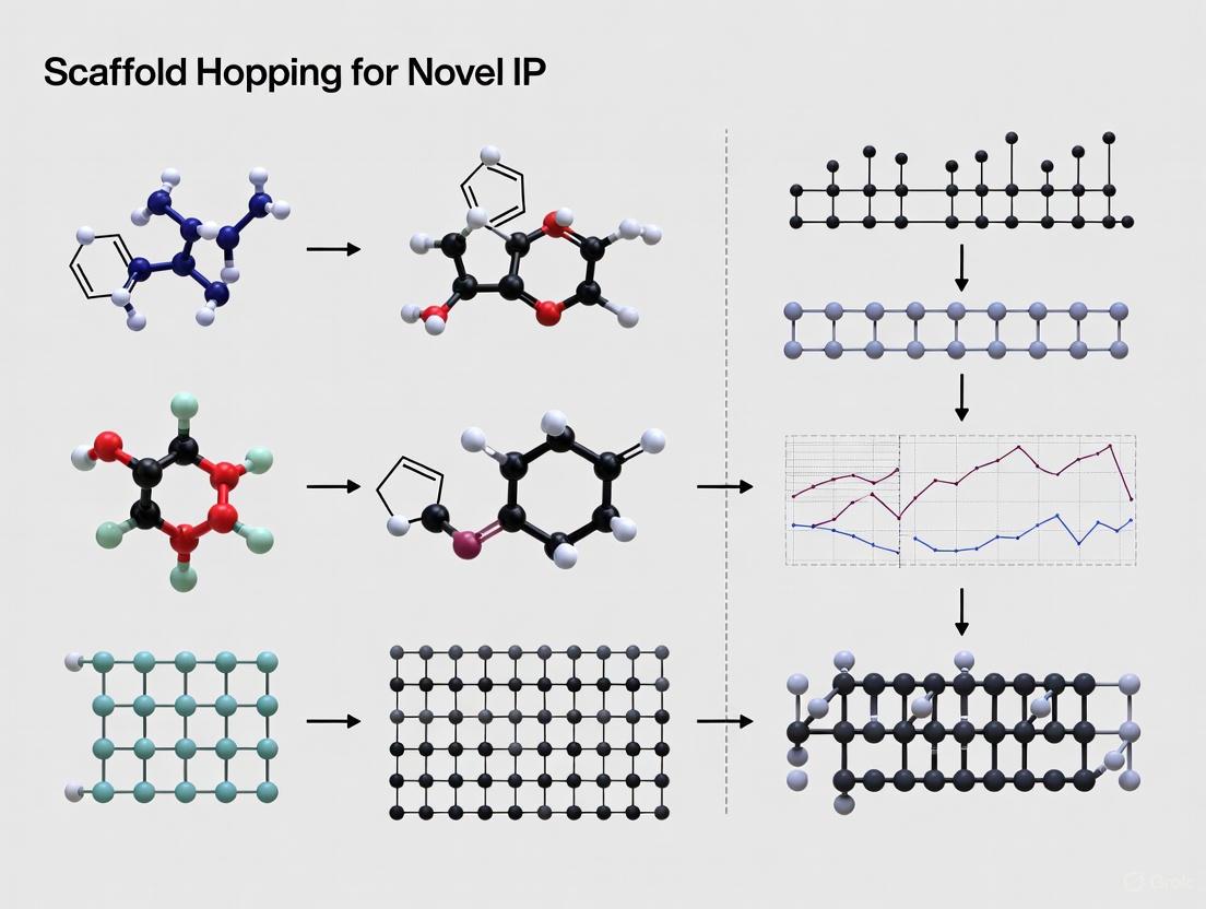
Visualizing Workflows and Relationships
The following diagrams illustrate the key classification system and a generalized experimental workflow for scaffold hopping, providing a visual summary of the concepts and processes described in this document.
Diagram 1: A classification of scaffold hopping into four degrees of structural change, from minor heteroatom substitutions (1°) to major topological overhauls (4°), with representative examples [4] [2] [3].
Diagram 2: A generalized workflow for identifying novel scaffolds through computational methods, leading to synthesis, experimental validation, and the generation of new intellectual property [1] [6] [7].
Scaffold hopping, a cornerstone strategy in modern medicinal chemistry, is defined as the structural modification of the molecular backbone of a known bioactive compound to create a novel chemotype while retaining or improving its biological activity [4]. This approach has emerged as a powerful solution to three critical challenges in pharmaceutical development: mitigating toxicity, optimizing suboptimal pharmacokinetic/pharmacodynamic (PK/PD) profiles, and navigating patent limitations to establish new intellectual property (IP) space [4] [8]. The fundamental premise of scaffold hopping relies on the understanding that structurally distinct compounds can maintain affinity for the same biological target if they preserve key ligand-target interactions present in the original molecule [4].
The strategic importance of scaffold hopping has grown substantially in recent years due to escalating drug development costs and the high failure rate of clinical candidates [9]. In the intensely competitive pharmaceutical industry, innovative methodologies that shorten research and development timelines while providing higher success rates are vital [8]. Scaffold hopping addresses this need by enabling researchers to start from validated molecular templates—including existing drugs, clinical candidates, and bioactive natural products—while systematically engineering out undesirable properties through strategic molecular modifications [8]. This approach has evolved from simple heterocyclic replacements to sophisticated computational and AI-driven design strategies that can explore broader chemical spaces and identify novel scaffolds with improved therapeutic profiles [9] [5].
Scaffold Hopping Classification and Strategic Framework
Degrees of Structural Modification
The scaffold hopping continuum is formally classified into four distinct categories based on the type and extent of structural modification to the parent molecule's core [4] [5]. This classification system, established by Sun and colleagues, provides a systematic framework for medicinal chemists to plan and execute scaffold hopping campaigns.
Heterocyclic Replacement (1° Scaffold Hopping): This simplest form involves substituting, adding, or removing heteroatoms within the molecular backbone, or replacing one heterocycle with another of high similarity [4] [8]. While these modifications are often minor, they retain the spatial arrangement of the unaltered pharmacophore and enable tuning of physicochemical properties [4]. A classic example includes the phosphodiesterase type 5 (PDE5) inhibitors sildenafil and vardenafil, which differ only in the position of a nitrogen atom yet are covered by separate patents [4].
Ring Opening and Closure (2° Scaffold Hopping): This approach introduces novel heterocyclic core scaffolds by either forming new rings (ring closure) or breaking existing ones (ring opening) [8] [10]. Ring closure increases molecular rigidity, which can enhance selectivity and reduce entropy costs upon binding, while ring opening increases flexibility, potentially improving absorption and membrane penetration [10].
Pseudopeptides and Peptidomimetics (3° Scaffold Hopping): This strategy addresses the limitations of natural peptides—such as poor metabolic stability and bioavailability—by designing synthetic analogs that mimic the bioactive conformation of peptides while incorporating non-peptide structural elements [10]. This is particularly valuable for targeting protein-protein interactions that are often intractable with conventional small molecules.
Topology-Based Scaffold Hopping (4° Scaffold Hopping): The most sophisticated approach involves significant structural overhaul where the molecular graph topology is altered while maintaining the spatial orientation of key pharmacophoric elements [8] [5]. This can result in scaffolds with minimal 2D structural similarity to the original compound yet preserved bioactivity, offering the greatest potential for novel IP generation.
Table 1: Classification of Scaffold Hopping Approaches with Applications
| Scaffold Hopping Degree | Structural Change | Primary Applications | IP Strength |
|---|---|---|---|
| Heterocyclic Replacement (1°) | Heteroatom substitution/swap within core ring [8] | PK/PD optimization, toxicity reduction [4] | Limited novelty; often provides minimal IP advantage [4] |
| Ring Opening/Closure (2°) | Altering ring systems (open/close) [8] | Enhance bioavailability, modify target selectivity [10] | Moderate; dependent on structural significance of change [8] |
| Pseudopeptides (3°) | Replacing peptide bonds with bioisosteres [10] | Improve metabolic stability of peptide therapeutics [10] | Strong for novel peptidomimetic scaffolds [10] |
| Topology-Based (4°) | Significant molecular graph alteration [5] | Circumvent existing patents, address multiple limitations [8] | Highest potential for groundbreaking IP [8] |
Experimental Design and Strategic Planning
Successful scaffold hopping requires meticulous pre-planning that aligns structural modification goals with specific project objectives. The strategic workflow begins with a comprehensive analysis of the parent compound's limitations—whether related to toxicity, PK/PD deficiencies, or IP constraints—followed by selection of the appropriate scaffold hopping degree to address these limitations.
For toxicity mitigation, the focus should be on modifying structural motifs associated with off-target interactions or metabolic activation to toxic species. This often involves 1° or 2° scaffold hopping to eliminate problematic substructures while maintaining target engagement. For PK/PD optimization, strategies may include altering logP through heterocycle replacement (1°) or modulating molecular flexibility through ring opening/closure (2°) to improve membrane permeability and metabolic stability. For patent circumvention, more extensive modifications (3° or 4°) are typically required to create sufficient structural novelty while preserving the essential pharmacophore.
The strategic planning phase must also consider synthetic feasibility, as even minor scaffold modifications can require entirely different synthetic routes [4]. Computational approaches, including molecular docking, pharmacophore modeling, and ADMET prediction, should be integrated early to prioritize the most promising scaffold modifications before committing resources to synthesis [11] [5].
Scaffold Hopping Strategy Selection Workflow
Experimental Protocols and Methodologies
Computational Screening and Design Protocols
Protocol 1: Integrated Virtual Screening for Scaffold Hopping
Objective: To identify novel scaffolds with preserved target affinity using a computational pipeline combining pharmacophore modeling, molecular docking, and ADMET prediction.
Materials and Software:
- Maestro Schrödinger Suite (or equivalent molecular modeling platform)
- Target protein structure (PDB source)
- Compound libraries (e.g., TargetMol Anticancer Library, ZINC, ChEMBL)
- High-performance computing cluster
Procedure:
- Compound and Protein Preparation:
- Curate known active compounds with experimental bioactivity data (IC50/EC50).
- Prepare ligands using LigPrep module: generate 3D conformations, assign protonation states at physiological pH (7.0±2.0), and apply OPLS3e or OPLS4 force field for energy minimization [11].
- Retrieve and prepare protein structure from PDB: add hydrogen atoms, assign bond orders, fill missing loops/side chains using Prime, remove crystallographic water molecules not involved in binding, and optimize hydrogen bonding network [11].
Pharmacophore Model Generation:
- Develop a multiligand consensus pharmacophore hypothesis using known active compounds.
- Set hypothesis coverage threshold to 15% to balance sensitivity and specificity.
- Constrain feature complexity to 4-7 pharmacophoric features (hydrogen-bond donors/acceptors, aromatic rings, hydrophobic regions) [11].
- Validate model using ROC curve analysis; select model with highest area under curve (AUC) value [11].
Pharmacophore-Based Virtual Screening:
- Screen compound libraries against validated pharmacophore model.
- Require minimum of four matched pharmacophoric features for compound retention [11].
- Output matched compounds for subsequent docking studies.
Hierarchical Molecular Docking:
- Generate receptor grid: define binding pocket using centroid of co-crystallized ligand or known active site residues (20Å box size).
- Perform high-throughput virtual screening (HTVS) for rapid sampling of large compound libraries.
- Advance top compounds to standard precision (SP) docking for more rigorous pose prediction.
- Submit final candidates to extra precision (XP) docking to eliminate false positives and refine binding pose predictions [11].
Binding Affinity Assessment:
- Calculate binding free energies for top-ranked poses using Molecular Mechanics/Generalized Born Surface Area (MM-GBSA).
- Compare MM-GBSA scores to reference compounds to prioritize scaffolds with improved theoretical affinity [11].
ADMET Profiling:
- Predict key ADMET parameters for lead candidates: aqueous solubility, Caco-2 permeability, cytochrome P450 inhibition, hERG liability, and human hepatocyte clearance.
- Apply QikProp or similar tool for rapid property screening.
- Eliminate compounds with predicted poor pharmacokinetics or toxicity signals [11].
Protocol 2: AI-Driven Scaffold Generation with Molecular Representation
Objective: To employ artificial intelligence and deep learning methods for generating novel molecular scaffolds with optimized properties.
Materials and Software:
- AI platforms with graph neural networks (GNNs), variational autoencoders (VAEs), or transformer architectures
- Curated dataset of bioactive molecules with associated properties
- SMILES or SELFIES representations of training compounds
Procedure:
- Data Preparation and Molecular Representation:
- Curate training set of known active compounds against target of interest.
- Convert structures to appropriate representation: SMILES strings for language models or molecular graphs for GNNs [5].
- For graph-based representations, nodes represent atoms (with features: element type, hybridization, valence) and edges represent bonds (with features: bond type, conjugation) [5].
Model Training:
- For language models (Transformer, BERT): tokenize SMILES strings and pre-train using masked language modeling objective [5].
- For graph models (GNN, VAE): train using supervised learning with bioactivity data or unsupervised learning with reconstruction loss [5].
- Incorporate multi-task learning to simultaneously predict multiple molecular properties (activity, solubility, toxicity).
Scaffold Generation and Optimization:
- Sample latent space of trained model to generate novel scaffold proposals.
- Apply transfer learning to fine-tune model for specific scaffold hopping objectives.
- Use reinforcement learning to optimize generated structures toward desired property profiles [5].
Output Evaluation and Validation:
- Filter generated structures using molecular docking to verify target engagement.
- Apply synthetic accessibility scoring (SAscore) to prioritize synthetically feasible scaffolds.
- Submit top candidates for experimental validation.
Table 2: Research Reagent Solutions for Computational Scaffold Hopping
| Reagent/Software Solution | Function | Application Context |
|---|---|---|
| Schrödinger Suite | Integrated drug discovery platform | Protein preparation, molecular docking, pharmacophore modeling, ADMET prediction [11] |
| OPLS Force Fields | Molecular mechanics parameter sets | Energy minimization and conformational sampling during structure preparation [11] |
| TargetMol Compound Libraries | Curated chemical libraries | Source of diverse compounds for virtual screening and scaffold inspiration [11] |
| Graph Neural Networks (GNNs) | Deep learning architecture | Learning molecular representations from graph structures for property prediction [5] |
| Variational Autoencoders (VAEs) | Generative deep learning model | Creating novel molecular structures in latent chemical space [5] |
| Molecular Fingerprints (ECFP) | Binary vector representation | Similarity searching and machine learning feature input [5] |
Medicinal Chemistry and Synthetic Protocols
Protocol 3: Systematic Heterocyclic Replacement (1° Scaffold Hopping)
Objective: To methodically replace heterocyclic rings in lead compounds to optimize properties while maintaining activity.
Materials:
- Anhydrous solvents (DMF, DMSO, THF, dioxane)
- Palladium catalysts (Pd(PPh3)4, Pd2(dba)3, Pd(dppf)Cl2)
- Ligands (XPhos, SPhos, BINAP)
- Building blocks (heterocyclic boronic acids/esters, halides, amines)
- Chromatography materials (silica gel, C18 reverse-phase)
Procedure:
- Retrosynthetic Analysis:
- Deconstruct target scaffold to identify key bond disconnections.
- Prioritize synthetic routes that enable late-stage diversification of the core heterocycle.
- Identify commercial availability of required heterocyclic building blocks.
Synthetic Implementation:
- Employ cross-coupling methodologies (Suzuki, Buchwald-Hartwig, Sonogashira) for carbon-carbon and carbon-heteroatom bond formation to assemble novel heterocyclic cores [8].
- Apply nucleophilic aromatic substitution for heteroatom incorporation.
- Utilize cyclization reactions to form new heterocyclic systems: (a) Pd-catalyzed C-H activation/cyclization for fused systems, (b) condensation reactions for azole formation, (c) cycloaddition reactions for complex ring systems [8].
Purification and Characterization:
- Purify compounds using flash chromatography (normal or reverse phase).
- Characterize all final compounds using NMR (1H, 13C), LC-MS, and HRMS.
- Determine purity by HPLC-UV/ELSD (>95% for biological testing).
Protocol 4: Ring Opening/Closure Strategies (2° Scaffold Hopping)
Objective: To modulate molecular rigidity and properties through strategic ring opening or closure.
Materials:
- Ring-closing metathesis catalysts (Grubbs II, Hoveyda-Grubbs)
- Cyclization reagents (POCl3, PPA, Eaton's reagent)
- Protecting groups (Boc, Cbz, Fmoc for amines; SEM, MOM for heterocycles)
Procedure:
- Ring Closure Approach:
- Design synthetic routes that incorporate alkenes for ring-closing metathesis to form medium/large rings.
- Employ palladium-catalyzed C-H activation for direct cyclization to fused heterocyclic systems.
- Implement intramolecular nucleophilic displacement for small ring formation (aziridines, epoxides, azetidines) [8].
Ring Opening Approach:
- Identify hydrolytically or enzymatically labile bonds in saturated heterocycles.
- Perform controlled ring opening of lactams, lactones, or cyclic carbamates under mild conditions.
- Functionalize opened structures to lock in bioactive conformations [8].
Conformational Analysis:
- Compare pre-organization of ring-closed analogs versus flexible open-chain analogs using molecular modeling.
- Assess bioactive conformation through docking studies and molecular dynamics simulations.
Experimental Approaches for Scaffold Modification
Application Notes: Case Studies and Data Analysis
Case Study 1: Overcoming Toxicity and Resistance in Tuberculosis Therapeutics
Background: The emergence of drug-resistant Mycobacterium tuberculosis strains has created an urgent need for novel anti-TB agents with improved safety profiles. Scaffold hopping has been successfully applied to optimize existing anti-TB drugs addressing toxicity and resistance mechanisms [4].
Experimental Data:
- Parent Compound: Bedaquiline analog with cardiotoxicity concerns (hERG inhibition) and emerging resistance.
- Scaffold Hopping Approach: 2° scaffold hopping (ring closure) combined with 1° (heteroatom replacement) to create novel diarylquinoline analogs.
- Results: Modified scaffold showed 5-fold reduced hERG inhibition while maintaining potent anti-mycobacterial activity (MIC90 = 0.06 µg/mL against drug-resistant strains) [4].
Key Insights:
- Strategic incorporation of nitrogen atoms in the quinoline core reduced lipophilicity, mitigating hERG liability.
- Ring closure in the side chain restricted conformational flexibility, enhancing target selectivity and reducing off-target interactions.
- The scaffold-hopped analog maintained activity against clinically isolated resistant strains, suggesting ability to overcome common resistance mechanisms [4].
Case Study 2: PK/PD Optimization of FGFR1 Inhibitors for Cancer Therapy
Background: FGFR1 inhibitors show promise in cancer therapy but often suffer from suboptimal target selectivity and dose-limiting toxicities. An integrated computational and medicinal chemistry approach was employed to discover novel FGFR1 inhibitors with improved profiles [11].
Experimental Data:
- Virtual Screening: From an initial library of 9,019 compounds, pharmacophore modeling and hierarchical docking identified 3 hits with superior FGFR1 binding affinity compared to reference ligand.
- Scaffold Hopping: Generated 5,355 structural derivatives through systematic 1° scaffold hopping.
- ADMET Optimization: Candidate compounds 20357a–20357c showed improved bioavailability and reduced toxicity in predictive models [11].
- Validation: Molecular dynamics simulations confirmed stable binding modes and favorable interaction energies for optimized candidates.
Key Insights:
- The combination of computational screening with scaffold hopping efficiently expanded chemical diversity while maintaining target engagement.
- Structural modifications focused on reducing molecular planarity decreased phospholipidosis risk, a common toxicity concern with FGFR inhibitors.
- Optimized compounds maintained nanomolar potency while improving drug-like properties, demonstrating the power of integrated computational-experimental approaches [11].
Case Study 3: Patent Circumvention in Antimalarial Drug Development
Background: The need for novel antimalarial agents has intensified with the spread of artemisinin resistance. Scaffold hopping provided a strategy to develop new intellectual property while maintaining antimalarial efficacy [12].
Experimental Data:
- Lead Identification: Discovered 1,2,4-triazole-containing carboxamide scaffold with antimalarial activity but suboptimal PK properties.
- Scaffold Hopping: Implemented 1° scaffold hopping to develop picolinamide analogs.
- Deuterium Incorporation: Strategic deuterium substitution improved metabolic stability (CLintapp HLM 17.3 μL/min/mg) while maintaining potency.
- Optimized Lead: Compound 110 exhibited EC50 < 200 nM against Plasmodium falciparum, moderate aqueous solubility (13.4 μM), and oral bioavailability (%F 16.2) in preclinical models [12].
Key Insights:
- Core modification from triazole to picolinamide created sufficient structural novelty for patent protection while maintaining key pharmacophore elements.
- Deuterium isotope effects provided subtle but impactful PK improvements without altering target engagement.
- The scaffold-hopped series displayed activity against various P. falciparum isolates with different genetic backgrounds, indicating potential for broad-spectrum application [12].
Table 3: Quantitative Outcomes of Scaffold Hopping Case Studies
| Case Study | Scaffold Hopping Approach | Primary Improvement | Quantitative Results | IP Status |
|---|---|---|---|---|
| TB Drug Optimization | 1° + 2° Scaffold Hopping | Reduced toxicity, overcome resistance | 5-fold ↓ hERG inhibition; MIC90 = 0.06 µg/mL [4] | Novel chemical series with distinct IP [4] |
| FGFR1 Inhibitor Design | Computational 1° Scaffold Hopping | Improved selectivity & ADMET | Nanomolar potency; enhanced predicted bioavailability [11] | Multiple novel chemotypes generated [11] |
| Antimalarial Development | 1° Scaffold Hopping + Deuterium | Enhanced metabolic stability | EC50 < 200 nM; CLintapp 17.3 μL/min/mg [12] | Patentable deuterated analogs [12] |
Intellectual Property Strategy and Patent Considerations
The strategic implementation of scaffold hopping is intrinsically linked to intellectual property generation in pharmaceutical development. A well-executed scaffold hopping campaign can create valuable new patent estates that extend market exclusivity while addressing limitations of existing compounds [8] [13].
Patent Fortress Strategy for Scaffold-Hopped Compounds
Successful IP protection for scaffold-hopped compounds requires construction of a comprehensive "patent fortress" that extends beyond basic composition of matter claims [13]. This multi-layered approach includes:
Composition of Matter (CoM) Patents: Foundational protection for the novel chemical structure itself, requiring demonstration of novelty, utility, and non-obviousness over prior art [13]. For scaffold-hopped compounds, non-obviousness is often demonstrated through unexpected improvements in properties (efficacy, safety, PK) compared to prior scaffolds.
Polymorph Patents: Protection of specific crystalline forms of the active pharmaceutical ingredient, characterized by XRPD peak listings, IR spectra, and melting points [13]. These patents create additional barriers to generic entry even after CoM patent expiration.
Formulation and Delivery Mechanism Patents: Claims covering specific dosage forms, excipient combinations, or delivery technologies that provide clinical benefits such as enhanced bioavailability or reduced dosing frequency [13].
Method of Use and Treatment Patents: Protection of specific therapeutic applications, dosing regimens, or patient subpopulations (e.g., biomarker-defined groups) [13]. These can provide exclusivity even for known compounds when new uses are discovered.
Strategic Patent Filing Timeline
Aligning IP strategy with R&D milestones is critical for maximizing protection. The optimal filing strategy involves:
Provisional Patent Application: File at lead optimization stage to establish early priority date, using the 12-month window to generate critical in vivo data strengthening the non-provisional application [13].
Non-Provisional (Utility) Application: File within 12 months of provisional application, incorporating newly generated data demonstrating superior properties and non-obviousness [13].
Secondary Patent Filings: Strategically file formulation, polymorph, and method-of-use patents throughout clinical development to build layered protection [13].
International Protection: Pursue patent coverage in key markets through PCT application or direct national filings, considering regional differences in patentability criteria.
This comprehensive IP strategy ensures that the innovations derived from scaffold hopping research receive maximal legal protection, creating sustainable competitive advantage and return on investment for pharmaceutical development programs.
Scaffold hopping is a strategic medicinal chemistry approach that involves modifying the core molecular structure, or scaffold, of a known bioactive compound to generate novel chemotypes with similar or improved biological activity [2] [14]. This methodology is fundamental to rational drug design, enabling the circumvention of existing intellectual property while optimizing pharmacological profiles [4]. The transitions from Morphine to Tramadol and from Sildenafil to Vardenafil represent seminal historical successes of this approach, demonstrating how deliberate core modification can yield therapeutics with distinct clinical advantages [2] [15].
Case Study 1: Morphine to Tramadol
Background and Rationale
Morphine, a potent natural product analgesic, acts as a μ-opioid receptor agonist [16]. Despite its efficacy, clinical use is limited by significant adverse effects, including respiratory depression, nausea, vomiting, and high addictive potential [2] [14]. The scaffold hop to Tramadol was pursued to develop an analgesic with a improved safety profile and reduced abuse liability [2].
Scaffold Hopping Strategy and Structural Analysis
The transformation from morphine to tramadol is a classic example of a ring-opening or closure (2° hop) strategy [2] [14]. This involved deconstructing morphine's complex, multi-ring system into a simpler, more flexible structure.
Table 1: Structural and Pharmacological Comparison of Morphine and Tramadol
| Feature | Morphine | Tramadol |
|---|---|---|
| Core Scaffold | Rigid pentacyclic structure (phenanthrene derivative) | Simple, flexible cyclohexanoid monocycle |
| Key Structural Change | Three fused rings | Ring opening of three fused rings |
| Primary Mechanism | μ-opioid receptor agonism | μ-opioid receptor agonism + Serotonin/Norepinephrine reuptake inhibition |
| Analgesic Potency | High (Potent) | Moderate (Approx. one-tenth of morphine) |
| Key Advantages | Potent analgesia | Reduced side effect profile (e.g., addiction, respiratory depression), good oral bioavailability [2] [14] |
Experimental Insight: Pharmacophore Conservation Analysis
Objective: To demonstrate that despite major 2D structural differences, morphine and tramadol share a conserved three-dimensional pharmacophore responsible for μ-opioid receptor engagement.
- Method: Molecular superposition using software such as the Flexible Alignment program in MOE [2] [14].
- Procedure:
- Generate low-energy 3D conformers for both morphine and the active metabolite of tramadol (O-desmethyltramadol).
- Align molecules based on key pharmacophore features.
- Analyze the spatial overlap of critical functional groups.
- Outcome: The superposition reveals conservation of three key pharmacophore elements [2] [14]:
- A positively charged tertiary amine nitrogen.
- An aromatic ring.
- A phenolic hydroxyl group (or the metabolically exposed ether in tramadol). This conserved spatial arrangement explains the retained μ-opioid receptor activity despite scaffold hopping [2].
Diagram 1: Logical workflow illustrating the rationale, strategy, and outcomes of the scaffold hop from Morphine to Tramadol.
Case Study 2: Sildenafil to Vardenafil
Background and Rationale
Sildenafil (Viagra) was the first-in-class phosphodiesterase-5 (PDE5) inhibitor approved for erectile dysfunction [17]. The development of Vardenafil (Levitra) represents a "me-too" drug discovery approach, where scaffold hopping was used to create a novel chemical entity with potential for improved potency and a distinct intellectual property position [15] [18].
Scaffold Hopping Strategy and Structural Analysis
The hop from sildenafil to vardenafil is a prime example of a heterocyclic replacement (1° hop) [2] [4]. The strategic modification involved a single atom swap in the core heterocyclic system.
Table 2: Structural and Pharmacological Comparison of Sildenafil and Vardenafil
| Feature | Sildenafil (Viagra) | Vardenafil (Levitra) |
|---|---|---|
| Core Scaffold | Pyrazolopyrimidinone | Imidazotriazinone |
| Key Structural Change | N-N swap in the 5-6 fused ring system | N-N swap in the 5-6 fused ring system |
| PDE5 Inhibitory Potency | Reference (IC₅₀ = 5 nM [17]) | Higher (IC₅₀ ~ 0.1-0.7 nM; 5-10x more potent in vitro [17]) |
| Clinical Dosage | 50-100 mg | 5-20 mg |
| Key Advantage | First-in-class | Improved potency allowing for lower dosage, distinct IP landscape [17] [15] [18] |
Experimental Insight: Crystallographic Binding Analysis
Objective: To determine the structural basis for Vardenafil's enhanced PDE5 inhibitory potency compared to Sildenafil using X-ray crystallography.
- Method: Protein Crystallography of PDE5-Inhibitor Complexes [17].
- Procedure:
- Protein Purification: Express and purify the catalytic domain of human PDE5A1.
- Complex Formation: Co-crystallize PDE5 with vardenafil.
- Data Collection & Structure Solution: Collect X-ray diffraction data and solve the crystal structure using molecular replacement.
- Outcome: The crystal structure (PDB: 3B2R) revealed key differences from sildenafil-bound PDE5 [17]:
- Conformational Change: The H-loop of PDE5 adopts a distinct conformation in the vardenafil complex.
- Metal Ion Displacement: Vardenafil binding causes a loss of divalent metal ions observed in other PDE5 structures.
- Molecular Configuration: Vardenafil exhibits a different bound configuration compared to sildenafil. These subtle but significant conformational variations in both the enzyme and the inhibitor provide the molecular basis for vardenafil's tighter binding affinity [17].
Diagram 2: Logical workflow illustrating the rationale, strategy, and outcomes of the scaffold hop from Sildenafil to Vardenafil.
Core Principles and Protocols for Scaffold Hopping
Classification of Scaffold Hopping Approaches
Scaffold hopping strategies can be systematically categorized based on the degree of structural alteration [2] [4] [14]:
- 1° Hop (Heterocycle Replacement): Involves substitution, addition, or removal of heteroatoms within a ring system, or replacement of one heterocycle with a similar one (e.g., Sildenafil → Vardenafil).
- 2° Hop (Ring Opening or Closure): Involves breaking bonds to open fused ring systems or forming new bonds to create rings, significantly altering molecular flexibility (e.g., Morphine → Tramadol).
- 3° Hop (Peptidomimetics): Replaces peptide backbones with non-peptide moieties to mimic the spatial arrangement of pharmacophoric elements while improving stability and oral bioavailability.
- 4° Hop (Topology-Based Hopping): Identifies novel scaffolds based on overall molecular shape or topology, often leading to the highest degree of structural novelty.
Computational Protocol for Scaffold Hopping
Modern scaffold hopping leverages computational tools to systematically explore chemical space. The following protocol outlines a typical virtual screening workflow.
Protocol: Ligand-Based Virtual Screening for Scaffold Hopping
Objective: To identify novel scaffold hops for a given lead compound using molecular descriptors and similarity searching.
Software/Tools: Molecular Operating Environment (MOE), RDKit, KNIME or Pipeline Pilot workflows.
Reagents & Computational Resources:
- Lead Compound: A known active molecule (e.g., Sildenafil).
- Compound Database: Commercially available or in-house database (e.g., ZINC, ChEMBL, PubChem).
- Descriptors: WHALES (Weighted Holistic Atom Localization and Entity Shape), ECFP (Extended Connectivity Fingerprints), or other 2D/3D molecular descriptors [19] [20].
- Computing Infrastructure: Standard desktop computer or high-performance computing cluster for large-scale screening.
Procedure:
- Query Preparation:
- Generate a low-energy 3D conformation of the lead compound.
- Calculate relevant molecular descriptors (e.g., WHALES, ECFP4) for the query.
Database Preparation:
- Curate the screening database by filtering for drug-like properties (e.g., Lipinski's Rule of Five).
- Generate canonical SMILES and compute the same molecular descriptors for all database compounds.
Similarity Searching & Scoring:
- Perform a similarity search by calculating the Tanimoto coefficient or Euclidean distance between the query descriptor and all database compound descriptors.
- Rank the database compounds based on their similarity score.
Analysis & Post-Processing:
- Apply a scaffold diversity filter (e.g., Bemis-Murcko scaffold analysis) to prioritize hits with novel core structures [20].
- Visually inspect top-ranking, structurally diverse hits to confirm pharmacophore feature conservation.
- Select promising candidates for in vitro biological evaluation.
The Scientist's Toolkit: Essential Research Reagents & Software
Table 3: Key Research Reagents and Computational Tools for Scaffold Hopping
| Tool / Reagent | Type | Primary Function in Scaffold Hopping |
|---|---|---|
| Molecular Operating Environment (MOE) | Software Suite | Provides comprehensive tools for molecular modeling, pharmacophore elucidation, and flexible molecular alignment [2]. |
| RDKit | Open-Source Cheminformatics | A toolkit for Cheminformatics used for descriptor calculation (e.g., ECFP), scaffold decomposition, and database curation [20]. |
| Spark (Cresset) | Software | Uses field-based technology to find bioisosteric replacements and generate novel scaffolds with similar 3D electrostatics and shape [18]. |
| Protein Data Bank (PDB) | Database | Repository for 3D protein structures; essential for structure-based design and analyzing ligand-target interactions (e.g., PDB: 3B2R for PDE5-Vardenafil) [17]. |
| ChEMBL / PubChem | Bioactivity Database | Provide access to vast amounts of bioactivity data for benchmarking, model training, and validating new scaffold hops [19] [20]. |
| WHALES Descriptors | Molecular Descriptors | Advanced 3D descriptors designed to identify isofunctional chemotypes with high scaffold-hopping potential [19]. |
The historic success stories of Morphine to Tramadol and Sildenafil to Vardenafil provide foundational proof-of-concept for scaffold hopping in drug discovery. These cases demonstrate that systematic modification of a central molecular scaffold—ranging from heterocycle replacement to ring opening—can successfully generate novel chemical entities with distinct intellectual property and optimized therapeutic profiles. As computational methodologies like advanced molecular descriptors and deep generative models continue to evolve [19] [20], the strategic application of scaffold hopping will remain a cornerstone of innovative research for developing new therapeutics within a robust IP framework.
Scaffold hopping, a cornerstone strategy in modern medicinal chemistry, refers to the modification of the central core structure of a known bioactive molecule to generate a novel chemotype while maintaining or improving its biological activity [2] [8]. This approach is critically employed to overcome limitations of existing lead compounds—such as poor pharmacokinetics, metabolic instability, toxicity, or insufficient efficacy—and to create new intellectual property (IP) space essential for sustained drug discovery research [8] [4]. The strategy is fundamentally guided by the principle that structurally diverse compounds can share key pharmacophore features, enabling them to interact with the same biological target [2]. This article provides a structured classification of major scaffold hopping techniques, supported by quantitative data, detailed protocols, and strategic insights, to equip researchers with a framework for pioneering novel IP in drug development.
A Tiered Classification of Scaffold Hopping Approaches
The methodology of scaffold hopping can be systematically classified into a tiered system based on the degree of structural alteration performed on the parent molecular scaffold [2] [14] [4]. This classification, originally proposed by Sun and co-workers, helps in rationalizing the design strategy and anticipating the resulting novelty and challenges [4].
- 1° Hop: Heterocycle Replacements involve the substitution, addition, or removal of heteroatoms within a ring system, or the replacement of one heterocycle with another of high similarity [2] [4]. This is the most common and often simplest form of scaffold hopping.
- 2° Hop: Ring Opening or Closure entails the strategic breaking (opening) or formation (closure) of bonds to alter the ring structure of the scaffold, directly manipulating molecular flexibility and conformational entropy [2] [14].
- 3° Hop: Peptidomimetics focuses on replacing peptide backbones with non-peptide moieties to mimic the spatial arrangement of key pharmacophoric groups, thereby improving the drug-like properties of bioactive peptides [2] [14].
- 4° Hop: Topology-Based Hopping represents the most profound alteration, where the new scaffold is structurally distinct from the original yet shares a similar overall shape or topology, often leading to a high degree of structural novelty [2] [14].
For the purpose of this application note, we will delve into the experimental protocols and considerations for the first, second, and fourth degrees of hopping.
Table 1: Characterization of Scaffold Hopping Tiers
| Hop Degree | Core Modification | Structural Novelty | Success Rate | Primary Application in IP Generation |
|---|---|---|---|---|
| 1°: Heterocycle Replacement | Swapping or replacing heteroatoms in a ring [4]. | Low | High | Tuning physicochemical properties; establishing key ligand-target interactions; creating patentably distinct analogs from prior art [8] [21]. |
| 2°: Ring Opening/Closure | Breaking or forming bonds to open or close rings [2]. | Medium | Medium | Significantly reducing synthetic redundancy; altering molecular flexibility and pharmacokinetic profiles [2] [14]. |
| 4°: Topology-Based | Identifying or designing cores with different connectivity but similar shape [2] [14]. | High | Low | Pioneering entirely novel chemotype classes; securing broad, strong IP for a target [2] [14]. |
Protocol 1: Heterocycle Replacement (1° Hop)
Rationale and Strategic Application
Heterocycle replacement is a foundational strategy for fine-tuning the properties of a lead compound. The primary motivation is often to mitigate metabolic liabilities, as replacing an electron-rich aromatic ring (e.g., benzene) with an electron-deficient heterocycle (e.g., pyridine) can significantly reduce its susceptibility to cytochrome P450-mediated oxidation [21]. This approach retains the spatial arrangement of the pharmacophore and adjacent substituents, allowing researchers to probe SAR, improve solubility, and enhance metabolic stability while generating novel, patentable entities [8] [4]. A classic example is the development of the antihistamine Azatadine from Cyproheptadine by replacing a phenyl ring with a pyrimidine, which improved solubility [2] [14].
Experimental Workflow for a 1° Hop
The following protocol outlines a systematic approach for conducting and validating a heterocycle replacement campaign.
Step 1: Pharmacophore and Vector Analysis
- Objective: Define the critical features of the lead molecule that must be conserved.
- Procedure: Using a tool like the Pharmacophore Generation module in Maestro (Schrödinger), identify key interaction features (e.g., Hydrogen Bond Acceptors/Donors, Aromatic Rings, Hydrophobic regions) from the parent molecule or a set of known actives [11]. Map the directionality (vectors) of substituents attached to the scaffold.
Step 2: Heterocycle Selection and Matched Pair Analysis
- Objective: Choose bioisosteric replacements that conserve geometry and electronics.
- Procedure:
- Consult databases of bioisosteres and heterocyclic properties.
- Prioritize replacements based on HOMO energy (see Table 2) to guide metabolic stability predictions. Lower HOMO energy often correlates with reduced oxidation potential [21].
- Perform a matched molecular pair analysis to predict the impact on key properties like cLogP, TPSA, and solubility [21].
Step 3: In Silico Validation
- Objective: Prioritize synthetic targets computationally.
- Procedure: Screen the virtual library of new analogs against a pharmacophore model. Subsequent molecular docking (e.g., using Glide) against the target protein structure (e.g., PDB ID) assesses the binding mode and conserved interactions [11]. Calculate predicted binding affinities (e.g., MM-GBSA) [11].
Step 4: Synthesis and In Vitro Profiling
- Objective: Experimentally validate the designed compounds.
- Procedure: Synthesize the top-ranking candidates. Evaluate their:
- Potency: Determine IC50 against the target.
- Metabolic Stability: Incubate with human liver microsomes (HLM) and calculate intrinsic clearance (CL~int~) [21].
- Passive Permeability: Perform Caco-2 or PAMPA assays.
Table 2: Electronic Properties and Metabolic Considerations of Common Heterocycles
| Heterocycle | HOMO Energy (eV) [21] | Relative Electron Density | Key Metabolic Consideration |
|---|---|---|---|
| Pyrrole | -8.66 | High | Prone to P450 oxidation; potential for reactive metabolite formation. |
| Benzene | -9.65 | Medium | Susceptible to arene oxidation. |
| Imidazole | -9.16 | Medium-High | Can coordinate to heme iron; may act as a P450 inhibitor. |
| Thiophene | -9.22 | Medium-High | Can be oxidized to reactive sulfoxides. |
| Pyridine | -9.93 | Low | Resistant to P450 oxidation; but may be a substrate for AO. |
| Pyrimidine | -10.58 | Low | Resistant to P450 oxidation; but may be a substrate for AO. |
The Scientist's Toolkit: Key Reagents for Heterocyclic Chemistry
| Research Reagent | Function in Scaffold Hopping |
|---|---|
| Boronic Acids and Pinacol Esters | Essential for Suzuki-Miyaura cross-coupling, enabling the rapid attachment of diverse aromatic and heteroaromatic groups to the new core [8]. |
| Palladium Catalysts (e.g., Pd(PPh₃)₄, Pd₂(dba)₃) | Catalyze key C-C and C-N bond-forming cross-coupling reactions for building heterocyclic systems [8]. |
| Chiral Ligands and Catalysts | Facilitate asymmetric synthesis to access enantiopure scaffolds, crucial for targeting chiral binding pockets [8]. |
| Building Blocks from Commercial Libraries (e.g., TargetMol) | Provide a source of diverse, often drug-like, fragments and cores for rapid analog generation and screening [11]. |
Protocol 2: Ring Opening and Closure (2° Hop)
Rationale and Strategic Application
Ring opening and closure strategies directly manipulate the conformational flexibility of a molecule. Ring closure (cyclization) is often employed to rigidify a flexible lead compound, pre-organizing it into its bioactive conformation. This reduces the entropic penalty upon binding to the target, which can lead to a significant increase in potency and selectivity [2] [14]. A historical example is the transformation of the flexible antihistamine Pheniramine into the rigidified Cyproheptadine via ring closure, which improved both binding affinity and absorption [2] [14]. Conversely, ring opening can be used to reduce potency in a controlled manner (e.g., to create a partial agonist) or to improve aqueous solubility by breaking up a large, planar hydrophobic system. The evolution of Morphine to Tramadol via ring opening is a classic example that resulted in a molecule with a better safety profile and oral bioavailability [2] [14].
Experimental Workflow for a 2° Hop
This protocol focuses on the strategic decision-making and experimental validation for ring closure, a common optimization tactic.
Step 1: Conformational Analysis and Bioactive Conformer Identification
- Objective: Determine the spatial orientation of functional groups in the bound state.
- Procedure: If a co-crystal structure of the lead with the target is available, use it directly. If not, perform a conformational search (e.g., using MOE or MacroModel) and align low-energy conformers to a pharmacophore model. Molecular dynamics simulations can also provide insight into populated conformations.
Step 2: Cyclization Strategy and Linker Design
- Objective: Design a synthetically accessible linker that connects two parts of the molecule without distorting the pharmacophore.
- Procedure: Identify two atoms in the lead molecule that are in proximity in the bioactive conformation. Design a linker (e.g., alkyl chain, amide, ether) that bridges these atoms to form a new ring (5-7 membered rings are typically preferred). Use in silico tools to model the cyclized analog and ensure it can adopt the desired conformation without high steric strain.
Step 3: Synthesis and Biophysical Characterization
- Objective: Confirm that the cyclized analog binds more tightly.
- Procedure: Synthesize the designed compounds. Use techniques like Surface Plasmon Resonance (SPR) or Isothermal Titration Calorimetry (ITC) to determine binding kinetics (k~on~, k~off~) and thermodynamics (ΔG, ΔH). A successful ring closure typically results in a slower k~off~ rate due to reduced conformational entropy loss upon binding.
Protocol 3: Topology-Based Hopping (4° Hop)
Rationale and Strategic Application
Topology-based hopping is the most ambitious scaffold hopping strategy. It aims to identify or design a core structure that is chemically distinct from the original but shares a similar overall shape or spatial distribution of key features, allowing it to interact with the same protein pocket [2] [14]. This approach can lead to breakthroughs in overcoming resistance, as the new scaffold may interact with different residues in the binding site, or in creating entirely new chemical series with superior properties and a strong, broad IP position [14]. Given the high degree of structural change, these hops are often discovered computationally rather than designed manually.
Experimental Workflow for a 4° Hop
This protocol relies heavily on advanced computational screening to identify potential topologically equivalent scaffolds.
Step 1: Query Definition
- Objective: Create a 3D representation of the essential binding features.
- Procedure: The query can be derived from:
- A known active molecule: Convert it into a 3D pharmacophore or use its molecular shape as a query.
- The protein binding site: Define a pharmacophore model directly from the key interactions in the protein active site (e.g., using the Receptor-Based Pharmacophore Generation tool in MOE).
Step 2: Shape-Based and Structure-Based Virtual Screening
- Objective: Search large chemical databases for structurally diverse molecules that match the query.
- Procedure:
- Ligand-Based: Use a tool like ROCS (Rapid Overlay of Chemical Structures) to screen databases for molecules that have a high shape similarity (Tanimoto Combo score) and feature overlap with the query [2].
- Structure-Based: Perform high-throughput molecular docking of millions of compounds from libraries like ZINC or Enamine into the target's binding site. Prioritize hits based on docking score and novel chemotype.
Step 3: Hit Triage and IP Assessment
- Objective: Select the most promising and patentable scaffolds for experimental testing.
- Procedure: Cluster the virtual hits by their Bemis-Murcko (BM) scaffolds to ensure diversity. Before synthesis, perform a thorough patent landscape analysis (e.g., using SciFinder or PatBase) on the top-scoring novel scaffolds to assess freedom to operate and the strength of the potential new IP.
The strategic application of scaffold hopping—from subtle heterocycle edits to bold topological leaps—provides a powerful and rational pathway for innovating beyond existing chemical matter. By systematically employing the classified tiers of 1° to 4° hops, researchers can deliberately navigate the trade-off between structural novelty and the probability of success [2] [8]. Integrating the detailed experimental protocols and computational workflows outlined in this document empowers drug development teams to efficiently generate novel, equipotent, or superior chemotypes. This not only addresses critical lead optimization challenges but also solidifies a robust and defensible intellectual property estate, which is the lifeblood of successful therapeutic research programs [8] [4].
The Similarity Property Principle is a foundational concept in medicinal chemistry, positing that structurally similar molecules are likely to exhibit similar biological activities. Scaffold hopping stands as a critical test and application of this principle, aiming to identify or design structurally diverse compounds that share biological function. This approach seeks to replace a molecule's core structure while preserving its pharmacophoric elements—the key functional groups responsible for its interaction with a biological target. Originally defined by Schneider et al. in 1999, scaffold hopping identifies isofunctional molecular structures with significantly different molecular backbones [22] [2] [14]. This technique has successfully produced marketed drugs such as Vadadustat, Bosutinib, Sorafenib, and Nirmatrelvir [22], demonstrating that the Similarity Property Principle can extend to structurally distinct chemotypes when critical interactions are maintained. The primary drivers for scaffold hopping include overcoming intellectual property constraints, improving poor physicochemical or pharmacokinetic properties, reducing toxicity, and enhancing metabolic stability [22] [8].
Core Principles and Classification of Scaffold Hopping
Scaffold hopping operates on the premise that while the core scaffold may change, the spatial arrangement of essential pharmacophoric features must be conserved to maintain binding affinity and biological activity. This conservation is often assessed through 3D molecular superposition, which reveals shared spatial positioning of key features like charged groups, aromatic rings, and hydrogen bond donors/acceptors, even when 2D structures appear vastly different [2] [14]. Successful scaffold hops maintain these critical interactions while potentially altering other properties.
Scaffold hops are systematically classified based on the degree and nature of structural modification, which correlates with the resulting structural novelty and potential for improved drug properties [2] [8] [14].
Table: Classification of Scaffold Hopping Approaches
| Hop Degree | Designation | Description | Key Objective |
|---|---|---|---|
| 1° Hop | Heterocycle Replacement [2] [8] [14] | Swapping or replacing atoms (e.g., C, N, O, S) within a ring system [14]. | Fine-tune properties like solubility or potency; create patentable variants [2]. |
| 2° Hop | Ring Opening or Closure [2] [8] [14] | Breaking bonds to open fused rings or adding bonds to rigidify flexible chains [2]. | Modulate molecular flexibility to impact binding entropy and ADMET properties [2]. |
| 3° Hop | Peptidomimetics [2] [14] | Replacing peptide backbones with non-peptide moieties to mimic bioactive peptides [2]. | Improve metabolic stability and oral bioavailability of peptide leads [2]. |
| 4° Hop | Topology-Based Hopping [2] [14] | Identifying cores with different connectivity but similar spatial orientation of vectors [2]. | Achieve high degrees of structural novelty for new IP space [2]. |
A classic example of a 2° hop (ring opening) is the transformation of the rigid, T-shaped Morphine into the more flexible Tramadol. Despite significant 2D structural differences, 3D superposition shows conservation of the key pharmacophore: a positively charged tertiary amine, an aromatic ring, and a polar hydroxyl group [2] [14]. Conversely, the development of the antihistamine Cyproheptadine from Pheniramine via ring closure (also a 2° hop) demonstrates how reducing flexibility can increase potency by pre-organizing the molecule for binding [2] [14].
Figure 1. Logical workflow for applying the Similarity Property Principle through different scaffold hopping strategies. The principle guides the selection of a hopping strategy to generate novel compounds with retained biological activity.
Computational Protocols for Scaffold Hopping
Computational methods are indispensable for modern scaffold hopping, enabling systematic exploration of chemical space. The following protocols detail key methodologies.
Protocol: Pharmacophore-Based Virtual Screening for Scaffold Hopping
This protocol uses a ligand-based pharmacophore model to identify novel scaffolds that share critical interaction points with a known active compound [11].
- Step 1: Model Generation. Curate a set of known active compounds for the target. Use software like Maestro (Schrӧdinger) to generate a consensus pharmacophore hypothesis. The model should include 4-7 features (e.g., Hydrogen Bond Donor (HBD), Hydrogen Bond Acceptor (HBA), Aromatic Ring (R), Positive Ionizable) [11].
- Step 2: Model Validation. Validate the model using a database containing known actives and inactives. Employ ROC curve analysis; a valid model should have an AUC (Area Under the Curve) significantly greater than 0.5, indicating an ability to distinguish active from inactive compounds [11].
- Step 3: Virtual Screening. Screen a large compound library (e.g., ChEMBL, ZINC, in-house collections) against the validated pharmacophore model. Retain compounds that match a user-defined minimum number of features (e.g., 4 out of 5) [11].
- Step 4: Hierarchical Docking. Subject the hits to hierarchical molecular docking (e.g., HTVS → SP → XP in Glide) to refine poses and score binding affinity. Use MM-GBSA calculations on top-scoring compounds for more rigorous binding free energy estimation [11].
- Step 5: Scaffold Analysis. Isolate the core scaffolds of the final hit compounds and compare them to the original scaffold using topological or fingerprint-based methods (e.g., Tanimoto similarity on ECFP4 fingerprints) to confirm a successful hop [22] [23].
Protocol: Shape Similarity Screening with ElectroShape
This ligand-based protocol identifies diverse scaffolds by matching the overall 3D shape and electron density of a query molecule, which is crucial for targets where shape complementarity is a primary driver of binding [22].
- Step 1: Query Preparation. Generate a low-energy 3D conformation of the query molecule. Optimize the geometry using a force field (e.g., MMFF94 or OPLS3e) and assign partial charges [22].
- Step 2: Shape Similarity Calculation. Using a tool like ElectroShape in the ODDT Python library, compute the electron shape similarity between the query and each molecule in a screening database. The ElectroShape method goes beyond atom-centered Gaussians to include electron density features [22] [1].
- Step 3: Result Filtering. Rank the database compounds by their shape similarity score (e.g., ElectroShape score). Apply a threshold (e.g., score > 0.7) to select candidates that are morphologically similar to the query.
- Step 4: Synthetic Accessibility Check. Evaluate the synthetic accessibility (SA) of the resulting hits using a tool like SAscore to prioritize compounds that are practical to synthesize [22].
- Step 5: Scaffold Replacement and Validation. For confirmed shape-similar hits, use a framework like ChemBounce to formally replace the original scaffold in the query molecule and generate new, proposed structures for synthesis [22].
Figure 2. A unified computational workflow for scaffold hopping integrating pharmacophore-based, shape-based, and structure-based screening methods. Key computational scoring steps are highlighted.
Protocol: Automated Scaffold Hopping with ChemBounce
ChemBounce is an open-source framework designed specifically for automated scaffold hopping, leveraging a large library of synthesis-validated fragments [22].
- Step 1: Input and Fragmentation. Provide the input molecule as a SMILES string. ChemBounce uses the HierS algorithm via ScaffoldGraph to systematically fragment the molecule, identifying all possible ring systems and linkers [22].
- Step 2: Scaffold Library Search. The tool uses the identified query scaffold to search its curated in-house library of over 3.2 million unique scaffolds derived from the ChEMBL database. Candidate scaffolds are identified based on Tanimoto similarity calculated from molecular fingerprints [22].
- Step 3: Molecule Generation and Rescreening. The query scaffold is replaced with candidate scaffolds from the library to generate new molecular structures. These structures are then rescreened based on both Tanimoto and electron shape similarities (using ElectroShape) to the original input to ensure retention of the global pharmacophore and activity [22].
- Step 4: Output and Customization. The final output is a set of novel compounds. Users can control the number of structures generated per fragment (
-n) and the similarity threshold (-t). Advanced options include retaining specific substructures (--core_smiles) or using a custom scaffold library (--replace_scaffold_files) [22].
Table: Comparison of Key Computational Tools for Scaffold Hopping
| Tool / Software | Primary Methodology | Key Features | Accessibility |
|---|---|---|---|
| ChemBounce [22] | Fragment-based replacement with shape similarity. | Curated ChEMBL scaffold library (3.2M compounds), ElectroShape similarity, high synthetic accessibility. | Open-source (GitHub), Google Colab. |
| FTrees (infiniSee) [1] | Feature Trees descriptor similarity. | "Fuzzy pharmacophore" search, identifies distant structural relatives, ligand-based. | Commercial (BioSolveIT). |
| SeeSAR (Inspirator Mode) [1] | Topological replacement with 3D vector matching. | ReCore function finds fragments with similar 3D connection points; structure-based. | Commercial (BioSolveIT). |
| Similarity Scanner [1] | Shape and pharmacophore superposition. | Ligand-based superposition based on shape and feature orientation. | Commercial (BioSolveIT). |
| Modern AI Models [5] | Graph Neural Networks, Transformers, VAEs. | Learns continuous molecular representations for generative scaffold hopping. | Various, some open-source. |
Experimental Validation and Case Studies
Computational predictions require rigorous experimental validation to confirm successful scaffold hops.
Case Study: FGFR1 Inhibitor Discovery
An integrated computational pipeline was used to discover novel FGFR1 inhibitors [11].
- Methods: A pharmacophore model (ADRRR_2) was built from known actives and used to screen an anticancer compound library. Hits underwent hierarchical docking (HTVS/SP/XP) and MM-GBSA analysis. The top hit was then used for scaffold hopping, generating 5,355 derivatives [11].
- Validation: The binding modes of the final candidates (20357a–20357c) were validated by Molecular Dynamics (MD) simulations, which confirmed stable binding interactions and favorable energies over a 100 ns simulation. ADMET profiling predicted improved bioavailability and reduced toxicity [11].
Case Study: From GLPG1837 to Novel CFTR Potentiators
This case demonstrates iterative optimization through scaffold hopping [8].
- Original Compound: GLPG1837 was a CFTR potentiator that required a high dose (500 mg twice daily), leading to adverse effects [8].
- Hop Strategy: Researchers performed a scaffold hop of the quinolinone core, replacing the aniline linkage with a benzyl ether and modifying the sulfonamide, culminating in a novel tetrahydroquinoline (THQ) scaffold [8].
- Resulting Compound: The optimized THQ-based compound showed a 3-fold improvement in binding free energy (ΔG = -8.9 kcal/mol) compared to GLPG1837 (ΔG = -8.5 kcal/mol). This enhanced potency allowed for a lower effective dose, mitigating the toxicity issues of the original candidate [8].
Experimental Validation Workflow
A standard workflow for validating a proposed scaffold hop includes:
- Step 1: Chemical Synthesis. Synthesize the top-ranked computationally designed compounds.
- Step 2: In Vitro Potency Assay. Determine the IC₅₀ or EC₅₀ value against the purified target protein or in a cellular assay. A successful hop typically shows potency within one order of magnitude of the original active compound.
- Step 3: Selectivity Profiling. Test against related off-targets (e.g., kinase panels) to ensure the hop did not introduce undesirable polypharmacology.
- Step 4: Structural Biology. Confirm the predicted binding mode by solving a co-crystal structure of the hopped compound with the target protein, if possible.
- Step 5: ADMET Profiling. Evaluate key properties in vitro: metabolic stability (e.g., in liver microsomes), membrane permeability (Caco-2 or PAMPA), and inhibition of key cytochromes P450 [8] [11].
Table: Key Research Reagents and Computational Tools for Scaffold Hopping
| Resource / Reagent | Type | Function in Scaffold Hopping | Example Sources / Providers |
|---|---|---|---|
| ChEMBL Database [22] | Bioactivity Database | Source for known active compounds to build models and for scaffold library construction. | https://www.ebi.ac.uk/chembl/ |
| TargetMol Anticancer Library [11] | Compound Library | Pre-curated library for virtual screening of potential anticancer scaffolds. | TargetMol |
| ChemBounce [22] | Software | Open-source tool for automated scaffold hopping with a focus on synthetic accessibility. | GitHub, Google Colab |
| Schrӧdinger Suite [11] | Software Platform | Integrated software for pharmacophore modeling (Maestro), molecular docking (Glide), and MM-GBSA. | Schrӧdinger |
| ODDT Python Library [22] | Software Library | Provides tools for calculating ElectroShape similarity and other cheminformatics tasks. | Open-source Python library |
| FGFR1 Kinase Assay Kit | Biochemical Assay | For experimental validation of FGFR1 inhibitor potency after a scaffold hop. | Various (e.g., Reaction Biology, Eurofins) |
| Human Liver Microsomes | In Vitro ADME Tool | For assessing metabolic stability of new scaffold-hopped compounds. | Various (e.g., Corning, XenoTech) |
| ZINC Database | Fragment Library | Source of commercially available fragments for topological replacement approaches. | http://zinc.docking.org/ |
Scaffold hopping is a powerful strategy that leverages the Similarity Property Principle to navigate the complex relationship between chemical structure and biological activity. By systematically classifying hops and employing robust computational protocols—from pharmacophore modeling and shape matching to tools like ChemBounce—researchers can deliberately design structurally novel compounds that retain desired biological function. This approach is crucial for generating new intellectual property, optimizing lead compounds, and ultimately delivering innovative therapeutics to the market. The continued integration of advanced AI-based molecular representation methods promises to further accelerate and expand the possibilities of scaffold hopping in drug discovery [5].
How to Perform Scaffold Hopping: Traditional and AI-Driven Methodologies
Structure-Based Virtual Screening (SBVS) has become a cornerstone in early drug and probe discovery, enabling researchers to rapidly and cost-effectively screen hundreds of millions of compounds against therapeutic targets with known three-dimensional structures [24]. This approach employs molecular docking to predict how small molecules interact with target binding sites, followed by scoring functions that estimate binding affinity [25]. In the context of scaffold hopping—a medicinal chemistry strategy that modifies the molecular backbone of known bioactive compounds to create novel chemotypes with improved properties—SBVS provides a powerful computational framework for exploring new intellectual property (IP) space while maintaining biological activity [8] [4]. The integration of SBVS with scaffold hopping techniques allows researchers to systematically navigate chemical space, identifying structurally distinct compounds that retain key ligand-target interactions of original active molecules, thereby facilitating the discovery of new patentable molecular entities with optimized pharmacodynamic, physicochemical, and pharmacokinetic (P3) profiles [8].
The fundamental premise of SBVS rests on exploiting the atomic-resolution 3D model of a target protein, typically generated through X-ray crystallography or predicted by algorithms like AlphaFold2 [26]. As the field has advanced, chemical and protein datasets containing integrated bioactivity information have grown substantially in both number and size, enabling the development of more sophisticated machine-learning approaches that often outperform their generic counterparts [26]. For scaffold hopping applications specifically, SBVS represents a validated tool that has received particular attention over the past decade due to significant advances in structural biology and genomics, which have facilitated a deeper understanding of the 3D structures of numerous validated biological targets [4].
Theoretical Framework of Docking and Scoring
Molecular Docking Fundamentals
Molecular docking constitutes the computational engine of SBVS, aiming to predict the optimal binding mode and affinity of a small molecule within the binding site of a target receptor [25]. The docking process involves two main components: pose generation and scoring. Search algorithms investigate a vast conformational space for each molecule in a compound library, generating multiple potential binding orientations (poses) through techniques such as systematic torsional searches, genetic algorithms, or molecular dynamics simulations [24]. The effectiveness of docking screens relies on adequate sampling of possible configurations, though approximations are necessarily employed to make large-scale screening computationally feasible [24].
The search algorithm must navigate the complex energy landscape of protein-ligand interactions, balancing computational efficiency with thoroughness. As noted in the practical guide to large-scale docking, "approximations are used that result in undersampling of possible configurations and inaccurate predictions of absolute binding energies" [24]. This challenge becomes particularly acute in scaffold hopping applications, where the core molecular structure differs significantly from known actives, potentially leading to novel binding modes that might be overlooked by overly restrictive search parameters.
Scoring Functions: Classification and Mechanisms
Scoring functions provide the quantitative assessment necessary to rank docking poses and prioritize compounds for further investigation. These mathematical models are designed to predict binding affinity by evaluating protein-ligand interactions. Traditional scoring functions are typically categorized into three main classes [25]:
Table 1: Classification of Scoring Functions in SBVS
| Type | Basis of Development | Examples | Strengths | Limitations |
|---|---|---|---|---|
| Force Field-Based | Sum of energy terms from classical force fields | DOCK, DockThor | Physically meaningful energy terms; Good transferability | Limited accuracy without solvation models; Sensitive to atomic parameters |
| Empirical | Regression against experimental binding affinity data | GlideScore, ChemScore | Fast calculation; Optimized for affinity prediction | Dependent on training set size and diversity; Limited to linear interactions |
| Knowledge-Based | Statistical analysis of atom pair frequencies in known structures | DrugScore, PMF | No need for experimental affinity data; Implicit solvation effects | Dependent on database size and quality; "Knowledge gaps" for novel complexes |
More recently, machine-learning-based scoring functions have emerged as a fourth category, using sophisticated algorithms like random forests, support vector machines, and deep learning to capture complex, nonlinear relationships between structural features and binding affinity [26] [25]. According to a 2023 protocol, "Often outperforming their generic and non-ML counterparts, target-specific ML-based SFs represent the state of the art for SBVS" [26].
The development of an empirical scoring function requires three key components: "(i) descriptors that describe the binding event, (ii) a dataset composed of three-dimensional structure of diverse protein–ligand complexes associated with the corresponding experimental affinity data, and (iii) a regression or classification algorithm to calibrate the model establishing a relationship between the descriptors and the experimental affinity" [25]. These models differ in the number and type of descriptors, the training algorithm, and the quality of the protein-ligand complexes used during parameterization.
SBVS Workflow and Experimental Protocols
Comprehensive SBVS Protocol
A robust SBVS protocol involves multiple stages, each requiring careful execution and validation. The following workflow diagram illustrates the key steps in a comprehensive structure-based virtual screening campaign:
Target Preparation and Binding Site Definition
The initial stage of any SBVS campaign involves meticulous preparation of the target protein structure. For a protein target (e.g., FGFR1 kinase domain, PDB ID: 4ZSA), the preparation protocol typically includes: adding hydrogen atoms in a physicochemically plausible manner considering physiological pH conditions; detecting and rectifying potential errors or incomplete residues, such as reconstructing missing atoms and adjusting side chain conformations; judiciously retaining or removing water molecules based on their structural and functional significance; assigning and validating disulfide bonds to maintain proper connectivity; and performing energy minimization using force fields such as OPLS3e to achieve a stable conformation [11]. For scaffold hopping applications, particular attention should be paid to the accurate definition of the binding pocket, as novel chemotypes may establish interactions with regions of the protein unexplored by known actives.
The construction of the docking grid represents a critical step that significantly impacts screening outcomes. The grid should encompass the entire binding site and adjacent regions that might accommodate novel scaffolds, with appropriate padding (typically 10-15Å beyond the known binding site) to ensure comprehensive sampling [24]. For targets with known conformational flexibility, multiple receptor conformations may be employed to account for induced-fit effects that could be particularly relevant for structurally diverse compounds identified through scaffold hopping.
Compound Library Preparation
Library preparation involves generating high-quality, energetically reasonable 3D conformations for each compound while implementing structural corrections, including Lewis structure validation, bond order normalization, stereochemical ambiguity resolution, and error checking to ensure molecular integrity [11]. For large-scale screening, it is essential to generate multiple conformers for each compound to account for molecular flexibility, though the extent of conformational sampling must be balanced against computational costs [24]. In the context of scaffold hopping, libraries may be specifically designed to include structurally diverse compounds with potential for novel IP, such as the TargetMol Anticancer Library containing 8,691 compounds or custom libraries generated through computational scaffold hopping approaches [11].
Hierarchical Docking Strategy
A hierarchical docking approach balances computational efficiency with accuracy by employing multiple tiers of increasing sophistication [11]. The protocol typically begins with high-throughput virtual screening (HTVS) using fast scoring functions and limited conformational sampling to rapidly filter large compound libraries (e.g., millions to billions of compounds). Surviving compounds then proceed to standard precision (SP) docking with more rigorous sampling and scoring, followed by extra precision (XP) docking for the top-ranked compounds [11]. This multi-stage filtration efficiently concentrates computational resources on the most promising candidates while maintaining statistical rigor in the early screening stages.
For scaffold hopping applications, it is advisable to employ more perclusive thresholds in the initial screening stages to avoid prematurely eliminating structurally novel compounds that might exhibit non-canonical binding modes. As noted in a study on FGFR1 inhibitors, "Following model selection, virtual screening was conducted using Maestro 11.8 with the ADRRR_2 pharmacophore. A minimum of four matched pharmacophoric features was required for compound retention during screening" [11].
Advanced Scoring and Binding Affinity Prediction
Following initial docking, more sophisticated scoring approaches should be applied to refine the ranking of top candidates. These include:
- MM-GBSA (Molecular Mechanics with Generalized Born and Surface Area solvation): A more computationally intensive method that provides improved binding affinity estimates by incorporating implicit solvation and entropy effects [11].
- Consensus Scoring: Combining multiple scoring functions to reduce individual method biases and improve hit rates [25].
- Machine-Learning Scoring: Utilizing target-specific machine-learning scoring functions that "often outperform their generic and non-ML counterparts" according to recent protocols [26].
For scaffold hopping applications, it is particularly important to visually inspect the predicted binding modes of top-ranked compounds to ensure that key interactions with the target are maintained despite structural changes to the molecular core.
Machine Learning-Enhanced SBVS Protocol
The integration of machine learning techniques has transformed SBVS by enabling the development of target-specific scoring functions with superior performance compared to traditional approaches. The following protocol, adapted from Tran-Nguyen et al. (2023), outlines a comprehensive framework for building and evaluating machine-learning scoring functions for SBVS [26]:
Protocol for Target-Specific ML Scoring Functions
Benchmark Existing Generic SFs: Begin by evaluating existing generic scoring functions against a public benchmark for your target to establish baseline performance metrics [26].
Prepare Experimental Data: Collect experimental bioactivity data for your target from public repositories such as ChEMBL, PubChem BioAssay, or BindingDB. Curate the data carefully, addressing potential issues such as inconsistent assay types, potential measurement errors, and duplicate entries [26].
Data Partitioning: Split the curated dataset into training and test sets using appropriate methods (e.g., temporal split, structural clustering) to ensure rigorous evaluation and avoid overoptimistic performance estimates [26]. The test set should represent a realistic scenario for prospective screening.
Model Generation and Evaluation: Generate target-specific machine-learning scoring functions using the prepared training-test partitions. Evaluate multiple supervised learning algorithms (e.g., random forests, support vector machines, deep learning) to identify the most suitable approach for your specific dataset [26]. The evaluation should encompass both pose prediction accuracy and virtual screening performance.
This protocol, which can typically be completed within one week using a single computer, makes use of accessible software tools such as Smina, CNN-Score, RF-Score-VS, and DeepCoy [26]. The authors emphasize that their aim is to "provide practical guidance on how to augment training data to enhance SBVS performance, how to identify the most suitable supervised learning algorithm for a data set, and how to build an SF with the highest likelihood of discovering target-active molecules within a given compound library" [26].
SBVS-Enabled Scaffold Hopping: Methods and Case Studies
Scaffold Hopping Classification and Strategies
Scaffold hopping encompasses a spectrum of structural modifications to the core of bioactive compounds, systematically categorized by the degree of structural change [8] [4]:
Table 2: Scaffold Hopping Classification and Applications in SBVS
| Degree | Structural Modification | SBVS Approach | IP Potential | Case Example |
|---|---|---|---|---|
| 1° (Heterocycle Replacement) | Substitution/swapping of heteroatoms in backbone ring | Pharmacophore-based screening; Shape similarity | Moderate (dependent on extent of modification) | Pyrazolo[1,5-a]pyrimidine-based TTK inhibitors derived from imidazo[1,2-a]pyrazine scaffold [8] |
| 2° (Ring Opening/Closure) | Opening or closing rings in molecular backbone | Flexible docking; Induced fit protocols | High (significant structural change) | ERK inhibitors designed via ring closure of pyrrole-2-carboxamide scaffold [8] |
| 3° (Peptidomimetics & Core Chain Modifications) | Replacing peptide bonds with bioisosteres; altering core connectivity | Geometric constraint docking; Interaction fingerprint analysis | High to Very High | Roxadustat analogs with modified hinge-binding regions [8] |
| 4° (Fragment Linking/ Merging) | Combining fragments from different scaffolds | Fragment-based docking; Structure-based assembly | Very High (novel chemotypes) | Sorafenib analogs with quinazoline-2-carboxylate backbone [8] |
The successful application of SBVS in scaffold hopping is exemplified by a study on FGFR1 inhibitors, where researchers "established a computational pipeline incorporating ligand-based pharmacophore modeling, multi-tiered virtual screening with hierarchical docking (HTVS/SP/XP), and MM-GBSA binding energy calculations to evaluate interactions within the FGFR1 kinase domain" [11]. From an initial library of 9,019 anticancer compounds, this approach identified three hit compounds with superior binding affinity compared to the reference ligand, followed by scaffold hopping to generate 5,355 structural derivatives with improved bioavailability and reduced toxicity profiles [11].
Molecular Dynamics for Binding Stability Assessment
For promising scaffold-hopped candidates identified through SBVS, molecular dynamics (MD) simulations provide critical validation of binding stability and interaction persistence. A typical protocol involves:
- System Setup: Placing the top-ranked protein-ligand complex in a solvation box with appropriate water models and ions to simulate physiological conditions.
- Equilibration: Gradually relaxing the system through stepwise minimization and heating phases to achieve stable temperature and pressure.
- Production Run: Conducting extended simulations (typically 50-100 ns or longer) to observe the stability of binding modes and identify key interactions.
- Trajectory Analysis: Calculating root-mean-square deviation (RMSD) of protein and ligand, interaction fingerprints, and binding free energies using methods such as MM-PBSA/GBSA.
In the FGFR1 inhibitor study, MD simulations "validated stable binding modes and favorable interaction energies for these candidates" identified through the scaffold hopping approach [11].
Research Reagent Solutions
The successful implementation of SBVS workflows requires access to specialized software tools and compound libraries. The following table details key resources mentioned in the literature:
Table 3: Essential Research Reagents and Computational Tools for SBVS
| Resource Type | Specific Tools/Libraries | Key Function in SBVS | Access Information |
|---|---|---|---|
| Docking Software | DOCK3.7, AutoDock Vina, Glide, GOLD | Pose generation and scoring | DOCK3.7 free for academic research [24]; Commercial packages available |
| Scoring Functions | RF-Score-VS, CNN-Score, DeepCoy, MM-GBSA | Binding affinity prediction | Open-source and commercial implementations [26] |
| Compound Libraries | ZINC15, TargetMol Anticancer Library, PubChem, ChEMBL | Source of screening compounds | Publicly accessible or commercially available [24] [11] |
| Structure Preparation | Protein Preparation Wizard (Schrödinger), LigPrep | System preparation for docking | Commercial software suites [11] |
| Pharmacophore Modeling | Maestro 11.8 (Schrödinger) | Ligand-based screening and scaffold hopping | Commercial software [11] |
| Scaffold Hopping Tools | MORPH, Scaffold Tree Algorithm | Systematic core modification | Various open-source and commercial implementations [8] |
Concluding Remarks
Structure-Based Virtual Screening represents a powerful methodology for accelerating drug discovery, particularly when integrated with scaffold hopping strategies for IP generation. By leveraging molecular docking and advanced scoring functions, researchers can efficiently explore vast chemical spaces to identify novel chemotypes with maintained biological activity and optimized properties. The continuous development of machine-learning scoring functions and the availability of large-scale compound libraries have further enhanced the effectiveness of SBVS approaches. As structural information continues to expand through experimental methods and computational prediction, and as virtual screening algorithms become increasingly sophisticated, SBVS is poised to remain an indispensable tool in the rational design of new therapeutic agents with strong intellectual property positions. For researchers focusing on scaffold hopping, the integration of hierarchical docking protocols with target-specific machine learning scoring functions, followed by rigorous validation through molecular dynamics simulations, provides a robust framework for navigating the complex landscape of structure-activity relationships while generating novel patentable chemical entities.
Ligand-Based Virtual Screening (LBVS) is a foundational computational technique in early drug discovery, employed when the three-dimensional structure of the target protein is unknown or unavailable. It operates on the similarity-property principle, which posits that structurally similar molecules are likely to exhibit similar biological activities [27]. In the context of novel intellectual property (IP) research, LBVS is particularly crucial for scaffold hopping—the identification of novel core structures (scaffolds) that retain the desired biological activity of a known active compound but are structurally distinct enough to circumvent existing patents [28] [5].
This Application Note details the use of two primary LBVS methodologies—pharmacophore modeling and molecular descriptor-based similarity searching—specifically for scaffold hopping. Pharmacophore models capture the essential, abstract features of an interaction, such as hydrogen bond donors/acceptors and hydrophobic regions, enabling the identification of structurally diverse compounds that fulfill the same spatial and electronic constraints [29] [30]. Conversely, molecular descriptors and fingerprints provide a quantitative representation of molecular structures, facilitating rapid similarity comparisons across vast chemical libraries to find compounds that are structurally different at the scaffold level but share key physicochemical properties [5] [27]. The integration of these methods provides a powerful strategy for exploring uncharted chemical space and generating novel, patentable chemical entities.
Theoretical Framework and Key Concepts
Molecular Representation for LBVS
The translation of a chemical structure into a computer-readable format is the critical first step in any LBVS workflow. The choice of representation directly influences the ability to identify novel scaffolds.
- String-Based Representations: The Simplified Molecular-Input Line-Entry System (SMILES) is a compact string notation that describes a molecule's topology and is widely used as an input for more complex representations [5].
- Molecular Fingerprints: These are binary vectors that encode the presence or absence of specific substructures or topological pathways in a molecule. Extended-Connectivity Fingerprints (ECFPs) are a leading example, capable of representing circular atom environments and are highly effective for similarity searching and machine learning models [28] [5]. They are designed to recognize local similarities even when the global scaffold differs.
- Pharmacophore Models: A pharmacophore is defined as the ensemble of steric and electronic features necessary to ensure optimal molecular interactions with a specific biological target and to trigger (or block) its biological response. It is an abstract representation that does not include actual molecular structures or atoms, focusing instead on generalized features like hydrogen bond donors (HBD), hydrogen bond acceptors (HBA), hydrophobic (H) regions, and positive/negative ionizable groups (P/I) [29] [31].
- Graph-Based Representations: Molecules can be represented as graphs, where atoms are nodes and bonds are edges. The Optimal Assignment Kernel (OAK) method, for instance, calculates molecular similarity by finding the best possible mapping of atoms between two molecules, considering their local chemical environments. This method has shown a pronounced ability to scaffold hop while maintaining a good enrichment of active structures [27].
The Scaffold Hopping Paradigm
Scaffold hopping aims to replace a known active scaffold with a novel one while preserving bioactivity. This process is critical for overcoming limitations of existing lead compounds, such as toxicity, metabolic instability, or to design around competitor patents [5] [32]. The strategies can be categorized into several types, including:
- Heterocyclic Replacements: Swapping one ring system for another with similar electronic distribution or geometry.
- Ring Opening or Closure: Altering the ring topology of the scaffold.
- Topology-Based Hops: Identifying scaffolds that present key pharmacophoric features in a similar spatial arrangement but through a different connectivity pattern [5].
Table 1: Classification of Scaffold Hopping Strategies with Examples
| Strategy | Description | Objective in IP Generation |
|---|---|---|
| Heterocyclic Replacement | Substituting a core ring system with a different heterocycle. | Creates chemically distinct entities with comparable activity. |
| Ring Opening/Closure | Converting a cyclic structure to an acyclic one, or vice versa. | Alters the core topology fundamentally. |
| Peptide Mimicry | Replacing peptide bonds with bioisosteres. | Improves metabolic stability and drug-likeness. |
| Topology-Based Hop | Changing the connectivity of rings or chains while maintaining feature orientation. | Generates structurally novel chemotypes that are not obvious. |
Computational Protocols and Methodologies
This section provides detailed, step-by-step protocols for implementing LBVS campaigns focused on scaffold hopping.
Protocol 1: Pharmacophore-Based Virtual Screening
The following protocol, derived from studies on topoisomerase I and KHK-C inhibitors, outlines the process for creating and using a ligand-based pharmacophore model [29] [30].
Step 1: Compound Selection and Preparation
- Gather a set of known active compounds with a range of potencies (typically 15-30 molecules) to serve as a training set [29].
- Prepare 3D structures of all compounds using software like Discovery Studio or MOE. This includes energy minimization and generating conformational models to account for molecular flexibility.
Step 2: Pharmacophore Model Generation (HypoGen Algorithm)
- Use the 3D QSAR pharmacophore generation module (e.g., in Discovery Studio).
- The algorithm identifies common features and correlates their spatial arrangement with the biological activity data.
- The output is a set of pharmacophore hypotheses, each consisting of features like HBA, HBD, and Hydrophobic regions, with associated geometric constraints [29].
Step 3: Model Validation
- Statistical Validation: Assess the hypothesis based on the cost function, correlation coefficient (( R^2 )), and root mean square deviation (RMSD).
- Test Set Validation: Use a separate set of known actives and inactives (not used in training) to calculate the predictive power of the model. A good model should correctly predict the activity of most test set compounds [29].
- Decoy Set Validation: Use an enrichment-based method like the Guner-Henry (GH) score to evaluate the model's ability to prioritize active compounds from a database of decoys.
Step 4: Database Screening
- Use the validated pharmacophore model as a 3D query to screen large, drug-like databases such as ZINC, NCI, or ChemDiv [29] [30].
- The screening process identifies molecules whose conformational ensemble can map onto all or most of the pharmacophore features.
Step 5: Post-Screening Filtration
- Subject the pharmacophore-matched hits to filtration based on:
- Lipinski's Rule of Five: To ensure drug-likeness [29].
- ADMET Filters: Predictive models for absorption, distribution, metabolism, excretion, and toxicity (e.g., using TOPKAT) to remove compounds with undesirable properties [29] [30].
- SMARTS-based Filters: To remove compounds with reactive or unwanted chemical groups.
The following workflow diagram illustrates the key steps and decision points in this protocol.
Protocol 2: Molecular Descriptor-Based Similarity Searching
This protocol leverages molecular descriptors and similarity coefficients to find structurally diverse analogs, as demonstrated in scaffold-focused virtual screens and QSAR modeling [28] [33].
Step 1: Query Selection and Scaffold Deconstruction
- Select a known, potent compound as the query.
- For a scaffold-focused search, deconstruct the molecule to its core scaffold. The Scaffold Tree method is highly effective, systematically breaking down molecules to level 1, which represents the Murcko framework—a medicinal chemistry-relevant representation of the core structure [28].
Step 2: Descriptor Calculation and Library Preparation
- Calculate molecular descriptors or fingerprints for both the query (or its scaffold) and every compound in the screening library.
- Common descriptors for scaffold hopping include:
- ECFP_4 Fingerprints: For 2D similarity searching [28].
- ROCS (Rapid Overlay of Chemical Structures): For 3D shape and feature similarity [28] [27].
- MACCS Keys: Structural keys for a fast preliminary screen [33].
- Optimal Assignment Methods: Graph-based methods that find the best atom-to-atom mapping between molecules [27].
Step 3: Similarity Calculation
- Compute the pairwise similarity between the query and each database molecule.
- For 2D fingerprints, use the Tanimoto coefficient, the most common metric. A value of 1.0 indicates identical fingerprints, while 0.0 indicates no similarity.
- For 3D shape-based methods like ROCS, use the TanimotoCombo score, which combines both shape and chemical feature overlap [28].
Step 4: Scaffold-Focused Ranking and Compound Selection
- Rank the entire database based on the similarity score.
- To explicitly promote scaffold diversity, prioritize the selection of compounds from the top-ranked scaffolds rather than just the top-ranked molecules. From each promising scaffold, select up to five diverse representative compounds to ensure sampling of different substituents [28].
Step 5: Clustering and Analysis
- Cluster the final hit list based on Tanimoto similarity (e.g., using k-means clustering) to visualize the chemical diversity achieved and to select a non-redundant set of compounds for further study [33].
Table 2: Key Molecular Descriptors and Their Application in Scaffold Hopping
| Descriptor / Method | Type | Mechanism | Scaffold-Hopping Potential | |
|---|---|---|---|---|
| ECFP_4 Fingerprints | 2D / Topological | Encodes circular atom environments up to a diameter of 4 bonds. | High | Recognizes local similarities despite global scaffold differences. |
| ROCS (TanimotoCombo) | 3D / Shape & Feature | Maximizes overlap of molecular volumes and pharmacophoric features. | Very High | Can identify molecules with different connectivities but similar 3D profiles. |
| Optimal Assignment (OAK) | Graph-Based | Finds best atom-to-atom mapping considering chemical environment. | High | Directly interprets mappings and is less reliant on predefined substructures. |
| MACCS Keys | 2D / Structural | 166-bit key based on predefined chemical substructures. | Low | Effective for finding close analogs, but limited in scaffold hopping. |
| Scaffold Tree (Level 1) | 2D / Framework | Represents the Murcko scaffold of a molecule. | Explicit | Focuses the search directly on core scaffold similarity. |
The following workflow diagram illustrates the parallel paths for 2D and 3D similarity searching in a scaffold-focused protocol.
The Scientist's Toolkit: Essential Research Reagents and Software
Table 3: Key Resources for Implementing LBVS for Scaffold Hopping
| Category / Item | Specific Examples | Function in LBVS/Scaffold Hopping |
|---|---|---|
| Commercial Software Suites | Discovery Studio (Biovia), MOE (Chemical Computing Group), Schrödinger Suite | Integrated platforms for pharmacophore modeling, molecular docking, QSAR, and descriptor calculation. |
| Open-Cheminformatics Tools | RDKit, OpenBabel | Open-source toolkits for manipulating molecules, calculating fingerprints (ECFP), and descriptor calculation. Essential for preprocessing and ML-based QSAR [33]. |
| Virtual Screening Tools | ROCS (OpenEye), EON | Specialized software for 3D shape-based and electrostatic similarity comparisons, crucial for scaffold hopping [28] [27]. |
| Compound Databases | ZINC, ChemDiv, NCI Database | Sources of commercially available and drug-like compounds for virtual screening [29] [33] [30]. |
| Scaffold Analysis Tools | Scaffold Tree generation in MOE or RDKit | Algorithms to systematically decompose molecules into hierarchical scaffolds, enabling scaffold-focused analysis [28]. |
| Machine Learning Libraries | Scikit-learn, PyTorch, TensorFlow | For building predictive QSAR models to score virtual screening hits or to generate novel scaffolds [5] [33]. |
Case Study: Integrated LBVS for Novel Topoisomerase I Inhibitors
A seminal study demonstrates the successful integration of these protocols to discover novel Topoisomerase I (Top1) inhibitors via scaffold hopping [29].
- Objective: Identify novel, non-camptothecin Top1 inhibitors with better efficacy and stability.
- Method:
- Pharmacophore Modeling (Hypo1): A 3D QSAR pharmacophore model was generated from 29 CPT derivatives.
- Validation: The model was validated with 33 test set molecules, showing strong predictive power.
- Virtual Screening: The Hypo1 model screened over 1 million drug-like molecules from the ZINC database.
- Filtration: Hits were filtered using Lipinski's Rule of Five, SMART filtration, and activity filters.
- Molecular Docking: Filtered hits were docked into the Top1 active site (PDB: 1T8I) to study binding modes.
- Toxicity Assessment & MD Simulations: TOPKAT toxicity prediction and molecular dynamics simulations identified three final "hit molecules" (ZINC68997780, ZINC15018994, ZINC38550809) with stable binding and low toxicity.
- Outcome: The campaign yielded three novel inhibitory scaffolds, distinct from camptothecin, demonstrating the power of a combined LBVS approach for successful scaffold hopping and hit identification [29].
Topological Replacement and Fragment-Based Approaches with Tools like SeeSAR
In the intensely competitive landscape of pharmaceutical research, the generation of novel intellectual property (IP) is paramount. Scaffold hopping has emerged as a critical strategy for creating new patentable molecular entities while maintaining desired biological activity. This approach involves the identification of isofunctional molecular structures with chemically distinct core structures, enabling medicinal chemists to overcome limitations of existing leads, including toxicity, promiscuity, unfavorable physicochemistry, or restricted IP space [1]. Among the various computational techniques available, topological replacement and fragment-based approaches represent particularly powerful methodologies for systematic scaffold modification. These methods enable researchers to explore uncharted chemical territory while preserving the essential pharmacophoric elements required for target engagement. The integration of sophisticated software tools like SeeSAR has dramatically accelerated these discovery workflows, providing intuitive platforms for visual, interactive drug design that bridges the gap between computational prediction and experimental implementation [34] [35].
Topological replacement specifically addresses the challenge of core scaffold modification by searching for molecular fragments that maintain the geometric orientation of substituents while altering the central architecture. This approach is particularly valuable when the core structure of a lead compound presents synthetic challenges, undesirable properties, or IP constraints. By focusing on the three-dimensional coordination of connection points, topological replacement enables bioisosteric substitutions that maintain the vectorial display of key functional groups [1]. When combined with fragment-based strategies that leverage small molecular building blocks to explore structure-activity relationships, researchers gain a powerful toolkit for IP generation. This application note details the practical implementation of these approaches using BioSolveIT's SeeSAR platform, providing structured protocols, quantitative comparisons, and visual workflows to guide researchers in their scaffold hopping campaigns.
Key Methodological Approaches and Their Applications
Topological Replacement with ReCore
The ReCore functionality, implemented within SeeSAR's Inspirator Mode, specializes in 3D-driven scaffold replacement by screening pre-processed fragment libraries for motifs with similar connection vector geometry [34] [1]. This method identifies potential scaffold hops by analyzing the three-dimensional orientation of attachment points in the original molecule and searching for alternative cores that can maintain the spatial arrangement of critical substituents. The algorithm screens libraries containing fragments from sources like the ZINC database and Protein Data Bank, ranking results according to their connecting vector similarity [1]. Pharmacophore constraints can be applied during this process to ensure that suggested replacements maintain key interactions with the biological target, significantly increasing the likelihood of preserving biological activity while achieving the desired structural novelty.
Experimental Protocol: Topological Replacement with ReCore
Initial Setup: Begin by loading your protein-ligand complex into SeeSAR. Prepare the structure by ensuring the binding site is properly defined and the ligand geometry is optimized.
Scaffold Identification: Enter the Inspirator Mode and select the ReCore functionality. Identify the specific scaffold region within your ligand that requires replacement, typically focusing on ring systems or core linkers that connect key pharmacophoric elements.
Vector Specification: Manually select the connection vectors (atoms or bonds) that define how substituents attach to the core scaffold. These vectors will be used to search for geometrically compatible replacements.
Constraint Application: Apply relevant pharmacophore constraints to maintain critical interactions. These may include hydrogen bond donors/acceptors, aromatic rings, or hydrophobic features essential for binding.
Library Selection: Choose appropriate fragment libraries for the search. BioSolveIT provides specialized index files optimized for ReCore, which are available free of charge.
Execution and Analysis: Execute the ReCore search and analyze the results based on geometric compatibility scores, estimated affinity changes (via HYDE scoring), and structural novelty. Select promising candidates for further optimization or synthesis.
The power of topological replacement lies in its ability to suggest chemically diverse scaffolds that maintain the spatial orientation of functional groups, making it particularly valuable for circumventing patent restrictions or addressing physicochemical limitations of existing lead compounds.
Fragment-Based Design with FastGrow
Fragment-based drug design (FBDD) utilizes small molecular fragments (typically <300 Da) as starting points for compound development, which are then elaborated or linked to enhance potency and optimize properties [34] [36]. SeeSAR implements this approach through its FastGrow technology, which rapidly screens hundreds of thousands of fragments against defined binding sites to suggest optimal decorations or extensions [35]. This method leverages a novel algorithm with shape-based directional descriptors to identify fragments that complement the binding cavity, providing medicinal chemists with structure-based suggestions for molecular optimization. The ultra-fast performance of FastGrow enables real-time interactive design, with screening results available within seconds on standard hardware [35].
Experimental Protocol: Fragment Growing with FastGrow
Complex Preparation: Load your protein-ligand complex into SeeSAR and define the binding site of interest. The software automatically detects binding sites but allows for manual refinement if necessary.
Growth Vector Selection: In Inspirator Mode, select the specific atom or fragment in your lead compound from which you wish to grow. This typically represents a position where structural elaboration could potentially form additional favorable interactions with the binding pocket.
Library Selection: Choose an appropriate fragment library based on your design objectives:
- Default set (12k fragments): Provides a representative collection of medchem-like fragments for general-purpose optimization.
- Medchem set (120k fragments): Offers expanded diversity derived from common drug motifs and PDB structures.
- sp3 set (28k fragments): Focuses on fragments with sp3-hybridized carbon atoms to enhance three-dimensionality.
- Hinge binder set (51k fragments): Specifically curated for kinase targets, containing computationally validated hinge-binding motifs [35].
Execution: Run the FastGrow calculation. The algorithm will rapidly screen the selected library and return ranked suggestions based on their potential to form favorable interactions in the binding cavity.
Evaluation: Assess suggested fragments using SeeSAR's visual HYDE analysis, which color-codes atoms based on their contributions to binding affinity (green=favorable, red=unfavorable). Prioritize fragments that fill hydrophobic pockets, form hydrogen bonds, or improve complementarity.
Integration: Select the most promising fragments and incorporate them into your lead compound using SeeSAR's molecule editing capabilities. Re-score the modified compound to evaluate the predicted improvement in binding affinity.
Fragment-based approaches are particularly valuable in the early stages of lead optimization, where systematic exploration of structure-activity relationships is required. The ability to rapidly screen large fragment libraries against the target structure enables data-driven decision-making and reduces reliance on chemical intuition alone.
Chemical Space Docking for Scaffold Discovery
Chemical Space Docking (C-S-D) represents a paradigm shift in virtual screening, enabling structure-based exploration of ultra-large, synthetically accessible compound collections spanning billions to trillions of molecules [34] [37]. This approach overcomes the limitations of traditional virtual screening by employing a combinatorial strategy that docks representative fragments and extends them within the binding site, rather than exhaustively docking pre-enumerated compounds [34]. The method is particularly valuable for scaffold hopping as it can identify completely novel chemotypes that would be missed by similarity-based approaches alone.
Experimental Protocol: Chemical Space Docking
Protein Preparation: Load and prepare your target protein in SeeSAR, ensuring proper protonation states and structural integrity. Recent updates in SeeSAR 14.2 allow for energy minimization of protein-ligand complexes using integrated YASARA functionality with 17 available force fields [37].
Binding Site Definition: Precisely define the binding site of interest, either based on a known ligand or through detection of unoccupied pockets.
Chemical Space Selection: Choose from available Chemical Spaces, which must be pre-uploaded to the HPSee server. Options include commercial spaces like eXplore and REAL Space, or proprietary in-house collections [34].
Workflow Configuration: Initiate the Space Docking Mode and configure appropriate parameters. Optionally augment the binding site definition with pharmacophore constraints or template molecules to guide the search.
Anchoring and Extension: Execute the multi-step docking process, beginning with fragment anchoring followed by iterative extension steps. The interface provides visual guidance for selecting optimal growth vectors [37].
Result Analysis: Evaluate generated compounds using ligand efficiency (LE) and lipophilic ligand efficiency (LLE) metrics, which are now default sorting parameters in recent SeeSAR versions [37].
Chemical Space Docking enables access to unprecedented chemical diversity while maintaining synthetic accessibility, as the compounds in these spaces are typically make-on-demand using robust reactions and available building blocks [34]. This makes it an invaluable tool for scaffold hopping campaigns aiming to generate novel IP with confirmed synthetic pathways.
Comparative Analysis of Scaffold Hopping Techniques
Table 1: Quantitative Comparison of Scaffold Hopping Approaches in SeeSAR
| Method | Library Size | Key Metrics | Typical Use Case | IP Generation Potential |
|---|---|---|---|---|
| ReCore (Topological Replacement) | Pre-processed indices from ZINC, PDB [1] | Vector similarity, HYDE affinity | Core replacement in established leads | High (focused 3D similarity) |
| FastGrow (Fragment-Based) | 12k-661k fragments [35] | ΔHYDE, LE, LLE | Lead optimization, R-group exploration | Medium (fragment elaboration) |
| Chemical Space Docking | Billions-trillions [34] | LE, LLE, synthetic accessibility | De novo scaffold discovery | Very High (novel chemotypes) |
| FTrees (Fuzzy Pharmacophores) | Ultra-large Chemical Spaces [1] | Feature Tree similarity | Finding distant structural relatives | High (fuzzy similarity) |
| Similarity Scanner | Custom compound sets | Shape and feature overlap | Lead hopping without structural data | Medium (shape similarity) |
Table 2: Fragment Libraries Available for FastGrow in SeeSAR
| Library Name | Fragment Count | Special Characteristics | Application Focus |
|---|---|---|---|
| Default Set | 12,000 | Medchem-like fragments | General purpose optimization |
| Medchem Set | 120,000 | Expanded drug-like motifs | Diverse decoration ideas |
| sp3 Set | 28,000 | sp3-hybridized α-carbons | 3D character enhancement |
| Hinge Binder Set | 51,000 | Computationally validated kinase motifs | Kinase-targeted projects |
| Dipeptide Set (N-Term) | 661,000 | Proteinogenic amino acids & bioisosteres | Peptidomimetic design |
| Dipeptide Set (C-Term) | 661,000 | C-terminal configuration | Peptidomimetic design |
The comparative analysis reveals a complementary relationship between these approaches, with each method offering distinct advantages for different stages of the scaffold hopping process. Topological replacement with ReCore excels when specific vector geometry must be maintained, while fragment-based approaches provide maximum flexibility for exploring binding interactions. Chemical Space Docking offers the broadest exploration capability but requires more computational resources. The combination of multiple methods often yields the best results, as different approaches can identify complementary scaffolds that might be missed when using a single methodology [34].
Visual Workflows for Implementation
Diagram 1: Integrated scaffold hopping workflow showing the relationship between different computational approaches and decision points in a typical campaign. The workflow begins with protein preparation and branches based on methodological selection before converging on analysis and experimental validation stages.
Diagram 2: Methodology-specific pathways for topological replacement and fragment-based approaches, highlighting the distinct computational processes for each technique while demonstrating their convergence toward the common goal of generating optimized compounds with novel scaffolds.
Table 3: Research Reagent Solutions for Scaffold Hopping Campaigns
| Resource Type | Specific Solution | Function in Research | Application Context |
|---|---|---|---|
| Software Platform | SeeSAR Drug Design Dashboard | Interactive visualization and analysis of protein-ligand complexes with HYDE scoring | Primary workbench for all scaffold hopping approaches |
| Fragment Libraries | FastGrow Libraries (Default, Medchem, sp3, Hinge Binder) | Provide validated molecular fragments for structure-based design | Fragment growing and linking in binding sites |
| Scaffold Replacement | ReCore Index Files | Pre-processed fragment databases for topological scaffold replacement | Core hopping with maintained vector geometry |
| Chemical Spaces | eXplore Space, REAL Space, in-house collections | Ultra-large compound collections for virtual screening | Chemical Space Docking for novel scaffold discovery |
| Computational Server | HPSee High-Performance Computing | Handles demanding docking calculations for large-scale screenings | Essential for Chemical Space Docking workflows |
| Validation Tools | HYDE Affinity Estimation, ADME Properties | Predict binding energy and drug-like properties | Prioritization of proposed scaffold hops |
The toolkit requirements vary depending on the selected approach. Topological replacement with ReCore requires specific index files containing pre-processed fragment information, while fragment-based design relies on curated fragment libraries optimized for the FastGrow algorithm. Chemical Space Docking demands the most extensive infrastructure, requiring both Chemical Space access and HPSee server installation for calculation handling [34] [35] [37]. Recent updates in SeeSAR 14.2 have enhanced usability across these tools, with improvements to editing workflows, filter management, and visualization of unresolved protein segments that impact binding site definition [37].
Topological replacement and fragment-based approaches implemented through platforms like SeeSAR provide robust methodologies for scaffold hopping in novel IP research. The integration of these complementary techniques enables comprehensive exploration of chemical space while maintaining the pharmacophoric elements essential for biological activity. The quantitative comparison presented in this application note demonstrates that each method offers distinct advantages, with topological replacement excelling in maintaining vector geometry, fragment-based approaches enabling systematic exploration of binding interactions, and Chemical Space Docking providing access to unprecedented structural diversity.
The continued evolution of these computational tools promises to further accelerate scaffold hopping campaigns. Recent developments in SeeSAR, including enhanced protein editing capabilities, force field-based minimization through YASARA integration, and improved Chemical Space Docking workflows, have already significantly expanded practical capabilities [37]. As artificial intelligence and machine learning approaches continue to mature, their integration with established structure-based methods will likely open new frontiers in scaffold hopping efficiency and effectiveness. By leveraging these sophisticated computational tools and following the detailed protocols outlined in this application note, researchers can systematically generate novel intellectual property while reducing the traditional risks associated with molecular optimization.
Scaffold hopping, a cornerstone strategy in medicinal chemistry, is defined as the design of novel molecular core structures (scaffolds) that retain the biological activity of a known lead compound but are structurally distinct. The primary objectives are to discover new chemical entities with improved efficacy, safety, pharmacokinetic profiles, or to circumvent existing intellectual property (IP) [9] [5]. In the context of novel IP research, successfully hopped scaffolds represent the foundation for building robust and defensible patent estates around new therapeutic compounds [38].
The advent of Artificial Intelligence (AI) has profoundly transformed this field. Traditional methods, which often relied on molecular fingerprinting and similarity searches, were limited by their dependency on predefined rules and expert knowledge [5]. AI-driven methods, particularly those based on Graph Neural Networks (GNNs), Variational Autoencoders (VAEs), and Diffusion Models, have enabled a data-driven revolution. These technologies can navigate the vast chemical space more efficiently, capturing non-linear and complex structure-activity relationships that are elusive to conventional techniques, thereby accelerating the discovery of novel, patentable chemotypes [9] [39] [5].
Key AI Methodologies and Quantitative Comparison
The following table summarizes the core AI architectures driving modern scaffold hopping, their unique mechanistic principles, and their specific applications in drug discovery.
Table 1: Key AI Models in Scaffold Hopping and Their Applications
| AI Model | Core Mechanism | Key Features for Scaffold Hopping | Exemplar Tools/Methods |
|---|---|---|---|
| Graph Neural Networks (GNNs) | Operates directly on molecular graph structures (atoms as nodes, bonds as edges) using message-passing between connected nodes [40]. | Excels at modeling molecular topology and interactions with protein targets; ideal for predicting properties and binding affinities [40]. | ScaffoldGVAE (Encoder component) [39], GraphGMVAE [39]. |
| Variational Autoencoders (VAEs) | A generative model that learns a compressed, continuous latent representation (latent space) of molecular structures. New structures are generated by sampling from this space [39]. | Explicitly separates scaffold and side-chain information; latent space can be optimized for desired properties [39]. | ScaffoldGVAE [39], JT-VAE, GVAE [39]. |
| Diffusion Models | Generates data by iteratively denoising a structure that starts as pure noise, learning a reverse process of a fixed forward noising process [39]. | State-of-the-art in generating high-quality, diverse molecular structures; used in atomic coordinate space for 3D-aware generation. | GEOLDM, MolDiff [39]. |
| Reinforcement Learning (RL) | An agent learns to make generation decisions (e.g., adding a molecular fragment) by receiving rewards for achieving desired objectives (e.g., high bioactivity, novel scaffold) [41]. | Unconstrained generation of full molecules optimized for specific, multi-objective rewards like 3D similarity and low 2D scaffold similarity [41]. | RuSH (Reinforcement Learning for Unconstrained Scaffold Hopping) [41]. |
To aid in the selection of the appropriate methodology, the following table provides a comparative analysis based on critical performance and application metrics.
Table 2: Comparative Analysis of AI Scaffold Hopping Methodologies
| Metric | GNN-Based Models | VAE-Based Models | Diffusion Models | Reinforcement Learning |
|---|---|---|---|---|
| Scaffold Diversity | Moderate | High (e.g., ScaffoldGVAE uses Gaussian mixture model for scaffold embedding) [39] | High [39] | High (Explicitly optimized for low 2D scaffold similarity) [41] |
| 3D/Pharmacophore Awareness | High (inherent from graph structure) [40] | Moderate | High (especially 3D diffusion models) [39] | High (Uses 3D and pharmacophore similarity as reward) [41] |
| Side-Chain Preservation | Not inherent | High (Explicitly separates side-chain embedding) [39] | Not inherent | Not inherent (Generates full molecules) [41] |
| Interpretability | Moderate (Message passing can be analyzed) | Low (Latent space is often opaque) | Low | Moderate (Governed by defined reward functions) |
| Reported Validation | Docking, MM/GBSA, Case studies (LRRK2 inhibitors) [39] | Docking, MM/GBSA, Case studies (LRRK2 inhibitors) [39] | Benchmark metrics | In silico comparison to known scaffold-hops [41] |
Application Notes & Experimental Protocols
Protocol 1: Scaffold Hopping with ScaffoldGVAE
Application Note: This protocol details the procedure for generating novel scaffolds while preserving desired side-chain functionalities using the ScaffoldGVAE model, a method that has been validated for generating inhibitors of targets like LRRK2 [39].
Materials & Pre-processing:
- Data Curation: Obtain a dataset of active molecules against your target of interest (e.g., from ChEMBL). Pre-process by standardizing charges, removing duplicates and metals, and applying medicinal chemistry filters (e.g., molecular weight, PAINS) [39].
- Scaffold Extraction: Employ the ScaffoldGraph method to decompose each molecule into its core scaffold(s), going beyond simple Bemis-Murcko extraction. Filter scaffolds to retain those with at least one non-benzene ring, ≤20 heavy atoms, and ≤3 rotatable bonds [39].
- Data Pairing: Create a dataset of molecule-scaffold pairs. For pre-training, a large dataset like ChEMBL (e.g., 800,000+ pairs) is used. For fine-tuning, use a smaller, target-specific activity dataset [39].
Methodology:
- Model Architecture:
- Encoder: A multi-view GNN encodes the molecular graph. It performs separate message-passing procedures with nodes (atoms) and edges (bonds) as centers. The final node and edge embeddings are concatenated to form a whole molecular embedding. This embedding is then split into a scaffold embedding and a side-chain embedding [39].
- Latent Space Modeling: The scaffold embedding is projected onto a Gaussian mixture model (GMM) prior distribution. The side-chain embedding remains a fixed vector [39].
- Decoder: A Recurrent Neural Network (RNN) decoder takes the concatenated scaffold and side-chain embeddings as its initial hidden state and reconstructs the scaffold's SMILES string [39].
- Training:
- Pre-training: Train the model on the large, diverse ChEMBL-derived molecule-scaffold dataset to learn general chemical rules [39].
- Fine-tuning: Transfer learning is performed on the smaller, target-specific dataset to bias the model towards generating scaffolds relevant to the target of interest [39].
- Generation:
- To generate novel molecules, sample a new scaffold embedding from the GMM prior.
- Concatenate this new scaffold embedding with the original side-chain embedding from a reference molecule.
- The decoder RNN generates a novel scaffold SMILES based on this combined vector, effectively performing a scaffold hop while retaining the side-chain context [39].
Validation:
- In-silico: Evaluate generated molecules using docking (e.g., LeDock), binding affinity prediction (e.g., MM/GBSA), and target-specific activity prediction models (e.g., GraphDTA) [39].
- In-vitro: Synthesize top-ranked novel scaffolds and test for biological activity and selectivity in relevant biochemical or cellular assays [39].
ScaffoldGVAE Workflow for Novel Scaffold Generation
Protocol 2: Unconstrained Scaffold Hopping with RuSH
Application Note: This protocol employs the RuSH framework, which utilizes reinforcement learning for unconstrained, full-molecule generation. This approach is designed to maximize 3D and pharmacophore similarity to a reference molecule while explicitly minimizing 2D scaffold similarity, ideal for exploring diverse chemical space [41].
Materials:
- Reference Molecule: A lead compound with known biological activity and, ideally, a 3D structure or pharmacophore model.
- Reward Metrics: Defined functions for calculating 3D molecular similarity (e.g., RMSD), pharmacophore overlap, and 2D scaffold dissimilarity (e.g., Bemis-Murcko scaffold comparison).
Methodology:
- Framework: RuSH is based on a Reinforcement Learning framework where a generative model is the agent.
- Action Space: The agent's actions involve the sequential generation of a full molecular structure, token-by-token (e.g., in SMILES), without being constrained to modifying a predefined substructure [41].
- Reward Function: The core of RuSH. The agent is rewarded based on a multi-objective function that combines:
- High 3D Shape Similarity to the reference molecule.
- High Pharmacophore Feature Similarity to the reference molecule.
- Low 2D Scaffold Topology Similarity to the reference molecule [41].
- Training Loop:
- The agent (generative model) proposes new molecules.
- For each generated molecule, the three reward metrics are computed.
- The rewards are combined into a single score, and the agent's policy is updated to maximize this composite reward over many iterations.
- This process steers the generation towards chemical space regions containing molecules that are similar in 3D shape and function (pharmacophore) but distinct in 2D scaffold structure [41].
Validation:
- In-silico: Compare generated molecules to known scaffold hops from literature. Validate that the binding mode and key interactions with the target are preserved despite scaffold changes [41].
- In-vitro: As with Protocol 1, top candidates require synthesis and experimental validation to confirm maintained or improved activity.
RuSH Reinforcement Learning Scaffold Hopping Cycle
The Scientist's Toolkit: Research Reagent Solutions
Table 3: Essential Resources for AI-Driven Scaffold Hopping Research
| Resource / Tool | Type | Primary Function in Workflow |
|---|---|---|
| ChEMBL Database | Public Data Repository | Provides curated bioactivity data for millions of molecules, used for model pre-training and fine-tuning [39]. |
| ScaffoldGraph | Software Library | Used for advanced molecular decomposition and scaffold analysis, beyond simple Bemis-Murcko extraction [39]. |
| RDKit | Cheminformatics Toolkit | Handles fundamental tasks: SMILES processing, molecular descriptor calculation, fingerprint generation, and substructure matching [39]. |
| LeDock | Computational Software | Performs molecular docking to predict binding poses and affinities of generated compounds for initial virtual validation [39]. |
| MM/GBSA | Computational Method | Calculates more refined binding free energy estimates post-docking to prioritize the most promising candidates [39]. |
| GraphDTA | AI Model | Predicts drug-target binding affinities directly from molecular structures, enabling rapid activity estimation [39]. |
Scaffold hopping is a central strategy in modern medicinal chemistry for generating novel, patentable chemical entities from existing bioactive compounds. It is defined as the modification of a molecule's core structure to create a new chemotype that retains or improves the desired biological activity while potentially overcoming deficiencies in its pharmacodynamic, physicochemical, and pharmacokinetic (P3) properties [8]. This approach is particularly valuable for creating new intellectual property (IP) space, especially when optimizing natural products or existing drug molecules whose core scaffolds may be constrained by prior art [2] [8].
This application note details a practical case study employing scaffold hopping to optimize aurones, a class of minor flavonoids with promising biological activities but suboptimal drug-like properties. The case study is presented within the context of a broader research thesis on IP generation, demonstrating a sequential workflow from lead identification to the creation of sulfated small molecules as potential glycomimetic therapeutics.
Aurone Scaffold Hopping: From Natural Product to Optimized Analogues
The Aurone Lead Compound
Aurones (2-benzylidenebenzofuran-3(2H)-ones) are plant-derived pigments recognized as "privileged scaffolds" in medicinal chemistry due to their broad-spectrum biological activities [42] [43]. Their core structure consists of a benzofuranone heterocycle with a 2-arylidene substituent. Despite demonstrating potent bioactivity, natural aurones face significant development challenges, including limited solubility, cellular permeability, metabolic instability, and promiscuous target interactions [42] [44].
Table 1: Key Challenges and Scaffold-Hopping Solutions for Aurone Optimization
| Challenge | Impact on Development | Scaffold-Hopping Solution |
|---|---|---|
| Limited Aqueous Solubility | Poor oral bioavailability | O-to-N/S heterocycle replacement [42] |
| Metabolic Instability | Short in vivo half-life | Introduction of heteroatoms (N, S) to modify metabolism [42] |
| Promiscuous Bioactivity | Off-target toxicity | Core scaffold refinement for target selectivity [43] |
| Restricted IP Space | Limited patentability | Creation of novel, bioisosteric chemotypes [8] |
Scaffold-Hopping Strategies and Synthetic Protocols
The following section outlines proven synthetic methodologies for generating novel aurone analogues via scaffold hopping.
Protocol 1: Synthesis of Azaaurones (Indolin-3-ones) via Knoevenagel Condensation
Principle: O-to-N bioisosteric replacement of the benzofuranone oxygen, resulting in an indolin-3-one core (azaaurone), which often exhibits improved solubility and metabolic profile [42].
Method I (Traditional Multi-step Synthesis):
- Synthesis of Intermediate 3 (Indolin-3-one):
- Start with ortho-acylated aniline (1).
- Perform N-acetylation followed by base-catalyzed cyclization.
- Alternative: Utilize a gold-catalyzed intermolecular oxidation of o-ethynylaniline (2) for a more rapid synthesis [42].
- Knoevenagel-Aldol Condensation:
- React intermediate 3 with an aromatic aldehyde.
- Conditions: Use piperidine, Et₃N, NaOH, or KOH as a base.
- Outcome: Selectively yields the desired (Z)-azaaurone scaffold [42].
Method III (Multi-step via Amino-Chalcone):
- Aldol Condensation: React 2-aminoacetophenone (5) with an aromatic aldehyde to form the amino-chalcone intermediate (6).
- Cyclization: Treat intermediate 6 with acetic acid in the presence of a cation-exchange catalyst (e.g., Amberlyst-15).
- Outcome: Affords the azaaurone scaffold via a selective 5-exo cyclic condensation [42].
Protocol 2: Synthesis of Thioaurones (Benzothiophenones)
Principle: O-to-S bioisosteric replacement, generating the benzothiophenone core (thioaurone), which can alter electronic properties and enhance selectivity [42] [44].
Table 2: Overview of Aurone Scaffold-Hopping Approaches
| Analog Class | Core Scaffold | Key Synthetic Method | Primary Advantage |
|---|---|---|---|
| Natural Aurone | Benzofuranone | Condensation of coumaranone with aldehydes [43] | Broad-spectrum activity |
| Azaaurone | Indolin-3-one | Knoevenagel condensation (Protocol 1.1) [42] | Improved solubility & metabolic stability |
| Thioaurone | Benzothiophen-3(2H)-one | O-to-S replacement on aurone core [42] | Novel IP, altered target specificity |
Biological Activity and IP Potential
Scaffold-hopped aurone analogues have demonstrated enhanced and more selective biological profiles, underpinning their value for novel IP generation.
- Anticancer Activity: Specific aurone derivatives have been developed as potent inhibitors of key oncology targets, including cyclin-dependent kinases (CDKs), cathepsin B, and tubulin polymerization [43]. For instance, certain synthetic aurones exhibit IC₅₀ values in the low micromolar to nanomolar range against various cancer cell lines (e.g., SH-SY5Y, HepG2, A549, and HeLa), showcasing their potential as targeted anticancer agents [43] [45].
- Enzyme Inhibition: The inherent versatility of the aurone scaffold makes it a potent framework for designing inhibitors against enzymes like cholinesterases, monoamine oxidases (MAO), and xanthine oxidase (XO) [46]. This provides therapeutic avenues for neurodegenerative and metabolic diseases.
- IP Generation: The transformation from the natural aurone scaffold to azaaurones or thioaurones represents a significant structural change. This degree of novelty is sufficient to support new patent claims, as evidenced by approved drugs where the core hop was the basis for distinct IP [8].
Sulfated Small Molecules: Discovery and Synthesis
Rationale for Sulfation in Drug Design
Sulfated small molecules serve as synthetic mimetics of glycosaminoglycans (GAGs), such as heparin and heparan sulfate [47] [48]. These endogenous sulfated polysaccharides regulate critical biological processes like coagulation, inflammation, and cell signaling by interacting with proteins. Designing small molecules that mimic their activity is a promising strategy for developing new therapeutics [48]. Key roles of sulfate groups include:
- Introducing Anionic Character: Enhancing water solubility and excretion properties [47].
- Modulating Biological Activity: Sulfated steroids can act as inactive hormone precursors, with activity regulated by sulfatase enzymes [47].
- Enabling Specific Molecular Recognition: Specific 3D constellations of sulfate groups can lead to high-affinity, specific binding to protein targets, as seen in the heparin-antithrombin interaction [47].
Virtual Screening Protocol for Sulfated Molecule Discovery
Objective: To identify novel, sulfated, non-saccharide scaffolds as allosteric modulators of thrombin, a key coagulation enzyme [48].
Challenge: Standard chemical databases contain very few sulfated non-saccharide molecules, making direct virtual screening difficult.
Sequential LBVS and SBVS Workflow:
- Ligand-Based Virtual Screening (LBVS):
- A pharmacophore model is developed based on known active sulfated benzofurans.
- This model screens a database of nearly 1 million non-sulfated small molecules (e.g., ZINC database) to identify candidate scaffolds with a compatible structure [48].
- Structure-Based Virtual Screening (SBVS):
- The top hits from the LBVS step are subjected to a genetic algorithm-based dual-filter docking and scoring screen against the target protein's structure (e.g., thrombin).
- This step prioritizes hits based on predicted binding affinity and pose [48].
- Synthesis and Evaluation:
- The top-scoring virtual hits are synthesized.
- The synthesized compounds are then tested in biochemical and plasma-based assays to validate their activity and mechanism (e.g., allosteric inhibition of thrombin) [48].
Chemical Sulfation Protocol
Principle: Introducing a sulfate group onto a pre-formed small molecule scaffold, typically as the final synthetic step due to the group's sensitivity [47] [49].
Protocol: Sulfation using Tributylsulfoammonium Betaine (Bu₃NSO₃, 1) [49] This method is noted for its simplicity and effectiveness, producing organosulfate salts with improved solubility in organic solvents, facilitating purification.
- Reagent Synthesis:
- Reaction: Mix tributylamine with chlorosulfonic acid.
- Yield: ~90% on a 60 g scale.
- Characterization: The betaine structure of 1 has been confirmed by NMR and single-crystal X-ray diffraction [49].
- Sulfation Reaction:
- React the alcohol-containing scaffold (2a, e.g., benzyl alcohol) with 2.0 equivalents of reagent 1.
- Conditions: The reaction can proceed at temperatures ranging from 30°C to 90°C, making it suitable for temperature-sensitive substrates. Completion is typically achieved within 2 hours at 90°C [49].
- Outcome: Near-quantitative conversion to the corresponding sulfate ester as the tributylammonium salt (3a).
- Salt Exchange (Optional):
- The intermediate tributylammonium salt can be converted to the more pharmaceutically acceptable sodium salt (4a) via simple salt exchange and precipitation [49].
- This protocol has been successfully applied to a diverse range of alcohols, including natural products and polyols, demonstrating high functional group tolerance.
Integrated Workflow and Visual Protocol
The following diagrams and tables summarize the key experimental strategies and tools discussed in this application note.
Workflow Visualization
Diagram 1: Integrated scaffold-hopping and sulfation discovery workflow. The path from lead identification to a novel IP-protected entity involves parallel strategies for core scaffold optimization and functionalization via sulfation.
The Scientist's Toolkit: Key Research Reagents
Table 3: Essential Reagents for Aurone Optimization and Sulfation
| Reagent / Tool | Function / Utility | Application Context |
|---|---|---|
| Bu₃NSO₃ (1) | Lipophilic sulfating agent that improves solubility of intermediate sulfate esters in organic solvents [49]. | Chemical sulfation of small molecules, natural products, and polyols. |
| Pd(PPh₃)₄ | Homogeneous palladium catalyst for Sonogashira coupling and cyclization in one-pot azaaurone synthesis [42]. | Synthesis of Z-azaaurone scaffolds. |
| Gold Catalysts | Catalyzes intermolecular oxidation of o-ethynylaniline and one-pot azaaurone formation [42]. | (e.g., BrettPhosAuNTf₂, JohnPhosAuCl/AgNTf₂) |
| Amberlyst-15 | Cation-exchange resin acting as an acid catalyst for cyclization. | Synthesis of azaaurones via amino-chalcone intermediate [42]. |
| Pharmacophore Model | Abstract representation of molecular features necessary for biological activity. | Ligand-based virtual screening (LBVS) to identify novel sulfatable scaffolds [48]. |
| Docking Software | Computational tool for predicting binding pose and affinity of a molecule to a protein target. | Structure-based virtual screening (SBVS) to prioritize synthesized hits [48]. |
This application note demonstrates a robust, multi-faceted approach to modern drug discovery, firmly grounded in the principle of scaffold hopping for IP generation. The case study successfully illustrates:
- The systematic optimization of a natural product lead (aurone) through rational bioisosteric core replacement (O-to-N, O-to-S) to address inherent P3 deficiencies and create novel chemical space.
- The application of a sequential LBVS/SBVS protocol to overcome database limitations and discover new sulfatable small-molecule scaffolds that mimic complex biologics like GAGs.
- The implementation of a practical, high-yielding chemical sulfation protocol using the reagent Bu₃NSO₃ to efficiently introduce sulfate groups onto target molecules.
The integration of these techniques provides a powerful framework for research scientists aiming to navigate the challenges of lead optimization and secure a strong, defensible position in the competitive landscape of pharmaceutical IP.
Virtual screening (VS) is a cornerstone of modern computer-aided drug design, enabling researchers to efficiently identify promising hit compounds from vast chemical libraries [50]. The two primary methodologies, ligand-based virtual screening (LBVS) and structure-based virtual screening (SBVS), each possess distinct strengths and limitations. LBVS relies on molecular similarity principles to identify new compounds based on known active ligands but may lack structural novelty [50] [51]. Conversely, SBVS utilizes the three-dimensional structure of the target protein to identify potential binders, offering greater potential for novel scaffold identification but at increased computational cost [50] [51]. The integration of these complementary approaches through hybrid strategies creates a powerful framework for hit identification, particularly valuable in scaffold hopping campaigns aimed at generating novel intellectual property (IP) while maintaining biological activity [3] [4].
Theoretical Foundation of Hybrid Virtual Screening
Hybrid LB/SB strategies can be systematically classified into three main architectural approaches, each with distinct implementations and advantages for scaffold hopping.
Classification of Hybrid Approaches
Table 1: Classification of Hybrid LB/SB Virtual Screening Approaches
| Approach | Definition | Key Characteristics | Advantages for Scaffold Hopping |
|---|---|---|---|
| Sequential | LBVS and SBVS methods applied in consecutive steps [50] | Progressive filtering of compound libraries; Typically LBVS first for cost efficiency, followed by SBVS [50] [51] | Rapid reduction of chemical space; Computational economic benefits [51] |
| Parallel | LBVS and SBVS conducted independently with results combined post-screening [50] | Uses data fusion algorithms to combine ranking scores from separate methods [50] [51] | Increased robustness; Mitigates limitations of individual methods [50] [52] |
| Hybrid (Integrated) | LB and SB information merged into a unified methodological framework [50] [52] | Creates novel descriptors or models combining ligand and structure data [51] [52] | Synergistic effects; Enhanced novelty identification [51] [52] |
Key Methodological Advancements
Recent technological advancements have significantly enhanced hybrid VS capabilities, particularly through machine learning (ML) and novel interaction descriptors. ML-enabled frameworks like A-HIOT demonstrate how chemical space-driven stacked ensembles can be combined with protein space-driven deep learning architectures to simultaneously identify and optimize hits for specific protein receptors [53]. This approach achieved remarkable performance, with tenfold cross-validation accuracies of 94.8% for hit identification and 81.9% for hit optimization for the CXCR4 receptor [53].
Interaction fingerprints (IFPs) represent another significant advancement, creating hybrid descriptors that encode protein-ligand interaction patterns. The recently developed fragmented interaction fingerprint (FIFI) exemplifies this approach, constructing fingerprints from extended connectivity fingerprint atom environments of ligands proximal to protein residues in the binding site [52]. This method retains amino acid sequence order information, enabling more accurate activity prediction compared to previous IFPs in retrospective evaluations across six biological targets [52].
Experimental Protocols
This section provides detailed methodologies for implementing hybrid virtual screening protocols in scaffold hopping campaigns.
Sequential Hybrid Protocol: Pharmacophore-Guided Docking
Protocol Objective: Identify novel scaffold FGFR1 inhibitors through sequential LBVS and SBVS filtration.
Step 1: Ligand-Based Pharmacophore Model Development
- Curate a collection of 39 bioactive small molecules with experimentally validated IC₅₀ values against FGFR1 [11].
- Import structures into Maestro 11.8 (Schrödinger) and prepare molecules using the LigPrep module to generate energetically optimized 3D conformations [11].
- Develop a multiligand consensus pharmacophore model using the Hypothesis Generation module. Set the Hypothesis Coverage Threshold to 15% and constrain feature complexity to 4-7 pharmacophoric features (hydrogen-bond donors/acceptors, aromatic systems) [11].
- Validate the pharmacophore model (e.g., ADRRR_2) using receiver operating characteristic (ROC) curve analysis, quantifying performance with area under curve (AUC) metrics [11].
Step 2: Pharmacophore-Based Virtual Screening
- Screen an anticancer compound library (e.g., 8,691 compounds from TargetMol Anticancer Library) against the validated pharmacophore model [11].
- Require a minimum of four matched pharmacophoric features (hydrogen-bond acceptors [A], donors [D], and aromatic rings [R]) for compound retention [11].
- Output matched compounds for subsequent structure-based screening.
Step 3: Structure-Based Hierarchical Docking
- Prepare the FGFR1 protein structure (PDB ID: 4ZSA) using the Protein Preparation Wizard in Maestro 11.8: add hydrogen atoms, correct missing residues, and minimize structure energy using the OPLS 3e force field [11].
- Generate receptor grids for molecular docking using the Glide module centered on the native ligand binding site [11].
- Implement hierarchical docking protocol: High-Throughput Virtual Screening (HTVS) mode for rapid preliminary screening, followed by Standard Precision (SP) and Extra Precision (XP) modes for progressive refinement of top-ranked compounds [11].
- Calculate binding free energies for top-ranked poses using Molecular Mechanics-Generalized Born Surface Area (MM-GBSA) calculations [11].
Step 4: Scaffold Hopping and ADMET Optimization
- Perform scaffold hopping on confirmed hit compounds using tools such as ReCore (BiosolveIT) or BROOD (OpenEye) to generate structural derivatives (e.g., 5,355 derivatives from initial hits) [11] [3].
- Evaluate absorption, distribution, metabolism, excretion, and toxicity (ADMET) properties of derived compounds using QikProp or similar tools [11].
- Validate stable binding modes of optimized candidates through molecular dynamics simulations (100 ns) [11].
Integrated Hybrid Protocol: Machine Learning with Interaction Fingerprints
Protocol Objective: Implement a hybrid VS workflow using interaction fingerprints combined with machine learning for enhanced hit identification with limited known actives.
Step 1: Data Set Curation and Preparation
- Collect structure-activity relationship matrices (SARMs) from public databases (ChEMBL, PubChem) for the target of interest, ensuring each SARM contains 10-50 active compounds with defined stereochemistry [52].
- Prepare decoy/inactive compounds from PubChem, ensuring they are distinct from active compounds (Tanimoto coefficient <0.2 using ECFP4) [52].
- For SBVS component: Obtain X-ray cocrystallized structures of the target from Protein Data Bank, prepare using standard protein preparation protocols (hydrogen addition, missing side-chain repair, energy minimization) [52].
Step 2: FIFI Fingerprint Generation
- For each compound, generate docked poses in the target binding site using molecular docking software (e.g., Glide, GOLD) [52].
- Generate Fragmented Interaction Fingerprints (FIFI) by encoding pairs of ligand substructures (represented as ECFP environments) and proximal protein residues [52].
- Retain sequence order of amino acid residues in fingerprint representation, distinguishing identical interaction types with different residues [52].
- Convert protein-ligand interaction patterns into fixed-length bit vectors for machine learning compatibility.
Step 3: Machine Learning Model Training
- Split data into training and test sets, ensuring test compounds have low similarity (<0.2 Tanimoto coefficient) to training compounds [52].
- Train supervised machine learning classifiers (e.g., random forest, deep neural networks) using FIFI vectors as input features and activity status as output variable [52].
- Optimize hyperparameters through cross-validation on the training set.
- Validate model performance on distinct test sets using enrichment factors, ROC curves, and precision-recall metrics [52].
Step 4: Virtual Screening and Hit Identification
- Apply trained models to screen large compound libraries (e.g., Enamine REAL, ZINC).
- Rank compounds by predicted probability of activity.
- Select top-ranked compounds for experimental validation, prioritizing those with novel scaffolds relative to training data.
Performance Comparison of Hybrid VS Approaches
Quantitative evaluation of hybrid VS strategies demonstrates their complementary strengths across different target classes and screening scenarios.
Table 2: Performance Comparison of Virtual Screening Approaches Across Multiple Targets
| Target | LBVS (ECFP4+ML) | SBVS (Docking) | Sequential (LB→SB) | Parallel (Rank Fusion) | Hybrid (FIFI+ML) |
|---|---|---|---|---|---|
| ADRB2 | 0.78 | 0.75 | 0.82 | 0.84 | 0.89 |
| Caspase-1 | 0.72 | 0.81 | 0.85 | 0.87 | 0.91 |
| KOR | 0.95 | 0.68 | 0.88 | 0.90 | 0.83 |
| LAG | 0.75 | 0.79 | 0.83 | 0.86 | 0.88 |
| MAPK2 | 0.80 | 0.77 | 0.84 | 0.86 | 0.90 |
| p53 | 0.77 | 0.82 | 0.86 | 0.88 | 0.92 |
Note: Performance metrics represent AUC-ROC values from retrospective screening studies using clustered test sets with similarity thresholds <0.2 to training compounds [52].
The exceptional performance of LBVS (ECFP4+ML) for the kappa opioid receptor (KOR) highlights target-dependent variations, where ligand-based methods may outperform structure-based approaches when high-quality structural data is limited or when known ligands share strong molecular patterns [52].
Research Reagent Solutions
Table 3: Essential Research Reagents and Computational Tools for Hybrid VS
| Category | Tool/Resource | Function | Application in Scaffold Hopping |
|---|---|---|---|
| LBVS Tools | ECFP/Morgan Fingerprints [52] [54] | Molecular similarity assessment | Identify structurally diverse compounds with similar properties to known actives |
| SBVS Tools | Molecular Docking (Glide [11], GOLD [52]) | Binding pose prediction and scoring | Evaluate novel scaffold complementarity to binding site |
| Hybrid Tools | FIFI Fingerprints [52] | Integrated structure-ligand descriptor | Enable ML models for activity prediction with limited training data |
| Scaffold Hopping Tools | ReCore [3], BROOD [3], Spark | Core structure replacement | Generate novel IP-space compounds retaining key interactions |
| Compound Libraries | Enamine REAL [51], TargetMol [11] | Source of screening compounds | Ultra-large libraries for diverse scaffold identification |
| ML Frameworks | A-HIOT [53], TAME-VS [54] | Automated hit identification and optimization | Combine chemical and protein space for enhanced screening |
Implementation Considerations for IP-Driven Research
Successful application of hybrid VS strategies for scaffold hopping and novel IP generation requires careful consideration of several practical factors. The choice between sequential, parallel, or integrated approaches should be guided by available data resources and project objectives. When high-quality protein structures are available and computational resources are limited, sequential approaches provide an effective balance of efficiency and effectiveness [50] [51]. For targets with limited known active compounds, hybrid methods incorporating interaction fingerprints with machine learning offer superior performance by maximizing information extraction from scarce data [52].
Scaffold hopping success depends critically on defining the appropriate level of structural modification. The classification system proposed by Sun et al. categorizes scaffold hops into four degrees: (1) heterocyclic replacements, (2) ring opening/closure, (3) scaffold similarity based on topology, and (4) gross scaffold changes with conserved pharmacophores [4]. For novel IP generation, higher-degree scaffold hops (3° and 4°) provide stronger patent positions but require more sophisticated hybrid VS approaches to maintain activity [3] [4].
Recent advances in machine learning have dramatically enhanced hybrid VS capabilities for scaffold hopping. Frameworks like A-HIOT demonstrate how integrated chemical space and protein space analysis can achieve hit identification accuracy exceeding 90% for specific targets [53]. Similarly, platforms like TAME-VS enable target-driven screening by leveraging homology-based target expansion and machine learning classification, making hybrid approaches accessible even for novel targets with limited direct ligand information [54]. These technological advancements position hybrid LB/SB strategies as essential components of modern IP-driven drug discovery pipelines.
Overcoming Scaffold Hopping Challenges: From Design to Synthesis
Scaffold hopping, also known as lead or core hopping, is a fundamental strategy in modern medicinal chemistry for rational drug design. It is defined as the modification of a known active compound by replacing its central molecular backbone (scaffold) to create a novel chemotype that retains similar biological activity against the target protein [2] [14]. The primary objectives are to overcome intellectual property (IP) constraints, improve poor physicochemical or pharmacokinetic properties, and circumvent toxicity issues or metabolic instability associated with an existing scaffold [22] [3].
The central challenge in scaffold hopping lies in navigating the novelty-potency trade-off. This trade-off describes the observed inverse relationship between the degree of structural novelty introduced into a compound and the likelihood that it will retain its biological activity [2] [14]. Small-step hops, such as heterocycle replacements, have a higher probability of maintaining potency but offer limited novelty. Conversely, large-step hops, such as topology-based redesign, can yield highly novel scaffolds but carry a greater risk of compromising the activity that made the original compound interesting [14]. This Application Note provides a detailed examination of this trade-off and offers structured protocols for successfully navigating it in novel IP research.
Defining the Novelty-Potency Paradigm
The scaffold hopping landscape can be systematically categorized based on the magnitude of structural change, each with distinct implications for the novelty-potency trade-off [2] [14]. Table 1 outlines this classification and its characteristics.
Table 1: Classification of Scaffold Hopping Approaches and Their Trade-offs
| Hop Degree | Structural Change Description | Typical Novelty Level | Expected Impact on Potency | Primary Objective |
|---|---|---|---|---|
| 1° (Small-step) | Heteroatom replacement or swap within a ring system [2]. | Low | High probability of retention | Fine-tuning properties (e.g., solubility, metabolic stability) [2]. |
| 2° (Medium-step) | Ring opening or ring closure to alter molecular flexibility [2]. | Medium | Moderate probability of retention | Optimizing binding entropy and absorption; overcoming IP [2] [14]. |
| 3° (Medium/Large-step) | Replacement of peptide backbones with non-peptidic moieties (peptidomimetics) [2]. | Medium to High | Variable probability of retention | Improving metabolic stability and oral bioavailability of peptides [2]. |
| 4° (Large-step) | Topology-based changes that alter the core scaffold's shape and connectivity [14]. | High | Lower probability of retention | Generating significant IP space and exploring novel chemotypes [14]. |
The trade-off exists because molecular recognition by a protein target is highly sensitive to the three-dimensional arrangement of functional groups and the molecule's overall shape and electrostatics. While significant changes in the two-dimensional (2D) scaffold are sought for novelty, the three-dimensional (3D) pharmacophore—the spatial arrangement of features critical for binding—must be conserved to maintain efficacy [2] [20]. This principle is illustrated by classic examples such as the transformation of the rigid, T-shaped morphine into the more flexible tramadol via ring opening. Despite major 2D structural differences, 3D superposition shows conservation of the key pharmacophore features: a positively charged tertiary amine, an aromatic ring, and a polar hydroxyl group [2] [14].
Quantitative Data on the Trade-off
Recent computational studies provide quantitative evidence that underpins the novelty-potency trade-off and offers benchmarks for successful hopping. The following table synthesizes key performance data from advanced scaffold hopping models.
Table 2: Quantitative Performance Metrics of Computational Scaffold Hopping Methods
| Model/Method | Core Approach | Reported Success Rate | Key Similarity Metrics | Implied Trade-off |
|---|---|---|---|---|
| DeepHop [20] | Supervised molecule-to-molecule translation using a multimodal transformer. | ~70% of generated molecules had improved bioactivity with high 3D/low 2D similarity [20]. | 2D Similarity (Tanimoto) ≤ 0.6; 3D Similarity (SC Score) ≥ 0.6 [20]. | Demonstrates that enforcing 3D similarity constraints can yield high novelty (low 2D) while improving potency. |
| ChemBounce [22] | Fragment replacement from a curated ChEMBL library with similarity filtering. | N/A (Prioritizes high synthetic accessibility and favorable drug-likeness scores) [22]. | Tanimoto & Electron Shape Similarity thresholds (default ≥ 0.5) [22]. | Balances novelty with practical synthesizability, a key aspect of the trade-off. |
| TurboHopp [55] | Accelerated 3D structure-based generation with Consistency Models. | Up to 30x faster inference than diffusion models, enabling rapid exploration [55]. | Pocket-conditioned 3D generation [55]. | Speed allows for broader sampling of the trade-off landscape, identifying viable hops more efficiently. |
The data from DeepHop is particularly instructive. The model's high success rate was contingent on a stringent similarity criteria: a 2D Tanimoto similarity of 0.6 or less to ensure scaffold novelty, coupled with a 3D similarity (SC Score) of 0.6 or more to preserve the essential geometry for binding [20]. This provides a quantitative guideline for researchers: aiming for this balance can tilt the novelty-potency trade-off in favor of success.
Experimental Protocols for Navigating the Trade-off
Protocol 1: 3D Pharmacophore-Preserving Hopping with DeepHop
This protocol uses a deep learning framework to generate novel scaffolds conditioned on 3D structure and protein target information, directly addressing the trade-off by design [20].
- I. Objective: To generate scaffold-hopped molecules with low 2D similarity but high 3D similarity to a reference compound, leading to maintained or improved bioactivity for a specified protein target.
- II. Research Reagent Solutions:
- III. Procedure:
- Data Curation and Pair Construction:
- Extract bioactivity data (e.g., IC50, Ki) for your target of interest from a source like ChEMBL. Convert activity to -log values (pChEMBL) [20].
- Identify Matched Molecular Pairs (MMPs) where two compounds share a high 3D similarity (SC Score ≥ 0.6) but low 2D scaffold similarity (Tanimoto ≤ 0.6), and the new compound shows a significant bioactivity improvement (ΔpCHEMBL ≥ 1) [20]. These pairs form the training set.
- Model Training & Fine-Tuning:
- Generation & Validation:
- Input a reference molecule and the target protein sequence into the trained model.
- The model outputs generated "hopped" molecules.
- Filter the outputs using the virtual profiling model (e.g., a Multi-Task Deep Neural Network) to predict bioactivity and prioritize molecules with improved predicted potency [20].
- Data Curation and Pair Construction:
- IV. Data Interpretation: The primary success metrics are a significant reduction in 2D scaffold similarity coupled with a high 3D molecular similarity and a predicted improvement in bioactivity. This confirms a successful navigation of the trade-off.
Protocol 2: Shape-Based Screening and Replacement with ChemBounce
This protocol leverages a large, synthesis-validated fragment library to perform scaffold replacements that are filtered by shape and electrostatic similarity, ensuring retained activity and synthetic feasibility [22].
- I. Objective: To replace the core scaffold of an input molecule with a novel scaffold from a reference library while conserving the overall molecular shape and charge distribution to maintain biological activity.
- II. Research Reagent Solutions:
- III. Procedure:
- Input and Fragmentation:
- Provide the input molecule as a valid SMILES string.
- ChemBounce fragments the molecule using the HierS methodology within ScaffoldGraph, decomposing it into ring systems, side chains, and linkers to identify the core scaffold(s) for replacement [22].
- Scaffold Identification and Replacement:
- The identified query scaffold is used to search the curated scaffold library. Scaffolds with Tanimoto similarity above a user-defined threshold (default 0.5) are retrieved as candidates [22].
- The query scaffold in the input molecule is replaced with each candidate scaffold to generate new molecular structures.
- Rescreening and Output:
- Generated structures are rescreened based on Tanimoto and electron shape similarities to the original input molecule.
- The final output is a list of novel compounds that meet the similarity thresholds and are likely synthesizable [22].
- Input and Fragmentation:
- IV. Data Interpretation: Successful hops are indicated by a high electron shape similarity score, suggesting conserved binding mode, and a Tanimoto similarity that indicates a definitively new core structure. The use of a synthesis-validated library increases the probability that successful designs can be practically realized.
Visualization of Methodologies and Relationships
The following workflow diagram encapsulates the core strategic process for managing the novelty-potency trade-off, integrating elements from the described protocols.
Figure 1: A generalized workflow for scaffold hopping that emphasizes the iterative process of defining objectives, selecting a computational strategy, and critically evaluating the resulting compounds against the dual constraints of novelty (low 2D similarity) and potential potency (high 3D similarity).
The hierarchical classification of scaffold hops is fundamental to understanding the strategic options available.
Figure 2: This diagram illustrates the classification of scaffold hops from small-step (1°) to large-step (4°), highlighting the inverse relationship between the degree of structural novelty and the probability of retaining biological activity—the core of the novelty-potency trade-off.
The Scientist's Toolkit
A successful scaffold hopping campaign relies on a combination of computational tools and conceptual frameworks. Table 3 details key resources.
Table 3: Essential Research Reagent Solutions for Scaffold Hopping
| Tool / Resource | Type | Primary Function in Scaffold Hopping | Relevance to Trade-off |
|---|---|---|---|
| DeepHop [20] | Generative AI Model | Supervised molecule-to-molecule translation integrating 3D and target information. | Directly optimizes for high 3D (potency) and low 2D (novelty) similarity. |
| ChemBounce [22] | Computational Framework | Replaces core scaffolds using a curated library, filtered by shape similarity. | Balances novelty with synthetic accessibility and shape conservation. |
| TurboHopp [55] | Generative AI Model | Accelerated 3D structure-based design for rapid scaffold exploration. | Enables high-speed sampling to efficiently map the novelty-potency landscape. |
| ElectroShape [22] | Similarity Algorithm | Calculates molecular similarity based on 3D shape and charge distribution. | Provides a superior metric for conserved binding potential vs. simple 2D fingerprints. |
| ReCore, BROOD, SHOP [3] | Commercial Software | Database searching and fragment replacement for scaffold hopping. | Provides robust, commercially supported platforms for identifying hop candidates. |
| Matched Molecular Pair (MMP) Analysis [20] | Data Mining Concept | Identifies pairs of compounds differing by a single structural change. | Used to build training sets that explicitly link structural change to activity change. |
Addressing Synthetic Accessibility (SA) of Novel Scaffolds
Scaffold hopping, a cornerstone strategy in modern medicinal chemistry, aims to discover novel molecular cores that retain the biological activity of a lead compound but offer improved properties or novel intellectual property (IP) potential [22] [5]. The ultimate success of any scaffold hopping campaign, however, hinges on the synthetic accessibility (SA) of the proposed structures. A theoretically ideal scaffold is of little practical value if its synthesis is prohibitively complex or low-yielding, creating a critical bottleneck in the hit-to-lead optimization process [56]. This Application Note details integrated computational and experimental protocols designed to prioritize and validate the synthetic accessibility of novel scaffolds from the outset, thereby de-risking drug discovery projects and accelerating the generation of valuable, defensible IP.
Computational Strategies for SA Assessment
Computational tools can pre-emptively flag potentially challenging structures and guide the generation of synthetically tractable candidates. The following table summarizes key methodologies.
Table 1: Computational Approaches for Ensuring Synthetic Accessibility
| Methodology | Core Principle | Key Features | Representative Tools |
|---|---|---|---|
| Reaction-Based Generative Models | Assembles molecules using predefined, validated chemical reactions to ensure synthetic feasibility [56]. | - Uses reaction rules (e.g., click chemistry, amide coupling)- Guarantees synthetic routes- High synthesizability success rates (e.g., ~80% for CuAAC-based libraries) | ClickGen [56], Synnet [56] |
| Fragment-Based Scaffold Replacement | Identifies the core scaffold of an input molecule and replaces it with synthetically-validated fragments from large databases [22]. | - Leverages curated libraries of synthesis-validated fragments (e.g., from ChEMBL)- Conserves peripheral pharmacophores- Evaluates shape and electrostatic similarity | ChemBounce [22] |
| Tangible Chemical Space Navigation | Searches vast, pre-enumerated chemical spaces where every molecule is derived from known reactions and available building blocks [57]. | - Access to billions of "make-on-demand" compounds (e.g., 25.8 billion in GalaXi)- Seamless integration with synthesis providers- Filters for drug-like properties and novelty | GalaXi with infiniSee [57] |
Application Note: Implementing the ChemBounce Protocol
ChemBounce is an open-source framework designed for scaffold hopping with high synthetic accessibility. The protocol below outlines its typical workflow [22].
Workflow Overview The diagram below illustrates the core scaffold hopping and evaluation process in ChemBounce.
Step-by-Step Protocol
Input Preparation
- Material: A SMILES string of the lead compound.
- Validation: Ensure the SMILES string is valid and represents a single, primary compound. Pre-process to remove salts or multiple components separated by "." [22].
Core Scaffold Identification
- Execution: Run the ChemBounce fragmentation algorithm. The tool typically uses the HierS methodology from the ScaffoldGraph library to decompose the input molecule into its ring systems, side chains, and linkers [22].
- Output: Identification of one or more core scaffolds within the input structure.
Scaffold Replacement
- Query: Select one specific scaffold from the previous step as the query.
- Library Search: ChemBounce searches its curated in-house library of over 3 million scaffolds derived from the ChEMBL database [22].
- Replacement: The query scaffold is replaced with candidate scaffolds from the library based on Tanimoto similarity.
Rescreening and Output
- Pharmacophore Conservation: Generated molecules are evaluated based on Electron Shape Similarity (computed using the ElectroShape method in the ODDT Python library) and Tanimoto similarity to the original input [22].
- Synthetic Accessibility Assessment: The curated library is built from synthesis-validated fragments, inherently favoring synthetically accessible candidates [22].
- Final Output: A set of novel compounds with retained pharmacophores and high predicted synthetic accessibility.
Command-Line Implementation
-o: Path to the output directory.-i: Input SMILES string.-n: Number of structures to generate per fragment.-t: Tanimoto similarity threshold (default 0.5) [22].
Experimental Validation of SA: The ClickGen Workflow
Computational predictions require experimental validation. The ClickGen model provides an integrated framework for generating and rapidly testing synthetically accessible molecules [56].
Workflow Overview The diagram below outlines the key stages from AI-driven design to wet-lab validation.
Step-by-Step Protocol
AI-Driven Molecular Generation
- Model: The ClickGen model utilizes modular reaction rules (e.g., copper-catalyzed azide-alkyne cycloaddition (CuAAC) and amide coupling) to assemble molecules from synthons [56].
- Optimization: A reinforcement learning module, guided by docking scores against the target protein (e.g., PARP1), directs the generation toward bioactive compounds [56].
Synthesis
- Reaction Execution: The proposed molecules are synthesized using the predefined, robust reaction schemes.
- CuAAC Reaction Protocol:
- Conditions: React azide and alkyne building blocks in a polar solvent (e.g., water, ethanol, DMSO, or THF) [56].
- Catalyst: Use a copper(I) catalyst such as CuBr or CuI, or generate copper(I) in situ from CuSO₄·5H₂O with a reducing agent like ascorbic acid [56].
- Ligands: Add ligands (e.g., triphenylphosphine or phenanthroline) to stabilize the copper catalyst.
- Temperature & Time: React at 25°C to 60°C for minutes to hours [56].
- Amide Coupling Protocol:
- Conditions: React carboxylic acid and amine building blocks in a polar solvent like dichloromethane or DMF [56].
- Coupling Agent: Use DCC (N,N'-Dicyclohexylcarbodiimide) or EDC (1-Ethyl-3-(3-dimethylaminopropyl)carbodiimide) to activate the carboxylic acid [56].
- Reaction Time: Typically proceeds within minutes to hours.
Bioactivity Assay
Key Performance Metrics: In a validation study targeting PARP1, ClickGen enabled the design, synthesis, and bioactivity testing of novel compounds within 20 days. Two lead compounds demonstrated nanomolar-level inhibitory activity and superior anti-proliferative efficacy [56].
The Scientist's Toolkit: Research Reagent Solutions
Table 2: Essential Research Reagents for SA-Focused Scaffold Hopping
| Reagent / Resource | Function in Protocol |
|---|---|
| CuBr, CuI, or CuSO₄·5H₂O | Serves as the copper catalyst or catalyst precursor for CuAAC "click" reactions, essential for cycloaddition [56]. |
| Ascorbic Acid | Acts as a reducing agent to generate reactive copper(I) in situ from copper(II) salts in CuAAC reactions [56]. |
| DCC or EDC | Carbodiimide-based coupling agents that activate carboxylic acids for amide bond formation with amines [56]. |
| Polar Solvents (e.g., DMSO, THF, DCM, DMF, Water) | Reaction medium for CuAAC and amide coupling reactions, chosen for their ability to dissolve reactants and support the reaction conditions [56]. |
| ChEMBL Database | A public repository of bioactive molecules with drug-like properties, used to build curated libraries of synthesis-validated fragments and scaffolds [22]. |
| GalaXi Chemical Space | A vast, synthesis-ready virtual compound library (≥25.8 billion molecules) for discovering novel, tangible scaffolds via tools like infiniSee [57]. |
Ensuring Favorable Physicochemical and Pharmacokinetic (P3) Profiles
Scaffold hopping is a strategic drug design approach that involves modifying the core structure of an existing bioactive molecule to create novel, patentable compounds with potentially improved molecular profiles [8]. The primary objective is to generate new molecular entities that maintain the desired pharmacological activity while enhancing their P3 properties—Pharmacodynamics, Physicochemical, and Pharmacokinetic characteristics [8]. This approach has become increasingly vital in modern drug discovery, particularly for navigating intellectual property landscapes and improving drug-like properties of lead compounds.
The success of scaffold hopping campaigns critically depends on maintaining or improving P3 profiles during structural transformation. Molecular properties such as solubility, metabolic stability, permeability, and target binding affinity must be carefully optimized throughout the scaffold design process [8]. This protocol outlines detailed methodologies for ensuring favorable P3 profiles during scaffold hopping operations, providing researchers with practical frameworks for novel IP generation.
Scaffold Hopping Classification and P3 Considerations
Fundamental Approaches
Scaffold hopping techniques can be systematically categorized based on the structural modifications employed, each with distinct implications for P3 profile optimization [2]:
- Heterocycle Replacements (1°-scaffold hopping): Involves substituting or swapping atoms within backbone rings while maintaining connectivity to peripheral groups. This approach minimally disrupts molecular topology, often preserving pharmacokinetic properties.
- Ring Opening or Closure (2°-scaffold hopping): Entails breaking or forming ring systems to alter molecular flexibility and conformation. Ring closure can reduce flexibility and potentially improve metabolic stability, while ring opening may enhance solubility.
- Peptidomimetics: Replaces peptide backbones with non-peptide moieties to improve metabolic stability and oral bioavailability while maintaining key pharmacophore interactions.
- Topology-based Hopping: Generates structurally diverse scaffolds that maintain similar three-dimensional spatial arrangements of key functional groups, potentially leading to significant intellectual property space while preserving pharmacodynamics.
Quantitative Analysis of Scaffold Hopping Impact on P3 Properties
Table 1: P3 Profile Changes in Successful Scaffold Hopping Case Studies
| Case Study | Structural Change | Potency (IC50) | Solubility | Metabolic Stability | Selectivity |
|---|---|---|---|---|---|
| BIIB-057 to XC608 [58] | Topology-based hopping | 3.9 nM → 3.3 nM (Maintained) | Not Reported | Not Reported | Reduced (2→14 kinases inhibited) |
| Imidazo[1,2-a]pyrazine to Pyrazolo[1,5-a][1,3,5]triazine [8] | Heterocycle replacement | Improved (IC50 = 1.4 nM) | Limited (Dissolution-limiting exposure) | Not Reported | Not Reported |
| Pyrazolo[1,5-a][1,3,5]triazine to Pyrazolo[1,5-a]pyrimidine (CFI-402257) [8] | Heterocycle replacement | Maintained | Improved (Resolved dissolution issues) | Not Reported | Not Reported |
Experimental and Computational Protocols
Integrated Workflow for P3-Optimized Scaffold Hopping
The following diagram illustrates the comprehensive workflow for implementing scaffold hopping with continuous P3 profile assessment:
Protocol 1: AI-Enhanced Scaffold Hopping with AAM Descriptors
Purpose: To identify scaffold-hopped compounds with similar target interactions but improved P3 profiles using Amino Acid interaction Mapping (AI-AAM) [58].
Materials and Reagents:
- Reference compound with known biological activity
- Chemical library (e.g., DrugBank, ZINC, in-house collections)
- Molecular docking software (e.g., AutoDock, Glide)
- Computing infrastructure for virtual screening
Procedure:
- Reference Compound Preparation:
- Obtain 3D structure of reference compound through crystallography or computational optimization
- Generate multiple conformations to account for flexibility
- Prepare structure for docking simulations (add hydrogens, assign charges)
AAM Descriptor Calculation:
- Dock reference compound into target protein binding site
- Map all ligand-amino acid interactions within 4Å distance cutoff
- Categorize interactions (hydrogen bonds, hydrophobic contacts, ionic interactions, π-π stacking)
- Encode interaction patterns as AAM descriptor vectors
Virtual Screening:
- Screen chemical library compounds using same AAM descriptor generation protocol
- Calculate similarity scores between reference and library compound descriptors
- Apply threshold (AAM similarity score ≥0.7) to identify potential hits [58]
P3 Profile Prediction:
- Subject hits to in silico ADMET prediction using QSAR models
- Prioritize compounds with predicted improved P3 profiles
- Select diverse scaffolds for experimental validation
Validation: Experimentally confirm target binding and measure IC50 values for top candidates. Perform kinase profiling or counter-screening to assess selectivity [58].
Protocol 2: Deep Learning-Guided Scaffold Generation
Purpose: To generate novel scaffolds with maintained bioactivity but improved properties using deep generative models [20] [59].
Materials and Reagents:
- Curated dataset of bioactive compounds (e.g., ChEMBL)
- Protein-ligand complex structures when available
- Computational resources with GPU acceleration
- Deep learning frameworks (PyTorch, TensorFlow)
Procedure:
- Data Curation:
- Collect bioactivity data for target protein family (e.g., kinases)
- Filter compounds with standardized activity measurements (pChEMBL values)
- Define scaffold hopping pairs with significant bioactivity improvement (pChEMBL Value ≥1), low 2D scaffold similarity (Tanimoto score ≤0.6), and high 3D similarity (SC score ≥0.6) [20]
Model Training:
- Implement multimodal transformer architecture integrating molecular 3D conformers and protein sequence information
- Train model to translate reference molecules to hopped counterparts with improved activities
- Validate model performance on held-out test sets
Scaffold Generation:
- Input reference molecule and target protein information
- Generate novel scaffolds with maintained pharmacophore geometry
- Filter generated molecules for synthetic accessibility and drug-likeness
P3 Optimization:
- Incorporate P3 property predictions as constraints during generation
- Use transfer learning to fine-tune models for specific P3 endpoints
- Apply reinforcement learning to balance activity and property objectives
Validation: Synthesize top-ranking generated compounds and experimentally profile against target and related off-targets. Measure key ADMET parameters in relevant assays [20].
Protocol 3: Structure-Based Scaffold Hopping with Core Replacement
Purpose: To replace molecular cores while maintaining critical binding interactions using 3D structural information [1].
Materials and Reagents:
- Protein-ligand complex structure or homology model
- Fragment libraries (e.g., PDB fragments, ZINC fragments)
- Structure-based design software (e.g., SeeSAR, molecular docking platforms)
- Structure-activity relationship data for reference series
Procedure:
- Binding Mode Analysis:
- Identify critical ligand-protein interactions from crystal structure or docking pose
- Define essential hydrogen bonds, hydrophobic contacts, and ionic interactions
- Determine scaffold regions amenable to modification
Core Replacement:
- Use topological replacement tools to identify alternative cores with similar vector geometry [1]
- Screen fragment libraries for geometric and chemical compatibility
- Prioritize cores with favorable physicochemical properties
Molecular Assembly:
- Connect retained substituents to new core using appropriate linkers
- Ensure maintained geometry of key pharmacophore elements
- Optimize linker length and composition for binding and properties
P3-Focused Optimization:
- Evaluate designed compounds for predicted solubility, permeability, and metabolic stability
- Modify substituents to fine-tune lipophilicity and other key parameters
- Select diverse candidates for synthesis based on balanced P3 profile
Validation: Determine crystal structures of key compounds with target protein to verify binding mode conservation. Measure potency, solubility, and metabolic stability in appropriate assays.
Essential Research Reagents and Computational Tools
Table 2: Key Research Reagent Solutions for Scaffold Hopping and P3 Profiling
| Category | Tool/Reagent | Specific Function | P3 Application |
|---|---|---|---|
| Computational Screening | SeeSAR [1] | Structure-based virtual screening with pharmacophore constraints | Identifies compounds with maintained target interactions |
| Fragment Databases | ReCore [1] | Provides 3D fragment libraries for topological replacement | Suggests synthetically accessible core replacements |
| Similarity Searching | FTrees [1] | Feature Tree-based similarity searching using fuzzy pharmacophores | Finds structurally diverse compounds with similar pharmacophores |
| Deep Learning | DeepHop [20] | Multimodal transformer for scaffold hopping | Generates novel scaffolds with improved bioactivity |
| Generative Models | DiffHopp [59] | Graph diffusion model for scaffold hopping | Creates novel molecular scaffolds conditioned on protein pockets |
| Activity Prediction | DMPNN/MTDNN [20] | Directed Message Passing Neural Networks for QSAR | Predicts target activity and selectivity profiles |
| P3 Prediction | In-house ADMET Models | Machine learning models for property prediction | Estimates solubility, permeability, metabolic stability |
Ensuring favorable P3 profiles during scaffold hopping requires integrated computational and experimental approaches. The protocols outlined provide systematic frameworks for generating novel intellectual property while maintaining drug-like properties. Success in this area depends on continuous P3 assessment throughout the design process, leveraging advanced computational methods while maintaining experimental validation. As scaffold hopping technologies evolve, particularly with advances in deep learning and structural biology, the ability to rationally optimize P3 profiles during scaffold transformation will continue to improve, accelerating the discovery of novel therapeutic agents with optimal pharmacological properties.
For researchers engaged in novel Intellectual Property (IP) generation, scaffold hopping represents a core strategy to create new chemical entities with desired biological activity while inventing around existing patents. This application note establishes that moving beyond traditional 2D similarity methods to approaches leveraging 3D molecular shape and pharmacophore alignment is critical for successful scaffold hopping. By focusing on the spatial arrangement of functional features rather than structural backbone similarity, researchers can identify truly novel chemotypes with reduced bias toward known chemical series, ultimately leading to stronger IP positions and improved drug properties.
Scaffold hopping, the strategy of discovering novel core structures (scaffolds) that retain the biological activity of a known lead compound, has become a fundamental technique in modern drug discovery [2]. Introduced in 1999, the concept emphasizes two key components: different core structures and similar biological activities relative to parent compounds [2]. The primary motivations for scaffold hopping include:
- IP Generation: Creating novel compounds that fall outside the scope of existing patents while maintaining therapeutic efficacy [5]
- Property Optimization: Improving pharmacokinetic, pharmacodynamic, or toxicity profiles associated with an original scaffold [2] [5]
- Chemical Diversity Exploration: Moving beyond corporate compound collections to access unexplored chemical space [60]
Traditional 2D similarity methods, which rely on structural fingerprints and substructure analysis, often fail to identify truly novel scaffolds due to their inherent bias toward structurally similar compounds [61]. This limitation directly impacts IP potential, as structurally similar compounds may remain within the "doctrine of equivalents" of existing patent claims. Approaches based on 3D molecular shape and pharmacophore alignment overcome this limitation by focusing on the essential spatial and functional requirements for biological activity rather than structural similarity alone.
Scientific Rationale: From Atomic Connectivity to Bioactive Volume
The Theoretical Foundation
The similarity property principle states that structurally similar compounds tend to have similar biological activities [2]. However, this principle does not exclude the possibility of structurally diverse compounds binding to the same target. 3D approaches exploit this nuance by recognizing that protein binding pockets recognize specific volumetric shapes and interaction patterns rather than two-dimensional atomic connectivity [62].
The molecular recognition landscape is highly nonlinear—minor changes in atomic position can dramatically affect binding affinity, while sometimes significant scaffold changes can be tolerated if key interaction features are maintained [2]. 3D methods capitalize on this understanding by prioritizing the conservation of bioactive conformation and feature positioning over structural similarity.
Historical Validation Through Clinical Successes
The migration from morphine to tramadol provides a classical demonstration of successful 3D-based scaffold hopping. Through ring opening of three fused rings, the rigid 'T'-shaped morphine scaffold was transformed into the more flexible tramadol structure [2]. Despite significant 2D structural differences, 3D superposition conserved the key pharmacophore features: a positively charged tertiary amine, an aromatic ring, and a hydroxyl group in equivalent spatial positions [2]. This scaffold hop reduced morphine's addictive potential and side effects while maintaining analgesic activity through the same μ-opioid receptor.
The evolution of antihistamines further illustrates this principle. Pheniramine's flexible structure was rigidified through ring closure to create cyproheptadine, significantly improving H1-receptor binding affinity [2]. Subsequent heterocyclic replacements led to pizotifen and azatadine, each demonstrating how 3D feature conservation with 2D structural changes can produce compounds with differentiated therapeutic profiles [2].
Quantitative Validation: 3D Methods Outperform 2D Approaches
Performance Metrics for Virtual Screening
Rigorous validation studies demonstrate the superior performance of 3D approaches for scaffold hopping applications. The following table summarizes benchmark results comparing various virtual screening methods across multiple protein targets, measured by early enrichment factors (EF) at 1% of the screened database—a critical metric for practical drug discovery where only limited numbers of compounds can be experimentally tested [62].
Table 1: Virtual Screening Performance Comparison Across Methods and Targets
| Target | 2D Fingerprint (Avg.) | ROCS (Shape+Color) | Shape Screening (Pharmacophore) |
|---|---|---|---|
| CA | 10.0-32.5 | 31.4 | 32.5 |
| CDK2 | 16.9-23.4 | 18.2 | 19.5 |
| COX2 | 16.7-21.4 | 25.4 | 21.0 |
| DHFR | 3.9-23.1 | 38.6 | 80.8 |
| ER | 9.5-17.6 | 21.7 | 28.4 |
| Average | 15.7 | 25.6 | 33.2 |
The data reveals that pharmacophore-based shape screening consistently outperforms both 2D fingerprint methods and other 3D approaches like ROCS across diverse protein targets [62]. Particularly noteworthy is the dramatic performance improvement for specific targets like DHFR, where the pharmacophore method achieved an 80.8 enrichment factor compared to 38.6 for ROCS and a maximum of 23.1 for 2D methods [62].
Scaffold Hopping Capability Assessment
A separate study evaluating the LigCSRre algorithm demonstrated its exceptional capability for scaffold hopping, correctly aligning co-crystalized ligands with different scaffolds in 71% of test cases across five protein targets [61]. The method successfully identified common interaction features despite significant 2D structural differences, particularly in challenging cases like Factor Xa inhibitors with high chemical diversity [61].
Experimental Protocols for 3D-Based Scaffold Hopping
Protocol 1: Shape-Based Screening with Pharmacophore Features
This protocol implements a robust shape-similarity screening approach incorporating pharmacophore constraints to identify novel scaffolds with conserved bioactivity.
Workflow: Shape-Based Screening with Pharmacophore Features
Materials and Reagents:
- Known active compound (query molecule with demonstrated biological activity)
- Commercial screening database (e.g., Life Chemicals Fragment Collection, ZINC, corporate collection)
- Computational software with shape screening capabilities (e.g., Schrödinger Shape Screening, OpenEye ROCS)
- Hardware: Multi-core processor workstation or computing cluster
Procedure:
- Query Preparation
- Generate a multi-conformer ensemble of the known active compound using conformational analysis software (e.g., ConfGen)
- Select the bio-active conformation if known from crystallography, or the lowest-energy conformer that presents key functional groups in their proposed binding orientation
Shape Query Definition
- Define the shape query using the selected conformer as a template
- Annotate critical pharmacophore features (hydrogen bond donors/acceptors, hydrophobic regions, charged groups, aromatic rings) using phase feature definitions
- For pure scaffold hopping applications, consider a "shape-only" approach initially to maximize structural diversity
Database Preparation
- Prepare the screening database as a multi-conformer ensemble
- Standardize structures, generate tautomers, and enumerate stereoisomers as appropriate
- Apply drug-like filters (e.g., molecular weight 250-500 Da, LogP ≤5) to focus on lead-like space
Screening Execution
- Execute shape screening using triplet alignment algorithm to identify database compounds with similar volume distribution
- Enable pharmacophore constraints to require matching of key interaction features
- Screen at high throughput rates (approximately 600 conformers/second on 2 GHz processor)
Hit Selection and Analysis
- Rank results by shape similarity score (normalized volume overlap)
- Apply excluded volume filters to eliminate compounds with steric clashes
- Visually inspect top-ranking compounds (typically top 100-500) for sensible feature alignment and scaffold novelty
- Select 10-50 diverse scaffolds for experimental validation
Troubleshooting:
- Low scaffold diversity: Reduce weighting of pharmacophore constraints or implement "shape-only" screening
- Poor conservation of key interactions: Increase specificity of pharmacophore feature definitions
- Computational bottlenecks: Reduce conformer count per compound or implement parallel processing
Protocol 2: Pharmacophore-Guided Deep Learning for Scaffold Generation
This protocol employs advanced deep learning methods to generate novel molecular scaffolds conditioned on 3D pharmacophore constraints, representing the cutting edge of AI-driven scaffold hopping.
Workflow: Pharmacophore-Guided Deep Learning
Materials and Reagents:
- Pre-trained PGMG model (available from academic publications or commercial implementations)
- Pharmacophore hypothesis (from receptor structure or multiple active ligands)
- Molecular docking software (e.g., Glide, AutoDock)
- Property calculation tools (e.g., RDKit, OpenEye toolkits)
Procedure:
- Pharmacophore Definition
- For structure-based design: Extract pharmacophore features from protein-ligand complex (key hydrogen bonding residues, hydrophobic pockets, charged interactions)
- For ligand-based design: Derive common pharmacophore from multiple active ligands with diverse scaffolds using hypothesis generation tools
- Represent pharmacophore as a graph with nodes as features and edges as inter-feature distances
Model Configuration
- Load pre-trained PGMG model with graph neural network encoder and transformer decoder architecture
- Configure generation parameters (batch size, sampling temperature, number of samples)
- Set validity filters to ensure generated structures are chemically feasible
Molecule Generation
- Sample latent variables from standard Gaussian distribution to enable diverse output from same pharmacophore input
- Generate molecules (SMILES representation) conditioned on the pharmacophore graph input
- Implement multiple generation cycles with different latent variable samples to maximize scaffold diversity
Output Evaluation and Selection
- Filter generated molecules for chemical validity (valid SMILES, correct atom valences)
- Assess novelty by comparing to known active compounds and existing corporate collections
- Calculate drug-like properties (QED, LogP, molecular weight) and synthetic accessibility
- Perform molecular docking to verify proposed binding modes and interaction conservation
- Select 20-100 compounds spanning multiple scaffold classes for synthesis and testing
Validation Metrics:
- Validity: >90% of generated structures should be chemically valid
- Novelty: >80% of generated scaffolds should be distinct from training set compounds
- Uniqueness: >70% of generated molecules should be structurally distinct from each other
- Pharmacophore matching: >85% of generated molecules should match input pharmacophore constraints
The Scientist's Toolkit: Essential Research Reagents and Solutions
Table 2: Key Computational Tools for 3D-Based Scaffold Hopping
| Tool Category | Example Solutions | Key Features | Scaffold Hopping Application |
|---|---|---|---|
| Shape Screening | Schrödinger Shape Screening, OpenEye ROCS | Triplet alignment algorithm, hard-sphere overlap calculations, pharmacophore feature encoding | Rapid identification of shape-similar scaffolds with conserved bioactivity [62] |
| Pharmacophore Modeling | Phase (Schrödinger), MOE Pharmacophore, LigandScout | Feature identification, hypothesis generation, 3D database searching | Definition of essential interaction features for scaffold design [60] |
| Deep Learning Generation | PGMG, RELATION, Pocket2Mol | Graph neural networks, transformer decoders, latent variable sampling | De novo generation of novel scaffolds matching 3D pharmacophore constraints [63] |
| Fragment Libraries | Life Chemicals Fragment Collection, ZINC Fragments | Lead-like compounds, high solubility, synthetic accessibility | Fragment-based scaffold hopping through growing, merging, or linking [64] |
| Molecular Docking | Glide, GOLD, AutoDock Vina | Flexible ligand docking, scoring functions, binding pose prediction | Validation of proposed scaffold binding modes and interaction conservation [65] |
Advanced Applications and Future Directions
Artificial Intelligence and Multi-Modal Representations
Modern scaffold hopping increasingly leverages artificial intelligence to navigate chemical space more efficiently. Deep learning models now employ graph neural networks to encode spatially distributed chemical features and transformer decoders to generate molecular structures [63]. These approaches learn continuous molecular representations that capture non-linear relationships beyond manual descriptors, enabling identification of scaffold hops that would be overlooked by traditional similarity metrics [5].
The PGMG (Pharmacophore-Guided deep learning approach for bioactive Molecule Generation) framework introduces latent variables to model the many-to-many relationship between pharmacophores and molecules, significantly improving output diversity [63]. In benchmark tests, PGMG achieved 93.7% validity and 85.4% uniqueness in generated molecules while maintaining strong adherence to input pharmacophore constraints [63].
Fragment-Based Scaffold Hopping
Fragment-based drug discovery provides a complementary approach to 3D-based scaffold hopping. By identifying small molecular fragments (MW <250 Da) that bind to different regions of a target protein, researchers can employ:
- Fragment growing: Adding chemical groups to increase potency while maintaining scaffold core [64]
- Fragment merging: Combining overlapping fragments into a single novel scaffold [64]
- Fragment linking: Connecting non-overlapping fragments with optimal linkers to create new chemotypes [64]
This approach proved successful in developing approved drugs including vemurafenib (BRAF inhibitor) and erdafitinib (FGFR inhibitor) [64].
The strategic migration from 2D similarity to 3D shape and pharmacophore-based methods represents a paradigm shift in scaffold hopping for IP generation. By focusing on the conserved spatial determinants of biological activity rather than structural similarity, these approaches enable medicinal chemists to identify truly novel scaffolds with reduced bias toward known chemical series. The experimental protocols and toolkits outlined in this application note provide researchers with practical frameworks for implementing these powerful approaches in their IP generation campaigns, ultimately leading to stronger patent positions and improved therapeutic candidates.
Limitations of Chemical Libraries and Strategies for In Silico Functionalization
The discovery of biologically active molecules is a central stage in small-molecule drug development [66]. Chemical libraries are indispensable tools in this process, yet they possess inherent limitations that can compromise the efficiency of lead discovery and optimization. This application note examines the critical constraints of conventional chemical libraries and details how in silico functionalization strategies, particularly scaffold hopping, can overcome these challenges. Framed within the context of novel intellectual property (IP) research, we provide detailed protocols for computational techniques that enable researchers to generate novel, patentable chemical entities with improved properties.
Key Limitations of Conventional Chemical Libraries
Traditional chemical libraries, while foundational to drug discovery, present several significant constraints that can lead to late-stage failures and increased development costs. A summary of these limitations and their implications is provided in the table below.
Table 1: Key Limitations of Conventional Chemical Libraries and Their Implications
| Limitation | Description | Impact on Drug Discovery |
|---|---|---|
| Structural Bias & Redundancy | Libraries often contain structurally similar compounds, leading to oversampling of familiar chemical space and undersampling of innovative regions [67]. | Reduced probability of identifying novel chemotypes; limited IP potential. |
| False Negatives in Screening | Technologically inherent issues, such as linker effects in DNA-encoded libraries (DECLs), can mask truly active compounds, leading to widespread false negatives [66]. | Missed opportunities for lead identification; skewed structure-activity relationship (SAR) data. |
| Restricted "Drug-Likeness" | Over-reliance on filters like Lipinski's Rule of Five can exclude complex natural product-inspired motifs with proven therapeutic value [68] [67]. | Exclusion of promising, albeit structurally complex, candidate molecules. |
| Synthetic & Supply Constraints | Access to physical compounds, especially natural products, is often limited by challenging synthesis, low natural abundance, or supply chain issues [68] [69]. | Hindered experimental validation and hit-to-lead progression. |
| Assay Interference Compounds | Libraries can contain pan-assay interference compounds (PAINS) that generate deceptive false-positive results in biological assays [68] [67]. | Waste of resources on invalid leads; misinterpretation of activity data. |
In Silico Functionalization as a Strategic Solution
In silico functionalization refers to the use of computational tools to predict, design, and optimize the physicochemical, pharmacokinetic, and pharmacodynamic (P3) properties of molecules before their physical synthesis [70] [8]. These approaches eliminate the need for physical samples during the design phase, offering a rapid and cost-effective alternative to expensive experimental cycles [68].
The core strategy for generating novel IP lies in scaffold hopping. This medicinal chemistry strategy involves modifying or replacing the core molecular structure (scaffold) of a known bioactive molecule to create new, patentable compounds with potentially improved P3 profiles [8]. Successful innovations achieved through this strategy include the development of marketed drugs and clinical candidates, such as Roxadustat and Sorafenib derivatives [8].
Table 2: Core Variants of Scaffold Hopping for IP Generation
| Variant | Description | IP & Property Impact |
|---|---|---|
| Heterocycle Replacement (1°) | Substituting or swapping atoms in the core ring of a heterocycle or carbocycle [8]. | Alters metabolic stability, solubility, and patent landscape. |
| Ring Closure or Opening (2°) | Forming new rings from side chains or linkers, or cleaving rings within the core [8]. | Modifies molecular rigidity and 3D shape, impacting binding affinity and selectivity. |
| Peptidomimetics & Surface Hopping | Replacing peptide bonds with bioisosteres or optimizing surface functionalities [8]. | Enhances metabolic stability and membrane permeability. |
| Fragment Hopping or Linking | Replacing a core fragment with a novel isofunctional fragment or linking two fragments [8]. | Generates significant structural novelty for new patent filings. |
Detailed Protocols for In Silico Functionalization
Protocol 1: Scaffold Hopping with ChemBounce
ChemBounce is an open-source computational framework designed to facilitate scaffold hopping by generating structurally diverse scaffolds with high synthetic accessibility [22].
Experimental Workflow:
Figure 1: The ChemBounce scaffold hopping workflow.
Step-by-Step Methodology:
- Input Preparation: Provide the input structure as a valid SMILES string. Preprocess to remove salts and validate the structure using standard cheminformatics tools [22].
- Scaffold Fragmentation: The tool processes the input SMILES and uses the HierS algorithm within ScaffoldGraph to systematically decompose the molecule into its core ring systems, side chains, and linkers [22].
- Query Selection: From the identified scaffolds, select one specific scaffold to use as the query for replacement.
- Candidate Scaffold Retrieval: ChemBounce identifies scaffolds similar to the query from its curated library of over 3 million synthesis-validated fragments derived from the ChEMBL database. This is done via Tanimoto similarity calculations based on molecular fingerprints [22].
- Molecule Generation & Rescreening: New molecules are generated by replacing the query scaffold with candidate scaffolds. These are rescreened to retain only compounds with similar pharmacophores, based on combined Tanimoto and ElectronShape similarity thresholds (default Tanimoto threshold is 0.5) to preserve biological activity [22].
- Output: The final output is a set of novel compounds that are structurally diverse yet predicted to retain the desired biological activity and possess high synthetic accessibility.
Command-Line Implementation:
Protocol 2: Integrated Virtual Screening & Scaffold Optimization
This protocol uses a combination of pharmacophore modeling, docking, and scaffold hopping to identify and optimize novel inhibitors, as demonstrated for FGFR1 [11].
Experimental Workflow:
Figure 2: Integrated virtual screening and optimization pipeline.
Step-by-Step Methodology:
- Pharmacophore Model Construction:
- Curate a set of known bioactive molecules (e.g., 39 FGFR1 inhibitors with experimental IC₅₀ values) [11].
- Use software like Maestro's "Hypothesis" module to generate a multiligand consensus pharmacophore model. A validated model (e.g., ADRRR_2) typically contains 4-7 features like hydrogen-bond acceptors (A), donors (D), and aromatic rings (R) [11].
- Virtual Screening & Docking:
- Screen a large compound library (e.g., an anticancer library of ~9,000 compounds) against the pharmacophore model [11].
- Subject matching compounds to hierarchical molecular docking (e.g., HTVS → SP → XP in Glide) against the prepared protein structure (e.g., FGFR1, PDB: 4ZSA) [11].
- Refine binding affinity predictions using MM-GBSA calculations.
- Scaffold Hopping & Optimization:
- Perform scaffold hopping on the top-ranked hit compounds to generate a large set of derivatives (e.g., 5,355 derivatives) [11].
- Re-evaluate these new candidates with docking and MM-GBSA to shortlist those with improved predicted binding affinity.
- ADMET Profiling & Validation:
- Conduct in silico ADMET prediction on the optimized candidates to filter out compounds with poor bioavailability or potential toxicity [11].
- Validate the stability of the ligand-protein complex for the final shortlisted candidates using molecular dynamics (MD) simulations (e.g., 100 ns simulations) [11].
The Scientist's Toolkit: Research Reagent Solutions
Table 3: Essential Computational Tools and Libraries for In Silico Functionalization
| Tool / Resource | Type | Function in Research |
|---|---|---|
| ChemBounce [22] | Open-source Software | Scaffold hopping framework for generating novel compounds with high synthetic accessibility. |
| Schrödinger Suite (Maestro, Glide) [11] | Commercial Software Platform | Integrated environment for pharmacophore modeling, protein preparation, molecular docking, and MM-GBSA calculations. |
| ChEMBL Database [22] | Public Chemical Database | Source of synthesis-validated bioactive molecules and scaffolds for building reference libraries. |
| TargetMol Anticancer Library [11] | Commercial Compound Library | Pre-curated library of structurally diverse compounds for virtual screening against oncology targets. |
| Google Colaboratory [22] | Cloud-Based Platform | Provides accessible, no-installation computational environment for running tools like ChemBounce. |
| Aggregator Platforms (e.g., Molport) [67] | Compound Sourcing | Streamlines procurement of commercially available compounds for experimental validation of computational hits. |
Validating Scaffold Hops: Assessing Success from In Silico to In Vitro
In the competitive landscape of drug discovery, scaffold hopping has emerged as a core strategy for generating novel intellectual property (IP) by designing new molecular backbones that retain the biological activity of existing leads. The success of these campaigns hinges on the rigorous application of key performance metrics to guide and validate the design process. This document provides detailed application notes and standardized protocols for the quantitative assessment of novelty, diversity, drug-likeness (Quantitative Estimate of Drug-likeness, QED), and binding scores. These metrics collectively ensure that newly generated scaffolds are not only structurally novel and patentable but also maintain strong therapeutic potential and binding affinity, thereby de-risking the path from initial design to preclinical development [8] [5].
Performance Metric Benchmarks and Interpretation
The following tables summarize target values, interpretation guidelines, and calculation methods for the core performance metrics used in scaffold hopping evaluation.
Table 1: Core Metric Benchmarks for Scaffold Hopping
| Metric | Target Value / Ideal Profile | Key Interpretation Guidelines | Common Calculation Methods |
|---|---|---|---|
| Novelty | 80-100% novel molecules relative to reference databases (e.g., ChEMBL, ZINC) [71]. | High novelty (>90%) indicates strong potential for new IP. Values below 80% may indicate insufficient exploration of chemical space. | Calculated as the percentage of generated molecules not found in established chemical databases [71]. |
| Diversity | Low structural similarity (Tanimoto < 0.3-0.4) to reference compounds [71] [41]. | A low Tanimoto coefficient for structural similarity, paired with high 3D/pharmacophore similarity, indicates successful hopping. | Structural: Tanimoto coefficient on MACCS keys or ECFP fingerprints [71]. 3D/Shape: ElectroShape similarity, RMSD from pharmacophore overlay [22] [41]. |
| Drug-likeness (QED) | QED > 0.5; ideal range 0.6-0.8 [71] [72]. | QED > 0.7 suggests a high probability of drug-like properties. A balance with other metrics is critical; very high QED may correlate with reduced novelty. | Quantitative Estimate of Drug-likeness, a weighted sum of molecular properties (e.g., molecular weight, LogP, H-bond donors/acceptors) [71] [72]. |
| Binding Scores | Docking score lower (more negative) than the reference active compound. | A more negative score suggests stronger predicted binding affinity. Must be interpreted in context of novelty and diversity to avoid over-optimization. | Molecular docking scores (e.g., from Glide, AutoDock Vina) or MM/GBSA binding free energy calculations (more accurate) [11] [72]. |
Table 2: Advanced and Composite Metrics
| Metric | Description | Application in Scaffold Hopping |
|---|---|---|
| Synthetic Accessibility (SA) Score | Estimate of how readily a molecule can be synthesized. | SA Score < 5 is desirable [22] [71]. Lower scores indicate higher synthetic feasibility, crucial for prioritizing candidates for synthesis. |
| Pharmacophore Similarity | Measure of conserved key interaction features (e.g., H-bond donors/acceptors, aromatic rings). | Used to ensure bioactivity is retained despite structural changes. Cosine similarity on CATS descriptors or 3D overlay methods are used [71] [11]. |
| Scaffold Diversity Index | Measures the variety of distinct molecular frameworks within a generated set. | A higher index indicates a more diverse exploration of core structures, increasing the chances of identifying superior backup series [8] [39]. |
Detailed Experimental Protocols
Protocol 1: Evaluating Generated Compounds with ChemBounce
ChemBounce is an open-source framework that uses a curated library of over 3 million synthesis-validated fragments from ChEMBL to perform scaffold hopping [22].
Workflow Overview
Step-by-Step Procedure
Input Preparation
- Input: A valid SMILES string of the lead compound.
- Validation: Preprocess the SMILES to ensure it represents a single, primary active compound. Remove salts and correct any malformed syntax (e.g., unbalanced brackets, invalid ring closures) to prevent parsing errors [22].
Command Line Execution
- Execute ChemBounce with the following base command, adjusting parameters as needed [22]:
- Key Parameters:
-n: Controls the number of structures to generate per fragment (e.g., 100-1000).-t: Sets the Tanimoto similarity threshold (default 0.5). A lower threshold encourages greater structural novelty.--core_smiles: (Optional) Specify substructures that must be retained to preserve critical pharmacophores.--replace_scaffold_files: (Optional) Use a custom, user-defined scaffold library instead of the default ChEMBL library.
Output Analysis
- The output directory will contain the generated compounds. Assess the results using the metrics in Table 1.
- Validation: Compute Electron Shape similarity using the
ElectroShapemethod in the ODDT Python library to ensure 3D pharmacophores are conserved [22].
Protocol 2: Multi-Objective Optimization with ScafVAE
ScafVAE is a graph-based variational autoencoder designed for multi-objective drug design, integrating scaffold-aware generation with property prediction [72].
Workflow Overview
Step-by-Step Procedure
Model Setup and Pre-training
- ScafVAE is pre-trained on a large dataset (e.g., ChEMBL) to learn a continuous, Gaussian-distributed latent representation of molecular structures [72].
Surrogate Model Training
- Train lightweight surrogate models (Multilayer Perceptrons - MLPs) on the latent space to predict key properties. These properties can include:
- Binding Affinity: Docking scores or experimentally measured binding data.
- Drug-likeness and Toxicity: QED, Synthetic Accessibility (SA) score, and ADMET properties [72].
- The model uses contrastive learning and molecular fingerprint reconstruction to enhance prediction accuracy with limited experimental data.
- Train lightweight surrogate models (Multilayer Perceptrons - MLPs) on the latent space to predict key properties. These properties can include:
Multi-Objective Molecule Generation
- Sample latent vectors that are optimized using the surrogate models' predictions.
- The decoder employs a bond scaffold-based generation process: it first assembles a molecular graph without specifying atom types (the "bond scaffold") and then decorates it with specific atoms. This approach expands the accessible chemical space while maintaining high chemical validity [72].
- Key Application: Generate dual-target drug candidates by optimizing for strong binding predictions against two target proteins simultaneously, while also optimizing for QED and SA scores [72].
Protocol 3: Unconstrained Scaffold Hopping with RuSH
The RuSH (Reinforcement Learning for Unconstrained Scaffold Hopping) framework uses generative reinforcement learning to design full molecules that exhibit high 3D/pharmacophore similarity but low scaffold similarity to a reference molecule [41].
Workflow Overview
Step-by-Step Procedure
Reinforcement Learning Loop
- The RL agent iteratively generates complete molecular structures.
Reward Function Calculation
- The core of RuSH is its reward function, which is designed to:
- Maximize 3D and Pharmacophore Similarity: Ensures the generated molecule maintains the key interaction features of the reference. This is calculated using 3D molecular overlay and pharmacophore feature alignment [41].
- Minimize Structural/Scaffold Similarity: Promotes novelty by penalizing molecules with high 2D fingerprint similarity (e.g., using Tanimoto coefficient on ECFP or MACCS keys) to the reference [41].
- The core of RuSH is its reward function, which is designed to:
Output and Validation
- The final output is a set of molecules that satisfy the dual objectives of the reward function.
- Advantage: This "unconstrained" generation avoids the need for pre-defined fragmentation or assembly rules, allowing for a more extensive exploration of chemical space and the discovery of scaffolds with greater diversity [41].
The Scientist's Toolkit: Research Reagent Solutions
Table 3: Essential Computational Tools for Scaffold Hopping
| Tool / Resource Name | Type | Primary Function in Scaffold Hopping |
|---|---|---|
| ChemBounce [22] | Open-source Software | Scaffold replacement via a large, synthesis-validated fragment library. |
| ScafVAE [72] | Deep Learning Model | Graph-based, multi-objective molecular generation with explicit scaffold control. |
| RuSH [41] | Reinforcement Learning Framework | Unconstrained generation of molecules optimized for 3D similarity and scaffold novelty. |
| ScaffoldGVAE [39] | Deep Learning Model | Scaffold generation and hopping using a variational autoencoder on graph neural networks. |
| Schrödinger Suite (e.g., Maestro) [11] | Commercial Software Platform | Integrated environment for pharmacophore modeling, hierarchical docking, and MM-GBSA calculations. |
| ChEMBL Database [22] | Public Database | Source of bioactive molecules for building reference sets and scaffold libraries. |
| ODDT Python Library [22] | Programming Library | Calculates Electron Shape similarity for 3D pharmacophore retention. |
| TargetMol Anticancer Library [11] | Commercial Compound Library | A source of structurally diverse compounds for virtual screening campaigns. |
In the context of scaffold hopping, a strategy aimed at discovering novel chemical cores with maintained bioactivity, computational validation is paramount [2] [3]. The successful identification of a new chemotype, or scaffold, requires rigorous confirmation that the novel compound interacts favorably with the biological target. This application note details a standardized protocol for the computational validation of scaffold-hopped compounds, leveraging molecular docking, molecular dynamics (MD) simulations, and binding free energy calculations to assess the stability and strength of the protein-ligand complex before committing to costly synthesis and experimental assays.
Application Notes & Protocols
The validation process is sequential, where the output of one stage informs the next. The following diagram illustrates the integrated workflow for computationally validating a scaffold-hopped compound, from initial pose prediction to final stability and affinity assessment.
Protocol 1: Molecular Docking for Pose Prediction
Objective: To predict the most probable binding conformation and orientation (pose) of the scaffold-hopped ligand within the target protein's binding site.
Detailed Methodology
Protein Preparation:
- Obtain the 3D structure of the target protein from the Protein Data Bank (PDB).
- Using software like Schrödinger's Protein Preparation Wizard, refine the structure by:
- Assigning bond orders and adding missing hydrogen atoms [73].
- Deleting crystallographic water molecules that are not involved in crucial binding interactions.
- Optimizing the protonation states of amino acid residues at physiological pH (e.g., 7.0 ± 2.0) [73].
- Performing energy minimization using a force field (e.g., OPLS 2005) to relieve steric clashes, with a heavy atom root-mean-square deviation (RMSD) convergence threshold of 0.30 Å [73].
Ligand Preparation:
- If the scaffold-hopped ligand is not in a ready-to-dock format, prepare it using a tool like Schrödinger's LigPrep.
- Generate possible 3D structures, assign correct bond orders, and generate possible ionization states and tautomers at the target pH (e.g., 7.0 ± 2.0) [73].
- Generate stereoisomers if the ligand contains chiral centers.
Receptor Grid Generation:
- Define the binding site for docking calculations. Typically, a grid is centered on the native ligand's centroid or the known active site residues.
- Set the grid box size to be large enough to accommodate the novel scaffold and any conformational changes.
Molecular Docking:
- Perform the docking calculation using software such as AutoDock 4.2.6, which uses a Lamarckian Genetic Algorithm (LGA), or Glide (Schrödinger) [74] [73].
- Execute a high-throughput virtual screening (HTVS) mode for initial filtering, followed by standard precision (SP) and/or extra precision (XP) modes for more refined pose prediction and scoring.
- Generate multiple poses (e.g., 10-50) per ligand.
Pose Assessment and Analysis:
- Visual Inspection: Critically examine the top-ranked poses. The scaffold-hopped ligand should form key interactions (e.g., hydrogen bonds, hydrophobic contacts, pi-pi stacking) that are consistent with the original pharmacophore model [2] [22].
- Similarity to Original Scaffold: Ensure that the substituents of the new scaffold orient themselves similarly to the original compound to maintain critical interactions with the protein, a key principle in scaffold hopping [3].
Table 1: Key Research Reagents and Software for Molecular Docking
| Item Name | Function/Description | Example Sources |
|---|---|---|
| Protein Structure | 3D coordinates of the target protein. | RCSB Protein Data Bank (PDB) |
| Ligand Structure | 3D structure of the scaffold-hopped compound. | Internal design, PubChem, ZINC |
| Structure Preparation Suite | Prepares and optimizes protein & ligand structures for computation. | Schrödinger Suite (Protein Prep Wizard, LigPrep) |
| Molecular Docking Software | Predicts the binding pose and orientation of a ligand in a protein binding site. | AutoDock 4.2.6 [74], Glide (Schrödinger) [73], MOE |
| Visualization Software | Visual analysis of protein-ligand complexes and interactions. | Schrödinger Maestro, Discovery Studio [74], PyMOL |
Protocol 2: Molecular Dynamics Simulation for Stability Assessment
Objective: To evaluate the stability of the docked protein-ligand complex and investigate the conformational dynamics and interactions under simulated physiological conditions over time.
Detailed Methodology
System Setup:
- Use the top-ranked docking pose as the initial structure for the MD simulation.
- Solvate the protein-ligand complex in an orthorhombic water box (e.g., using TIP3P water model) with a buffer distance (e.g., 10 Å) between the protein and the box edge.
- Add counterions (e.g., Na⁺, Cl⁻) to neutralize the system's charge.
Simulation Parameters:
- Use a force field such as OPLS 2005 or AMBER for describing atomic interactions [73].
- Employ software like Desmond (Schrödinger) or GROMACS [74] [73].
- Apply periodic boundary conditions.
- Maintain a constant temperature (e.g., 300 K) using a thermostat (e.g., Nose-Hoover) and a constant pressure (e.g., 1 atm) using a barostat (e.g., Martyna-Tobias-Klein).
Simulation Run:
- Perform an energy minimization of the system to remove bad contacts.
- Equilibr the system in two phases: first with positional restraints on heavy atoms of the protein and ligand (NVT and NPT ensembles), then without restraints.
- Run a production simulation for a sufficient timeframe to capture relevant dynamics. For initial stability checks, a simulation of 50-100 nanoseconds is often used, though longer times may be required for specific systems [74] [73].
Trajectory Analysis:
- Root-Mean-Square Deviation (RMSD): Calculate the RMSD of the protein backbone and the ligand heavy atoms relative to the initial structure to assess the overall stability of the complex. A stable complex will typically reach a plateau.
- Root-Mean-Square Fluctuation (RMSF): Calculate the RMSF of protein residues to identify flexible regions and check if binding site residues remain stable.
- Interaction Analysis: Monitor specific protein-ligand interactions (hydrogen bonds, hydrophobic contacts, salt bridges) over the simulation time to identify persistent, key interactions.
Table 2: Quantitative Stability Metrics from a Sample MD Simulation (100 ns)
| Metric | Description | Interpretation of a Stable Complex |
|---|---|---|
| Protein Backbone RMSD | Measures the average change in protein atom positions over time. | Plateaus at a low value (e.g., < 2-3 Å), indicating no major conformational shifts. |
| Ligand Heavy Atom RMSD | Measures the stability of the ligand within the binding pocket. | Remains low (e.g., < 2 Å) after initial equilibration, suggesting a stable binding pose. |
| Intermolecular H-bonds | Number of hydrogen bonds between the protein and ligand. | Consistent or frequent hydrogen bonds with key binding site residues. |
| Protein-Ligand Contacts | Timeline of hydrophobic, ionic, and water-bridged interactions. | Presence of persistent, specific contacts throughout the simulation. |
Protocol 3: Binding Free Energy Calculation using MM/GBSA
Objective: To obtain a quantitative estimate of the binding affinity between the scaffold-hopped ligand and the target protein, complementing the qualitative insights from docking and MD.
Detailed Methodology
Trajectory Selection:
- Use snapshots extracted from the stable phase of the MD production trajectory (e.g., every 100 ps over the last 50 ns) for the calculation. This accounts for flexibility and solvation effects, providing a more rigorous estimate than a single static structure.
MM/GBSA Calculation:
- Perform the Molecular Mechanics with Generalized Born and Surface Area Solvation (MM/GBSA) calculation using tools like the
MMPBSA.pymodule in AMBER or Schrödinger's Prime module. - The method decomposes the binding free energy (ΔGbind) as:
- ΔGbind = EMM + Gsolv - TΔS
- EMM: Gas-phase molecular mechanics energy (electrostatic + van der Waals).
- Gsolv: Solvation free energy (polar + non-polar).
- TΔS: Conformational entropy change (often omitted for large systems due to high computational cost and error, leading to a relative ranking of ligands rather than absolute affinity) [73].
- Perform the Molecular Mechanics with Generalized Born and Surface Area Solvation (MM/GBSA) calculation using tools like the
Analysis:
- The final output is an average binding free energy value (in kcal/mol). A more negative value indicates stronger binding.
- Per-residue Decomposition: Analyze the contribution of individual amino acid residues to the total binding energy. This identifies "hotspot" residues critical for binding the new scaffold [73].
- Selectivity Analysis: To assess selectivity, run parallel MM/GBSA calculations for the scaffold-hopped ligand bound to an off-target or orthologous protein (e.g., a human glucose transporter hGLUT1 versus the Plasmodium PfHT1) [73]. A more favorable ΔG_bind for the target indicates potential selectivity.
Table 3: Sample MM/GBSA Binding Free Energy Results for Hypothetical Scaffold-Hopped Compounds
| Compound ID | MM/GBSA ΔG_bind (kcal/mol) | Key Interacting Residues (from Decomposition) | Selectivity vs. Off-Target (kcal/mol) |
|---|---|---|---|
| SH-001 | -52.4 ± 3.8 | Val314, Gly183, Thr49, Asn52 [73] | +8.2 (Favorable) |
| SH-002 | -48.1 ± 4.1 | Ser315, Ser317, Asn48 [73] | +5.5 (Favorable) |
| SH-003 | -45.9 ± 5.2 | Gly183, Ile311, Asn52 | +2.1 (Favorable) |
| Original Scaffold | -50.2 ± 3.5 | Val314, Thr49, Ser317 | +7.8 (Favorable) |
This integrated protocol of molecular docking, molecular dynamics simulations, and binding free energy calculations provides a robust framework for the computational validation of scaffold-hopped compounds. By systematically applying these methods, researchers can prioritize the most promising novel scaffolds with a high probability of maintaining biological activity and favorable binding properties, thereby de-risking the subsequent experimental phases of drug discovery and strengthening the foundation for novel intellectual property.
In the competitive landscape of drug discovery, scaffold hopping has emerged as a pivotal strategy for generating novel chemical entities with improved properties and freedom-to-operate. This technique involves identifying isofunctional molecular structures with chemically distinct core motifs while maintaining key pharmacological activity [1]. The success of any scaffold hopping campaign, however, hinges on rigorous experimental corroboration to ensure that newly designed compounds not only retain desired activity but also exhibit favorable selectivity and safety profiles. This application note provides detailed protocols for three critical validation stages: IC50 determination, selectivity profiling, and mechanism of action studies, framed within the context of scaffold hopping for novel intellectual property research.
The fundamental premise of scaffold hopping lies in replacing an undesired molecular scaffold while preserving the essential pharmacophore responsible for biological activity. This approach addresses critical limitations in lead compounds, including toxicity, promiscuity, unfavorable physicochemical properties, or patent restrictions [1]. As the pharmaceutical industry increasingly focuses on new modalities—which now account for nearly 60% of the total pipeline value—robust validation methods become even more essential for assessing novel scaffolds [75].
IC50 Determination: Evaluating Compound Potency
The half-maximal inhibitory concentration (IC50) quantifies compound potency by measuring the concentration required to inhibit a biological process by 50%. This parameter serves as a crucial benchmark for evaluating the efficacy of antitumor agents and other therapeutics [76]. Traditional methods for IC50 determination often face limitations including time dependency, lack of physiological relevance, and inability to capture dynamic cellular responses.
Advanced Methodologies for IC50 Determination
Contrast Surface Plasmon Resonance (SPR) Imaging
Surface Plasmon Resonance imaging offers a label-free, real-time approach for monitoring cellular responses to therapeutic compounds. This method is particularly valuable for assessing cytotoxicity of anticancer drugs on various cancer cell lines, including lung (CL1-0, A549), liver (Huh-7), and breast (MCF-7) cancer cells [76].
- Sensor Fabrication: Utilize gold-coated periodic nanowire array sensors with 400 nm periodicity fabricated via injection molding technology. These sensors produce a reflective SPR dip at 580 nm, positioned at the overlap between red and green channels of a color CCD sensor [76].
- Image Acquisition: Capture differential SPR responses through contrast imaging of red and green channels. This reflects changes in cell adhesion strength following compound treatment.
- Data Analysis: Calculate the γ value using the equation: (γ = (IG - IR)/(IG + IR)) where (IR) and (IG) represent the intensity of the red and green channels, respectively. This measurement exhibits a low noise floor (<10⁻⁴), achieving high signal-to-noise ratio in image-based SPR detection [76].
- IC50 Calculation: Track variations in cell attachment over time at predefined intervals (initial seeding, immediately after drug administration, and 24 hours post-treatment) to construct dose-response curves for IC50 determination.
Table 1: Comparison of IC50 Determination Methods
| Method | Key Principle | Advantages | Limitations | Suitable for Scaffold Hopping |
|---|---|---|---|---|
| Contrast SPR Imaging [76] | Measures drug-induced changes in cell adhesion via spectral shifts | Label-free, real-time monitoring, high-throughput capability | Requires specialized equipment and sensor fabrication | Excellent for comparing cellular effects of different scaffolds |
| In-Cell Western Assay [77] | Immunoassay-based protein quantification within intact cells | Physiological relevance, high-throughput, multiplex capability | Requires specific antibodies, moderate throughput | Good for target engagement confirmation |
| Growth Rate Analysis [78] | Calculates effective growth rate (r) from exponential proliferation | Time-independent parameters, reveals cytostatic/cytotoxic effects | Requires multiple time points, specialized analysis | Excellent for distinguishing scaffold effects on proliferation |
| Traditional MTT Assay [78] | Measures metabolic activity via tetrazolium salt reduction | Cost-effective, widely established, simple workflow | End-point measurement, indirect viability proxy, time-dependent IC50 | Moderate; common for initial screening |
In-Cell Western (ICW) Assays
In-cell Western assays combine principles of immunoassays and Western blotting to directly assess protein expression and phosphorylation within intact cells [77].
- Cell Culture and Treatment: Plate cells in multi-well plates and treat with increasing concentrations of scaffold-hopped compounds for designated time periods.
- Cell Fixation and Permeabilization: Fix cells with paraformaldehyde to preserve protein expression and phosphorylations, then permeabilize with Triton X-100 to allow antibody penetration.
- Antibody Incubation: Incubate with primary antibodies specific to target proteins, followed by secondary antibodies conjugated to fluorescent labels (e.g., AzureSpectra dyes).
- Image Acquisition and Analysis: Image cells using systems such as the Sapphire FL Biomolecular Imager and quantify signal intensity using analysis software (e.g., AzureSpot Pro). Generate dose-response curves from which IC50 values can be determined using nonlinear regression analysis [77].
Time-Independent Growth Rate Analysis
This innovative approach addresses the time-dependent limitations of traditional IC50 by calculating the effective growth rate for both control and treated cells [78].
- Exponential Growth Modeling: Model cell proliferation as (N(t) = N0 \cdot e^{r \cdot t}), where (N(t)) is cell population at time (t), (N0) is initial population, and (r) is the effective growth rate.
- Growth Rate Determination: Calculate the effective growth rate for a range of drug concentrations by fitting exponential functions to cell population data measured at short time intervals.
- New Parameters Introduction:
- Concentration-Response Fitting: Fit the concentration dependence of the effective growth rate to determine IC50 and the new time-independent parameters.
The following workflow diagram illustrates the key decision points in selecting and applying the appropriate IC50 determination method within a scaffold hopping campaign:
Selectivity Profiling: Assessing Target Specificity
Selectivity profiling is fundamental during chemical probe or drug development to define the precision with which a compound engages its intended target(s). For scaffold-hopped compounds, understanding the selectivity profile is crucial to ensure that desired activity is maintained while minimizing off-target interactions that could lead to toxicity or adverse effects [79].
Cellular Selectivity Profiling Methods
While biochemical selectivity profiling panels have been commonly used, they often fail to reflect compound selectivity in live cells due to differences in compound permeability, subcellular localization, and competition by cellular components like ATP [79]. The following cellular approaches provide more physiologically relevant selectivity assessment:
NanoBRET Target Engagement (TE) Assays
This approach leverages bioluminescence resonance energy transfer (BRET) between NanoLuc-tagged target proteins and target-binding fluorescent probes to directly and quantitatively measure apparent compound affinity and target occupancy via probe displacement in live cells [79].
- Cell Preparation: Engineer cells to express NanoLuc-tagged target proteins for a panel of related targets (e.g., 192 kinases).
- Probe Incubation: Incubate cells with a cell-permeable, fluorescently-labeled probe that binds to the target proteins.
- Compound Treatment: Treat cells with increasing concentrations of scaffold-hopped compounds.
- BRET Measurement: Measure BRET signals between NanoLuc-tagged proteins and the bound probe. Displacement of the probe by test compounds reduces BRET signal in a dose-dependent manner.
- Data Analysis: Calculate EC₅₀ values for probe displacement and generate selectivity profiles across the target panel. Compare selectivity profiles between original and scaffold-hopped compounds [79].
Chemical Proteomics
Chemical proteomics interrogates proteome-wide binding interactions using probes derived from compounds of interest, enabling unbiased identification of on- and off-target interactions [79].
- Probe Design: Derive probes from scaffold-hopped compounds by incorporating a capture handle (e.g., biotin) or a bioorthogonal reactive group for live-cell applications.
- Cell Treatment: Treat intact cells or cell lysates with the compound-derived probes.
- Target Enrichment: Lyse cells (if using live-cell approach) and couple to capture handle if necessary. Enrich probe-bound proteins using affinity chromatography (e.g., streptavidin beads for biotinylated probes).
- Mass Spectrometry Analysis: Identify enriched proteins using liquid chromatography-tandem mass spectrometry (LC-MS/MS).
- Competition Experiments: Validate specific targets by competing probe binding with parent scaffold-hopped compounds.
Cellular Thermal Shift Assay (CETSA)
CETSA is a probe-free technique that assesses compound binding to target proteins in cells by measuring the ability of a compound to stabilize a protein to thermal challenge [79].
- Cell Treatment: Treat intact cells with scaffold-hopped compounds or vehicle control.
- Heat Challenge: Subject cells to a range of temperatures.
- Protein Aggregation Assessment: Centrifuge to separate aggregated proteins (precipitate) from soluble proteins.
- Target Detection:
- Immunoassay-based: Detect non-aggregated target protein in soluble fraction using specific antibodies.
- CETSA-MS: Analyze soluble fractions by quantitative mass spectrometry to detect compound-induced changes in protein thermal stability across the proteome.
- Data Analysis: Calculate thermal shift (ΔTₘ) and generate melting curves for target proteins.
Table 2: Comparison of Cellular Selectivity Profiling Methods
| Method | Key Principle | Throughput | Target Coverage | Key Advantage for Scaffold Hopping |
|---|---|---|---|---|
| NanoBRET TE Assays [79] | Direct probe displacement measured via BRET in live cells | High | Defined panel of tagged targets | Quantitative live-cell affinity measurements; direct comparison across scaffolds |
| Chemical Proteomics [79] | Proteome-wide pull-down with compound-derived probes | Medium | Entire proteome | Unbiased discovery of novel off-targets for new scaffolds |
| CETSA/CETSA-MS [79] | Compound-induced protein thermal stabilization | Medium (Immunoassay)Lower (MS) | Defined targets (Immunoassay)Proteome-wide (MS) | Probe-free; detects membrane-associated and complex-bound targets |
| Cellular Functional Assays [79] | Downstream functional response (reporter gene, ion flux, etc.) | High | Defined signaling pathways | Confirms functional selectivity in relevant cellular context |
Application in Scaffold Hopping: Case Example
When the kinase inhibitor Sorafenib was profiled against 192 kinases in live cells using NanoBRET TE assays, the cellular selectivity profile showed improved selectivity compared to the biochemical profile, while also revealing two new off-targets (NTRK2 and RIPK2) not detected biochemically [79]. This highlights how cellular selectivity profiling can refine the understanding of scaffold-specific interactions and potentially identify new therapeutic opportunities or safety concerns for scaffold-hopped compounds.
Mechanism of Action Studies: Elucidating Target Engagement and Pathways
Mechanism of action (MoA) describes the process by which a molecule produces a pharmacological effect, including its interaction with direct biomolecular targets and subsequent effects on biological pathways [80]. For scaffold-hopped compounds, confirming that the desired mechanism of action is maintained despite structural changes is paramount.
Key Approaches for MoA Elucidation
Target Engagement Validation
Confirming direct interaction between the scaffold-hopped compound and its intended target provides the foundation for MoA understanding.
- Cellular Thermal Shift Assay (CETSA): As described in Section 3.1.3, CETSA directly demonstrates target engagement in a cellular context by measuring compound-induced thermal stabilization of the target protein [79].
- Cellular Labeling Techniques: Utilize techniques like NanoBRET TE assays (Section 3.1.1) to quantitatively measure target engagement and occupancy in live cells [79].
Pathway Modulation Analysis
Determine the downstream consequences of target engagement by assessing effects on key signaling pathways.
- Phospho-Proteomics: Analyze global changes in protein phosphorylation following treatment with scaffold-hopped compounds to identify affected signaling networks.
- Transcriptomic Analysis: Conduct RNA sequencing to evaluate changes in gene expression profiles.
- Multiplex Immunoassays: Utilize In-Cell Western assays [77] or other multiplexed protein detection methods to simultaneously monitor phosphorylation or expression of multiple pathway components.
Phenotypic Consequences Assessment
Link target engagement and pathway modulation to functional outcomes.
- Cell Viability and Proliferation: Employ the IC50 determination methods outlined in Section 2 (e.g., growth rate analysis [78]) to connect MoA to functional effects.
- High-Content Imaging: Use automated microscopy to quantify morphological changes, protein translocation, and other phenotypic endpoints.
The relationship between scaffold hopping and the subsequent experimental corroboration of MoA can be visualized as an iterative cycle of hypothesis and validation:
Emerging MoA Paradigms
Recent advances have revealed novel mechanisms of action particularly relevant for assessing scaffold-hopped compounds:
- Targeted Protein Degradation: Small-molecule degraders such as PROTACs (Proteolysis-Targeting Chimeras) catalyze the ubiquitination and degradation of target proteins via the ubiquitin-proteasome system. CIP-DEL (Chemical Inducers of Proximity DNA-Encoded Library) screening enables high-throughput discovery of compounds that induce protein-protein interactions, including PROTACs [81]. This is particularly valuable for profiling the selectivity of degraders across protein paralogs.
- Inhibitor-Induced Kinase Degradation: Some kinase inhibitors supercharge native proteolytic circuits, leading to kinase degradation as an alternative mechanism to classical inhibition [80].
- Molecular Glue Degraders: These compounds induce protein homodimerization and degradation through degron mimicry, revealing distinct glue mechanisms [80].
The Scientist's Toolkit: Essential Research Reagents and Platforms
Successful experimental corroboration of scaffold-hopped compounds relies on specialized reagents and platforms. The following table details key solutions for implementing the protocols described in this application note.
Table 3: Research Reagent Solutions for Experimental Corroboration
| Category / Reagent/Platform | Primary Function | Key Application in Scaffold Hopping |
|---|---|---|
| IC50 Determination | ||
| Gold-coated nanowire array sensors [76] | Transductive element for SPR imaging | Label-free monitoring of cell adhesion changes induced by novel scaffolds |
| AzureSpectra fluorescent labels & Sapphire FL Imager [77] | Detection and imaging for In-Cell Western assays | Multiplexed analysis of target modulation in intact cells |
| Selectivity Profiling | ||
| NanoBRET TE Assay Systems [79] | Live-cell target engagement quantification | Direct comparison of binding affinity and selectivity across scaffold series |
| Chemical Proteomics Probes [79] | Proteome-wide target identification | Unbiased discovery of off-targets unique to a new scaffold |
| CETSA/CETSA-MS Platforms [79] | Probe-free cellular target engagement | Confirmation of target engagement for membrane-impermeable scaffolds |
| Mechanism of Action | ||
| DNA-Encoded Libraries (DELs) [81] | High-throughput discovery of protein-protein interaction inducers | Screening for molecular glues or bivalent degraders from scaffold libraries |
| Phospho-Specific Antibody Panels | Multiplexed signaling pathway analysis | Verification of intended pathway modulation after scaffold replacement |
| Computational Support | ||
| SeeSAR & ReCore Software [1] | Structure-based scaffold replacement and analysis | Virtual screening and topological replacement for scaffold design |
| FTrees/InfiniSee [1] | Fuzzy pharmacophore similarity searching | Identification of isofunctional scaffolds with different core structures |
Robust experimental corroboration through IC50 determination, selectivity profiling, and mechanism of action studies forms the critical path for validating scaffold-hopped compounds in novel IP research. The methods detailed in this application note—from label-free SPR imaging and time-independent growth rate analysis to live-cell target engagement assays and proteome-wide selectivity profiling—provide a comprehensive framework for demonstrating that newly designed scaffolds maintain desired pharmacological activity while potentially offering improved properties. As scaffold hopping continues to evolve as a key strategy in drug discovery, these rigorous validation protocols ensure that intellectual property positions are built on a foundation of solid experimental evidence, de-risking the development of novel therapeutic agents and paving the way for successful translation to clinical applications.
Comparative Analysis of Scaffold Hopping Methodologies
Scaffold hopping, a cornerstone strategy in modern drug discovery, refers to the design of novel molecular core structures (scaffolds) that retain or improve the biological activity of a known reference compound while exhibiting significant structural differences in their backbone frameworks [5] [2]. This methodology is critically important for overcoming intellectual property (IP) constraints, improving pharmacokinetic properties, and reducing toxicity issues associated with existing lead compounds [22] [20]. The fundamental challenge in scaffold hopping lies in balancing the exploration of novel chemical space with the preservation of essential pharmacophoric features responsible for biological activity, a delicate equilibrium that conflicts with the traditional similarity-property principle in medicinal chemistry [2].
The evolution of scaffold hopping has been significantly accelerated by advancements in computational chemistry and artificial intelligence (AI). Traditional methods relied heavily on expert knowledge and database searching, while modern AI-driven approaches leverage deep learning, generative models, and free energy calculations to systematically explore the vast chemical space beyond human intuition and existing compound libraries [5] [20]. This review provides a comprehensive comparative analysis of contemporary scaffold hopping methodologies, focusing on their underlying principles, experimental protocols, and applications in novel IP research, with particular emphasis on their performance in generating patentable chemical entities with optimized properties.
Classification and Methodological Frameworks
Scaffold hopping strategies can be systematically classified based on the degree of structural modification and the underlying computational approach. Understanding these classifications provides researchers with a framework for selecting appropriate methodologies for specific drug discovery challenges.
Structural Classification of Hopping Approaches
Traditional classification systems categorize scaffold hops based on the nature of the structural transformation applied to the parent molecule. Sun et al. (2012) established a widely recognized framework dividing scaffold hopping into four principal categories of increasing complexity [5] [2]:
Table 1: Structural Classification of Scaffold Hopping Approaches
| Category | Structural Transformation | Degree of Novelty | Example Applications |
|---|---|---|---|
| Heterocyclic Replacements | Swapping atoms within rings or replacing entire heterocycles | Low to Moderate | PDE5 inhibitors Sildenafil to Vardenafil (C/N swap) [2] |
| Ring Opening/Closure | Breaking cyclic bonds to create acyclic structures or forming new rings | Moderate | Morphine to Tramadol (ring opening) [2] |
| Peptidomimetics | Replacing peptide backbones with non-peptide moieties | Moderate to High | Various protease inhibitors [2] |
| Topology-Based Hopping | Fundamental changes in molecular graph connectivity | High | Kinase inhibitor scaffold diversification [20] |
This classification system demonstrates a key trade-off in scaffold hopping: as the degree of structural novelty increases, the success rate of maintaining biological activity typically decreases, though successful hops at higher levels can yield more significant IP advantages [2].
Computational Method Paradigms
Modern computational approaches to scaffold hopping have evolved beyond structural classifications to encompass diverse methodological paradigms, each with distinct strengths and applications in IP-focused research.
Table 2: Computational Method Paradigms for Scaffold Hopping
| Method Paradigm | Core Principle | Key Advantages | IP Generation Potential |
|---|---|---|---|
| AI-Driven Generative Models | Deep learning generation of novel scaffolds conditioned on 3D pharmacophores or protein structures | Explores vast chemical space beyond existing databases; generates truly novel scaffolds [5] [20] | High - creates previously undocumented chemotypes |
| Free Energy Calculations | Physical modeling of binding affinity changes during scaffold modification | High accuracy for predicting activity retention; physics-based insights [82] | Moderate - optimizes existing scaffolds with precision |
| Similarity-Based Screening | Database searching using 2D/3D similarity metrics or pharmacophore matching | Computationally efficient; leverages existing chemical libraries [22] | Low to Moderate - limited to known chemical space |
| Fragment Replacement | Systematic swapping of molecular fragments from curated libraries | High synthetic accessibility; controlled structural diversity [22] | Moderate - novel combinations of known fragments |
The selection of an appropriate computational paradigm depends on multiple factors, including the desired degree of novelty, available structural information about the target, computational resources, and synthetic capabilities. For novel IP generation, AI-driven generative models typically offer the highest potential for breakthrough discoveries, while free energy calculations provide valuable validation and optimization for promising scaffolds [41] [55].
Experimental Protocols and Workflows
Implementation of scaffold hopping methodologies requires well-defined experimental protocols. This section details standardized workflows for key approaches, enabling researchers to apply these techniques effectively in IP-driven drug discovery projects.
AI-Driven Generative Scaffold Hopping Protocol
DeepHop Multimodal Transformer Framework [20]
The DeepHop model reformulates scaffold hopping as a supervised molecule-to-molecule translation task, generating novel scaffolds with dissimilar 2D structures but similar 3D configurations and improved bioactivity.
Materials and Reagents:
- Reference Molecule: Active compound with known bioactivity (SMILES format)
- Target Protein: Sequence or structure of the biological target
- Training Data: Curated bioactivity dataset (e.g., ChEMBL kinase subset)
- Software: DeepHop implementation (Python/PyTorch), RDKit for cheminformatics, DMPNN or MTDNN for QSAR prediction
Procedure:
Data Preparation and Preprocessing
- Filter bioactivity data for target proteins of interest (≥300 bioactivity instances)
- Normalize molecular structures using RDKit (remove salts, isotopes, neutralize charges)
- Convert activity values to pChEMBL scale [-Log(molar IC50, Ki, Kd)]
Scaffold Hopping Pair Construction
- Identify Matched Molecular Pairs (MMPs) with significant bioactivity improvement (ΔpCHEMBL ≥ 1)
- Apply similarity filters: 2D scaffold similarity (Tanimoto on Morgan fingerprints) ≤ 0.6 AND 3D similarity (shape and color score) ≥ 0.6
- Generate conformer ensembles (100 conformations per molecule) using RDKit
Model Architecture and Training
- Implement multimodal transformer integrating:
- Molecular 3D conformer information via spatial graph neural network
- Protein sequence information through transformer encoder
- Bioactivity optimization objective
- Train model on constructed scaffold-hopping pairs using teacher forcing
- Implement multimodal transformer integrating:
Inference and Generation
- Input reference molecule and target protein to trained model
- Generate novel scaffold-hopped molecules via sequence decoding
- Filter outputs based on validity, 2D dissimilarity, and predicted bioactivity
Validation and Selection
- Evaluate generated molecules using virtual profiling model (MTDNN recommended)
- Select candidates with predicted improved activity and maintained 3D similarity
- Prioritize molecules with novel Bemis-Murcko scaffolds for IP protection
This protocol has demonstrated the generation of approximately 70% molecules with improved bioactivity alongside high 3D similarity but low 2D scaffold similarity to template molecules, significantly outperforming traditional methods [20].
RBFE calculations provide a physics-based approach to predict binding affinity changes during scaffold modifications, particularly valuable for validating potential hops identified through generative methods.
Materials and Reagents:
- Ligand Structures: 3D coordinates of initial (L0) and final (L1) scaffolds
- Protein Structure: High-resolution receptor structure (X-ray crystal preferred)
- Software: Molecular dynamics package (Amber, OpenMM), force field (GAFF2, OPLS3), free energy analysis tools
Procedure:
System Preparation
- Prepare protein structure: add hydrogens, assign protonation states, optimize side chains
- Parameterize ligands using appropriate force field (GAFF2 recommended)
- Solvate system in explicit water model (TIP3P) with counterions for neutrality
Transformation Pathway Design
- Identify bond changes between L0 and L1 (ring opening/closure, linker modification)
- For ring opening: select bond whose removal yields topology most similar to L1
- Apply auxiliary dihedral restraints (N-3 for N-membered ring) with 10 kcal/mol strength
- Reference dihedral angles obtained from equilibrated L0 structure
Multistage Free Energy Calculation
- Stage 1: Simultaneously break selected bond and apply dihedral restraints (λ: 0→1)
- Stage 2: Remove auxiliary dihedral restraints (λ: 1→0)
- Stage 3: Alchemical transformation of remaining differences using traditional RBFE
- Use thermodynamic integration or Bennett acceptance ratio for free energy estimation
Analysis and Interpretation
- Calculate total ΔΔG = ΔG1 + ΔG2 + ΔG3 + ΔG4 from thermodynamic cycle
- Interpret results: ΔΔG < -1 kcal/mol indicates improved binding affinity
- Validate method using hydration free energy calculations on core fragments
This protocol has demonstrated success in modeling challenging scaffold hops including ring opening/closure, ring contraction/expansion, and linker modifications with accuracy comparable to experimental measurements [82].
Fragment-Based Scaffold Hopping Protocol
ChemBounce Framework Implementation [22]
ChemBounce enables systematic scaffold hopping through fragment replacement from a curated library of synthesis-validated scaffolds, balancing novelty with synthetic accessibility.
Materials and Reagents:
- Input Molecule: Query compound in SMILES format
- Scaffold Library: Curated collection of >3 million fragments from ChEMBL
- Software: ChemBounce implementation, RDKit, ODDT Python library for ElectroShape similarity
Procedure:
Input Processing and Scaffold Identification
- Input query molecule as SMILES string
- Fragment molecule using HierS algorithm via ScaffoldGraph
- Generate basis scaffolds (ring systems only) and superscaffolds (with linkers)
- Exclude single benzene rings due to ubiquity
Similarity Searching and Candidate Generation
- Calculate Tanimoto similarity between query scaffold and library scaffolds
- Select top-N candidate scaffolds based on similarity threshold (default: 0.5)
- Generate new molecules by replacing query scaffold with candidate scaffolds
Shape-Based Rescreening
- Compute electron shape similarity using ElectroShape in ODDT
- Filter generated compounds maintaining similar pharmacophores
- Apply synthetic accessibility scoring (SAscore) to prioritize feasible compounds
Output and Selection
- Rank output compounds by combined Tanimoto and shape similarity
- Apply optional property filters (Lipinski's Rule of Five, QED)
- Export diverse scaffold-hopped candidates for further evaluation
ChemBounce has demonstrated particular utility in generating compounds with favorable drug-likeness (QED) and synthetic accessibility profiles compared to commercial scaffold hopping tools [22].
Visualization of Methodologies
The following workflow diagrams illustrate the logical relationships and experimental processes involved in key scaffold hopping methodologies, providing visual guidance for implementation.
AI-Driven Scaffold Hopping Workflow
Free Energy Calculation Workflow
The Scientist's Toolkit: Essential Research Reagents and Solutions
Successful implementation of scaffold hopping methodologies requires specialized computational tools and resources. The following table details essential components of the scaffold hopping research toolkit.
Table 3: Essential Research Reagents and Solutions for Scaffold Hopping
| Tool/Resource | Type | Function | Application Context |
|---|---|---|---|
| ChEMBL Database | Bioactivity Database | Provides curated bioactivity data for training and validation | Data preparation for AI models; bioactivity benchmarking [22] [20] |
| RDKit | Cheminformatics Library | Molecular normalization, fingerprint calculation, conformer generation | Preprocessing; similarity calculations; structural operations [22] [20] |
| ScaffoldGraph | Scaffold Analysis Tool | Implements HierS algorithm for molecular fragmentation | Scaffold identification and decomposition [22] |
| ElectroShape/ODDT | Shape Similarity Tool | Calculates electron shape similarity incorporating charge distribution | 3D pharmacophore similarity assessment [22] |
| OpenMM/Amber | Molecular Dynamics Engine | Performs alchemical free energy calculations | RBFE calculations for scaffold validation [82] |
| DeepHop Model | Multimodal Transformer | Generates novel scaffolds conditioned on 3D and target information | AI-driven scaffold hopping [20] |
| ChemBounce | Fragment Replacement Tool | Systematic scaffold swapping from curated library | Fragment-based scaffold exploration [22] |
| TurboHopp | Consistency Model | Accelerated 3D structure-based scaffold generation | High-throughput scaffold hopping with protein pocket conditioning [55] |
Performance Comparison and Applications
Quantitative Performance Metrics
Different scaffold hopping methodologies exhibit distinct performance characteristics, which influence their suitability for various research objectives, particularly in IP generation.
Table 4: Performance Comparison of Scaffold Hopping Methods
| Method | Success Rate | Novelty Potential | Computational Cost | Synthetic Accessibility |
|---|---|---|---|---|
| DeepHop Multimodal Transformer [20] | ~70% with improved activity | High | High | Moderate |
| RBFE with Auxiliary Restraints [82] | High accuracy for affinity prediction | Moderate | Very High | High |
| ChemBounce Fragment Replacement [22] | Moderate to High | Moderate | Low | High |
| TurboHopp Consistency Model [55] | Comparable to diffusion models | High | Moderate (30× faster than diffusion) | Moderate |
Case Study Applications in Novel IP Research
PDE2A Inhibitor Scaffold Hopping [83] The transformation from pyrazolopyrimidine to imidazotriazine core in PDE2A inhibitors exemplifies successful scaffold hopping driven by hydrogen-bond basicity predictions. LMP2/cc-pVTZ calculations predicted strengthened hydrogen bonding with the protein active site, leading to the clinical candidate PF-05180999 with improved affinity and brain penetration. This case demonstrates how computational predictions of specific molecular interactions can guide successful scaffold hops with significant IP and pharmacological advantages.
Kinase Inhibitor Scaffold Diversification [20] DeepHop application across 40 kinase targets demonstrated the model's capability to generate scaffolds with novel Bemis-Murcko frameworks while maintaining or improving potency. This approach is particularly valuable in the kinase field where patent literature is dense and novel chemotypes provide significant IP advantages. The model successfully generated scaffolds with low 2D similarity (Tanimoto ≤ 0.6) but high 3D similarity (SC score ≥ 0.6), achieving a 1.9× higher success rate compared to traditional methods.
Accelerated Scaffold Hopping with TurboHopp [55] The TurboHopp framework addresses a critical bottleneck in 3D structure-based drug design by achieving up to 30× faster inference speeds compared to diffusion models while maintaining generation quality. This acceleration enables more extensive exploration of chemical space and integration with reinforcement learning (RLCM) for property optimization. Such efficiency gains are particularly valuable in early-stage IP research where rapid iteration and comprehensive space coverage are essential for securing patent protection.
Scaffold hopping methodologies have evolved from expert-guided structural modifications to sophisticated AI-driven generative approaches, significantly expanding capabilities for novel IP generation in drug discovery. The comparative analysis presented herein demonstrates that method selection involves strategic trade-offs between novelty, success rate, computational requirements, and synthetic feasibility. For maximum IP impact, integrated approaches combining AI-driven exploration with physics-based validation offer the most promising path forward. As these methodologies continue to advance, particularly through acceleration techniques like consistency models and improved conditioning on structural and target information, scaffold hopping will play an increasingly central role in navigating the complex landscape of chemical space for breakthrough therapeutic discoveries with strong patent protection.
In modern drug discovery, the technique of scaffold hopping is a fundamental strategy for generating novel chemical entities with improved properties and new intellectual property (IP) space. Scaffold hopping, the process of identifying isofunctional molecular structures with significantly different molecular backbones, enables medicinal chemists to create patentable compounds with potentially enhanced pharmacodynamic, physiochemical, and pharmacokinetic (P3) profiles [2] [8]. This Application Note provides a detailed framework for evaluating success in both preclinical models and patent landscapes when employing scaffold hopping strategies. We present integrated methodologies that allow researchers to systematically assess the therapeutic potential of novel scaffolds through rigorous preclinical validation while simultaneously evaluating their commercial viability through comprehensive patent landscape analysis. This dual-focused approach ensures that promising candidates are not only biologically active but also positioned within a favorable IP environment for development and commercialization.
Scaffold Hopping: Classification and Strategic Implementation
Classification of Scaffold Hopping Approaches
Scaffold hopping strategies are systematically classified based on the degree of structural modification applied to the original molecular framework. This classification helps researchers select the appropriate approach based on their project goals, balancing structural novelty with the likelihood of retaining biological activity [2].
Table: Classification of Scaffold Hopping Approaches
| Hop Degree | Structural Modification | Structural Novelty | Success Rate | Primary Applications |
|---|---|---|---|---|
| 1° (Small-step) | Heterocycle replacements; atom swapping | Low | High | Lead optimization; property improvement |
| 2° (Medium-step) | Ring opening or closure; peptidomimetics | Medium | Medium | Bioavailability enhancement; reducing flexibility |
| 3° (Large-step) | Topology-based changes; core structure replacement | High | Low | Generating novel chemotypes; creating new IP space |
The classification system enables strategic decision-making in scaffold hopping campaigns. Small-step hops (1°), represented by swapping carbon and nitrogen atoms in an aromatic ring or replacing carbon with other heteroatoms, result in a low degree of structural novelty but high probability of maintaining biological activity [2]. This approach is exemplified by the development of Vardenafil from Sildenafil, where a single atom change in the core structure created a new patentable entity [2]. Medium-step hops (2°) involve more extensive modifications such as ring opening or closure, often aimed at reducing molecular flexibility to enhance binding entropy or improve pharmacokinetic properties [2]. The transformation of morphine to tramadol through ring opening represents a classic example of this approach, resulting in reduced side effects and improved oral bioavailability [2]. Large-step hops (3°) involve topology-based changes that generate structurally distinct chemotypes with high novelty but carry greater risk of altered biological activity [8].
Experimental Workflow for Scaffold Hopping and Evaluation
The following diagram illustrates the integrated experimental workflow for scaffold hopping, preclinical evaluation, and patent assessment:
Preclinical Evaluation Framework for Scaffold-Hopped Compounds
Principles of Rigorous Preclinical Study Design
Robust preclinical evaluation is essential for validating scaffold-hopped compounds. Hypothesis-testing preclinical studies must be designed, conducted, analyzed, and reported to the highest levels of scientific rigour to ensure reliable results and successful translation to clinical applications [84]. Key principles include:
- Clear Protocol Development: Researchers should prepare detailed protocols including statistical analysis plans before experiment initiation, documenting methods to reduce experimental bias [84]. These protocols should ideally be preregistered to enhance rigor and transparency.
- Experimental Unit Identification: Correct identification of the experimental unit is fundamental to reliable experimental design. The experimental unit is the entity subjected to an intervention independently of all other units; it must be possible to assign any two experimental units to different comparison groups [84]. For example, if treatment is applied to individual mice by injection, the experimental unit is the animal. However, if contamination between cage mates is possible, the cage becomes the experimental unit.
- Control Groups: Appropriate control groups (negative control, vehicle control, positive control, sham control, comparative control, or naïve control) must be implemented to distinguish treatment effects from confounding variables [84]. All control groups should be treated identically to treatment groups except for the intervention being studied.
Quantitative Assessment of Preclinical Data
Preclinical data analysis requires appropriate statistical methods to determine treatment effects and their biological relevance. The following statistical approaches are commonly employed in preclinical studies [85]:
Table: Statistical Methods for Preclinical Data Analysis
| Statistical Method | Application Context | Key Outputs | Considerations |
|---|---|---|---|
| t-test | Comparison between two means/groups | t-value, p-value | Cannot be used for more than two groups |
| ANOVA | Testing differences in means of three or more groups with one dependent variable | F-statistic, p-value | Requires post-hoc testing for specific group comparisons |
| MANOVA | Extension of ANOVA for two or more dependent variables | Wilks' lambda, p-value | More complex interpretation required |
| Power Analysis | Sample size determination before study initiation | Required sample size, effect size | Prevents underpowered studies; typically uses pilot data |
Statistical significance (typically p<0.05) indicates that the data provide sufficient evidence to reject the null hypothesis (H₀) of no treatment effect [85]. However, statistical significance alone is insufficient; researchers must also consider effect size and biological relevance. The minimum effect size is the smallest biologically meaningful difference the experiment is designed to detect and should be declared in the protocol before study initiation [84].
Essential Research Reagents and Platforms
The following toolkit represents essential resources for implementing scaffold hopping and preclinical evaluation protocols:
Table: Research Reagent Solutions for Scaffold Hopping and Preclinical Evaluation
| Reagent/Platform | Function | Application Context |
|---|---|---|
| ChemBounce | Computational scaffold hopping framework | Generates structurally diverse scaffolds with high synthetic accessibility [86] |
| Schrödinger Suite | Molecular modeling and drug discovery platform | Protein preparation, pharmacophore modeling, molecular docking [11] |
| TargetMol Anticancer Library | Curated compound collection | Source of diverse chemical entities for virtual screening [11] |
| IKOSA AI Platform | Automated image analysis | Preclinical data analysis with deep learning capabilities [85] |
| Patsnap Analytics | Patent intelligence platform | Patent landscape analysis and competitive intelligence [87] |
| OPLS 3e Force Field | Molecular mechanics parameter set | Energy minimization and conformational analysis [11] |
Patent Landscape Analysis for Scaffold-Hopped Compounds
Methodology for Comprehensive Patent Assessment
Patent landscape analysis provides critical intelligence for strategic decision-making in scaffold hopping campaigns. A systematic approach to patent analysis involves several key stages [87]:
- Define Scope and Strategic Objectives: Clearly establish technology boundaries, geographic coverage, time frames, and specific questions the analysis must address. Objectives may include freedom-to-operate assessment, white space identification, or competitive intelligence [87].
- Develop Comprehensive Search Strategies: Implement systematic searches using technical terminology, classification codes (IPC/CPC), assignee variations, inventor networks, and citation mapping. Modern platforms with semantic expansion capabilities can identify relevant patents even when different terminology is used [87].
- Collect, Validate, and Normalize Data: Execute searches across multiple patent databases (USPTO, EPO, WIPO, CNIPA, JPO) and consolidate results with attention to normalization of assignee names, classifications, and technical details [87].
- Analyze Patterns and Visualize Insights: Employ multiple analytical lenses including trend analysis, geographic distribution, technology clustering, citation network analysis, and competitive positioning to transform raw data into strategic intelligence [87].
- Generate Strategic Recommendations: Translate analytical findings into actionable recommendations addressing freedom-to-operate, white space opportunities, competitive threats, portfolio gaps, and licensing possibilities [87].
Quantitative Metrics in Patent Landscape Analysis
The following table outlines key quantitative metrics used in patent landscape analysis to evaluate the competitive environment and innovation potential for scaffold-hopped compounds:
Table: Key Metrics for Patent Landscape Analysis in Drug Discovery
| Metric Category | Specific Metrics | Strategic Interpretation |
|---|---|---|
| Innovation Volume | Number of patents/families; Filing trends over time | Indicates technology maturity and investment level [87] |
| Innovation Quality | Citation counts; Patent strength indices; Legal status | Reflects technological influence and commercial relevance [88] |
| Competitive Landscape | Market share by assignee; Emerging players; Filing patterns | Reveals strategic priorities and potential partnerships [87] |
| Geographic Coverage | Jurisdictional filing patterns; Geographic protection maps | Indicates market priorities and commercial potential [87] |
| Technology Clustering | IPC/CPC code distribution; Semantic clustering | Identifies innovation hotspots and white space opportunities [87] |
Integrated Success Assessment Framework
The following diagram illustrates the integrated workflow for concurrent preclinical and patent success evaluation of scaffold-hopped compounds:
Case Study: Integrated Evaluation of FGFR1 Inhibitors
Experimental Protocol: Scaffold Hopping and Preclinical Validation
A recent study demonstrates the integrated approach to scaffold hopping and evaluation for Fibroblast Growth Factor Receptor 1 (FGFR1) inhibitors [11]. The following detailed protocol can be adapted for similar targets:
Phase 1: Computational Design and Virtual Screening
- Compound Preparation: Curate known active compounds (e.g., 39 FGFR1 inhibitors with experimental IC₅₀ values). Prepare structures using LigPrep module (Schrödinger Suite) to generate energetically optimized 3D conformations with corrected stereochemistry and bond orders [11].
- Protein Preparation: Retrieve target structure (e.g., FGFR1, PDB ID: 4ZSA) from Protein Data Bank. Process using Protein Preparation Wizard (Maestro 11.8) to add hydrogen atoms, correct missing residues, and minimize structure energy using OPLS 3e force field [11].
- Pharmacophore Modeling: Develop multiligand consensus pharmacophore model using 5 critical features (hydrogen-bond donors/acceptors, aromatic systems). Validate model using ROC curve analysis with AUC threshold >0.8 [11].
- Virtual Screening: Screen compound libraries (e.g., 8,691 compounds from TargetMol Anticancer Library) using validated pharmacophore model. Require minimum 4 matched pharmacophoric features for retention [11].
- Hierarchical Docking: Implement multi-tiered molecular docking (HTVS/SP/XP) with Glide module. Calculate binding energies using MM-GBSA to prioritize compounds with optimal FGFR1 interactions [11].
Phase 2: Scaffold Hopping and ADMET Optimization
- Scaffold Identification: Identify core scaffolds of top-ranked compounds using computational fragmentation algorithms.
- Scaffold Replacement: Employ scaffold hopping tools (e.g., ChemBounce) to generate structural derivatives (e.g., 5,355 compounds) using fragment libraries derived from ChEMBL database [11] [86].
- ADMET Prediction: Calculate key properties including aqueous solubility, cytochrome P450 inhibition, hepatotoxicity, and plasma protein binding using QSAR models [11].
- Molecular Dynamics Validation: Conduct 100ns MD simulations to validate binding mode stability and interaction energy profiles of top candidates [11].
Phase 3: Experimental Preclinical Validation
- In Vitro Potency Assays: Determine IC₅₀ values against FGFR1 using biochemical kinase assays. Evaluate selectivity against related kinases (FGFR2, FGFR3, VEGFR2) [11].
- Cellular Efficacy Studies: Assess anti-proliferative activity in FGFR1-dependent cancer cell lines. Measure apoptosis induction and cell cycle effects [11].
- ADME Profiling: Conduct in vitro metabolic stability studies in liver microsomes, permeability assays (Caco-2, PAMPA), and plasma protein binding determination [11].
- In Vivo Efficacy: Evaluate antitumor activity in FGFR1-driven xenograft models. Determine pharmacokinetic parameters (Cmax, Tmax, AUC, t½) and establish PK/PD relationships [11].
Patent Landscape Assessment Protocol
Concurrent with preclinical studies, implement the following patent assessment protocol:
- Freedom-to-Operate Analysis: Map all relevant FGFR1 inhibitor patents in target jurisdictions. Analyze claim scope for potential overlap with scaffold-hopped candidates [87].
- White Space Identification: Identify gaps in competitor patent portfolios through systematic analysis of IPC/CPC classifications and semantic clustering of patent claims [87].
- Competitive Intelligence: Profile key players in FGFR inhibitor space, analyzing their filing patterns, portfolio strength, and strategic directions [88].
- Novelty Assessment: Evaluate patentability of scaffold-hopped compounds based on structural novelty, unexpected properties, and therapeutic advantages over prior art [2].
This Application Note provides a comprehensive framework for evaluating scaffold-hopped compounds through integrated preclinical and patent landscape assessment. The strategic convergence of these disciplines enables researchers to simultaneously optimize for biological activity and commercial viability. By implementing the detailed protocols for scaffold hopping design, rigorous preclinical validation, and comprehensive patent analysis, drug discovery teams can enhance their success rates in generating novel therapeutic agents with strong IP protection. The case study on FGFR1 inhibitors demonstrates the practical application of this integrated approach, showcasing how systematic evaluation across multiple domains leads to informed decision-making and successful project outcomes. As scaffold hopping methodologies continue to evolve with advances in computational chemistry and AI-based design, the principles outlined in this document will remain essential for maximizing the real-world impact of drug discovery programs.
Conclusion
Scaffold hopping has evolved from a conceptual framework to an indispensable strategy in the medicinal chemist's toolkit, primarily for generating novel intellectual property and optimizing lead compounds. The successful application of this technique requires a deep understanding of its foundational principles, a mastery of both traditional and modern AI-driven methodologies, and a proactive approach to troubleshooting inherent challenges. The future of scaffold hopping is inextricably linked to advances in computational power and artificial intelligence, with models like DiffHopp and ScaffoldGVAE paving the way for more efficient and creative exploration of chemical space. As these technologies mature, they promise to significantly accelerate the discovery of novel clinical candidates for a wide range of diseases, particularly in areas of high unmet medical need like rare and intractable disorders, by systematically generating valuable new IP from existing chemical knowledge.
References
- 1. Scaffold Hopping [https://www.biosolveit.de/application-academy/scaffold-hopping/]
- 2. Classification of Scaffold Hopping Approaches - PMC [https://pmc.ncbi.nlm.nih.gov/articles/PMC3328312/]
- 3. Hop To It! The World of Scaffold Hopping | Eureka blog [https://www.criver.com/eureka/hop-it]
- 4. Scaffold Hopping in Tuberculosis Drug Discovery [https://pmc.ncbi.nlm.nih.gov/articles/PMC12557394/]
- 5. Recent advances in molecular representation methods and ... [https://www.nature.com/articles/s44386-025-00017-2]
- 6. Scaffold-Hopped Compound Identification by Ligand-Based ... [https://pmc.ncbi.nlm.nih.gov/articles/PMC11267578/]
- 7. Free energy perturbation (FEP)-guided scaffold hopping [https://pmc.ncbi.nlm.nih.gov/articles/PMC9072250/]
- 8. Molecular medicinal insights into scaffold hopping-based ... [https://www.sciencedirect.com/science/article/abs/pii/S1359644623003616]
- 9. Approaches to Scaffold Hopping for Identifying New ... [https://pubmed.ncbi.nlm.nih.gov/41208066/]
- 10. Targeting cancer using scaffold-hopping approaches [https://www.sciencedirect.com/science/article/abs/pii/S135964462400240X]
- 11. Discovery of Novel FGFR1 Inhibitors via Pharmacophore ... [https://www.mdpi.com/2813-3137/3/4/35]
- 12. Discovery and optimization of a novel carboxamide ... [https://www.sciencedirect.com/science/article/pii/S022352342500337X]
- 13. Patenting Strategies for Small Molecule Drugs to Maximize ... [https://www.drugpatentwatch.com/blog/patenting-strategies-for-small-molecule-drugs-to-maximize-roi-and-market-exclusivity/?srsltid=AfmBOoopUBqpFUuCD4zxw75iWMrfiONOjfMniz-1WrsByERQQ_8ICTFt]
- 14. Classification of scaffold-hopping approaches [https://www.sciencedirect.com/science/article/abs/pii/S1359644611003813]
- 15. The inhibitors of phosphodiesterase type 5A effectively ... [https://www.sciencedirect.com/science/article/abs/pii/B978044327402200022X]
- 16. The Mechanisms Involved in Morphine Addiction [https://www.mdpi.com/1422-0067/20/17/4302]
- 17. Conformational Variations of Both Phosphodiesterase-5 ... [https://pmc.ncbi.nlm.nih.gov/articles/PMC2950070/]
- 18. Using scaffold hopping to search nearby chemical space [https://cresset-group.com/about/news/using-scaffold-hopping-to-search-nearby-chemical-space/]
- 19. Scaffold-Hopping from Synthetic Drugs by Holistic ... [https://www.nature.com/articles/s41598-018-34677-0]
- 20. Deep scaffold hopping with multimodal transformer neural ... [https://jcheminf.biomedcentral.com/articles/10.1186/s13321-021-00565-5]
- 21. Scaffold-hopping as a strategy to address metabolic liabilities ... [https://pmc.ncbi.nlm.nih.gov/articles/PMC7451012/]
- 22. ChemBounce: a computational framework for scaffold hopping ... [https://pmc.ncbi.nlm.nih.gov/articles/PMC12449259/]
- 23. Systematic assessment of scaffold hopping versus activity ... [https://www.sciencedirect.com/science/article/abs/pii/S0968089615003752]
- 24. A practical guide to large-scale docking [https://www.nature.com/articles/s41596-021-00597-z]
- 25. Empirical Scoring Functions for Structure-Based Virtual ... [https://www.frontiersin.org/journals/pharmacology/articles/10.3389/fphar.2018.01089/full]
- 26. A practical guide to machine-learning scoring for structure- ... [https://pubmed.ncbi.nlm.nih.gov/37845361/]
- 27. Optimal assignment methods for ligand-based virtual screening [https://jcheminf.biomedcentral.com/articles/10.1186/1758-2946-1-14]
- 28. Scaffold-Focused Virtual Screening: Prospective Application ... [https://pmc.ncbi.nlm.nih.gov/articles/PMC3665241/]
- 29. Ligand-based Pharmacophore Modeling, Virtual Screening ... [https://www.sciencedirect.com/science/article/pii/S2001037018302617]
- 30. Pharmacophore-based virtual screening and in silico ... [https://www.frontiersin.org/journals/pharmacology/articles/10.3389/fphar.2025.1531512/full]
- 31. Structure- and Ligand-Based Virtual Screening for ... [https://www.mdpi.com/1420-3049/30/1/100]
- 32. The use of ligand-based de novo design for scaffold ... [https://www.sciencedirect.com/science/article/abs/pii/S0968089607008127]
- 33. Structure-based virtual screening, molecular docking, and MD ... [https://pmc.ncbi.nlm.nih.gov/articles/PMC12312920/]
- 34. Drug Discovery Insights [https://www.biosolveit.de/drug-discovery-insights/]
- 35. SeeSAR • Everyday Drug Design Dashboard [https://www.biosolveit.de/products/seesar/]
- 36. Fragment-Based Drug Design: From Then until Now, and ... [https://pubmed.ncbi.nlm.nih.gov/39992814/]
- 37. Changelog - SeeSAR [https://www.biosolveit.de/products/seesar/changelog/]
- 38. Identification of novel PI3K inhibitors through a scaffold ... [https://pubmed.ncbi.nlm.nih.gov/29017786/]
- 39. ScaffoldGVAE: scaffold generation and hopping of drug ... [https://jcheminf.biomedcentral.com/articles/10.1186/s13321-023-00766-0]
- 40. Graph neural networks driven acceleration in drug discovery [https://www.sciencedirect.com/science/article/pii/S2211383525006884]
- 41. Scaffold Hopping with Generative Reinforcement Learning [https://chemrxiv.org/engage/chemrxiv/article-details/67b74e176dde43c908b99585]
- 42. Scaffold-Hopping Strategies in Aurone Optimization [https://www.mdpi.com/1420-3049/29/12/2813]
- 43. Review Article Aurone and its analogues as targeted drugs ... [https://www.sciencedirect.com/science/article/abs/pii/S0968089625003165]
- 44. Scaffold-Hopping Strategies in Aurone Optimization [https://pmc.ncbi.nlm.nih.gov/articles/PMC11206683/]
- 45. A New Method of Preparing Aurone by Marine ... [https://pmc.ncbi.nlm.nih.gov/articles/PMC9822012/]
- 46. Aurones as versatile enzyme Inhibitors [https://www.sciencedirect.com/science/article/pii/S2772417425000366]
- 47. Chemical Sulfation of Small Molecules – Advances and ... [https://pmc.ncbi.nlm.nih.gov/articles/PMC2913517/]
- 48. On scaffold hopping: Challenges in the discovery of ... [https://www.sciencedirect.com/science/article/abs/pii/S0960894X12013704]
- 49. a strategy for synthesising sulfated molecules - RSC Publishing [https://pubs.rsc.org/en/content/articlehtml/2019/cc/c9cc01057b]
- 50. Merging Ligand-Based and Structure-Based Methods in ... [https://pmc.ncbi.nlm.nih.gov/articles/PMC7587536/]
- 51. Combined usage of ligand- and structure-based virtual ... [https://www.sciencedirect.com/science/article/abs/pii/S0223523424010444]
- 52. Bridging Structure- and Ligand-Based Virtual Screening ... [https://pmc.ncbi.nlm.nih.gov/articles/PMC11411525/]
- 53. Machine intelligence-driven framework for optimized hit ... [https://jcheminf.biomedcentral.com/articles/10.1186/s13321-022-00630-7]
- 54. Target-driven machine learning-enabled virtual screening ... [https://www.frontiersin.org/journals/molecular-biosciences/articles/10.3389/fmolb.2023.1163536/full]
- 55. TurboHopp: Accelerated Molecule Scaffold Hopping with ... [https://arxiv.org/abs/2410.20660]
- 56. ClickGen: Directed exploration of synthesizable chemical ... [https://www.nature.com/articles/s41467-024-54456-y]
- 57. GalaXi [https://www.biosolveit.de/chemical-spaces/galaxi/]
- 58. A novel in silico scaffold - hopping method for drug repositioning in... [https://www.nature.com/articles/s41598-023-46648-1?error=cookies_not_supported&code=7451b877-5ed6-4c71-9385-85559d30023b]
- 59. Introducing DiffHopp: A New Approach to Scaffold Hopping [https://scisimple.com/en/articles/2025-10-01-introducing-diffhopp-a-new-approach-to-scaffold-hopping--a3wnpqm]
- 60. The scaffold hopping potential of pharmacophores [https://www.sciencedirect.com/science/article/abs/pii/S1740674910000284]
- 61. Ligand scaffold hopping combining 3D maximal substructure ... [https://bmcbioinformatics.biomedcentral.com/articles/10.1186/1471-2105-10-245]
- 62. Shape-based screening - Schrödinger [https://www.schrodinger.com/life-science/learn/white-papers/shape-based-screening/]
- 63. A pharmacophore-guided deep learning approach for ... [https://www.nature.com/articles/s41467-023-41454-9]
- 64. Fragment-based Lead Preparation in Drug Discovery and ... [https://lifechemicals.com/blog/computational-chemistry/415-fragment-based-lead-preparation-in-drug-discovery-and-development]
- 65. Molecular dynamics simulation, 3D-pharmacophore and ... [https://pubmed.ncbi.nlm.nih.gov/34405765/]
- 66. Widespread false negatives in DNA-encoded library data [https://pubs.rsc.org/en/content/articlehtml/2025/sc/d5sc00844a]
- 67. The evolution of chemical collections and drug-like diversity [https://www.ddw-online.com/the-evolution-of-chemical-collections-and-drug-like-diversity-36277-202509/]
- 68. In Silico ADME Methods Used in the Evaluation of Natural ... [https://pmc.ncbi.nlm.nih.gov/articles/PMC12389637/]
- 69. In Silico strategies for drug discovery: optimizing natural ... [https://link.springer.com/article/10.1007/s44371-025-00201-3]
- 70. In-silico strategies in nano-drug design [https://www.sciencedirect.com/science/article/pii/S2790676025000226]
- 71. Pharmacophore-Guided Generative Design of Novel Drug- ... [https://arxiv.org/html/2510.01480v1]
- 72. Multi-objective drug design with a scaffold-aware variational ... [https://pubs.rsc.org/en/content/articlehtml/2025/sc/d4sc08736d]
- 73. Molecular docking, simulation and binding free energy ... [https://journals.plos.org/plosone/article?id=10.1371/journal.pone.0268269]
- 74. Molecular docking, validation, dynamics simulations, and ... [https://pmc.ncbi.nlm.nih.gov/articles/PMC7573242/]
- 75. Emerging New Drug Modalities in 2025 [https://www.bcg.com/publications/2025/emerging-new-drug-modalities]
- 76. Determination of Half-Maximal Inhibitory Concentration ... [https://pmc.ncbi.nlm.nih.gov/articles/PMC12489899/]
- 77. In-cell Western Assays for IC50 Determination [https://azurebiosystems.com/blog/in-cell-western-assays-for-ic50-determination/]
- 78. Assessment of Cell Viability in Drug Therapy: IC50 and ... [https://www.mdpi.com/1999-4923/17/2/247]
- 79. Cellular Selectivity Profiling: Unveiling Novel Interactions ... [https://www.promegaconnections.com/cellular-selectivity-profiling-unveiling-novel-interactions-and-more-accurate-compound-specificity/]
- 80. Mechanism of action - Latest research and news [https://www.nature.com/subjects/mechanism-of-action]
- 81. Selectivity Profiling of Bromodomain PROTACs Using ... [https://pubmed.ncbi.nlm.nih.gov/40792622/]
- 82. Scaffold Hopping Transformations Using Auxiliary ... [https://pmc.ncbi.nlm.nih.gov/articles/PMC8215533/]
- 83. Hydrogen-Bond-Basicity Predictions for Scaffold Hopping in ... [https://www.rowansci.com/blog/pde2a-scaffold-hopping-hydrogen-bond-basicity]
- 84. General Principles of Preclinical Study Design - PMC [https://pmc.ncbi.nlm.nih.gov/articles/PMC7610693/]
- 85. Our Guide to Success in Preclinical Data Analysis [https://www.kolaido.com/the-key-to-robust-translational-results-in-preclinical-data-analysis/]
- 86. ChemBounce: a computational framework for scaffold ... [https://pubmed.ncbi.nlm.nih.gov/40973478/]
- 87. Patent Landscape Analysis: Complete Guide for 2025 [https://www.patsnap.com/resources/blog/articles/patent-landscape-analysis-guide-2025/]
- 88. How Patent Landscape Analysis Drives Business Growth [https://caldwelllaw.com/news/how-patent-landscape-analysis-drives-business-growth/]