RP-TLC and RP-HPTLC for Lipophilicity Measurement: A Complete Guide for Drug Development
Lipophilicity, a key determinant of a compound's absorption, distribution, metabolism, excretion, and toxicity (ADMET), is crucial in modern drug discovery.
RP-TLC and RP-HPTLC for Lipophilicity Measurement: A Complete Guide for Drug Development
Abstract
Lipophilicity, a key determinant of a compound's absorption, distribution, metabolism, excretion, and toxicity (ADMET), is crucial in modern drug discovery. This article provides a comprehensive overview of Reversed-Phase Thin-Layer Chromatography (RP-TLC) and Reversed-Phase High-Performance Thin-Layer Chromatography (RP-HPTLC) as efficient, cost-effective tools for lipophilicity assessment. We explore the foundational principles of lipophilicity and its role in pharmacokinetics and pharmacodynamics, detail the core methodologies and practical applications of RP-TLC/HPTLC for analyzing diverse drug classes, address common troubleshooting and method optimization strategies for enhanced accuracy, and validate these chromatographic techniques by comparing them with computational and other experimental methods. Aimed at researchers and drug development professionals, this guide serves as an essential resource for leveraging planar chromatography in rational drug design.
Lipophilicity Fundamentals: Why It's a Cornerstone of Drug Discovery
Lipophilicity is one of the most fundamental physicochemical properties in pharmaceutical research, governing a compound's ability to dissolve in fats, lipids, and non-polar solvents versus aqueous environments. This balance directly influences all pharmacokinetic processes—absorption, distribution, metabolism, and excretion (ADME)—while also impacting toxicity and pharmacodynamic activity [1] [2]. In early drug discovery, lipophilicity serves as a key screening parameter to eliminate unpromising candidates, thereby shortening development timelines and reducing costs [1].
Lipophilicity is quantitatively expressed through two primary parameters: the partition coefficient (log P) and the distribution coefficient (log D). While often used interchangeably, these terms represent distinct concepts. Log P describes the partition of the unionized form of a compound between octanol and water phases, whereas Log D accounts for the distribution of all species (ionized and unionized) at a specific pH, providing a more physiologically relevant measure for ionizable compounds [2] [3]. For drug candidates, maintaining lipophilicity within an optimal range is crucial; excessively lipophilic compounds demonstrate poor aqueous solubility, while overly hydrophilic compounds struggle to penetrate lipid membranes [1]. According to prevailing guidelines, successful drug candidates typically possess log P values between 0-3 and log D₇.₄ values between 1-3, with Lipinski's Rule of Five specifying Clog P <5 as a key criterion for oral bioavailability [1] [2].
This application note delineates the theoretical foundations and practical methodologies for lipophilicity assessment, with particular emphasis on reversed-phase thin-layer chromatography (RP-TLC) and reversed-phase high-performance thin-layer chromatography (RP-HPTLC) within a comprehensive analytical framework.
Theoretical Foundations: Log P vs. Log D
Partition Coefficient (Log P)
The partition coefficient (log P) represents the equilibrium concentration ratio of a single, unionized species between 1-octanol (organic phase) and water (aqueous phase). Mathematically, it is expressed as:
where [Drug]octanol and [Drug]water represent the concentrations of the unionized compound in the octanol and aqueous phases, respectively [3]. Log P is a constant value dependent solely on the intrinsic properties of the neutral molecule and is independent of pH [2] [4].
Distribution Coefficient (Log D)
The distribution coefficient (log D) extends this concept by accounting for all forms of a compound present at a specific pH—including ionized, partially ionized, and unionized species. It is defined as:
where [Ion]_water represents the concentration of ionized species in the aqueous phase [3]. Unlike log P, log D is pH-dependent and provides a more accurate representation of a compound's lipophilicity under physiological conditions [2].
The Critical Distinction in Drug Development
The distinction between log P and log D becomes critically important for compounds with ionizable functional groups, which constitute the majority of pharmaceutical substances. Whereas log P describes the lipophilicity of only the neutral form, log D reflects the actual partitioning behavior at biologically relevant pH values, such as pH 7.4 for blood or varying pH throughout the gastrointestinal tract [2].
For instance, a basic compound like 5-methoxy-2-[1-(piperidin-4-yl)propyl]pyridine exhibits significantly different distribution profiles across physiological pH ranges. While its log P suggests high lipophilicity and membrane permeability, its log D profile reveals high hydrophilicity and low permeability at physiologically relevant pH (<8) due to ionization [2]. This discrepancy underscores why log D provides more meaningful predictions for in vivo behavior, particularly for ionizable compounds.
Table 1: Comparative Analysis of Log P and Log D
| Parameter | Partition Coefficient (Log P) | Distribution Coefficient (Log D) |
|---|---|---|
| Definition | Ratio of unionized compound concentrations between octanol and water | Ratio of all compound species (ionized + unionized) between octanol and water at specific pH |
| pH Dependence | No | Yes |
| Ionization Consideration | No | Yes |
| Measurement Scope | Single molecular species | Multiple ionization states |
| Physiological Relevance | Limited | High |
| Mathematical Form | Log P = log([C]octanol/[C]water) | Log D = log([C]octanol/([C]water + [Ion]_water)) |
| Theoretical Relationship | - | Log D = Log P - log(1 + 10^(pH-pKa)) for acidsLog D = Log P - log(1 + 10^(pKa-pH)) for bases |
Methodological Approaches for Lipophilicity Assessment
Computational (In Silico) Methods
In silico methods leverage mathematical algorithms and existing experimental data to predict lipophilicity from molecular structure alone. These approaches range from simple group contribution methods to sophisticated deep neural networks [5]. While computationally efficient and cost-effective for screening large compound libraries, their predictive accuracy depends heavily on the quality and relevance of their training data, with reliability diminishing for novel or structurally complex molecules [5]. Calculated values should therefore be utilized for preliminary screening, experimental method selection, and plausibility checks of experimentally obtained values rather than as definitive measurements [5].
Direct Experimental Methods
Shake-Flask Method
The shake-flask method represents the gold standard for direct log P determination, officially recommended by the Organisation for Economic Co-operation and Development (OECD) [5]. This method involves dissolving the compound in a pre-saturated octanol-water system, vigorous shaking to establish partitioning equilibrium, phase separation, and quantitative analysis of solute concentrations in both phases typically using HPLC or UV-Vis spectroscopy [4] [5].
Protocol: Standard Shake-Flask Method
- System Preparation: Pre-saturate n-octanol with water and water with n-octanol by mixing equal volumes and shaking for 24 hours followed by phase separation.
- Sample Preparation: Dissolve the compound of interest in the aqueous phase at a concentration below its solubility limit.
- Equilibration: Combine equal volumes (typically 10-50 mL each) of octanol-saturated water and compound solution in a separation flask with water-saturated octanol. Shake mechanically for predetermined time (30 minutes to 24 hours) at constant temperature (25°C).
- Phase Separation: Allow phases to separate completely; centrifugation may be employed if emulsions persist.
- Quantitative Analysis: Withdraw aliquots from both phases and analyze compound concentration using HPLC with appropriate detection (UV, MS) [5].
While providing a direct partition measurement, the shake-flask method suffers from limitations including lengthy equilibration times (1-24 hours), substantial solvent and compound consumption, limited dynamic range (-2 < log P < 4), and susceptibility to emulsion formation [4] [5].
High-Throughput Modifications
Recent innovations have adapted traditional methods to meet the demands of modern drug discovery:
Miniaturized Shake-Flask in 96-Well Format: This approach measures partition coefficients between a polymer phase (plasticized polyvinyl chloride) and aqueous phase in 96-well microplates, enabling simultaneous determination of 15 compounds with 6 replicates in approximately 4 hours [6].
Vortex-Assisted Liquid-Liquid Microextraction (VALLME): This technique employs vortex agitation to disperse microvolumes of n-octanol in aqueous phase, creating an emulsion that dramatically increases interfacial area and reduces equilibrium time to approximately 2 minutes [5].
Chromatographic Methods
Chromatographic techniques provide indirect lipophilicity measurements by correlating retention parameters with partition coefficients, offering advantages of rapid analysis, minimal sample requirements, and broad applicability range.
Reversed-Phase High Performance Liquid Chromatography (RP-HPLC)
RP-HPLC represents one of the most widely employed chromatographic methods for lipophilicity assessment, officially recognized by IUPAC as equivalent to the shake-flask method [1] [4]. The retention factor (log k) correlates with lipophilicity through the relationship:
where φ represents the volume fraction of organic modifier, log kw is the extrapolated retention value for pure water as mobile phase, and b is the slope representing the compound's sensitivity to organic modifier [1] [4].
Protocol: RP-HPLC Lipophilicity Determination (OECD-Compliant)
Method 1: Rapid Screening
- Chromatographic System: C18 column (e.g., 4.6 × 100 mm, 3 μm); mobile phase: methanol-water or acetonitrile-water gradient; flow rate: 1.0 mL/min; detection: UV at compound-specific wavelength.
- Calibration: Inject reference compounds with known log P values (see Table 2). Calculate capacity factor k = (tᵣ - t₀)/t₀, where tᵣ is compound retention time and t₀ is column void time.
- Standard Curve: Plot log k versus reference compound log P values to establish linear relationship: log P = a × log k + b.
- Sample Analysis: Inject test compound under identical conditions, calculate log k, and determine log P from standard curve [4].
Method 2: Enhanced Accuracy
- Multiple Gradient Measurements: Determine retention factors (log k) using at least three different mobile phase compositions (e.g., 60%, 70%, 80% organic modifier).
- log kw Determination: Plot log k versus organic modifier percentage (φ) and extrapolate to 0% organic modifier to obtain log kw.
- Standard Curve: Plot log kw versus reference compound log P values: log P = a × log kw + b.
- Sample Analysis: Determine log kw for test compound and calculate log P from standard curve [4].
Method 1 provides rapid analysis (<30 minutes per compound) suitable for early screening, while Method 2 offers superior accuracy (R² = 0.996) for late-stage development despite longer analysis time (2-2.5 hours per compound) [4].
Table 2: Reference Compounds for HPLC Lipophilicity Calibration [4]
| Compound Name | log P |
|---|---|
| 4-Acetylpyridine | 0.5 |
| Acetophenone | 1.7 |
| Chlorobenzene | 2.8 |
| Ethylbenzene | 3.2 |
| Phenanthrene | 4.5 |
| Triphenylamine | 5.7 |
Reversed-Phase High Performance Thin-Layer Chromatography (RP-HPTLC)
RP-HPTLC has emerged as a robust, cost-effective alternative for lipophilicity assessment, particularly valuable for complex samples and method development. The retention parameter Rₘ is related to lipophilicity through:
where Rf represents the retardation factor, φ is the volume fraction of organic modifier, and Rₘw is the extrapolated value for pure water mobile phase [1].
Protocol: RP-HPTLC Lipophilicity Determination
- Stationary Phase: Select pre-coated HPTLC plates (silica gel 60 F₂₅₄) with C8 or C18 modified layers. Pre-wash plates with methanol and activate at 110°C for 5 minutes prior to use.
- Sample Application: Apply test compounds as 6-mm bands using automated sample applicator (e.g., Camag Linomat V) with microsyringe, maintaining 10-mm inter-band distance.
- Mobile Phase: Prepare binary mixtures of water with organic modifiers (methanol, acetonitrile, acetone, or dioxane) at multiple concentrations. Dioxane and methanol have demonstrated particular utility as modifiers for lipophilicity estimation [1].
- Chromatographic Development: Perform ascending development in twin-trough glass chambers pre-saturated with mobile phase vapor for 10 minutes. Development distance: 8.5 cm; development time: approximately 60 minutes.
- Detection and Quantification: Dry plates thoroughly and perform densitometric scanning at compound-specific wavelength (e.g., 254 nm) using TLC scanner. For mycophenolate mofetil analysis, the optimal mobile phase composition was toluene-acetone-methanol (6:2:2 v/v/v) with Rf value of 0.55 ± 0.02 [7].
- Data Analysis: Calculate Rₘ values at multiple modifier concentrations and extrapolate to 0% organic modifier to obtain Rₘw as the chromatographic lipophilicity parameter [1].
HPTLC offers distinct advantages including parallel analysis of multiple samples, minimal solvent consumption, and the ability to analyze crude mixtures without purification [7]. The method has been successfully validated for pharmaceutical analysis, demonstrating linearity (100-500 ng/band for mycophenolate mofetil), precision (RSD <2%), and accuracy (recovery 98-102%) [7].
Method Selection and Workflow Integration
The selection of appropriate lipophilicity assessment methods depends on multiple factors including development stage, required throughput, accuracy, compound availability, and physicochemical properties.
Diagram Title: Lipophilicity Method Selection Workflow
Table 3: Comprehensive Comparison of Lipophilicity Assessment Methods
| Method | Measurement Range (log P) | Throughput | Sample Requirements | Advantages | Limitations |
|---|---|---|---|---|---|
| In Silico | Broad (theoretically unlimited) | Very High | Structure only | Rapid, low cost, no compound needed | Accuracy depends on training data; unreliable for novel scaffolds |
| Shake-Flask | -2 to 4 | Low | ~1 mg, high purity | Direct measurement, gold standard | Time-consuming, emulsion formation, limited range |
| Slow-Stirring | -2 to 4.5 | Very Low | ~1 mg, high purity | No emulsions, accurate for high log P | Very long equilibration (2-3 days) |
| RP-HPLC Method 1 | 0 to 6 | High | Minimal, tolerates impurities | Rapid (<30 min), automated | Moderate accuracy (R²=0.97) |
| RP-HPLC Method 2 | 0 to 6 | Moderate | Minimal, tolerates impurities | High accuracy (R²=0.996) | Longer analysis (2-2.5 h) |
| RP-HPTLC | 0 to 6 | Moderate | Minimal, crude mixtures | Parallel analysis, minimal solvent use | Less automated than HPLC |
| Polymer-Water (96-well) | -2 to 4 | High | Minimal | High-throughput, small volumes | Indirect method requiring calibration |
The Researcher's Toolkit: Essential Materials and Reagents
Table 4: Key Research Reagent Solutions for Lipophilicity Assessment
| Reagent/ Material | Function/Application | Specification Notes |
|---|---|---|
| n-Octanol | Organic phase for shake-flask methods | HPLC grade; pre-saturate with water before use |
| Buffer Solutions | Aqueous phase for log D determination | Phosphate buffer (pH 7.4) for physiological relevance |
| C18/C8 Columns | Stationary phases for RP-HPLC | 3-5 μm particle size; end-capped for better reproducibility |
| HPTLC Plates | Stationary phases for RP-HPTLC | Silica gel 60 F₂₅₄ with C8/C18 modification; pre-washed with methanol |
| Organic Modifiers | Mobile phase components | Methanol, acetonitrile, dioxane (HPLC grade) |
| Reference Compounds | Calibration standards | Set covering log P range 0.5-5.7 (see Table 2) |
| Plasticized PVC | Polymer phase for 96-well method | 2:1 (w/w) DOS:PVC in THF |
| Microplates | High-throughput formats | 96-well polypropylene plates for miniaturized methods |
Lipophilicity assessment remains a cornerstone of modern pharmaceutical development, with log P and log D serving as critical parameters for compound optimization. While the shake-flask method provides the gold standard for direct measurement, chromatographic approaches—particularly RP-HPLC and RP-HPTLC—offer robust, efficient alternatives suitable for various stages of drug discovery. The distinction between log P (pH-independent partition of neutral species) and log D (pH-dependent distribution of all species) is essential for accurate prediction of in vivo behavior, especially for ionizable compounds.
RP-HPTLC specifically presents distinct advantages for method development and analysis of complex mixtures, with demonstrated applicability across diverse compound classes including 5-heterocyclic 2-(2,4-dihydroxyphenyl)-1,3,4-thiadiazoles [1]. By integrating these methodologies into a strategic workflow that aligns technique selection with specific research objectives, scientists can efficiently obtain reliable lipophilicity data to guide compound design and optimization efforts, ultimately contributing to the development of safer and more effective therapeutics.
The Critical Role of Lipophilicity in ADMET Properties and Pharmacodynamics
Lipophilicity, the physicochemical property defining a compound's affinity for a non-polar environment relative to an aqueous one, serves as a fundamental parameter in drug discovery and development. Expressed as the decimal logarithm of the partition coefficient (log P) for neutral compounds or the distribution coefficient (log D) at a specific pH for ionizable compounds, this single descriptor profoundly influences both pharmacokinetic behavior and pharmacodynamic activity [8] [9]. In modern pharmaceutical research, controlling lipophilicity within an optimal range is crucial for designing drug candidates with balanced absorption, distribution, metabolism, excretion, and toxicity (ADMET) profiles [10] [11]. The following application note details the significance of lipophilicity and provides standardized protocols for its determination, contextualized within broader research on Reversed-Phase Thin-Layer Chromatography (RP-TLC) and Reversed-Phase High-Performance Thin-Layer Chromatography (RP-HPTLC).
Lipophilicity in Drug Disposition and Action
Influence on ADMET Properties
Lipophilicity is a key determinant in every stage of a drug's journey through the body, directly impacting critical ADMET properties:
Absorption: For passive diffusion across biological membranes, a drug must first partition into the lipid bilayer. Compounds with very low log P values may have poor membrane permeability, while excessively high log P can lead to poor aqueous solubility and dissolution, limiting absorption from the gastrointestinal tract [12] [8] [9]. A balanced lipophilicity is essential for optimal absorption.
Distribution: Lipophilicity influences the volume of distribution and plasma protein binding. Highly lipophilic compounds tend to have a larger volume of distribution due to affinity for adipose tissue and greater plasma protein binding, which can reduce the fraction of free, active drug [9]. Crucially, penetration across the blood-brain barrier (BBB) is enhanced for small, lipophilic compounds, with an ideal log P around 2 considered optimal for CNS targeting [8] [9].
Metabolism and Excretion: The body often metabolizes lipophilic drugs into more polar metabolites to facilitate elimination. Consequently, higher lipophilicity can be associated with higher metabolic rates and potential drug-drug interactions [9].
Toxicity: Increased lipophilicity is correlated with a higher risk of promiscuous binding and off-target interactions, including inhibition of the hERG potassium channel, which is associated with cardiac QT interval prolongation [11] [9]. The phenomenon of "molecular obesity" describes the trend toward developing large, lipophilic molecules that, while potent, often possess unfavorable pharmacokinetic and safety profiles [9].
Role in Pharmacodynamics
Beyond pharmacokinetics, lipophilicity directly affects pharmacodynamics by influencing ligand-target interactions. The presence of lipophilic moieties in a molecule can enhance binding affinity to hydrophobic pockets on protein targets [9]. This property is a key parameter in countless Quantitative Structure-Activity Relationship (QSAR) studies, helping to rationalize and predict biological activity [8] [9]. However, increasing lipophilicity solely to gain potency can compromise selectivity, leading to off-target effects and toxicity [9].
The Optimal Lipophilicity Space
Extensive analyses of successful drugs have established a well-defined optimal range for lipophilicity. As summarized in [10], the ideal space is defined by a narrow log D range of approximately 1 to 3. This range supports a favorable balance of solubility and permeability, maximizing the likelihood of oral absorption and overall drug-likeness. Operating outside this range, particularly with high log P/log D, increases the risk of attrition due to poor solubility, promiscuity, and toxicity [10] [9].
Table 1: Impact of Lipophilicity on Key Drug Properties
| Property | Low Lipophilicity (Log P < 0) | High Lipophilicity (Log P > 5) |
|---|---|---|
| Aqueous Solubility | Good | Poor |
| Membrane Permeability | Poor (unless actively transported) | Good |
| Plasma Protein Binding | Lower | Higher |
| Volume of Distribution | Smaller | Larger |
| Metabolic Rate | Lower | Higher |
| Risk of Toxicity | Lower | Higher (e.g., hERG inhibition) |
| BBB Penetration | Poor | Enhanced (up to a point) |
Analytical Methods for Determining Lipophilicity
Computational (In Silico) Methods
A multitude of software platforms and algorithms exist for predicting log P from molecular structure. These methods are invaluable for high-throughput virtual screening in the early stages of drug discovery [13] [5] [9]. However, their predictions are approximations and can vary significantly—sometimes by up to two log units—depending on the algorithm and the chemical series [5] [9]. Therefore, their primary use is for initial filtering and prioritization, and they should be supplemented with experimental data as soon as possible.
Table 2: Common Platforms for In Silico Lipophilicity Prediction
| Platform/Algorithm | Brief Description | Typical Application |
|---|---|---|
| AlogPs | Algorithm-based prediction | General use in drug discovery |
| XlogP3 | Atom-additive method | Broad applicability |
| MlogP | Moriguchi log P | QSAR studies |
| logPconsensus | Average of multiple methods | Improved reliability |
| logPchemaxon | Tool from ChemAxon suite | Integrated cheminformatics |
| Molinspiration | Online calculation platform | Drug-likeness screening |
Experimental Methods
Shake-Flask Method
The shake-flask method is the gold standard for log P determination, as endorsed by the OECD. It involves dissolving the compound in a biphasic system of n-octanol and water, shaking to equilibrium, and then measuring the concentration in each phase, typically using HPLC [5] [4] [9]. While accurate, it is labor-intensive, requires high compound purity, and is generally limited to a log P range of -2 to 4 [5] [4].
Chromatographic Methods
Chromatographic techniques are widely used due to their speed, reproducibility, and ability to handle a wider lipophilicity range.
Reverse-Phase High-Performance Liquid Chromatography (RP-HPLC): RP-HPLC is recommended by IUPAC and OECD as an equivalent to the shake-flask method [14] [8]. The retention factor (log k) correlates with log P. Using a gradient of an organic modifier (e.g., methanol, acetonitrile), the retention time is used to calculate log k, which is then correlated with known log P values of standards to create a calibration curve [4]. More accurate methods extrapolate retention to zero organic modifier content (log kw*) to better mimic the partitioning in an octanol-water system [4].
Reverse-Phase Thin-Layer Chromatography (RP-TLC & RP-HPTLC): RP-TLC is a simple, efficient, and cost-effective "green" analytical technique ideal for lipophilicity screening [13] [8]. It requires minimal sample preparation, uses tiny amounts of compound, and is not sensitive to impurities [13] [15]. The retention parameter RM* is derived from the RF* value and can be extrapolated to zero organic modifier concentration to obtain a chromatographic lipophilicity index (RMW*), which is well-correlated with log P [13] [14] [15].
Table 3: Comparison of Key Experimental Methods for Log P Determination
| Method | Measured Parameter | Lipophilicity Range | Advantages | Disadvantages |
|---|---|---|---|---|
| Shake-Flask | log P (direct) | -2 to 4 | Gold standard, direct measurement | Slow, requires pure compound, narrow range |
| RP-HPLC | log k / log kw* | 0 to 6+ | Fast, broad range, handles impure samples | Indirect measurement, requires calibration |
| RP-TLC/HPTLC | RM / RMW* | Broad | Very fast, low cost, "green", high-throughput | Indirect measurement |
Application Notes & Experimental Protocols
Protocol: Lipophilicity Determination by RP-HPTLC
This protocol is adapted from studies on neuroleptics and other bioactive compounds [13] [14] [15].
1. Materials and Equipment
- HPTLC Plates: RP-18 F254 or RP-8 F254 stationary phases (e.g., from Merck).
- Organic Modifiers: HPLC-grade methanol, acetonitrile, acetone, and 1,4-dioxane.
- Sample Solutions: Standard solutions of test compounds and reference compounds in a volatile solvent (e.g., methanol) at ~1 mg/mL.
- Equipment: HPTLC chamber, micropipettes, UV lamp or other visualization system.
2. Chromatographic Procedure
- Spotting: Apply 0.2-2 µL of each sample solution to the RP-HPTLC plate in triplicate.
- Mobile Phase: Prepare binary mixtures of water and organic modifier. A typical series uses the organic modifier at volume fractions (φ) from 0.5 to 0.9, in increments of 0.05 or 0.1.
- Development: Develop the chromatograms in a saturated chamber using the ascending technique to a distance of 5-7 cm.
- Detection: Visualize spots under UV light (λ=254 nm) or using an appropriate derivatization agent.
3. Data Analysis and Calculation
- Calculate the RF* value for each spot: RF* = distance traveled by compound / distance traveled by solvent front.
- Convert RF* to RM: *RM* = log(1/RF* - 1).
- For each compound, plot RM* values against the volume fraction (φ) of the organic modifier.
- The y-intercept of the linear regression line (RM* = RMW* + Sφ) is the chromatographic lipophilicity parameter RMW*, which is correlated with log P [13] [15].
Protocol: Lipophilicity Determination by RP-HPLC
This protocol summarizes methods established according to OECD guidelines [8] [4].
1. Materials and Equipment
- HPLC System: With UV detector.
- Columns: C18 or C8 columns (e.g., 150 mm x 4.6 mm, 5 µm).
- Mobile Phase: Phosphate buffer (pH 7.4) and methanol or acetonitrile.
- Reference Compounds: A set of 5-6 compounds with known log P values covering a broad lipophilicity range (e.g., from 4-acetylpyridine (log P 0.5) to triphenylamine (log P 5.7)).
2. Standard Calibration
- Run the reference compounds under isocratic conditions with at least three different mobile phase compositions (e.g., 60%, 70%, 80% methanol).
- For each compound and condition, calculate the capacity factor: k = (tR* - t0) / *t0, where *tR* is the compound's retention time and t0* is the column dead time.
- Plot log k against the known log P for each reference compound at a fixed mobile phase composition to create a one-point calibration curve. For higher accuracy (Method 2), plot log k against the organic modifier content (φ) for each reference compound, extrapolate to φ=0 to get log kw, and then create a calibration curve of log P vs. log *kw* [4].
3. Analysis of Test Compounds
- Inject the test compound under the same chromatographic conditions used for calibration.
- Calculate its log k (or log kw* for Method 2).
- Determine the log P value by interpolating from the standard calibration equation.
The Scientist's Toolkit: Essential Research Reagents & Materials
Table 4: Key Reagents and Materials for Chromatographic Lipophilicity Determination
| Item | Function/Description | Example Use Case |
|---|---|---|
| RP-18 HPTLC Plates | Non-polar stationary phase for reversed-phase separation. Standard phase for lipophilicity screening. | Primary stationary phase for RP-HPTLC protocol [13] [15]. |
| RP-8 HPTLC Plates | Less hydrophobic alternative to RP-18. Useful for very hydrophilic compounds. | Comparing retention on different phases [13]. |
| Methanol (HPLC Grade) | Organic modifier for mobile phase. Mimics hydrogen-bonding properties of n-octanol. | Most common organic modifier in RP-HPTLC and RP-HPLC [14] [4]. |
| Acetonitrile (HPLC Grade) | Organic modifier for mobile phase. Different selectivity compared to methanol. | Alternative modifier in RP-HPLC for different selectivity [14]. |
| 1,4-Dioxane | Organic modifier for mobile phase. | Used in RP-TLC studies of neuroleptics [13]. |
| log P Reference Standards | Compounds with known, reliably measured log P values. | Calibrating RP-HPLC systems (e.g., acetophenone, chlorobenzene, phenanthrene) [4]. |
Visualizing Relationships and Workflows
The following diagrams summarize the critical role of lipophilicity and the experimental workflow for its determination.
Diagram 1: Lipophilicity influences on ADMET and pharmacodynamics. Maintaining lipophilicity within the optimal range (log P/D ~1-3) is crucial for balanced drug properties [10] [8] [9].
Diagram 2: Integrated workflow for lipophilicity assessment. The path combines in silico predictions with complementary experimental methods to guide compound optimization [13] [8] [5].
Lipophilicity remains one of the most critical and informative physicochemical parameters in drug discovery. A thorough understanding of its profound impact on both ADMET properties and pharmacodynamics is essential for rational drug design. Chromatographic techniques, particularly RP-TLC/HPTLC and RP-HPLC, provide robust, efficient, and reliable means to determine this key descriptor. The protocols outlined herein, when integrated into a comprehensive screening strategy, enable researchers to rapidly identify and optimize drug candidates with a higher probability of success, thereby reducing late-stage attrition and accelerating the development of new therapeutics.
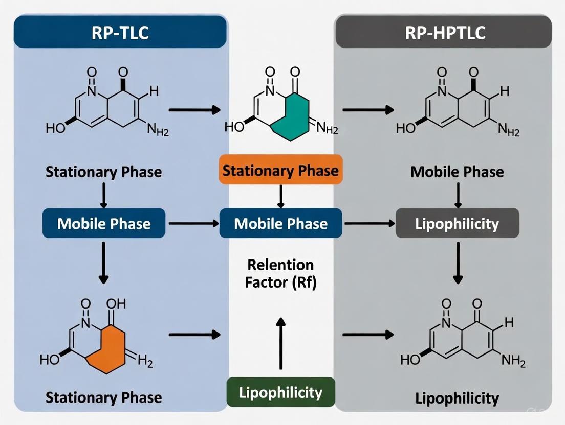
Reversed-phase thin-layer chromatography (RP-TLC) and its high-performance counterpart (RP-HPTLC) represent sophisticated planar chromatographic techniques where the stationary phase is non-polar and the mobile phase is polar, operating on the principle of hydrophobic interactions [16]. This reversal of the classical normal-phase mode makes these techniques particularly suited for the separation and analysis of moderate to non-polar compounds, with extensive applications in pharmaceutical analysis, natural product chemistry, and notably, in the assessment of molecular lipophilicity—a critical parameter in drug design and development [17] [18]. Lipophilicity, quantified as the partition coefficient (log P), profoundly influences a drug candidate's absorption, distribution, metabolism, excretion, and toxicity (ADMET) properties [17]. RP-TLC and RP-HPTLC have emerged as powerful, reliable, and cost-effective tools for modeling this crucial property, offering significant advantages over traditional shake-flask methods and other chromatographic techniques [17] [19].
Core Principles and Theoretical Foundation
The Reversed-Phase Mechanism
In RP-TLC, the separation is governed by the differential partitioning of analytes between a non-polar stationary phase and a polar mobile phase [16]. Common stationary phases include silica gel chemically bonded with long-chain alkyl groups such as C18 (octadecylsilane) or C8 [20] [18]. The mobile phase typically consists of water mixed with a water-miscible organic modifier like methanol, acetonitrile, or tetrahydrofuran [16] [21].
The fundamental retention parameter in all planar chromatography is the retardation factor (Rf), calculated as: Rf = (distance traveled by solute) / (distance traveled by solvent front) [20] [16]. In reversed-phase systems, less polar (more lipophilic) compounds have a stronger affinity for the non-polar stationary phase and thus migrate more slowly, resulting in lower Rf values. Conversely, more polar (hydrophilic) compounds are more readily eluted by the polar mobile phase, yielding higher Rf values [16]. This inverse relationship forms the basis for using RP-TLC to assess lipophilicity.
Correlation with Lipophilicity
The connection between chromatographic retention and lipophilicity is established through the relationship between the Rf value and the partition coefficient (log P). The Rf value is transformed into the RM value, which is linearly related to log P [17] [19]: RM = log ( (1 / Rf) - 1 )
In practice, a solute's RM value is determined using mobile phases with varying concentrations of the organic modifier (e.g., methanol or acetone in water). The RM value is then extrapolated to 0% organic modifier (referred to as RM0 or RMW) to estimate the partition coefficient in a pure aqueous environment, which correlates directly with the solute's lipophilicity [17]. This chromatographically-determined lipophilicity parameter has been shown to be a excellent predictor of a molecule's behavior in biological systems [17] [19].
Comparative Advantages of RP-TLC and RP-HPTLC
Advantages Over Other Lipophilicity Assessment Methods
Table 1: Comparison of Lipophilicity Assessment Methods
| Method | Key Advantages | Key Limitations | Suitability for Lipophilicity |
|---|---|---|---|
| Shake-Flask (classical log P) | Considered the "gold standard" [19] | Labor-intensive, time-consuming, requires high purity compounds [17] [19] | Direct measurement, but impractical for high-throughput |
| RP-TLC | High throughput, low cost, minimal sample prep, green (low solvent use) [17] [18] [19] | Lower efficiency than HPLC | Excellent for initial screening and compounds with a wide range of lipophilicity [17] |
| RP-HPTLC | Higher resolution, better accuracy, validated & cGMP-compliant, superior sensitivity [22] [21] | Higher cost than TLC | Ideal for precise quantification and regulatory analysis [22] |
| Computational (in silico log P) | Very fast, no lab resources required | Accuracy varies, dependent on algorithms and training sets [17] | Good for preliminary screening, requires experimental validation |
Advantages Over Normal-Phase Planar Chromatography
For lipophilicity assessment, RP-TLC is fundamentally superior to normal-phase TLC (NP-TLC). In NP-TLC, the stationary phase is polar (e.g., silica gel) and the mobile phase is non-polar. This setup is ideal for separating compounds based on polarity, but it does not effectively model the partitioning between aqueous and lipid environments, which is the definition of lipophilicity. The reversed-phase system directly mimics the n-octanol/water system used in the classical log P determination, making it a more relevant and accurate model for this key physicochemical property [17] [19].
Advantages Over Column Chromatography (HPLC)
While RP-HPLC is also widely used for lipophilicity assessment, RP-TLC/HPTLC offers several distinct benefits:
- Higher Sample Throughput: Multiple samples and standards can be run simultaneously on a single plate under identical conditions, drastically increasing analytical capacity [18] [23].
- Lower Solvent Consumption: Mobile phase consumption is remarkably low, making it an environmentally friendly ("green") and cost-effective technique [18] [19].
- Flexibility in Detection: The off-line nature of TLC allows for the use of destructive detection reagents (e.g., charring with sulfuric acid) and multiple detection methods on the same separation [18].
- No Sample Carryover: Since the plate is used only once, there is no risk of cross-contamination or carryover between analyses, often reducing the need for extensive sample clean-up [18].
- Superior Separation of Weakly Retained Analytes: Theoretical and experimental studies have shown that for mixtures of weakly retained analytes (low k values), TLC can provide a more regular distribution of spots and better resolution than HPLC under similar reversed-phase conditions [23].
Essential Protocols for Lipophilicity Assessment
Protocol 1: Determining Lipophilicity (RMW) via RP-TLC
This protocol is adapted from studies assessing the lipophilicity of antiparasitic, antihypertensive, and non-steroidal anti-inflammatory drugs [17].
Principle: The retention parameter (RM) of a compound is determined in several methanol-water mobile phases. The RM value is then extrapolated to 0% methanol to obtain RMW, which serves as a chromatographic descriptor of lipophilicity.
Materials & Reagents:
- Stationary Phase: RP-18W HPTLC plates (e.g., Merck) [17] [23].
- Mobile Phase: Methanol and water mixtures (e.g., 50:50, 60:40, 70:30, 80:20 v/v). Prepare by accurate volumetric mixing of HPLC-grade solvents.
- Sample Solutions: Dissolve test compounds in a suitable solvent (e.g., methanol) at a concentration of ~1 mg/mL [17].
- Equipment: TLC chamber, micropipettes or capillaries, UV lamp or densitometer.
Procedure:
- Plate Preparation: Pre-wash the RP-18 plates by developing with methanol to the top. Activate by heating at 120°C for 20-30 minutes if necessary [24].
- Sample Application: Using a micropipette, apply 0.5-2 µL of each sample solution as small spots (~2 mm diameter) on a pencil baseline 1.0 cm from the bottom edge. Maintain adequate spacing between spots.
- Chamber Equilibration: Pour the mobile phase (e.g., methanol-water 60:40) into the TLC chamber to a depth of 0.5 cm. Place a filter paper wick along the chamber wall and seal it for 20 minutes to saturate the atmosphere [24].
- Chromatogram Development: Place the spotted plate in the saturated chamber and develop until the solvent front has migrated 6-8 cm from the origin.
- Detection & Calculation: Mark the solvent front, dry the plate, and visualize spots under UV light (254 nm) or by appropriate derivatization. For each compound and each mobile phase composition:
- Measure the distance from the origin to the center of the spot.
- Calculate the Rf value.
- Calculate the RM value using the formula: RM = log ( (1 / Rf) - 1 ) [17].
- Data Analysis: Plot the RM values for each compound against the volume fraction of methanol (φ) in the mobile phase. The y-intercept (RM at φ = 0), denoted RMW, is the chromatographic lipophilicity index [17].
Protocol 2: Stability-Indicating Assay via RP-HPTLC
This protocol is based on a validated method for the quantitation of flibanserin in pharmaceutical dosage forms, demonstrating the application of RP-HPTLC for precise analysis [21].
Principle: A green RP-HPTLC method is used to separate an active pharmaceutical ingredient from its degradation products formed under stress conditions (e.g., acid, base, oxidation), allowing for the specific quantification of the intact drug.
Materials & Reagents:
- Stationary Phase: HPTLC silica gel 60 RP-18 F254 plates (e.g., Merck).
- Mobile Phase: Acetone/water (80:20, v/v) – a green solvent system [21].
- Standard and Sample Solutions: Prepared in methanol or a mobile phase-compatible solvent.
- Equipment: HPTLC system with automatic applicator (e.g., Linomat 5), horizontal developing chamber (e.g., HDC 2), TLC scanner (e.g., TLC Scanner 4), visionCATS software.
Procedure:
- Plate Pre-conditioning: Pre-wash the HPTLC plates with the mobile phase and dry thoroughly.
- Precision Application: Using an automatic applicator, apply standards and samples as 8 mm bands onto the HPTLC plate (8-10 mm from the bottom edge). The application volume for quantification is typically 100-1600 ng/band [21].
- Development: Develop the plate in a twin-trough chamber or automatic developing chamber (ADC 2) pre-saturated with the acetone/water (80:20) mobile phase for 20 minutes. The development distance is typically 70-80 mm.
- Densitometric Analysis: After development and drying, scan the plate at the appropriate wavelength (e.g., 204 nm for flibanserin) using a deuterium lamp in the reflectance mode [21].
- Validation & Quantification:
- Construct a calibration curve by plotting peak area against the concentration of the standard.
- Determine the concentration of the analyte in the sample solutions from the calibration curve.
- Validate the method for linearity, accuracy, precision, robustness, and specificity as per ICH guidelines [21].
The Scientist's Toolkit: Key Research Reagents and Materials
Table 2: Essential Materials for RP-TLC/RP-HPTLC Research
| Item | Function/Description | Common Examples & Notes |
|---|---|---|
| Reversed-Phase Plates | The non-polar stationary phase for separation. | RP-18 (C18): Most common, highest hydrophobicity. RP-8 (C8): Moderate hydrophobicity. RP-2: Lowest hydrophobicity. Plates with F254 indicator allow UV visualization [20] [18]. |
| Organic Modifiers | Component of the mobile phase to adjust strength/selectivity. | Methanol: Common, good solvating power. Acetonitrile: Strong eluent, different selectivity. Acetone: Used in "green" methods [16] [21]. |
| Application Instrument | Precisely deposits sample onto the plate. | Microcapillaries: For manual, low-cost spotting. Automatic Applicators (e.g., Linomat): For precise, reproducible band application essential for HPTLC quantification [18]. |
| Development Chamber | Controlled environment for chromatographic development. | Twin-Trough Chamber: Allows saturation and small solvent volumes. Automatic Developing Chamber (ADC): Provides full control over development conditions [18]. |
| Detection/Documentation | Visualizes and records separated analyte zones. | UV Cabinet (254/366 nm): For UV-active/fluorescent compounds. TLC Densitometer: For in-situ scanning and quantification. Derivatization Reagents: (e.g., anisaldehyde-sulfuric acid) for chemical visualization [18] [16]. |
Critical Applications in Pharmaceutical Research
Lipophilicity Profiling of Drug Candidates
RP-TLC is extensively used for the high-throughput determination of lipophilicity for series of drug candidates. A key study demonstrated its utility by assessing antiparasitic drugs (metronidazole, ornidazole), antihypertensive drugs (nilvadipine, felodipine), and NSAIDs (mefenamic acid, ketoprofen) [17]. The RMW parameter obtained was found to be a robust alternative to traditional log P for predicting biological activity and ADMET properties in QSAR studies [17]. The technique is particularly valuable in the early stages of drug discovery where rapid profiling of numerous compounds is required.
Stability-Indicating Methods and Forced Degradation Studies
RP-HPTLC excels in the analysis of pharmaceutical dosage forms, especially for stability testing. The method for flibanserin is a prime example, where it successfully separated the drug from its degradation products formed under various stress conditions [21]. This application highlights a key advantage of the planar format: the entire sample, including any irreversibly adsorbed degradation products or impurities, remains on the plate and can be visualized, providing a complete picture of the sample's composition [18].
Green Analytical Chemistry
The move towards sustainable laboratory practices has increased the adoption of RP-HPTLC methods that utilize green solvent systems, such as acetone-water mixtures [21]. These methods have been assessed using metrics like the AGREE analytical scale and demonstrate that it is possible to achieve high-performance analysis while minimizing environmental impact and toxicity, without compromising the reliability required for pharmaceutical analysis [21].
Workflow and Decision Pathway
The following diagram illustrates the logical workflow for developing and executing an RP-TLC/HPTLC method for lipophilicity assessment, integrating the core principles and protocols discussed.
Figure 1: RP-TLC Lipophilicity Assessment Workflow. This flowchart outlines the sequential steps for determining the lipophilicity of compounds using the RP-TLC technique, from initial setup to final data analysis.
RP-TLC and RP-HPTLC stand as robust, versatile, and highly efficient analytical techniques uniquely positioned for modern pharmaceutical research, particularly in the critical area of lipophilicity measurement. Their core principles, based on hydrophobic interactions in a reversed-phase mode, provide a direct and reliable correlation with the partition coefficient (log P). The distinct advantages of these methods—including unparalleled sample throughput, minimal solvent consumption, flexibility in detection, and the ability to analyze crude or impure samples—make them superior to many alternative techniques for specific applications like high-throughput lipophilicity screening and stability-indicating assays. As the field progresses, the integration of RP-HPTLC with advanced detection systems like mass spectrometry and its alignment with green chemistry principles will further solidify its role as an indispensable tool in the drug development pipeline.
In the field of modern pharmaceutical research, lipophilicity stands as a fundamental physicochemical property that significantly influences the absorption, distribution, metabolism, excretion, and toxicity (ADMET) profiles of drug candidates [13]. As a rapid and cost-effective analytical technique, Reverse-Phase Thin-Layer Chromatography (RP-TLC) and its high-performance counterpart (RP-HPTLC) have emerged as powerful tools for lipophilicity assessment [13] [25]. These methods rely on key chromatographic parameters—Rf, RM, and the extrapolated RMW—which provide a quantitative basis for understanding molecular behavior and predicting biological activity. This application note details the theoretical foundations, experimental protocols, and practical applications of these parameters within the context of lipophilicity measurement research, providing researchers and drug development professionals with a standardized framework for implementation.
Theoretical Foundations of Key Parameters
The Retention Factor (Rf)
The Retention Factor (Rf) is a dimensionless parameter that represents the relative distance a compound travels compared to the solvent front in a chromatographic system [26] [20]. It is calculated using the formula: [ R_f = \dfrac{\text{distance traveled by the compound}}{\text{distance traveled by the solvent front}} ] The Rf value always lies between 0 and 1, where a value of 0 indicates the compound remains at the origin (very polar) and a value of 1 indicates the compound migrates with the solvent front (very non-polar) [26]. For optimal results, a desirable Rf value lies between 0.3 and 0.7 [27]. The Rf value is primarily used for compound identification, purity assessment, and monitoring reaction progress [27].
The Hydrophobicity Parameter (RM)
The RM value is a derivative parameter specifically used in reversed-phase chromatography to assess compound hydrophobicity [13] [25]. It is calculated from the Rf value using the formula: [ RM = \log \left( \frac{1 - Rf}{R_f} \right) ] Unlike Rf, the RM value increases with increasing compound hydrophobicity [13]. This linearizing transformation provides a more convenient parameter for establishing quantitative structure-activity relationships (QSARs).
The Extrapolated Lipophilicity Descriptor (RMW)
The RMW is an extrapolated lipophilicity parameter derived from the relationship between RM values and the concentration of organic modifier in the mobile phase [13] [25]. In practice, the RM value is determined for a compound using several mobile phases containing different concentrations of organic modifier (e.g., methanol, acetonitrile, 1,4-dioxane). The RM values are then plotted against the concentration of the organic modifier. The resulting relationship is often linear, described by the equation: [ RM = R{MW} + bC ] where C is the concentration of the organic modifier, b is the slope of the regression line, and RMW is the intercept on the RM axis, corresponding to the theoretical RM value in pure water [13]. This RMW value is interpreted as a chromatographic representation of the partition coefficient (log P) and serves as a reliable experimental measure of a compound's intrinsic lipophilicity [13].
The logical and experimental workflow connecting these three parameters is outlined in the following diagram:
Experimental Protocols
Protocol 1: Determining the Rf Value
Principle: The Rf value is the fundamental measurement in TLC, representing the relative migration of a compound between the stationary and mobile phases [26] [20].
Materials:
- Stationary Phase: TLC or HPTLC plates (e.g., silica gel, RP-18, RP-8) [13] [20] [28]
- Mobile Phase: Appropriate solvent system based on compound polarity
- Sample: Compound dissolved in a volatile, non-polar solvent (e.g., dichloromethane) [27]
- Equipment: TLC chamber, capillary micropipettes, UV lamp, or other visualization methods
Procedure:
- Plate Preparation: Mark a baseline about 1 cm from the bottom edge of the TLC plate with a pencil. Do not touch the surface of the plate [20].
- Sample Application ("Spotting"): Using a capillary micropipette, apply a small spot of the sample solution onto the baseline. Keep the spot as small as possible (1-2 mm diameter) to prevent diffusion [27].
- Chromatogram Development ("Running"): Place the TLC plate in a saturated TLC chamber containing the mobile phase. The mobile phase level must be below the baseline. Allow the solvent to ascend via capillary action until it is about 0.5 cm from the top of the plate [26] [27].
- Visualization and Measurement: Immediately upon removing the plate from the chamber, mark the solvent front with a pencil. Visualize the spots under UV light or using an appropriate derivatization agent. Measure the distance from the baseline to the center of the spot (compound) and from the baseline to the solvent front [26] [20].
- Calculation: Calculate the Rf value using the formula provided in Section 2.1.
Protocol 2: Determining RM and RMW for Lipophilicity Assessment
Principle: This protocol uses Reverse-Phase (RP) TLC/HPTLC with multiple mobile phase compositions to determine the RMW value, a key descriptor for lipophilicity [13] [25].
Materials:
- Stationary Phase: Reversed-phase plates (e.g., RP-18F254, RP-8F254, RP-2F254) [13] [28]
- Mobile Phase: Aqueous solutions of organic modifiers (e.g., acetone, acetonitrile, methanol, 1,4-dioxane). Prepare a series of at least 5-6 different concentrations for each modifier [13].
- Standard and Test Compounds: Pure samples of the compounds of interest.
- Equipment: HPTLC chamber, automatic sample applicator (e.g., CAMAG LINOMAT V), densitometer (e.g., CAMAG TLC Scanner) [29].
Procedure:
- Sample Application: Using an automatic applicator, apply standard and test samples as bands (e.g., 6 mm width) on the RP-TLC/HPTLC plate. Application positions should be at least 15 mm from the sides and 10 mm from the bottom [29].
- Chromatogram Development: Develop the plate in a pre-saturated twin-trough chamber with different mobile phases, each containing a specific volume fraction of organic modifier (e.g., 50%, 55%, 60%, 65%, 70% methanol in water). Develop until the solvent front migrates a fixed distance (e.g., 7 cm) [29].
- Densitometry Scanning: After development and drying, scan the plate using a densitometer in absorbance mode at a suitable wavelength (e.g., 276 nm) [29].
- Rf and RM Calculation: For each compound and each mobile phase composition, determine the Rf value and calculate the corresponding RM value.
- RMW Extrapolation: For each compound, plot the RM values against the concentration (C, % v/v) of the organic modifier. Perform linear regression analysis. The y-intercept (where C=0, representing pure water) is the RMW value [13].
Data Presentation and Analysis
Table 1: Key chromatographic parameters for lipophilicity assessment.
| Parameter | Definition | Calculation | Application | Range/Properties |
|---|---|---|---|---|
| Rf | Retention Factor | ( R_f = \frac{\text{distance traveled by compound}}{\text{distance traveled by solvent}} ) [26] [20] | Compound identification, purity assessment, monitoring reaction progress [27] | 0 to 1; Higher value = less polar [20] |
| RM | Hydrophobicity Parameter | ( RM = \log \left( \frac{1 - Rf}{R_f} \right) ) [13] [25] | Intermediate calculation for RMW; QSAR studies | -∞ to +∞; Higher value = more hydrophobic [13] |
| RMW | Extrapolated Lipophilicity Descriptor | Intercept of RM vs. organic modifier concentration plot (( RM = R{MW} + bC )) [13] | Primary lipophilicity index correlating with log P; ADMET prediction [13] [25] | Correlates with octanol-water partition coefficient (log P) |
Exemplary Experimental Data
The following table illustrates typical results from a lipophilicity study of neuroleptics using RP-TLC, demonstrating the relationship between mobile phase composition, RM values, and the final RMW.
Table 2: Exemplary RM data for a neuroleptic compound (e.g., Fluphenazine) on RP-18 plates with methanol-water mobile phases, and the resulting RMW value. (Data adapted from [13])
| Organic Modifier (Methanol) Concentration (% v/v) | Experimental Rf Value | Calculated RM Value |
|---|---|---|
| 50 | 0.25 | 0.48 |
| 55 | 0.35 | 0.27 |
| 60 | 0.46 | 0.07 |
| 65 | 0.58 | -0.14 |
| 70 | 0.68 | -0.33 |
| RMW (Extrapolated) | --- | 1.02 |
The Scientist's Toolkit: Essential Research Reagents and Materials
Successful determination of Rf, RM, and RMW relies on the selection of appropriate materials. The table below lists key solutions and their functions.
Table 3: Key Research Reagent Solutions for RP-TLC/HPTLC Lipophilicity Studies.
| Item | Function/Description | Examples/Specifications |
|---|---|---|
| RP-TLC/HPTLC Plates | The stationary phase for reversed-phase separation. | RP-18, RP-8, RP-2 plates (e.g., from Merck) [13] [28]. RP-18 is most common for lipophilicity. |
| Organic Modifiers | Component of the mobile phase to modulate retention. | Acetone, Acetonitrile, Methanol, 1,4-Dioxane [13]. Choice affects selectivity and RMW correlation. |
| Sample Solvent | Volatile solvent to dissolve and apply samples. | Dichloromethane, Methanol [27]. Should be volatile and not interfere with the mobile phase. |
| Visualization Agents | To detect compounds that are not UV-active. | Iodine vapor, vanillin/sulfuric acid spray, ninhydrin for amino acids [20]. |
| Calibration Standards | Compounds with known lipophilicity for method validation. | Often a homologous series (e.g., alkyl phenyl ketones) or standard drugs with known log P values. |
The chromatographic parameters Rf, RM, and RMW form a critical triad for the efficient and reliable assessment of molecular lipophilicity using RP-TLC and RP-HPTLC techniques. The RMW value, in particular, serves as a robust experimental descriptor that correlates well with the traditional octanol-water partition coefficient (log P), a cornerstone of pharmaceutical research and QSAR modeling [13] [25]. The protocols and guidelines outlined in this application note provide a clear pathway for researchers to generate high-quality, reproducible data, thereby accelerating drug candidate design and development by enabling more accurate predictions of ADMET properties early in the discovery pipeline.
Practical Guide: Running and Applying RP-TLC/HPTLC Lipophilicity Assays
In Reversed-Phase Thin-Layer Chromatography (RP-TLC) and Reversed-Phase High-Performance Thin-Layer Chromatography (RP-HPTLC), the selection of an appropriate stationary phase is fundamental for achieving optimal separation, particularly in lipophilicity measurement research. The stationary phase serves as the non-polar, hydrophobic component in the chromatographic system, interacting differentially with analytes based on their chemical affinities. Among the various chemically bonded silica gel stationary phases available, RP-2 (dimethyl), RP-8 (octyl), and RP-18 (octadecyl) represent a series of increasing hydrocarbon chain length and surface coverage, resulting in progressively stronger hydrophobic character. These phases are characterized by siloxane bonds (Si-O-Si-R) where the R group represents hydrocarbon chains of differing lengths chemically bonded to the silica gel support matrix. The growing use of instrumental TLC and HPTLC has enabled these techniques to meet stringent guidelines for validated analytical methods in current Good Laboratory Practice (cGLP) and current Good Manufacturing Practice (cGMP), making them invaluable in pharmaceutical and environmental research where lipophilicity data is critical [18].
Comparative Characteristics of RP-2, RP-8, and RP-18
Structural and Chemical Properties
The fundamental difference between RP-2, RP-8, and RP-18 stationary phases lies in the length of their alkyl chains and the consequent degree of hydrophobicity. RP-2 phases feature short dimethylsilyl (C2) chains, offering the lowest hydrophobic character due to minimal carbon content. RP-8 phases contain octyl (C8) chains, providing intermediate hydrophobicity, while RP-18 phases with their octadecyl (C18) chains present the longest alkyl ligands and highest hydrophobicity among the three. This structural variation directly impacts their retention mechanisms, with longer chains providing more extensive hydrophobic interaction sites for non-polar compounds. The silica gel support particle size also differs between conventional TLC (10-15 μm) and HPTLC (5-6 μm), with the latter providing better separation efficiency and lower detection limits [18] [30].
Lipophilicity Assessment Performance
Recent research has systematically evaluated the performance of these stationary phases for lipophilicity assessment. A comprehensive study investigating eight cephalosporin antibiotics on different stationary phases revealed distinct retention behaviors across RP-2, RP-8, and RP-18 phases using both methanol-water and acetone-water mobile phases. The retention behavior of analyzed molecules was defined by the RM0 constant, which represents the theoretical RM value at 0% organic modifier, obtained by extrapolation from measurements at multiple mobile phase compositions [31].
Table 1: Comparison of RM0 Lipophilicity Parameters for Cephalosporins on Different Stationary Phases
| Stationary Phase | Chain Length | Hydrophobicity | Typical RM0 Range (Cephalosporins) | Retention Strength |
|---|---|---|---|---|
| RP-2 | C2 (Short) | Low | Lower RM0 values | Weakest |
| RP-8 | C8 (Intermediate) | Medium | Intermediate RM0 values | Moderate |
| RP-18 | C18 (Long) | High | Higher RM0 values | Strongest |
The study concluded that RP-8, CN, and RP-18 plates were appropriate stationary phases for lipophilicity investigation, with similar retention behaviors observed on RP-18, RP-8, and CN stationary phases. This similarity suggests that for many applications, RP-8 phases may provide optimal balance between retention strength and analysis time [31]. Another investigation on neuroleptics further confirmed the utility of all three phases while highlighting the superior correlation between experimental data and computational models for RP-8 and RP-18 systems [13].
Experimental Protocols for Lipophilicity Assessment
Standardized Methodology for RM0 Determination
The following protocol details the experimental procedure for determining lipophilicity parameters using RP-2, RP-8, and RP-18 stationary phases, adapted from multiple research studies [31] [13] [17].
Materials and Equipment
- Stationary Phases: Commercial HPTLC plates precoated with RP-2F254, RP-8F254, and RP-18F254 (e.g., from Merck)
- Mobile Phase: Binary mixtures of water with organic modifiers (methanol, acetone, or acetonitrile) in varying proportions (typically 30-80% organic modifier)
- Application Device: Automated applicator (e.g., CAMAG Automatic TLC Sampler 4) or manual microcapillary pipettes
- Development Chamber: Saturated glass twin-trough chamber or automatic developing chamber (e.g., CAMAG ADC 2)
- Detection System: TLC/HPTLC scanner with UV/Vis detection at appropriate wavelengths
- Data Analysis: Software for calculating RM values and statistical analysis
Procedure
Sample Preparation: Prepare standard solutions of analytes (approximately 1 mg/mL) in appropriate solvents (methanol or acetone).
Plate Preconditioning: If necessary, precondition plates by developing with methanol and drying at room temperature.
Sample Application: Apply samples as bands (6 mm length) 10 mm from the bottom edge of the plate using an automated applicator or manual pipettes. Maintain application rate of 150 nL/s.
Mobile Phase Preparation: Prepare mobile phases with varying proportions of organic modifier (e.g., 30%, 40%, 50%, 60%, 70%, 80% methanol-water or acetone-water). Mix thoroughly and degas if necessary.
Chromatogram Development: Saturate development chamber with mobile phase vapor for 20-30 minutes. Develop plates by ascending technique to a distance of 70 mm from the application position under controlled temperature conditions (22±2°C).
Plate Drying: Dry developed plates completely using a stream of warm air (hair dryer) or in a plate heater.
Detection: Detect analyte zones under UV light at 254 nm or 366 nm, or using appropriate derivatization reagents.
Data Recording: Document chromatograms digitally and measure migration distances of solvent front and analyte zones.
Calculation: Calculate RM values using the formula: RM = log(1/RF - 1), where RF = (distance traveled by analyte)/(distance traveled by solvent front). Determine RM0 values by linear extrapolation to 0% organic modifier.
Data Analysis and Interpretation
The Soczewiński-Wachtmeister equation forms the theoretical basis for lipophilicity determination: RM = RM0 + bφ, where φ represents the volume fraction of organic modifier in the mobile phase. The RM0 parameter serves as the chromatographic lipophilicity index comparable to the traditional logP value. Statistical analysis including correlation studies and principal component analysis (PCA) should be performed to evaluate relationships between chromatographic data and computed logP values [31] [17].
Table 2: Optimal Chromatographic Conditions for Different Stationary Phases
| Parameter | RP-2 | RP-8 | RP-18 |
|---|---|---|---|
| Recommended Mobile Phase | Acetone-water or methanol-water | Methanol-water or acetonitrile-water | Methanol-water or acetonitrile-water |
| Organic Modifier Range | 40-80% | 30-70% | 20-60% |
| Development Time | Shorter (15-25 min) | Intermediate (20-30 min) | Longer (25-40 min) |
| Suitability | Low-mid lipophilicity compounds | Mid lipophilicity compounds | Mid-high lipophilicity compounds |
Research Reagent Solutions
Table 3: Essential Materials for RP-TLC Lipophilicity Studies
| Reagent/Material | Function/Application | Examples/Specifications |
|---|---|---|
| HPTLC Plates (RP-2, RP-8, RP-18) | Stationary phases with different hydrophobic characteristics | Merck RP-2F254, RP-8F254, RP-18F254; particle size 5-6 μm for HPTLC |
| Organic Modifiers | Mobile phase components for elution strength adjustment | Methanol, acetonitrile, acetone, 1,4-dioxane (HPLC grade) |
| Application System | Precise sample deposition for quantitative analysis | CAMAG Automatic TLC Sampler 4 (ATS4); 100-500 nL application volumes |
| Development Chamber | Controlled chromatogram development environment | CAMAG ADC 2; twin-trough glass chamber for saturation control |
| Detection System | Visualization and quantification of separated analytes | CAMAG TLC Scanner; UV/Vis detection at 254 nm, 275 nm, or 366 nm |
| Derivatization Reagents | Enhanced detection of non-UV absorbing compounds | Iodine vapor, sulfuric acid, ninhydrin, specific chromogenic reagents |
Applications in Pharmaceutical Research
The comparative use of RP-2, RP-8, and RP-18 stationary phases has proven particularly valuable in pharmaceutical research for lipophilicity assessment of drug candidates. Studies on diverse compound classes including cephalosporins [31], neuroleptics [13], antiparasitics, antihypertensives, NSAIDs [17], and 1,3,4-thiadiazoles [14] have demonstrated the critical importance of stationary phase selection. For cephalosporin antibiotics, comprehensive chromatographic investigation revealed similar retention behavior on RP-18, RP-8 and CN stationary phases, with RP-8, CN and RP-18 plates identified as appropriate for lipophilicity investigation [31]. Research on neuroleptics including fluphenazine, triflupromazine, and flupentixol further confirmed that chromatographic parameters obtained from RP-8 and RP-18 systems showed superior correlation with computational models compared to RP-2 [13].
The lipophilicity data obtained from these studies provides crucial information for predicting ADMET (Absorption, Distribution, Metabolism, Excretion, and Toxicity) properties of drug candidates. According to recent research, compounds with balanced lipophilic-hydrophilic character (log P 0-3) typically demonstrate optimal membrane permeability and aqueous solubility [14]. The versatility of RP-TLC with different stationary phases allows researchers to fine-tune separations for compounds across a wide lipophilicity range, making it an indispensable tool in modern drug development pipelines.
Figure 1: Decision workflow for selecting appropriate stationary phases in lipophilicity assessment studies.
The comparative evaluation of RP-2, RP-8, and RP-18 stationary phases reveals distinct advantages and applications for each in lipophilicity measurement research. RP-2 phases, with their shorter alkyl chains and lower hydrophobicity, are suitable for compounds with lower lipophilicity, providing faster analysis times but potentially less retention for highly non-polar compounds. RP-18 phases offer the strongest retention and are ideal for separating compounds with medium to high lipophilicity, though they may require stronger organic modifiers or longer development times. RP-8 phases frequently represent an optimal compromise, balancing sufficient retention for most pharmaceutical compounds with reasonable analysis times, and have demonstrated excellent correlation with computational lipophilicity models in multiple studies. The selection of an appropriate stationary phase should be guided by the specific compound characteristics, required sensitivity, and the intended application of the lipophilicity data in subsequent QSAR analyses or ADMET predictions. As planar chromatography continues to evolve with improved instrumentation and stationary phase modifications, the strategic selection from among these phases will remain fundamental to accurate lipophilicity assessment in drug development.
In reversed-phase thin-layer chromatography (RP-TLC) and high-performance thin-layer chromatography (RP-HPTLC), the selection of an organic modifier in the mobile phase is a critical parameter that directly impacts the retention, separation efficiency, and lipophilicity measurement of analytes. For researchers and drug development professionals, understanding the distinct properties of common modifiers—methanol (MeOH), acetonitrile (ACN), 1,4-dioxane, and acetone—is fundamental to designing robust chromatographic methods, particularly for determining the lipophilicity parameters of new chemical entities. Lipophilicity, expressed as log P, is a fundamental physicochemical property that influences the pharmacokinetic and pharmacodynamic profiles of therapeutic substances, guiding the selection of promising drug candidates in early development stages [32] [14]. This application note provides a structured comparison of these four organic modifiers and detailed protocols for their application in lipophilicity measurement research.
Properties and Selectivity of Organic Modifiers
The retention behavior of analytes in RP-TLC is governed by hydrophobic interactions and the solvophobic theory. The organic modifier influences this process by altering the elution strength of the mobile phase and engaging in specific molecular interactions with both the solute and the stationary phase. Selectivity changes observed when switching modifiers can be explained primarily by the modifier's interaction with the stationary phase, which subsequently affects the solute's partitioning behavior [33].
Table 1: Key Properties of Common Organic Modifiers in RP-TLC/HPTLC
| Organic Modifier | Elution Strength in RP-TLC | Hydrogen-Bonding Properties | Dipolarity/Polarizability | Primary Applications & Notes |
|---|---|---|---|---|
| Methanol (MeOH) | Moderate | Strong proton donor (acidity=0.93), strong proton acceptor (basicity=0.62) | Moderate (0.60) | - Well-suited for lipophilicity determination [14].- Provides high retention mean values (RMw) [14]. |
| Acetonitrile (ACN) | Strong | Weak proton donor (acidity=0.19), weak proton acceptor (basicity=0.31) | High (0.75) | - Common for pharmaceutical analysis [33].- Higher elution strength than methanol [34]. |
| 1,4-Dioxane | Weak | Proton acceptor only (basicity=0.55), no proton donor properties | Moderate (0.58) | - Particularly beneficial for lipophilicity estimation via HPTLC [14].- Can increase retention of proton-donor solutes relative to ACN [33]. |
| Acetone | Information Missing | Proton acceptor, weak proton donor | Information Missing | - Used in validated lipophilicity screening methods [32]. |
The data in Table 1 indicates that the retention mean values (RMw) for analytes follow the order MeOH > dioxane > acetone > ACN on both C8 and C18 stationary phases [14]. This hierarchy is crucial for predicting and manipulating analyte retention. Furthermore, the different hydrogen-bonding capabilities of each modifier are key drivers of selectivity. For instance, replacing ACN with tetrahydrofuran (THF), which has proton-acceptor properties similar to dioxane, can increase the relative retention of solutes possessing proton-donor groups [33]. This principle is directly applicable to the modifiers discussed here, where a switch from ACN to dioxane would be expected to enhance the retention of phenolic compounds relative to non-hydrogen-bonding analytes.
Figure 1: Decision workflow for selecting an organic modifier based on the desired chromatographic outcome in RP-TLC/HPTLC.
Application in Lipophilicity Measurement
Lipophilicity is a critical parameter in drug design, influencing absorption, distribution, metabolism, and excretion (ADME) [14]. RP-TLC and RP-HPTLC are established techniques for determining the experimental lipophilicity of compounds, expressed as RMw, which is derived from the relationship between the retention factor (RM) and the concentration of the organic modifier (ϕ) [32] [14].
The relationship is described by the Soczewinski–Wachtmeister equation: RM = RMw + bϕ. Here, RMw is the extrapolated value to zero organic modifier concentration, representing the partition coefficient, and b is the slope indicating the sensitivity of retention to the modifier [14]. Experimental data shows that the choice of modifier significantly influences the derived RMw value and its correlation with computed log P values.
Table 2: Comparison of Lipophilicity Determination Efficacy Using Different Modifiers
| Organic Modifier | Stationary Phase | Key Finding in Lipophilicity Studies | Correlation with Computational log P |
|---|---|---|---|
| Methanol | C18, C8 | Provides high RMw values; suitable for a wide range of analytes [14]. | Strong correlation observed [14]. |
| Acetonitrile | C18, C8 | Lower RMw values compared to MeOH; useful for different selectivity [14]. | Strong correlation observed [14]. |
| 1,4-Dioxane | C18, C8 | Considered particularly beneficial for lipophilicity estimation via HPTLC [14]. | Information Missing |
| Acetone | RP-2, RP-8, RP-18 | Used in combination with other modifiers for neuroleptics; part of validated systems [32]. | Information Missing |
For accurate lipophilicity determination, it is recommended to use multiple modifier systems to validate the results. Studies on heterocyclic thiadiazoles found that dioxane and MeOH were particularly beneficial as organic modifiers for lipophilicity estimation, with chromatographic parameters showing good correlation with calculated values [14].
Detailed Experimental Protocols
Protocol 1: Determining Lipophilicity (RMw) of a New Chemical Entity
This protocol outlines the steps for determining the lipophilicity of a drug candidate using RP-TLC/HPTLC with different organic modifiers [32] [14].
- Principle: The retention parameter RMw, determined by extrapolating RM values to 0% organic modifier, serves as an experimental measure of lipophilicity.
Materials:
- Samples: Solutions of the test compound(s) in a volatile solvent like methanol (e.g., 1 mg/mL).
- Stationary Phases: HPTLC plates RP-18 F254, RP-8 F254.
- Mobile Phases: Binary mixtures of water with MeOH, ACN, 1,4-dioxane, and acetone. Prepare at least 5 different compositions for each modifier (e.g., for MeOH: 50%, 55%, 60%, 65%, 70% v/v).
- Equipment: Standard TLC/HPTLC development chamber, micropipette, UV lamp or scanner, and documentation system.
Procedure:
- Conditioning: Saturate the chromatographic chamber with the mobile phase for 30 minutes [35].
- Application: Spot 1-2 µL of the sample solution on the HPTLC plate, approximately 1 cm from the bottom edge.
- Development: Develop the plate in the pre-saturated chamber until the mobile phase front migrates a fixed distance (e.g., 8 cm).
- Drying: Air-dry the developed plate completely.
- Detection: Detect the spots under UV light at 254 nm or using a TLC scanner.
- Data Collection: Measure the retention factor (Rf) for each spot. Calculate RM using the formula: RM = log (1/Rf - 1).
- Analysis: For each modifier system, plot RM values against the volume fraction (ϕ) of the organic modifier in the mobile phase. Perform linear regression. The y-intercept of the resulting line is the RMw value.
Figure 2: Experimental workflow for determining the lipophilicity parameter RMw using RP-TLC/HPTLC.
Protocol 2: Simultaneous Quantification of Drugs in Spiked Human Plasma
This protocol adapts a green HPTLC method for quantifying multiple drugs, demonstrating the practical application of modifier selection for complex separations [35].
- Objective: To simultaneously quantify an antiviral drug (Remdesivir) with a co-administered antibiotic (Linezolid) and anticoagulant (Rivaroxaban) in spiked human plasma.
- Chromatographic Conditions:
- Stationary Phase: TLC silica gel aluminum plates 60 F254.
- Mobile Phase: Dichloromethane-Acetone (8.5:1.5, v/v).
- Detection: Densitometric detection at 254 nm.
- Procedure:
- Sample Preparation: Precipitate plasma proteins by mixing plasma with an organic solvent (e.g., acetonitrile). Centrifuge and collect the supernatant.
- Chromatography: Follow steps 1-6 from Protocol 1 using the specified mobile phase.
- Results: Expected retardation factors (Rf) are 0.23, 0.53, and 0.72 for Remdesivir, Linezolid, and Rivaroxaban, respectively [35]. The use of acetone in the mobile phase provides the necessary selectivity to resolve these three drugs effectively.
The Scientist's Toolkit: Essential Research Reagents
Table 3: Key Reagents and Materials for RP-TLC/HPTLC Lipophilicity Studies
| Item | Function/Description | Example from Literature |
|---|---|---|
| RP-18 F254 HPTLC Plates | Most common reversed-phase stationary phase; silica gel bonded with octadecyl chains. | Used for lipophilicity determination of neuroleptics and 1,3,4-thiadiazoles [32] [14]. |
| RP-8 F254 HPTLC Plates | Less hydrophobic alternative to C18; silica gel bonded with octyl chains. | Used for comparative lipophilicity studies to understand retention mechanisms [14]. |
| Methanol (HPLC Grade) | Versatile organic modifier with strong proton-donor and proton-acceptor capabilities. | Used as a component of the mobile phase for determining RMw [14]. |
| Acetonitrile (HPLC Grade) | High elution strength modifier with high dipolarity but weak hydrogen-bonding. | Applied in mobile phases for pharmaceutical analysis and lipophilicity screening [33] [14]. |
| 1,4-Dioxane (HPLC Grade) | Organic modifier with proton-acceptor properties and weak elution strength. | Found to be particularly beneficial for lipophilicity estimation of heterocyclic compounds [14]. |
| Acetone (HPLC Grade) | Ketone modifier used for its specific selectivity and elution properties. | Employed in mobile phases for analyzing neuroleptics and drug combinations [32] [35]. |
| Microsyringe (e.g., 100 µL) | For precise application of sample spots onto the HPTLC plate. | Used with an autosampler for accurate and reproducible sample application [35]. |
| Densitometry Scanner | Instrument for quantifying the intensity of separated bands directly on the TLC plate. | Used for detection and quantification in drug analysis and method validation [35] [36]. |
The strategic selection of methanol, acetonitrile, dioxane, or acetone as an organic modifier is a powerful tool for optimizing RP-TLC and RP-HPTLC methods, especially for determining lipophilicity in drug development. Each modifier imparts a unique combination of elution strength and molecular interactions, directly influencing retention and selectivity. Methanol and dioxane are particularly effective for lipophilicity studies, often showing strong correlations with computational models. By applying the structured protocols and decision frameworks provided in this application note, researchers can systematically develop robust, reliable, and informative chromatographic methods for profiling the physicochemical properties of new drug candidates.
Within drug discovery and development, the lipophilicity of a compound is a critical physicochemical parameter that profoundly influences its absorption, distribution, metabolism, excretion, and toxicity (ADMET) properties [17]. Reversed-Phase Thin-Layer Chromatography (RP-TLC) and its high-performance counterpart (RP-HPTLC) have emerged as robust, cost-effective, and high-throughput techniques for reliably determining the lipophilicity of biologically active compounds [17]. This protocol provides a detailed, step-by-step guide for researchers and drug development professionals to determine the lipophilicity parameter RMW using RP-TLC/RP-HPTLC. The RMW value, derived from chromatographic retention data, serves as a reliable experimental descriptor for lipophilicity, often correlating well with the traditional n-octanol/water partition coefficient (log P) [17]. The method outlined here is framed within broader thesis research on utilizing chromatographic techniques for lipophilicity measurement, emphasizing practical application and data integrity.
Principles and Theoretical Background
In RP-TLC, the classical normal-phase system is reversed: the stationary phase is non-polar (e.g., silica gel modified with C18 chains), while the mobile phase is polar, typically consisting of water and a water-miscible organic solvent such as methanol or acetonitrile [37]. Under these conditions, the retention mechanism is governed by hydrophobic interactions. Consequently, more hydrophobic compounds exhibit stronger retention on the stationary phase and migrate a shorter distance, while hydrophilic compounds migrate further [37].
The fundamental retention parameter in TLC is the Rf value, which is calculated as the ratio of the distance traveled by the compound to the distance traveled by the solvent front [38]. For lipophilicity studies, the Rf value is often transformed into the RM value using the following relationship: RM = log (1/Rf – 1)
The lipophilicity parameter RMW is obtained by extrapolating the RM values to 0% organic modifier in the mobile phase. This is achieved by measuring RM values at several different concentrations of the organic modifier and applying the Soczewiński–Wachtmeister equation: RM = RMW + Sφ, where φ is the volume fraction of the organic modifier in the mobile phase, and S is a constant [17]. A plot of RM versus φ yields a straight line, and the intercept (RMW) at φ = 0 provides the chromatographic lipophilicity index.
Materials and Equipment
The Scientist's Toolkit: Research Reagent Solutions
Table 1: Essential materials and reagents for RP-TLC/RP-HPTLC lipophilicity determination.
| Item Category | Specific Examples & Specifications | Function/Purpose in the Protocol |
|---|---|---|
| Stationary Phase | HPTLC plates precoated with silica gel 60 RP-18 F₂₅₄S [39], RP-8, RP-2 [40]. | Provides the non-polar (hydrophobic) surface for reversed-phase separation. The chain length (C18, C8, C2) affects retention and tolerance to aqueous mobile phases [40]. |
| Organic Modifiers | Acetonitrile, Methanol, Ethanol (all HPLC grade) [37] [39]. | Component of the mobile phase; modifies elution strength and polarity. Acetonitrile is often preferred for low UV detection [37]. |
| Aqueous Component | Deionized water (e.g., Milli-Q grade) [39]. | The polar component of the mobile phase. Water is the weakest eluent in RP-TLC. |
| Standard & Samples | Drug compounds (e.g., Antiparasitics, NSAIDs, Antihypertensives) [17]. | The analytes whose lipophilicity is to be determined. Must be soluble in a volatile solvent compatible with the mobile phase. |
| Application Solvent | Chloroform, Methanol [39]. | Used to dissolve samples for spotting onto the TLC plate. |
| Detection System | UV lamp (e.g., 254 nm, 366 nm) [38], Densitometer [39]. | Visualizes compounds that are UV-active or fluorescent. Densitometry allows for precise quantification of band intensity. |
| Development Chamber | Twin-trough glass chamber, Automatic Developing Chamber (ADC) [39]. | A sealed, saturated environment where the mobile phase ascends the plate via capillary action, separating the applied samples. |
Experimental Protocol
Step 1: Sample and Standard Solution Preparation
- Standard Stock Solution: Accurately weigh approximately 10 mg of the pure analytical standard of the compound under investigation. Transfer this to a 10 mL volumetric flask and dissolve it using an appropriate volatile organic solvent such as chloroform or methanol. Dilute to the mark with the same solvent to obtain a stock solution of approximately 1 mg/mL [39].
- Sample Solutions (for formulation analysis): For tablet formulations, accurately weigh and powder not less than ten tablets. Transfer a portion of the powder, equivalent to the weight of one tablet, to a volumetric flask. Add a suitable solvent (e.g., chloroform), sonicate for 10-15 minutes to facilitate extraction, and dilute to volume. Filter the solution to remove insoluble excipients [39].
- Working Solutions: Prepare a series of dilutions from the stock solution using the mobile phase or a compatible solvent to obtain a concentration range suitable for application and calibration (e.g., 50-600 ng/band) [39].
Step 2: Chromatographic Plate Preparation and Sample Application
- Plate Selection: Use commercially available RP-TLC or RP-HPTLC plates, such as glass-backed RP-18 silica gel 60 F₂₅₄S plates. If needed, cut the plate to the desired size using a plate cutter.
- Application Strategy: Mark the application line lightly with a pencil, typically 8-10 mm from the bottom edge of the plate [41]. Mark the solvent front line, 80-100 mm from the application line [39].
- Sample Application:
- Manual Application: Using a calibrated micropipette or capillary, apply the sample and standard solutions as compact, small spots (diameter ~3 mm) on the marked application line. Maintain a consistent track distance (e.g., 8.5 mm) between application points [41].
- Automatic Application: For higher precision and reproducibility, use an automatic sample applicator (e.g., CAMAG ATS4). Set the parameters such as band length (e.g., 6-8 mm), application rate (e.g., 150 nL/s), and application volume (e.g., 1-3 µL/band) [41] [39].
Step 3: Mobile Phase Selection and Chromatographic Development
- Mobile Phase Preparation: Prepare the mobile phase in a binary mixture of water and a polar organic solvent. A typical system for initial method development could be a mixture of acetonitrile and water or ethanol and water (70:30, v/v) [39]. The choice of organic modifier and its proportion will significantly impact the retention and resolution.
- Prepare at least 5-7 different mobile phase compositions with varying volume fractions (φ) of the organic modifier (e.g., 40%, 50%, 60%, 70%, 80% organic solvent in water) to enable RMW calculation [17].
- Chamber Saturation: Pour the prepared mobile phase into a twin-trough development chamber. Line the chamber walls with filter paper to aid saturation. Allow the chamber to equilibrate for at least 20-30 minutes at room temperature to ensure a uniform vapor atmosphere [39].
- Chromatographic Development: Place the spotted plate vertically into the chamber, ensuring the mobile phase level is below the application line. Close the chamber lid securely. Allow the mobile phase to ascend the plate via capillary action until it reaches the pre-marked solvent front line (e.g., 8-10 cm from the point of application) [41] [39].
- Plate Drying: After development, carefully remove the plate from the chamber and air-dry it in a fume hood or using a gentle stream of air to completely evaporate the mobile phase solvents.
Step 4: Detection and Densitometric Analysis
- Visualization: Examine the dried plate under a UV lamp at appropriate wavelengths (e.g., 254 nm or 366 nm). Circle the migrated spots/bands with a sharp pencil for identification.
- Densitometric Scanning: For precise quantification, place the plate in a TLC scanner (densitometer). Set the scanner parameters, including the wavelength (e.g., 257 nm for voriconazole [42] or 253 nm for rivaroxaban [39]), and scan each track. The densitometer will generate a chromatogram for each track, providing the Rf value and the peak area for each band.
Step 5: Data Analysis and RMW Calculation
- Calculate RM Values: For each compound and at each mobile phase composition (φ), calculate the RM value using the measured Rf value and the formula: RM = log (1/Rf – 1).
- Plot and Determine RMW: Create a table of RM values versus the volume fraction (φ) of the organic modifier for each compound.
- Perform linear regression analysis for each data set (RM vs. φ).
- The equation of the line will be of the form RM = RMW + Sφ, where the y-intercept is the RMW value (the lipophilicity parameter at zero organic modifier), and S is the slope of the line, indicating the compound's sensitivity to changes in mobile phase composition [17].
Figure 1: Workflow for RMW Determination via RP-TLC/HPTLC.
Application Notes and Data Presentation
Example Data and Calculations
Table 2: Exemplary RM data for a hypothetical compound at different methanol-water mobile phases.
| Volume Fraction of Methanol (φ) | Rf Value | RM Value [log(1/Rf - 1)] |
|---|---|---|
| 0.40 | 0.25 | 0.48 |
| 0.50 | 0.38 | 0.20 |
| 0.60 | 0.55 | -0.09 |
| 0.70 | 0.72 | -0.41 |
| 0.80 | 0.85 | -0.76 |
A plot of the RM values from Table 2 against the volume fraction of methanol (φ) yields a regression line: RM = 1.205 - 2.415φ. The R² value is 0.998. The RMW value, which is the intercept at φ = 0, is 1.205.
Troubleshooting and Best Practices
- Spot Tailing: This can be caused by overloading or secondary interactions with residual silanols on the stationary phase. To mitigate, reduce the application concentration or use a mobile phase additive like a buffer [37].
- High Rf Values (>0.9) or Low Rf Values (<0.1): The mobile phase is too strong or too weak, respectively. Adjust the ratio of organic modifier to water to bring the Rf into an optimal range (0.2-0.8) for accurate RM calculation.
- Irregular Solvent Front: This indicates improper chamber saturation. Ensure adequate saturation time and that the chamber is level and draft-free.
- Reproducibility: For robust RMW determination, it is critical to maintain consistent conditions across all runs, including chamber saturation time, development distance, and temperature. Automated equipment greatly enhances reproducibility [39].
This protocol provides a comprehensive guide for determining the lipophilicity parameter RMW using RP-TLC/RP-HPTLC. The technique is a powerful, efficient, and economical alternative to the shake-flask method for lipophilicity assessment. The generated RMW values are invaluable in quantitative structure-activity relationship (QSAR) studies and in optimizing the ADMET profiles of drug candidates during the early stages of pharmaceutical development [17]. By following this standardized protocol, researchers can generate reliable and reproducible lipophilicity data to advance their drug discovery projects.
Lipophilicity is a fundamental physicochemical property that significantly influences the absorption, distribution, metabolism, excretion, and toxicity (ADMET) profile of therapeutic substances. Within drug discovery and development, reversed-phase thin-layer chromatography (RP-TLC) and its high-performance counterpart (RP-HPTLC) provide efficient, cost-effective platforms for experimental determination of lipophilicity parameters. These techniques enable rapid profiling of drug candidates by simulating partitioning between polar mobile phases and non-polar stationary phases. This application note details specific protocols and case studies applying RP-TLC and RP-HPTLC to analyze neuroleptics, anti-inflammatories, and anti-cancer agents, providing researchers with validated methodologies for lipophilicity assessment within drug development workflows.
Application Case Studies
Analysis of Neuroleptics
Background: Neuroleptics, or antipsychotics, are a chemically diverse group of heterocyclic compounds containing nitrogen atoms, used primarily to manage psychiatric conditions such as schizophrenia and bipolar disorder. Their long-term use is associated with adverse effects, making rapid ADMET profiling crucial [32].
Experimental Lipophilicity Determination via RP-TLC: A 2025 study demonstrated the application of RP-TLC to determine the lipophilicity of five neuroleptics: fluphenazine, triflupromazine, trifluoperazine, flupentixol, and zuclopenthixol [32].
- Stationary Phases: Three silica plates with different non-polar coatings were used: RP-2F254, RP-8F254, and RP-18F254.
- Mobile Phases: Binary systems containing phosphate buffer (pH 7.4) and an organic modifier (acetone, acetonitrile, or 1,4-dioxane) were employed.
- Detection: The compounds were visualized under UV light at 254 nm.
- Lipophilicity Parameter: The chromatographic parameter RMW, derived from the relationship between the compound's retention factor (Rf) and the concentration of organic modifier, was used as a direct measure of lipophilicity (log P) [32].
Comparative Computational Analysis: The experimental RMW values were compared with computational log P predictions from multiple algorithms, including AlogPs, iLogP, XlogP3, and MlogP. This hybrid approach validated the chromatographic method and provided a comprehensive lipophilicity profile [32].
The table below summarizes the key findings from the neuroleptics study, illustrating the relationship between structure and lipophilicity.
Table 1: Experimental Lipophilicity (RMW) of Neuroleptics Determined by RP-TLC on Different Stationary Phases [32]
| Compound | Chemical Class | RP-2F254 (RMW) | RP-8F254 (RMW) | RP-18F254 (RMW) |
|---|---|---|---|---|
| Fluphenazine | Phenothiazine | 2.41 | 3.02 | 3.85 |
| Trifluoperazine | Phenothiazine | 2.35 | 2.95 | 3.78 |
| Triflupromazine | Phenothiazine | 1.98 | 2.52 | 3.30 |
| Flupentixol | Thioxanthene | 2.38 | 2.99 | 3.82 |
| Zuclopenthixol | Thioxanthene | 2.45 | 3.06 | 3.89 |
Key Findings: Thioxanthene derivatives (e.g., zuclopenthixol) exhibited slightly higher lipophilicity than phenothiazine derivatives. Triflupromazine, with its aliphatic substituent, was the least lipophilic. The RP-18 stationary phase provided the highest resolution for lipophilicity determination [32].
Analysis of Anti-Inflammatory Agents
Background: Anti-inflammatory drugs, such as Non-Steroidal Anti-Inflammatory Drugs (NSAIDs), are commonly analyzed for quality control and residue monitoring in food products. Meloxicam (MEL) is one such NSAID used in veterinary medicine.
HPTLC Protocol for Anti-Inflammatory and Antibiotic Combination: A validated HPTLC method was developed for the simultaneous quantification of meloxicam (anti-inflammatory) and florfenicol (antibiotic) in bovine tissue, demonstrating the technique's applicability in complex matrices [43].
- Stationary Phase: Aluminum HPTLC plates pre-coated with silica gel 60 F254.
- Mobile Phase: Glacial acetic acid, methanol, triethylamine, and ethyl acetate (0.05:1.00:0.10:9.00, v/v).
- Application: Samples were applied as 5 mm bands.
- Development & Detection: Plates were developed in a twin-trough chamber pre-saturated with mobile phase for 15 minutes. Densitometric detection was performed at 230 nm [43].
Method Performance: The method was validated per ICH guidelines, showing linearity for MEL in the range of 0.03–3.00 µg/band. It proved to be a reliable tool for regulatory monitoring of veterinary drug residues in edible tissues, ensuring public health and food safety [43].
Analysis of Anti-Cancer Agents
Background: The identification of bioactive compounds from natural products is a critical pathway in anti-cancer drug discovery. A major challenge is the repeated isolation of known compounds, which can be mitigated by efficient pre-isolation screening.
Integrated NMR-HPTLC Workflow for Bioactivity Screening: The "PLANTA" protocol represents an advanced integrated workflow for detecting and identifying bioactive compounds in complex natural extracts prior to isolation [44]. While not exclusively a lipophilicity study, it powerfully demonstrates the role of HPTLC in a multidimensional analytical approach relevant to anti-cancer agent discovery.
- Workflow: The protocol combines 1H NMR profiling, HPTLC separation, and bioassays with statistical correlation strategies.
- HPTLC's Role: HPTLC provides high-throughput separation and visualization of complex mixtures. A novel cross-correlation technique, Statistical Heterocovariance–SpectroChromatographY (SH-SCY), links HPTLC bands with specific NMR peaks, enhancing confidence in compound identification [44].
- Bioassay Integration: The protocol uses bioassays like the DPPH free radical scavenging assay to identify active fractions. NMR-HetCA then correlates spectral data with bioactivity trends to pinpoint active constituents [44].
Performance: In a proof-of-concept study using an artificial extract of 59 compounds, the PLANTA protocol achieved an 89.5% detection rate of active metabolites and 73.7% correct identification, streamlining the discovery process for anti-cancer and other bioactive natural products [44].
Essential Protocols
Standard RP-TLC Protocol for Lipophilicity Determination
This protocol is adapted from the neuroleptics study [32] for the determination of lipophilicity parameters (RMW) for small organic molecules.
Materials & Reagents:
- Stationary Phases: RP-TLC plates (e.g., RP-2, RP-8, RP-18 F254s).
- Mobile Phase: Phosphate buffer (e.g., pH 7.4) and organic modifiers (HPLC-grade acetone, acetonitrile, methanol, or 1,4-dioxane).
- Standard Solutions: 1 mg/mL of each analyte dissolved in a suitable solvent (e.g., methanol).
- Development Chamber: A standard twin-trough TLC chamber.
- Detection System: UV lamp or TLC scanner.
Procedure:
- Mobile Phase Preparation: Prepare a series of mobile phases with varying concentrations of organic modifier (e.g., from 40% to 70% v/v) in the aqueous buffer.
- Sample Application: Using a micropipette, apply 1-2 µL of each standard solution as spots on the baseline of the RP-TLC plate (1.0 cm from the bottom).
- Chromatographic Development: Place the plate in the chamber pre-saturated with the mobile phase for 15-30 minutes. Allow the mobile phase to ascend vertically until it reaches a predetermined mark (e.g., 8 cm from the origin).
- Plate Drying & Visualization: Air-dry the developed plate in a fume hood. Visualize the spots under UV light at 254 nm.
- Data Recording: Measure the retention factor (Rf) for each spot.
- Calculation of RMW: The RMW value is determined from the equation of the line representing the relationship between the Rf value and the volume fraction of the organic modifier in the mobile phase.
HPTLC Protocol for Quantitative Analysis in Biological Matrices
This protocol is based on the analysis of antiviral drugs in spiked human plasma [45] and is applicable for quantifying drugs in complex biological samples.
Materials & Reagents:
- Plates: HPTLC silica gel 60 F254 aluminum plates (20 x 20 cm).
- Mobile Phase: Optimized for the target analytes. Example: Dichloromethane:acetone (8.5:1.5, v/v) for remdesivir, linezolid, and rivaroxaban [45].
- Standard Solutions: Primary stock solutions (1 mg/mL) of analytes in methanol or acetonitrile.
- Biological Matrix: Human plasma.
- Instrumentation: CAMAG Linomat autosampler, TLC scanner controlled by winCATS software.
Procedure:
- Sample Preparation: Spike the biological matrix (e.g., plasma) with standard solutions. Precipitate proteins by adding a solvent like acetonitrile, vortex, and centrifuge. Collect the supernatant.
- Application: Using an autosampler (e.g., Linomat 5), apply the samples and standards as bands (e.g., 5 mm width) onto the HPTLC plate.
- Development: Develop the plate in a twin-trough chamber pre-saturated with the mobile phase for 20-30 minutes.
- Densitometry: After development and drying, scan the plate with a TLC scanner in absorbance mode at the optimized wavelength (e.g., 254 nm).
- Quantification: Construct a calibration curve by plotting the peak area against the concentration of the standard bands. Determine the concentration of analytes in the unknown samples from the calibration curve.
The Scientist's Toolkit: Research Reagent Solutions
Table 2: Essential Materials and Reagents for RP-TLC/HPTLC Lipophilicity Studies
| Item | Function/Description | Example Use Case |
|---|---|---|
| RP-18F254 HPTLC Plates | Stationary phase with C18-chain modification; offers high hydrophobicity for separating medium to high lipophilicity compounds. | Provided highest resolution for neuroleptic lipophilicity ranking [32]. |
| RP-8F254 & RP-2F254 Plates | Stationary phases with C8 and C2 modifications; less hydrophobic than C18, used for more polar compounds. | Used for comparative lipophilicity assessment of neuroleptics [32]. |
| Organic Modifiers (ACN, Acetone, 1,4-Dioxane) | Component of mobile phase to adjust elution strength and selectivity; different modifiers can affect compound interaction. | Used in binary mixtures with buffer to determine RMW values for neuroleptics [32]. |
| TLC/HPTLC Scanner | Instrument for densitometric quantification of analyte bands on the plate by measuring absorbance or fluorescence. | Enabled quantification of drugs in pharmaceutical and biological samples at µg/band levels [45] [46]. |
| Automated Sample Applicator (e.g., Linomat) | Provides precise and reproducible application of samples as narrow bands, critical for quantitative HPTLC. | Used for band-wise application in the quantification of remdesivir and meloxicam [43] [45]. |
| Twin-Trough Development Chamber | Chamber for chromatographic development; pre-saturation with mobile phase vapor ensures reproducible development conditions. | Standard equipment for development in all cited HPTLC methods [43] [45] [32]. |
Workflow and Pathway Diagrams
The following diagram illustrates the integrated workflow for bioactivity screening and lipophilicity assessment, combining elements from the PLANTA protocol [44] and standard RP-TLC practice [32].
Figure 1: Integrated workflow for bioactivity screening and lipophilicity assessment, combining TLC separation with spectroscopic and biological assays.
RP-TLC and RP-HPTLC are powerful, versatile, and cost-effective techniques for lipophilicity determination and quantitative analysis within pharmaceutical research. The case studies presented demonstrate their direct application in profiling neuroleptics, quantifying anti-inflammatories in complex matrices, and identifying potential anti-cancer agents from natural products. The provided detailed protocols and workflows offer researchers a practical framework to implement these techniques, accelerating drug discovery and development by providing critical physicochemical and bioactivity data at early stages.
Enhancing Accuracy: Troubleshooting and Advanced Optimization Strategies
Optimizing Mobile Phase Composition with Systems like the PRISMA Model
In the field of medicinal chemistry, lipophilicity is a fundamental physicochemical parameter that significantly influences a drug's absorption, distribution, metabolism, excretion, and toxicity (ADMET) profile. It is commonly expressed as the logarithm of the n-octanol/water partition coefficient (log P) [47] [9]. Reverse-phase thin-layer chromatography (RP-TLC) and reverse-phase high-performance thin-layer chromatography (RP-HPTLC) have emerged as efficient, reproducible, and cost-effective techniques for the experimental determination of lipophilicity, particularly during the early stages of drug design and development [13] [47]. The core principle involves correlating the chromatographic retention parameters of analytes with their lipophilicity, where more lipophilic compounds exhibit higher retention on non-polar stationary phases.
The accuracy and resolution of these chromatographic separations are profoundly influenced by the composition of the mobile phase. Selecting the optimal mobile phase is critical for achieving precise and reproducible lipophilicity measurements. The PRISMA model provides a systematic, three-dimensional approach for optimizing the mobile phase without requiring prior knowledge of analyte structures or properties [48] [49] [50]. This model enables researchers to methodically select and combine solvents to achieve the desired selectivity and resolution for complex mixtures, making it particularly valuable for analyzing structurally diverse neuroleptics and their derivatives [13] [48]. This application note details the integration of the PRISMA optimization system within RP-TLC and RP-HPTLC workflows dedicated to lipophilicity assessment.
The PRISMA Optimization System
Theoretical Foundation
The PRISMA model is a systematic optimization strategy that conceptualizes the solvent selection process as a three-dimensional geometric figure. This model allows chromatographers to generate a wide range of solvent combinations from a limited set of initial experiments and visually identify the optimal mobile phase composition [49] [50]. The system is highly versatile and has been successfully applied across various chromatographic techniques, including TLC, HPLC, and OPLC [49].
The model operates on the principle that the selectivity and eluting strength of a mobile phase can be precisely tuned by adjusting the proportions of different solvents from various selectivity groups. This is particularly crucial in lipophilicity measurement, where the goal is to achieve adequate separation between the compound of interest and any impurities to accurately determine its retention factor (R₍F₎) or the derived lipophilicity parameter (R₍MW₎) [13] [9].
The Three-Step Workflow
The PRISMA optimization process is structured into three distinct stages:
Step 1: Selection of Basic Parameters. This initial stage involves choosing the stationary phase, the vapor phase, and a set of individual solvents from different selectivity groups. These solvents should exhibit a range of chemical properties (e.g., proton-donor, proton-acceptor, dipole-dipole interactions) to cover a broad selectivity space [50].
Step 2: Solvent Combination Optimization using the PRISMA Model. In this core stage, the solvent system is optimized by determining the ideal combination of selected solvents. The process involves defining the solvent strength, adjusting selectivity, and fine-tuning the final ratio, often visualized through a three-dimensional prism [49] [50].
Step 3: Selection of Chromatographic Mode and Technique. The final stage involves choosing the appropriate development mode (e.g., linear, circular) and transferring the optimized mobile phase to the chosen chromatographic technique, such as RP-TLC or RP-HPTLC [50].
The following workflow diagram illustrates the practical application of these stages in RP-TLC/HPTLC method development.
Experimental Protocols
Protocol 1: PRISMA Method for General RP-TLC Mobile Phase Optimization
This protocol is adapted from applications in radiopharmaceutical analysis [48] and stationary phase optimization [49], tailored for lipophilicity studies.
Materials and Reagents:
- Solvents: Select at least 3-4 solvents from different selectivity groups (see Table 1). Acetone, acetonitrile, and 1,4-dioxane are commonly used for neuroleptic lipophilicity screening [13].
- Stationary Phase: RP-18F₂₅₄, RP-8F₂₅₄, or RP-2F₂₅₄ TLC/HPTLC plates.
- Analytes: Standard solutions of the target compounds (e.g., neuroleptics such as fluphenazine, triflupromazine) and potential impurities.
Procedure:
- Preliminary Solvent Screening: Spot the analyte mixture on the RP-TLC plates. Develop separately with each pre-selected neat solvent. Observe the migration distance (R₍F₎ value).
- Define Solvent Strength: Identify solvents that produce R₍F₎ values between 0.2 and 0.8. These solvents have appropriate eluting strength. If a solvent is too strong (R₍F₎ > 0.8), it is diluted with n-hexane or cyclohexane to reduce strength. If too weak (R₍F₎ < 0.2), a stronger solvent is added [48] [50].
- Optimize Selectivity: Create binary and ternary mixtures of the strength-adjusted solvents. A typical approach is to create a volume ratio of 1:1:1 for a ternary mixture. Test these mixtures and evaluate the resolution (R₍s₎) between the critical pair of analytes.
- Fine-tune the Ratio: Systematically vary the proportions of the solvents in the mixture (e.g., from 10:10:80 to 80:10:10) to maximize the resolution. The optimal ratio is the one that provides baseline separation (R₍s₎ ≥ 1.5).
- Validation: Validate the optimized mobile phase by running replicates to ensure reproducibility of the R₍F₎ values.
Protocol 2: Application to Lipophilicity Determination of Neuroleptics
This specific protocol is derived from a recent study investigating phenothiazine and thioxanthene derivatives [13].
Materials and Reagents:
- Stationary Phases: RP-2F₂₅₄, RP-8F₂₅₄, and RP-18F₂₅₄ TLC plates.
- Organic Modifiers: Acetone, acetonitrile, 1,4-dioxane, and methanol of HPLC grade.
- Analytes: Standard solutions of neuroleptics (fluphenazine, triflupromazine, trifluoperazine, flupentixol, zuclopenthixol) at 1 mg/mL in methanol.
Procedure:
- Mobile Phase Preparation: Prepare mobile phases with varying volume fractions of the organic modifier (e.g., from 40% to 80% in water) for each solvent type.
- Chromatography: Apply 1 µL of each analyte solution to the RP-TLC plates. Develop the plates in a pre-saturated chamber with the respective mobile phase. Allow the solvent front to migrate 6 cm from the origin.
- Detection: Dry the plates and visualize under UV light at 254 nm.
- Data Analysis: Measure the R₍F₎ value for each spot. Calculate the lipophilicity parameter R₍MW₎ using the equation: R₍MW₎ = R₍F₎ - (k * log [organic modifier]), where k is a constant. Alternatively, plot log 1/R₍F₎ -1 against the volume fraction of the organic modifier to obtain the lipophilicity index [13].
- Correlation with Computational log P: Compare the experimental R₍MW₎ values with in silico log P predictions from various algorithms (e.g., ALogPs, iLogP, XLogP3) to validate the method [13].
Data Presentation and Analysis
Solvent Selectivity and Properties
Table 1: Common Organic Modifiers for RP-TLC Lipophilicity Measurement
| Solvent | Selectivity Group | Common Use in RP-TLC | Remarks |
|---|---|---|---|
| Acetonitrile [13] [51] | VI (Dipole) | Primary organic modifier | High eluting strength, low viscosity, UV transparent. |
| Methanol [48] [51] | II (Proton Donor) | Organic modifier, often mixed | Can significantly alter separation selectivity in HILIC. |
| Acetone [13] | VI (Dipole) | Organic modifier | Strong eluting power, useful for medium-polarity compounds. |
| 1,4-Dioxane [13] | VI (Dipole) | Organic modifier | Good for separating complex mixtures. |
| Tetrahydrofuran (THF) [48] | III (Dipole/Donor) | Secondary modifier | Strong eluting power, good for very hydrophobic analytes. |
Exemplary Lipophilicity Data
Table 2: Lipophilicity Parameters (R₍MW₎) of Neuroleptics Determined by RP-TLC with Different Mobile Phases [13]
| Compound | RP-18/ Acetone | RP-18/ Acetonitrile | RP-18/ 1,4-Dioxane | RP-8/ Acetone | Computational log P (consensus) |
|---|---|---|---|---|---|
| Fluphenazine | 2.45 | 2.51 | 2.38 | 2.40 | 3.50 - 4.80 |
| Triflupromazine | 3.10 | 3.05 | 2.95 | 3.02 | 4.50 - 5.20 |
| Trifluoperazine | 2.75 | 2.80 | 2.68 | 2.71 | 4.10 - 5.00 |
| Flupentixol | 3.25 | 3.30 | 3.18 | 3.22 | 4.20 - 5.10 |
| Zuclopenthixol | 3.55 | 3.60 | 3.45 | 3.51 | 4.80 - 5.50 |
The data in Table 2 demonstrates how the experimental lipophilicity parameter R₍MW₎ can vary with different stationary and mobile phase combinations. While the absolute values differ from in silico predictions, the relative order of lipophilicity among the compounds is consistently maintained, which is crucial for comparative QSAR studies [13]. The correlation between chromatographic R₍MW₎ and computational log P values validates RP-TLC as a reliable tool for rapid lipophilicity screening.
The Scientist's Toolkit: Research Reagent Solutions
Table 3: Essential Materials for PRISMA-Optimized RP-TLC Lipophilicity Measurement
| Item | Function/Role | Example Specifications |
|---|---|---|
| RP-TLC/HPTLC Plates | Non-polar stationary phase for reverse-phase separation. | RP-18F₂₅₄, RP-8F₂₅₄, RP-2F₂₅₄; 10x10 cm or 10x20 cm, with fluorescent indicator [13]. |
| Organic Solvents (HPLC Grade) | Components of the mobile phase; their combination dictates selectivity. | Acetone, acetonitrile, methanol, 1,4-dioxane. Stored in sealed bottles to prevent water absorption [13] [48]. |
| Chromatographic Chamber | A sealed tank for mobile phase vapor saturation and plate development. | Twin-through or linear development chamber, preferably with glass lid for saturation [48]. |
| Microsyringe or Capillary | For precise application of analyte solutions onto the TLC plate. | 1-5 µL capacity, allowing for spot diameters of 1-3 mm. |
| UV Lamp/Camera/Densitometer | For detection and quantification of separated analytes on the TLC plate. | UV light at 254 nm and 366 nm; documentation system; densitometer for in-situ scanning of spot intensity [48]. |
The PRISMA model provides a robust, systematic framework for optimizing mobile phase composition in RP-TLC and RP-HPTLC, directly enhancing the accuracy and efficiency of lipophilicity measurements for drug candidates. By methodically exploring the solvent selectivity space, researchers can develop high-resolution methods capable of separating structurally similar compounds, such as neuroleptics and their derivatives, even without prior knowledge of all impurities [13] [48]. The integration of this optimized chromatographic data with in silico predictions and topological indices creates a powerful hybrid approach for early ADMET profiling, accelerating the rational design of new therapeutic agents with desirable pharmacokinetic properties.
Within the context of lipophilicity measurement research using Reversed-Phase Thin-Layer Chromatography (RP-TLC) and Reversed-Phase High-Performance Thin-Layer Chromatography (RP-HPTLC), the selection of an organic modifier is a critical, data-driven decision. Lipophilicity, a key physicochemical property governing a compound's absorption, distribution, metabolism, and excretion (ADME), is conventionally expressed as the logarithm of the n-octanol/water partition coefficient (log P) [47]. Chromatographic techniques, particularly RP-TLC and RP-HPTLC, have emerged as mainstream experimental methods for its rapid and reproducible determination, endorsed by organizations like IUPAC [14].
The retention mechanism in reversed-phase systems is primarily based on hydrophobic interactions, where the affinity of a solute for the non-polar stationary phase versus the polar mobile phase determines its retention [14]. The organic modifier, as a component of the mobile phase, directly disrupts this equilibrium. Its type and concentration profoundly influence the retention behavior, separation selectivity, and ultimately, the accuracy of the derived lipophilicity parameter (e.g., ( R{Mw} ) or ( \log kw )) [52] [53]. This application note provides a data-driven comparison of methanol and dioxane, two commonly used modifiers, to guide researchers in making an informed selection for their lipophilicity studies.
Comparative Modifier Analysis
The choice between methanol and dioxane impacts key aspects of the chromatographic process, from the retention mechanism to the resulting lipophilicity parameters. The table below summarizes their core characteristics and performance data derived from recent studies.
Table 1: Data-Driven Comparison of Methanol and Dioxane as Organic Modifiers in RP-TLC/HPTLC
| Feature | Methanol (MeOH) | Dioxane (DIO) | Experimental Evidence |
|---|---|---|---|
| Primary Mechanism | Strong hydrogen-bond acidity and moderate basicity; reduces solvophobic interaction [52]. | High hydrogen-bond basicity; significantly disrupts hydrogen bonding networks [52]. | LSER studies show DIO has a greater effect on HBD acidity of the mobile phase [52]. |
| Retention Strength | Stronger eluent; results in lower ( R{Mw} ) / ( \log kw ) values for the same compound [14]. | Weaker eluent; results in higher ( R{Mw} ) / ( \log kw ) values [14]. | On C18 plates, the mean ( R_{Mw} ) for a set of thiadiazoles was 2.32 for MeOH and 2.95 for DIO [14]. |
| Separation Selectivity | Provides selectivity based on solute hydrogen-bond basicity [52]. | Alters selectivity by modifying the system's hydrogen-bond acidity [52]. | Selectivity changes are attributed to displacement of water from the stationary phase and altered buffer acidity/basicity [52]. |
| Linearity with ( \log P) | Correlations with log P can be excellent but are system-dependent. | Correlations with log P can be excellent but are system-dependent. | For neuroleptics, ( R_{Mw} ) from MeOH/water and DIO/water systems showed strong correlation with computational log P [13]. |
| Recommended Use | Preferred for its similarity to water and as a first-choice modifier for general lipophilicity screening [14] [53]. | Particularly beneficial for profiling polar compounds and as a complementary modifier for selectivity optimization [14]. | A study on analgesics found DIO beneficial for lipophilicity estimation, while MeOH was a standard choice [53]. |
Experimental Protocols
Protocol 1: Determining Lipophilicity (( R_{Mw} )) using a Methanol-Water Gradient
This protocol is optimized for determining the chromatographic lipophilicity parameter ( R_{Mw} ) using methanol as the organic modifier, following the Soczewiński-Wachtmeister equation [14] [53].
Principle: The retention factor (( RM )) is linearly related to the volume fraction ((\phi)) of the organic modifier in the mobile phase. Extrapolation to zero organic modifier (( R{Mw} )) provides a standardized lipophilicity index.
Materials & Reagents:
- Stationary Phase: HPTLC plates (e.g., RP-18 ( F{254} ), RP-8 ( F{254} ), or RP-2 ( F_{254} )) [13] [53].
- Mobile Phase: Pre-mixed solutions of HPLC-grade methanol and purified water. A typical series includes 50%, 60%, 70%, 80%, and 90% (v/v) methanol.
- Analytes: Standard solutions of the compounds of interest, dissolved in a suitable solvent (e.g., methanol) at a concentration of ~1 mg/mL [53].
- Equipment: Standard HPTLC system (development chamber, micropipette, UV cabinet).
Procedure:
- Plate Preparation: Cut the HPTLC plate to the required size. Lightly mark the application line and the solvent front line with a pencil.
- Sample Application: Using a micropipette, apply 1-2 µL of each analyte solution as spots on the application line. Allow the spots to dry completely.
- Chromatographic Development: Pour approximately 20 mL of the first mobile phase (e.g., 50% MeOH) into a twin-trough chamber and equilibrate for 15-20 minutes. Place the plate in the chamber and develop until the solvent front reaches the marked line.
- Drying and Detection: Remove the plate from the chamber, allow it to dry completely, and visualize the spots under UV light (e.g., at 254 nm).
- Data Recording: Measure the migration distance of the solvent front (Zf) and the center of each analyte spot (Zx). Calculate the retention factor ( RF ) (( RF = Zx / Zf )) and then derive ( RM ) using the formula: ( RM = \log[(1 - RF)/RF] ) [53].
- Replicate and Repeat: Repeat steps 3-5 for each mobile phase composition in the gradient series. Perform each analysis in triplicate to ensure reproducibility.
- Data Analysis: For each analyte, plot the ( RM ) values against the volume fraction ((\phi)) of methanol. Perform linear regression analysis. The y-intercept of the regression line (when (\phi = 0)) is the lipophilicity parameter ( R{Mw} ) [14].
Protocol 2: Complementary Profiling using a Dioxane-Water System
This protocol uses dioxane to provide an alternative lipophilicity measurement, which is particularly useful for polar compounds or for verifying results from the methanol system.
Materials & Reagents:
- Stationary Phase: HPTLC plates (e.g., RP-18 ( F_{254} )) [14].
- Mobile Phase: Pre-mixed solutions of 1,4-dioxane and purified water. A typical series includes 40%, 50%, 60%, 70%, and 80% (v/v) dioxane.
- Analytes & Equipment: As described in Protocol 1.
Procedure:
- Plate and Sample Preparation: Follow steps 1 and 2 from Protocol 1.
- Development with Dioxane: Use the prepared dioxane-water mobile phases for chromatographic development. Due to the different eluotropic strength of dioxane, the concentration range used typically starts lower than that for methanol [14].
- Detection and Data Recording: Follow steps 4 and 5 from Protocol 1 to calculate the ( R_M ) value for each analyte in each dioxane concentration.
- Data Analysis: Plot ( RM ) versus the volume fraction of dioxane for each analyte and perform linear regression. The y-intercept is the ( R{Mw(DIO)} ) parameter.
The Scientist's Toolkit
Table 2: Essential Research Reagents and Materials for RP-TLC/HPTLC Lipophilicity Measurement
| Item | Function/Description | Application Note |
|---|---|---|
| RP-18 HPTLC Plates | Silica gel plates bonded with octadecylsilyl (C18) groups; the most hydrophobic common stationary phase. | Provides a standardized non-polar surface for partitioning; the most frequently used phase for log P correlation [14] [13]. |
| RP-8 HPTLC Plates | Silica gel plates bonded with octyl (C8) groups; less hydrophobic than C18. | Useful for retaining very hydrophobic compounds or when a wider retention window is needed [14] [53]. |
| Methanol (HPLC Grade) | Polar protic solvent; strong hydrogen-bond donor and acceptor. | The primary organic modifier for general lipophilicity screening due to its strong elution strength and similarity to water [14]. |
| 1,4-Dioxane (HPLC Grade) | Polar aprotic solvent; strong hydrogen-bond acceptor. | A complementary modifier for selectivity optimization, especially for polar compounds; yields higher R_Mw values [14] [13]. |
| Microsyringe / Capillary | Precise application of sample solutions onto the TLC plate. | Ensures small, uniform initial spot size, which is critical for achieving high-resolution separation and accurate R_F measurement. |
| UV Cabinet (254 nm/366 nm) | For visualization of UV-active compounds after chromatographic development. | Non-destructive detection method; allows for quantification of migration distances [53]. |
The selection between methanol and dioxane is not a matter of identifying a single "best" modifier, but of choosing the most appropriate tool for a specific research goal. Methanol, with its stronger elution power and proven performance, serves as an excellent first choice for general lipophilicity screening and ranking of drug candidates. Dioxane, yielding higher ( R_{Mw} ) values and offering different selectivity, is a powerful complementary tool, particularly valuable for profiling polar compounds or when verifying results from a single-modifier system.
A robust lipophilicity research strategy should employ both modifiers. The correlation of the resulting chromatographic parameters (( R{Mw(MeOH)} ) and ( R{Mw(DIO)} )) with each other and with calculated log P values provides a comprehensive and data-driven profile of the compounds' hydrophobic character, thereby enhancing the reliability of research outcomes in drug development [14] [13].
In drug discovery and development, lipophilicity is a fundamental physicochemical parameter that significantly influences a compound's absorption, distribution, metabolism, excretion, and toxicity (ADMET) profile [9]. It is conventionally expressed as the logarithm of the n-octanol/water partition coefficient (log P) or the distribution coefficient (log D) at a specific pH [47]. Reversed-phase thin-layer chromatography (RP-TLC) and reversed-phase high-performance thin-layer chromatography (RP-HPTLC) have emerged as powerful, reliable, and cost-effective techniques for the experimental determination of lipophilicity [54] [12]. These chromatographic methods offer significant advantages, including speed, reproducibility, insensitivity to impurities, broad dynamic range, and minimal sample requirements [47]. However, researchers often encounter analytical challenges such as spot tailing, poor resolution, and data outliers that can compromise the accuracy and reliability of lipophilicity measurements. This application note details the common pitfalls in RP-TLC/HPTLC-based lipophilicity studies and provides standardized protocols for their identification, management, and resolution, framed within the context of advanced medicinal chemistry research.
Understanding and Addressing Spot Tailing
Causes and Identification of Spot Tailing
Spot tailing manifests as a comet-like or streaked appearance of chromatographic spots, leading to inaccurate Rf calculation and imprecise lipophilicity determination. The primary causes can be categorized as follows:
- Chemical Interactions: Undesirable secondary interactions between analytes and the stationary phase are a predominant cause. For basic compounds, interactions with residual acidic silanol groups on the silica-based stationary phase can cause severe tailing [55]. Similar hydrophobic interactions can occur with highly lipophilic compounds.
- Overloading Effects: Applying excessive sample concentration or volume can saturate the stationary phase, resulting in non-linear chromatography and pronounced tailing [55]. This is particularly relevant for strong UV-absorbing compounds where concentrated solutions are often spotted.
- Mobile Phase Issues: Incorrect mobile phase pH or strength can exacerbate undesirable analyte-stationary phase interactions. The use of contaminated or degraded mobile phases can also contribute to tailing.
- Technique-Related Factors: These include improper spotting technique (damaging the stationary phase), using a sample solvent that is too strong relative to the mobile phase, and developing in an unsaturated chamber, which can cause edge effects [55].
The following workflow provides a systematic approach for diagnosing and correcting spot tailing:
Experimental Protocol for Mitigating Spot Tailing
Objective: To systematically eliminate chemical and physical causes of spot tailing in RP-TLC/HPTLC analyses.
Materials:
- RP-18 TLC or HPTLC plates (e.g., Silica gel 60 RP-18F254S [42])
- Analytical grade solvents (methanol, acetonitrile, water)
- Buffer salts (e.g., Tris, sodium acetate, acetic acid) [54]
- Horizontal or twin-trough TLC chamber
Procedure:
- Initial Condition Check:
- Sample Dilution: Prepare a series of sample dilutions (e.g., 0.1, 0.5, 1.0 mg/mL). Re-spot and develop. A significant reduction in tailing with higher dilution indicates sample overloading.
- Spotting Technique: Ensure microcapillaries or automated applicators do not scratch the plate surface. Spots should be small, compact, and dried completely before development.
Mobile Phase Optimization:
- pH Adjustment: For ionizable compounds, adjust the aqueous buffer's pH to suppress ionization. For bases, use a pH at least 2 units below their pKa (e.g., acetate buffer pH 4.8 [54]); for acids, use a pH at least 2 units above their pKa (e.g., Tris buffer pH 7.4 [54]).
- Additive Incorporation: Incorporate 0.1-0.5% (v/v) of a competing amine like triethylamine (TEA) in the mobile phase to block silanol interactions for basic compounds [55]. Note: TEA is not compatible with MS detection.
- Organic Modifier Strength: Systematically vary the concentration of the organic modifier (acetone, acetonitrile, methanol, 1,4-dioxane [54] [12]) to find the optimal strength that provides compact spots.
Stationary Phase and Chamber Considerations:
- Plate Quality: Use high-purity, end-capped RP-TLC plates to minimize residual silanol activity.
- Chamber Saturation: Pre-saturate the development chamber with mobile phase vapor for at least 20-30 minutes before plate development to achieve uniform solvent front and minimize edge effects.
Troubleshooting Poor Resolution
Fundamentals of Resolution Enhancement
Poor resolution between adjacent spots compromises the accuracy of Rf and RM measurements, which are critical for deriving the lipophilicity parameter RM0 [54]. Resolution (Rs) is a function of efficiency (N), selectivity (α), and retention (k). The diagram below illustrates the interconnected parameters affecting resolution:
Experimental Protocol for Optimizing Resolution
Objective: To achieve baseline separation of compound mixtures for reliable lipophilicity determination.
Materials:
- RP-18 TLC/HPTLC plates
- Multiple organic modifiers (methanol, acetonitrile, acetone, tetrahydrofuran) [12]
- Buffer solutions
Procedure:
- Scouting Runs with Different Modifiers:
- Prepare mobile phases with the same nominal strength (e.g., 60% organic) but different modifiers: Methanol-Water, Acetonitrile-Water, Acetonitrile-Tetrahydrofuran-Water [12].
- Develop the plate with each system. The different selectivities of these modifiers can dramatically alter the elution order and spot separation.
Fine-Tuning with Mobile Phase Gradients:
Two-Dimensional Chromatography:
- For exceptionally complex mixtures, employ 2D-TLC. Develop the plate in the first direction with one mobile phase system.
- Dry the plate thoroughly and develop at a 90° angle with a different mobile phase system of orthogonal selectivity (e.g., changing pH and modifier type). This maximizes the separation power.
Managing Outliers in Lipophilicity Data
Systematic Approach to Outlier Detection
Outliers in lipophilicity data, derived from chromatographic measurements (RM0, log PTLC), can arise from experimental error, compound-specific properties, or model limitations. A robust outlier management strategy is essential. The process involves detection, investigation, and decision-making, as outlined below:
Protocol for Outlier Investigation and Handling
Objective: To establish a consistent and scientifically defensible method for handling data points that deviate from the established lipophilicity trend.
Materials:
- Standard compounds with known log P values (e.g., acetanilide, benzophenone, anthracene) [54]
- Statistical software package (e.g., STATISTICA [54])
- In-silico log P prediction tools (e.g., ALOGPS, AClogP, XLOGP3 [54])
Procedure:
- Experimental Verification:
- Re-run the RP-TLC analysis for the suspected outlier compound in triplicate, paying meticulous attention to mobile phase preparation, spotting, and chamber conditions.
- Re-calculate the RM values and re-plot the RM vs. organic modifier concentration (φ) graph. Check for linearity (r > 0.95) [54]. A poor fit for a specific compound suggests issues with its behavior in the chromatographic system.
Structural and Physicochemical Interrogation:
- Examine the molecular structure of the outlier. Look for features that may lead to atypical interactions, such as strong hydrogen bonding donors/acceptors, the presence of charges at the mobile phase pH, or metal-complexing ability [47].
- Compare the experimental log PTLC value with multiple in-silico predictions (e.g., ALOGPS, miLogP, XLOGP3 [54]). A consistent deviation across different algorithms may validate the experimental value as a true property of the compound rather than an error.
Statistical and Correlation Analysis:
- Use statistical tests like Grubbs' test to identify a single outlier or the Dixon Q-test for a small dataset.
- In Quantitative Structure-Retention Relationship (QSRR) studies, analyze the residuals (difference between predicted and observed RM0). A compound with a standardized residual exceeding ±2.5 standard deviations warrants investigation.
- Perform principal component analysis (PCA) on ADME properties or structural descriptors [12]. An outlier compound will appear isolated from the main cluster in the scores plot, providing a visual confirmation of its unique characteristics.
Reporting:
- Never discard an outlier without documentation. Clearly report all outliers, the methods used for their identification, and the rationale for their exclusion or inclusion in the final model. This ensures the transparency and reproducibility of the research.
The Scientist's Toolkit: Essential Research Reagents and Materials
Table 1: Key reagents and materials for RP-TLC/HPTLC lipophilicity determination.
| Item | Function/Description | Application Example |
|---|---|---|
| RP-18 TLC/HPTLC Plates | Stationary phase with C18 chains bonded to silica gel; basis for hydrophobic interactions. | Standard support for reversed-phase separations [54] [42]. |
| Organic Modifiers | Methanol, Acetonitrile, Acetone, 1,4-Dioxane, Tetrahydrofuran. Modifies mobile phase strength and selectivity. | Creating binary mobile phases with water/buffer for lipophilicity gradient [54] [12]. |
| Buffer Systems | Acetate buffer (pH ~4.8), Tris buffer (pH ~7.4). Controls pH to manipulate ionization of analytes. | Ion suppression for ionizable compounds to measure true log P [54]. |
| Standard Compounds | Acetanilide, Benzophenone, Anthracene, DDT with known log P. | Calibration curve for converting RM0 to log PTLC [54]. |
| In-silico Tools | ALOGPS, AClogP, XLOGP3. Predicts log P for comparison and outlier identification. | Benchmarking experimental results and verifying data plausibility [54]. |
Successfully navigating the common pitfalls of spot tailing, poor resolution, and outlier management is paramount for generating high-quality, reliable lipophilicity data using RP-TLC and RP-HPTLC. By implementing the systematic troubleshooting workflows, standardized experimental protocols, and rigorous data handling procedures outlined in this application note, researchers can enhance the accuracy and reproducibility of their findings. Mastering these aspects is crucial for advancing drug discovery projects, where precise lipophilicity data directly informs the optimization of lead compounds towards favorable pharmacokinetic and safety profiles.
In modern pharmaceutical research, the measurement of lipophilicity via reversed-phase thin-layer chromatography (RP-TLC) and reversed-phase high-performance thin-layer chromatography (RP-HPTLC) is crucial for predicting the absorption, distribution, metabolism, and excretion (ADME) properties of drug candidates [12] [56]. Chemometric analysis transforms this raw chromatographic data into meaningful biological insights, with Principal Component Analysis (PCA) serving as a fundamental multivariate statistical tool for revealing hidden patterns and relationships within complex datasets [12] [56].
This application note provides detailed protocols for implementing PCA and related chemometric techniques to enhance the interpretation of lipophilicity data obtained from RP-TLC and RP-HPTLC studies. These methodologies enable researchers to efficiently prioritize compound selection and optimize drug development workflows.
Theoretical Foundations
The Role of Lipophilicity in Drug Development
Lipophilicity, frequently quantified as the partition coefficient (log P), is a critical physicochemical parameter that significantly influences a compound's pharmacokinetic profile [56]. It affects passive cellular uptake, plasma protein binding, blood-brain barrier penetration, and metabolic stability [12]. Traditionally determined using shake-flask methods, chromatographic techniques like RP-TLC and RP-HPTLC offer a high-throughput, reliable alternative for lipophilicity estimation [56] [57].
In these systems, the chromatographic parameter ( RM ), calculated as ( RM = \log(1/RF - 1) ), correlates with lipophilicity. The relationship between ( RM ) and the volume fraction of organic modifier (φ) in the mobile phase is described by the equation: ( RM = R{M0} + Sφ ), where ( R_{M0} ) represents the extrapolated lipophilicity index and S denotes the slope indicating solvent interaction [12].
Principal Component Analysis (PCA) in Chromatography
PCA is a dimensionality reduction technique that transforms complex, multidimensional datasets into a new coordinate system defined by principal components (PCs) [12]. These PCs are linear combinations of the original variables and are calculated to capture the maximum variance within the data [12].
The PCA workflow in chromatographic applications typically involves: (1) assembling a data matrix where rows represent analytes and columns represent retention parameters or ADME properties; (2) standardizing the data to normalize variables; (3) computing the covariance matrix; (4) determining eigenvalues and eigenvectors; and (5) projecting the data onto the new principal component space [12] [56].
The resulting scores plot visualizes sample clustering and patterns, while the loadings plot illustrates how original variables contribute to these patterns, enabling intuitive interpretation of complex relationships [12].
Experimental Protocols
Protocol 1: Sample Preparation and Chromatography
This protocol outlines the procedure for generating lipophilicity data suitable for subsequent chemometric analysis [12] [56].
Materials and Reagents
- Analytical standards: Drug substances or compounds of interest (e.g., s-triazine derivatives, anti-androgens, uric acid-lowering agents) [12] [56].
- Stationary phases: Select appropriate plates based on research goals:
- Mobile phases: Prepare binary mixtures of water with organic modifiers:
- Equipment: HPTLC/TLC chambers, micropipettes (e.g., 0.2-5 µL), UV visualization cabinet (λ = 254 nm), densitometer (e.g., Camag TLC Scanner 3) [58] [12].
Procedure
- Standard Solution Preparation: Precisely weigh and dissolve each compound in a suitable solvent (e.g., methanol) to prepare stock solutions of 1 mg/mL [12] [59]. Prepare serial dilutions as needed for calibration.
- Sample Application: Using an automated applicator (e.g., Camag Linomat 5), spot 0.2-5 µL of each solution onto the baseline of the chromatographic plate [12] [60]. Maintain consistent spotting positions and band widths.
- Chromatographic Development: Develop plates in twin-trough chambers pre-saturated with mobile phase vapor for approximately 20 minutes at room temperature using the ascending technique [60] [59].
- Detection and Imaging: Visualize dried plates under UV light at 254 nm. Document ( R_F ) values (distance traveled by compound/distance traveled by solvent front) [12]. For quantification, perform densitometric scanning at appropriate wavelengths (e.g., 236-276 nm) [58] [59].
- Data Recording: Record ( RF ) values for each compound across multiple mobile phase compositions. Calculate corresponding ( RM ) values [12].
Protocol 2: Data Analysis and PCA Implementation
This protocol details the steps for organizing chromatographic data and performing PCA.
Data Preprocessing
- Data Matrix Construction: Create a matrix where rows represent different compounds and columns represent variables. These variables can include:
- Experimental lipophilicity parameters (( R_{M0} ), slope S) from different chromatographic systems [56] [57].
- Calculated ADME properties from in-silico tools (e.g., Human Intestinal Absorption-HIA, Plasma Protein Binding-PPB, Blood-Brain Barrier-BBB penetration) [12].
- Computed log P values from various software (AClogP, AlogPs, MlogP, etc.) [56].
- Data Standardization: Autoscale the data by subtracting the mean and dividing by the standard deviation for each variable to ensure all parameters contribute equally to the analysis [12].
Principal Component Analysis
- Software Setup: Utilize statistical software capable of multivariate analysis (e.g., NCSS, Statistica, PASS, or R with appropriate packages) [12].
- PCA Execution: Perform PCA on the standardized data matrix. Extract eigenvalues and eigenvectors for the principal components.
- Results Interpretation:
- Examine the scree plot to determine the number of meaningful PCs to retain (typically those with eigenvalues >1).
- Analyze the loadings plot to identify which original variables contribute most significantly to each PC and interpret the chemical meaning of the components.
- Study the scores plot to observe natural clustering of compounds and detect outliers [12] [56].
Advanced Chemometric Techniques
- Cluster Analysis (CA): Apply hierarchical clustering to group compounds with similar lipophilicity and ADME profiles, complementing PCA findings [56].
- Sum of Ranking Differences (SRD): Use this method to compare and rank the performance of different chromatographic systems or computational algorithms for lipophilicity assessment [56].
- Sparse Heterocovariance (sHetCA): For complex mixtures, implement sHetCA with HPTLC data to detect bioactive compounds by correlating chemical profiles with biological activity data, achieving success rates up to 85.7% in identifying active substances [61].
Applications and Data Interpretation
Case Study: Lipophilicity Assessment of s-Triazine Derivatives
A study on s-triazine derivatives demonstrated the power of PCA in analyzing chromatographic behavior and ADME properties [12]. The analysis used ( R_{M0} ) values determined from RP-HPTLC on RP-18W/UV₂₅₄ plates with five different mobile phases.
Table 1: Experimental Lipophilicity (R({}_{M0})) and Selected ADME Properties of s-Triazine Derivatives [12]
| Compound Group | R({}_{M0}) (Mean ± SD) | HIA (%) | PPB (%) | BBB Penetration | Caco-2 Permeability |
|---|---|---|---|---|---|
| Group A | 1.25 ± 0.15 | 94.5 ± 2.1 | 78.3 ± 5.2 | Moderate | High |
| Group B | 2.41 ± 0.23 | 82.3 ± 3.5 | 91.5 ± 3.8 | High | Moderate |
| Group C | 0.87 ± 0.11 | 96.8 ± 1.2 | 65.2 ± 4.7 | Low | High |
| Group D | 1.96 ± 0.19 | 87.6 ± 2.8 | 85.4 ± 4.1 | High | Moderate |
PCA performed on eight calculated ADME properties (HIA, PPB, BBB, etc.) revealed clear grouping patterns [12]. The first two principal components accounted for 68.72% of the total variance (PC1: 47.31%, PC2: 21.41%). The loadings plot indicated that parameters like kinase inhibition (KI), protease inhibition (PI), and nuclear receptor ligand (NRL) binding negatively correlated with PC1, while plasma protein binding (PPB) and skin permeability (SP) showed positive correlation [12]. The scores plot successfully grouped the compounds into four distinct clusters corresponding to their structural classifications, demonstrating PCA's ability to differentiate compounds based on pharmacokinetic profiles derived from chromatographic data [12].
Case Study: Comparative Lipophilicity of Anti-Androgens and Blood Uric Acid Lowering Agents
A comparative study of two pharmacological classes utilized both RP-TLC and RP-HPTLC on three stationary phases (RP18F254, RP18WF254, RP2F254) with multiple mobile phases [56]. The experimental lipophilicity (( R_{MW} )) was compared with eight different computed log P values.
PCA and cluster analysis applied to this dataset clearly distinguished anti-androgenic compounds from uric acid lowering agents based on their lipophilicity characteristics [56]. The SRD analysis provided a straightforward ranking of the various computational and chromatographic methods for lipophilicity assessment, identifying the most reliable approaches for these compound classes [56].
Table 2: Key Advantages of Multivariate Methods in Lipophilicity Studies
| Method | Key Function | Application in Lipophilicity Research | Reference |
|---|---|---|---|
| Principal Component Analysis (PCA) | Dimensionality reduction and pattern recognition | Identifying natural groupings of compounds based on retention and ADME properties | [12] [56] |
| Cluster Analysis (CA) | Organizing objects into meaningful subgroups | Confirming compound classification based on chromatographic behavior | [56] |
| Sum of Ranking Differences (SRD) | Comparing and ranking multiple methods | Evaluating the performance of different chromatographic systems and computational algorithms | [56] |
| Sparse Heterocovariance (sHetCA) | Detecting bioactive components in mixtures | Correlating HPTLC chemical profiles with bioactivity data for target discovery | [61] |
The Scientist's Toolkit
Research Reagent Solutions
Table 3: Essential Materials for RP-TLC/HPTLC Lipophilicity Studies with Chemometric Analysis
| Item | Function/Description | Example Use Case |
|---|---|---|
| HPTLC RP-18 F₂₅₄S Plates | Stationary phase for reversed-phase chromatography with UV indicator for band visualization | Primary support for lipophilicity screening of drug candidates [56] [59] |
| Methanol, Acetonitrile, 2-Propanol | Organic modifiers for creating binary mobile phases with water | Creating solvent gradients of varying polarity to determine ( R_{M0} ) [12] |
| Automated Sample Applicator | Precise sample spotting (e.g., Camag Linomat 5) for reproducible band application and shape | Ensuring high-quality, quantitative data suitable for robust statistical analysis [60] [59] |
| TLC Densitometer Scanner | In-situ quantification of band intensity at multiple wavelengths (e.g., 236-276 nm) | Generating digital chromatographic profiles for multivariate analysis [58] [60] |
| Statistical Software Suite | Software for multivariate analysis (e.g., PASS, Statistica, NCSS, R) | Performing PCA, cluster analysis, and other chemometric techniques [12] |
| PreADMET / Molinspiration | In-silico prediction tools for calculating ADME properties and molecular descriptors | Generating supplementary data for correlation with chromatographic retention [12] |
Workflow and Data Analysis Visualization
Figure 1: Integrated workflow for chemometric analysis of chromatographic lipophilicity data, showing the progression from experimental data generation to multivariate pattern recognition.
Figure 2: Data analysis pipeline for Principal Component Analysis, illustrating the transformation of raw data into interpretable scores and loadings plots for pattern recognition.
Benchmarking Reliability: Validating TLC Data Against Established Methods
Correlating Chromatographic RMW with Calculated log P from Various Algorithms
Lipophilicity, a fundamental physicochemical property influencing drug absorption, distribution, metabolism, and toxicity (ADMET), is commonly expressed as the logarithm of the n-octanol/water partition coefficient (log P). This application note details a standardized protocol for determining the lipophilicity of drug substances by correlating experimental reversed-phase thin-layer chromatography (RP-TLC) parameters, notably the extrapolated RMW value, with in silico log P predictions from various algorithms. Framed within broader thesis research on RP-TLC and RP-HPTLC for lipophilicity measurement, this document provides validated methodologies for obtaining reliable lipophilicity data, which are crucial for quantitative structure-activity relationship (QSAR) studies and early drug development.
In modern drug discovery, lipophilicity is a key parameter that must be optimized to ensure favorable pharmacokinetic and pharmacodynamic profiles [9]. The gold standard for expressing lipophilicity is log P, which describes the partition of a solute between n-octanol and water. Experimental determination can be performed directly using the shake-flask method or indirectly via chromatographic techniques [9] [47].
Reversed-phase chromatographic methods, such as RP-TLC and RP-HPTLC, offer a rapid, reproducible, and cost-effective alternative to the labor-intensive shake-flask method [47] [62]. In these methods, the chromatographic parameter RMW—the theoretical retention value in a pure water mobile phase, derived by extrapolating from measurements with binary organic-water mobile phases—serves as an experimental lipophilicity index [13] [63]. The core principle of this approach is that a solute's retention on a non-polar stationary phase is governed by the same hydrophobic interactions that drive its partitioning into n-octanol [47].
While numerous computational algorithms (e.g., AlogPs, XlogP3, milogP) exist to predict log P directly from molecular structure, their accuracy can vary significantly [13] [64]. Therefore, correlating the experimentally derived RMW with calculated log P values serves two critical purposes: it validates the predictive power of in silico tools and provides a robust experimental benchmark for medicinal chemists. This application note establishes a standardized protocol for this correlation within a drug discovery context.
Experimental Design and Workflow
The following diagram illustrates the integrated experimental and computational workflow for correlating chromatographic data with calculated log P.
Materials and Reagents
The Scientist's Toolkit
| Category | Item / Reagent | Specification / Function |
|---|---|---|
| Stationary Phases | RP-18 F254s HPTLC plates [62] | Most common non-polar phase; C18-chain bonded silica |
| RP-8 F254 plates [13] | Less hydrophobic alternative; C8-chain bonded silica | |
| RP-2 F254 plates [13] | Least hydrophobic; C2-chain bonded silica | |
| Organic Modifiers | Acetonitrile (ACN) [62] | Common modifier; aprotic, strong eluting strength |
| Methanol (MeOH) [63] | Common modifier; protic, weaker eluting strength | |
| Acetone, 1,4-Dioxane [13] | Alternative modifiers for method optimization | |
| Software & Algorithms | AlogPs, iLogP, XlogP3 [13] | Commonly used algorithms for log P prediction |
| MlogP, milogP, logPconsensus [13] | Additional algorithms for comparative analysis | |
| ChemSketch, Molinspiration [13] | Software for calculating molecular descriptors |
Detailed Experimental Protocols
Protocol 1: Computational log P Prediction
This protocol outlines the procedure for obtaining and comparing in silico log P values.
- Input Structure Preparation: Draw the canonical Simplified Molecular Input Line Entry System (SMILES) string or 2D structure of the analyte using chemical drawing software (e.g., ChemSketch) [13].
- Algorithm Selection: Submit the prepared structure to a minimum of five different log P prediction algorithms. A recommended selection includes AlogPs, ilogP, XlogP3, MlogP, and milogP to cover diverse calculation methodologies [13].
- Data Collection: Record all predicted log P values in a structured table. The use of a consensus value (e.g., logPconsensus) from platforms like Molinspiration Cheminformatics is also highly recommended [13].
- Statistical Summary: For each compound, calculate the mean, standard deviation, and range of the predicted log P values to assess algorithmic agreement and identify outliers.
Protocol 2: Experimental RMW Determination by RP-TLC/HPTLC
This protocol details the steps for determining the chromatographic lipophilicity index, RMW.
- Sample Preparation: Dissolve pure analytes in a volatile solvent (e.g., methanol) to obtain a ~1% (w/v) solution [62].
- Chromatographic Procedure:
- Application: Apply 1-10 µL of the sample solution as bands or spots onto the RP-TLC/HPTLC plate (e.g., RP-18 F254s) using an automated applicator [62] [65].
- Mobile Phase: Prepare a series of binary mobile phases. A typical system uses acetonitrile and water (or a buffer), with the volume fraction of acetonitrile varying from 50% to 80% in 5-10% increments [62].
- Development: Develop the chromatograms in a saturated twin-trough chamber at ambient temperature (e.g., 22 ± 2 °C) via ascending development [62] [65].
- Detection and Visualization: After development and drying, visualize the analyte bands under UV light at an appropriate wavelength (e.g., 254 nm or 366 nm) [62].
- Data Calculation:
- Measure the migration distance of the solute front (x) and the solvent front (y). Calculate the retardation factor (Rf) for each mobile phase composition: Rf = x / y [62].
- Calculate the Rm value for each mobile phase composition using the Bate-Smith and Westall equation: Rm = log(1/Rf - 1) [62].
- For each analyte, plot the Rm values against the volume fraction (C) of the organic modifier in the mobile phase (e.g., C_ACN).
- Perform linear regression analysis (Rm = a × C + Rm0) and obtain the extrapolated Rm0 value (RMW) as the y-intercept (when C = 0, i.e., pure water as the mobile phase) [62]. The RMW is the chromatographic lipophilicity index.
Data Analysis and Correlation
The following table provides a template for compiling the lipophilicity data obtained from the protocols above. The sample data for neuroleptics is based on published research [13].
Table 1: Sample Lipophilicity Data for Selected Neuroleptic Drugs
| Compound | Experimental RMW | Calculated log P (AlogPs) | Calculated log P (XlogP3) | Calculated log P (milogP) | Calculated log P (Consensus) |
|---|---|---|---|---|---|
| Fluphenazine | 2.45 | 4.30 | 4.80 | 3.92 | 4.34 |
| Triflupromazine | 2.88 | 5.10 | 5.45 | 4.85 | 5.13 |
| Trifluoperazine | 2.60 | 4.75 | 5.02 | 4.51 | 4.76 |
| Flupentixol | 2.52 | 4.52 | 4.95 | 4.33 | 4.60 |
| Zuclopenthixol | 2.91 | 5.25 | 5.68 | 4.97 | 5.30 |
Statistical Correlation and Model Building
- Regression Analysis: Construct a scatter plot with the calculated consensus log P values on the x-axis and the experimental RMW values on the y-axis.
- Model Fitting: Perform linear least-squares regression to establish the correlation equation: RMW = m × logP + c.
- Validation: Assess the quality of the correlation using the correlation coefficient (r > 0.95 is desirable), coefficient of determination (R²), and standard error of the estimate [62]. A high correlation coefficient confirms that the chromatographic RMW is a reliable predictor of the computationally derived partition coefficient.
Application Notes
- Stationary Phase Selection: The choice of stationary phase (RP-2, RP-8, RP-18) influences the retention mechanism and the resulting RMW values. The RP-18 phase is recommended for its wide application range and strong correlation with log P [13] [62].
- Organic Modifier: Acetonitrile is often preferred over methanol due to its sharper peak profiles and different selectivity, but the modifier should be selected based on the chemical nature of the analytes [13] [62].
- Algorithm Discrepancies: Significant variations between calculated log P values are common. The consensus value from multiple algorithms generally provides a more robust benchmark for correlation than any single method [13] [64].
- Limitations: The linear Rm vs. C relationship may not hold for all compounds, particularly very hydrophilic or very lipophilic substances. In such cases, a different model (e.g., quadratic) may be required for accurate RMW extrapolation.
{ article }
Comparing RP-TLC with Shake-Flask and RP-HPLC Methods
Lipophilicity, quantified as the partition coefficient (log P), is a fundamental physicochemical property critical in drug design and development, influencing a compound's absorption, distribution, metabolism, excretion, and toxicity (ADMET) profile. This application note provides a detailed comparative analysis of three primary methods for determining lipophilicity: the classical shake-flask method, Reversed-Phase Thin-Layer Chromatography (RP-TLC), and Reversed-Phase High-Performance Liquid Chromatography (RP-HPLC). Within the broader research on RP-TLC and RP-HPTLC for lipophilicity measurement, we present structured quantitative data, detailed experimental protocols, and standardized workflows to guide researchers in selecting and implementing the most appropriate method for their drug development projects.
In medicinal chemistry, the lipophilicity of a potential drug candidate is a key parameter that provides crucial information about its binding to receptors and its ADMET properties [17]. Lipophilicity is most commonly expressed as log P, the decimal logarithm of the partition coefficient of a compound in the immiscible two-phase system of n-octanol and water [4] [14]. While the shake-flask method is the historical gold standard for directly measuring log P, chromatographic techniques like RP-TLC and RP-HPLC have emerged as powerful, reliable, and high-throughput alternatives [17] [66]. These methods model the distribution of a compound between a non-polar stationary phase and a polar mobile phase, allowing for the estimation of lipophilicity parameters [17]. This document systematically compares these three core methodologies, framing them within the context of advanced lipophilicity screening, to equip scientists with the practical knowledge needed for efficient application in early-stage drug discovery and development.
Comparative Analysis of Lipophilicity Determination Methods
The choice of method for lipophilicity determination depends heavily on the project's stage, required throughput, accuracy, and the nature of the compounds. The table below summarizes the key characteristics of the shake-flask, RP-TLC, and RP-HPLC methods.
Table 1: Comprehensive comparison of methods for lipophilicity determination.
| Method | Measurement Principle | Key Lipophilicity Parameter | Typical Measurement Range (log P) | Key Advantages | Key Limitations |
|---|---|---|---|---|---|
| Shake-Flask | Direct partitioning between n-octanol and water phases [4]. | Experimentally determined log P [66]. | -2 to 4 [4] | Considered the gold standard; direct measurement [4]. | Time-consuming; requires high sample purity; not suitable for unstable compounds; limited range [4]. |
| RP-TLC / HPTLC | Partitioning between a non-polar stationary phase (e.g., C18, C8) and a polar mobile phase [17]. | RMw (from Soczewiński–Wachtmeister equation) [17] [14]. | Varies with system | Higher reproducibility vs. shake-flask; rapid; low cost; simple; multiple samples in parallel [17]. | Less automated than HPLC; data processing can be less straightforward. |
| RP-HPLC | Partitioning between a non-polar stationary phase (e.g., C18, C8, IAM) and a polar mobile phase [4] [14]. | log kw (intercept of log k vs. organic modifier plot) [4] [14]. | 0 to 6+ [4] | High speed; mild conditions; small sample volume; low purity requirements; broad detection range [4]. | Requires specialized equipment and software; method development can be complex. |
Beyond these general characteristics, the chromatographic parameters RM and log k are derived from fundamental retention measurements. In RP-TLC, the RM value is calculated as RM = log(1/RF - 1), where RF is the retention factor [17]. In RP-HPLC, the capacity factor is calculated as k = (tR - t0)/t0, where tR is the analyte retention time and t0 is the column dead time [4]. To estimate a log P-like lipophilicity value, the retention parameters (RM for TLC, log k for HPLC) are determined using mobile phases with different concentrations of organic modifier (φ). A linear relationship is established, described by the Soczewiński–Wachtmeister equation: RM = RMw + bφ or log k = log kw + bφ [17] [14]. The intercept (RMw or log kw), which represents the theoretical retention in a pure water mobile phase, is then used as the chromatographic lipophilicity index [17] [14].
Table 2: Comparison of RP-HPLC method validation data for lipophilicity determination [4].
| RP-HPLC Method | Standard Equation | Correlation Coefficient (R²) | Run Time per Compound | Cost/Speed | Application Scenario |
|---|---|---|---|---|---|
| Method 1 | log P = a × log k + b | 0.970 | Within 0.5 h | Low / Fast | Early drug screening of >30 compounds, presence of time constraints. |
| Method 2 | log P = a × log kw + b | 0.996 | 2–2.5 h | High / Slow | Drug screening in late stages of drug development, absence of time constraints. |
Detailed Experimental Protocols
Protocol for Lipophilicity Determination using RP-TLC
This protocol is adapted from studies on antiparasitic, antihypertensive, and neuroleptic drugs [17] [13].
I. Materials and Reagents
- Stationary Phase: Pre-coated TLC plates (e.g., silica gel 60 RP-18 F254 or RP-8 F254).
- Organic Modifiers: HPLC-grade methanol, acetonitrile, acetone, or 1,4-dioxane.
- Sample Solutions: Dissolve test compounds in a suitable volatile solvent (e.g., acetone, methanol) at a concentration of ~1 mg/mL [24].
II. Equipment
- TLC chamber (glass twin-trough chamber recommended)
- Micropipettes (e.g., 0.5-10 µL)
- UV lamp (at 254 nm and 366 nm)
- Densitometer (for quantitative analysis)
III. Procedure
- Plate Preparation: Pre-wash the RP-TLC plates by developing them in methanol. Activate the plates by heating in an oven at 120°C for 20-30 minutes prior to use [24].
- Sample Application: Using a micropipette, spot 0.5-2 µL of the sample solution onto the baseline, drawn 1.0 cm from the bottom edge of the plate. Maintain spot diameters under 2 mm to prevent resolution loss [24].
- Mobile Phase Preparation: Prepare at least five different mobile phases for each organic modifier-water system (e.g., acetone-water, methanol-water). The organic modifier concentration should typically range from 40% to 80% (v/v) [17].
- Chromatographic Development: Equilibrate the TLC chamber saturated with the mobile phase for a minimum of 20 minutes. Develop the plate in the saturated chamber using the ascending technique until the solvent front travels to 0.5 cm from the top of the plate [24].
- Detection and Visualization: After development, air-dry the plate. Detect spots under UV light at 254 nm or using appropriate derivatization reagents [24].
- Data Calculation: For each mobile phase composition:
- Measure the distance from the baseline to the center of the spot and to the solvent front.
- Calculate the RF value: RF = (distance traveled by compound) / (distance traveled by solvent front).
- Calculate the RM value: RM = log(1/RF - 1) [17].
- Lipophilicity Parameter (RMw) Determination: Plot the RM values against the volume fraction (φ) of the organic modifier in the mobile phase for each compound. Perform linear regression analysis. The intercept of the regression line (at φ = 0) is the RMw value, which serves as the chromatographic lipophilicity index [17].
Figure 1: Experimental workflow for lipophilicity determination using RP-TLC.
Protocol for Lipophilicity Determination using RP-HPLC
This protocol is based on established methods compliant with OECD guidelines [4].
I. Materials and Reagents
- HPLC System: Equipped with a pump, autosampler, column oven, and UV/Vis detector.
- Columns: C18 or C8 column (e.g., 150 mm x 4.6 mm, 5 µm). IAM or cholesterol columns can be used for membrane affinity studies [14].
- Mobile Phase: HPLC-grade methanol or acetonitrile, and high-purity water.
- Reference Compounds: A set of at least 6 compounds with known log P values covering a broad lipophilicity range (e.g., from 4-acetylpyridine (log P 0.5) to triphenylamine (log P 5.7)) [4].
II. Procedure
- System Calibration and Standard Equation Establishment:
- Inject each reference compound under isocratic conditions using at least three different mobile phase compositions (e.g., 60%, 70%, 80% methanol).
- For each compound and each mobile phase, calculate the capacity factor, k = (tR - t0)/t0.
- Plot log k against the volume fraction of organic modifier (φ) for each reference compound. The intercept at φ=0 is log kw.
- Establish a standard calibration curve by plotting the known log P values of the reference compounds against their experimentally determined log k (for a single mobile phase) or log kw values. Perform linear regression to obtain the standard equation: log P = a × log k + b (Method 1) or log P = a × log kw + b (Method 2) [4].
- Analysis of Test Compounds:
- Inject the test compound under the same isocratic conditions used for the reference compounds in Method 1, or under multiple isocratic conditions to determine its log kw for Method 2.
- Calculate the log k (Method 1) or log kw (Method 2) for the test compound.
- Substitute the calculated log k or log kw value into the standard equation to determine its log P value [4].
Figure 2: Decision workflow for RP-HPLC lipophilicity determination, showing two method options.
Protocol for Lipophilicity Determination using the Shake-Flask Method
This protocol outlines the classical approach for direct log P measurement [4] [66].
I. Materials and Reagents
- n-Octanol (saturated with water)
- Buffer solution (e.g., phosphate buffer, pH 7.4) or high-purity water (saturated with n-octanol)
- Test compound
- Centrifuge tubes (e.g., 10-15 mL volume with screw caps)
- Centrifuge
II. Equipment
- Mechanical shaker
- Centrifuge
- UV-Vis spectrophotometer or HPLC system for quantification
III. Procedure
- Phase Saturation: Pre-saturate n-octanol with the aqueous phase (water or buffer) and vice versa by mixing the two phases thoroughly and allowing them to separate overnight.
- Partitioning:
- Prepare a known concentration of the test compound in one of the pre-saturated phases (typically the aqueous phase).
- Combine a known volume of the drug solution with an equal volume of the other pre-saturated phase in a centrifuge tube. The standard phase ratio is 1:1, but this may be adjusted.
- Seal the tube and shake the mixture mechanically for a sufficient time (e.g., 1-2 hours) to reach partitioning equilibrium at a constant temperature.
- Phase Separation: After shaking, allow the phases to separate completely. Centrifugation may be used to aid separation.
- Quantification:
- Carefully separate the two phases.
- Analyze the concentration of the compound in each phase using a suitable analytical method, such as UV spectrophotometry or HPLC.
- Data Calculation:
- Calculate the partition coefficient, P = [Compound]octanol / [Compound]water.
- The lipophilicity is expressed as log P = log (P).
The Scientist's Toolkit: Essential Research Reagents and Materials
Successful lipophilicity screening requires careful selection of materials and reagents. The following table details key components for the featured experiments.
Table 3: Essential research reagents and materials for lipophilicity determination methods.
| Item Category | Specific Examples & Specifications | Primary Function & Rationale |
|---|---|---|
| Stationary Phases (Chromatography) | RP-18F254: Octadecyl-silylated silica gel [17] [14]. RP-8F254: Octyl-silylated silica gel [13] [14]. IAM (HPLC): Immobilized Artificial Membrane (phosphatidylcholine) [14]. | Models hydrophobic interactions; RP-18 is most common, RP-8 is less retentive, IAM better mimics biological membranes [14]. |
| Organic Modifiers | Methanol, Acetonitrile, Acetone, 1,4-Dioxane [17] [13] [14]. | Component of the mobile phase; modulates elution strength and selectivity. Methanol is often preferred for its similarity to water [14]. |
| Reference Standards (HPLC) | 4-Acetylpyridine (log P 0.5), Acetophenone (log P 1.7), Chlorobenzene (log P 2.8), Triphenylamine (log P 5.7) [4]. | Calibrates the HPLC system; establishes the correlation between retention time and known log P [4]. |
| Partitioning Solvents (Shake-Flask) | n-Octanol (water-saturated), Aqueous Buffer (n-octanol saturated) [4] [66]. | Forms the biphasic system for direct partitioning; n-octanol/water is the standard system for modeling biological membranes [66]. |
| Sample Preparation | Volatile Solvents (Acetone, Methanol), 0.22 µm Syringe Filters [24]. | Dissolves and purifies samples for spotting or injection; filtration removes particulates that could damage plates or columns [24]. |
The shake-flask method, RP-TLC, and RP-HPLC each offer distinct advantages for lipophilicity assessment in drug development. The shake-flask method remains the definitive standard for direct log P measurement but is less suited for high-throughput or unstable compounds. RP-TLC provides an excellent balance of simplicity, cost-effectiveness, and parallel processing capability, making it ideal for initial compound ranking. RP-HPLC offers superior automation, accuracy over a very wide lipophilicity range, and the ability to use specialized stationary phases like IAM for better biomimetic modeling. The choice between Method 1 (fast) and Method 2 (accurate) in RP-HPLC further allows for tailoring the approach to the specific stage of the drug discovery pipeline. Ultimately, the integration of data from these complementary techniques, within a framework that recognizes their respective strengths and limitations, provides the most robust strategy for guiding the selection and optimization of drug candidates with favorable ADMET properties.
{ /article }
Utilizing Immobilized Artificial Membrane (IAM) Phases to Model Biomembrane Penetration
Immobilized Artificial Membrane (IAM) chromatography has emerged as a powerful biomimetic tool for predicting drug membrane permeability during early-stage drug discovery. IAM phases are prepared by covalently immobilizing phosphatidylcholine (PC)—the major phospholipid found in cell membranes—onto silica particles, creating a surface that closely mimics the environment of a biological cell membrane [67]. Unlike traditional reversed-phase columns (e.g., ODS silica), which retain analytes based primarily on hydrophobicity, IAM phases enable a combination of hydrophobic, ion pairing, and hydrogen bonding interactions, more accurately simulating the complex process of biomembrane penetration. This combined interaction measurement, known as phospholipophilicity, provides superior correlation with cellular permeability data compared to octanol-water partition coefficients [67] [68].
The integration of IAM data with traditional lipophilicity measurements, such as those obtained by Reversed-Phase Thin-Layer Chromatography (RP-TLC) and Reversed-Phase High-Performance Thin-Layer Chromatography (RP-HPTLC), creates a robust framework for understanding a compound's absorption potential. This Application Note details practical protocols for using IAM chromatography and contextualizes its value within a broader research thesis focused on RP-TLC/RP-HPTLC for lipophilicity measurement.
Principles and Applications of IAM Chromatography
Core Principles and Advantages
IAM chromatography functions by simulating the phospholipid bilayer of cell membranes. The stationary phase consists of monolayer of phospholipids covalently bound to a solid silica support. When a drug molecule passes through the column, it interacts with this membrane-like surface, and its retention time reflects its potential to penetrate real biological membranes [67].
The key advantage of IAM over other lipophilicity measures is its biological relevance. The octanol-water system (LogP) serves as a useful general lipophilicity index but fails to capture specific electrostatic and hydrogen-bonding interactions that occur at the membrane interface. IAM chromatography successfully incorporates these interactions, making it particularly valuable for predicting passive diffusion through cellular barriers [68]. Data obtained from IAM columns correlates well with traditional but more laborious in-vitro methods, such as experiments using intestinal tissue or Caco-2 cell line cultures, yet is significantly faster and cheaper to perform [67].
Application in Drug Discovery
IAM chromatography is primarily used in Lead Optimization to screen new chemical entities for their membrane permeability potential. This directly informs decisions on which compounds to advance as New Drug Candidates, thereby impacting speed to market [67]. Specific applications include:
- Predicting Gastrointestinal Absorption: IAM retention data can model passive diffusion across the intestinal epithelium [68].
- Estimating Blood-Brain Barrier (BBB) Penetration: The technique helps identify compounds likely to reach the central nervous system, which is crucial for developing neurotherapeutics [69] [68].
- Understanding Drug-Membrane Interactions: IAM phases are useful in membrane protein purification and fundamental studies of how drugs interact with membrane components [67].
Table 1: Comparison of Techniques for Assessing Membrane Permeability and Lipophilicity
| Technique | Principle | Measured Parameter | Throughput | Correlation to Biological Systems |
|---|---|---|---|---|
| IAM Chromatography | Partitioning into a phosphatidylcholine-coated surface | Retention factor (k |
High | Excellent correlation with Caco-2/intestinal tissue models [67] |
| RP-TLC/RP-HPTLC | Partitioning between non-polar stationary phase (e.g., RP-18) and mobile phase | RM value; RMW (extrapolated to 0% organic modifier) | High | Good for general lipophilicity; less biomimetic than IAM [13] |
| Shake-Flask (LogP/LogD) | Equilibrium partitioning between n-octanol and water | LogP (neutral species) / LogD (pH-dependent) | Low | Established gold standard, but low throughput [68] |
| Surface Plasmon Resonance (SPR) | Real-time binding to lipid membranes deposited on a sensor chip | Partition constant (Kp); kinetics (koff) [70] | Medium | Direct measurement; provides kinetic and equilibrium data [70] |
Experimental Protocols
Essential Research Reagents and Materials
Table 2: Key Research Reagent Solutions for IAM Chromatography
| Item | Function/Description | Example Specifications |
|---|---|---|
| IAM Column | Core analytical column with immobilized phosphatidylcholine. | IAM.PC.DD2 (e.g., Regis Technologies) [67] |
| HPLC/UHPLC System | Liquid chromatography instrument for analyte separation. | Binary pump, autosampler, column oven, and detector (DAD or UV/Vis) |
| Mobile Phase Buffers | Aqueous buffer to control pH and ionic strength. | 10-50 mM phosphate or ammonium acetate buffer, pH 5.0-7.4 |
| Organic Modifier | Modifier to elute strongly retained analytes. | Acetonitrile or Methanol (HPLC grade) |
| Standard Compounds | For system suitability testing and calibration. | Compounds with known IAM retention and permeability (e.g., propranolol, caffeine) |
Protocol: Measuring Drug Permeability Using IAM.HPLC
A. Column Conditioning and Equilibration
- Install the IAM column (e.g., IAM.PC.DD2) in the column oven set to a constant temperature (e.g., 25°C or 37°C).
- Flush the column with a minimum of 10 column volumes of a starting mobile phase (e.g., 100% aqueous buffer) at a slow flow rate (e.g., 0.2 mL/min) to condition the membrane surface.
- Equilibrate the column with the initial isocratic mobile phase (e.g., 5-10% acetonitrile in aqueous buffer) until a stable baseline is achieved.
B. Mobile Phase Preparation
- Prepare a stock of aqueous buffer, such as 20 mM 4-(2-hydroxyethyl)piperazine-1-ethanesulfonic acid (HEPES) or phosphate-buffered saline (PBS), and adjust to the physiologically relevant pH of 7.4.
- Prepare the mobile phase by mixing the aqueous buffer with HPLC-grade organic modifier (acetonitrile is preferred) to the desired concentration. Filter and degas all mobile phases before use.
C. Sample Analysis
- Prepare stock solutions of test compounds and standard controls in a suitable solvent (e.g., DMSO), ensuring final injection concentrations are known and the injection solvent does not strongly interfere with the chromatographic peak shape.
- Inject the sample onto the IAM column. A typical gradient or isocratic method can be used. For initial screening, a linear gradient from 5% to 80% acetonitrile over 20 minutes is a common starting point.
- Monitor the elution with a UV/Vis or DAD detector at an appropriate wavelength for the analytes.
D. Data Calculation and Interpretation
- Record the retention time (t
) for each analyte and the void time (t<0>), determined using a non-retained compound like uracil or sodium nitrate. - Calculate the retention factor (k
) using the formula: k = (t .- t<0>) / t<0> - The value of k
serves as a direct metric of phospholipophilicity. Higher k values indicate greater affinity for the membrane model and thus higher predicted passive membrane permeability.
Diagram 1: IAM.HPLC Experimental Workflow
Protocol: Integrating IAM Data with RP-TLC Lipophilicity Studies
Within a research thesis focused on RP-TLC, IAM data provides a complementary, biomimetic perspective.
- Perform Parallel Lipophilicity Measurement: For a set of test compounds (e.g., neuroleptics like fluphenazine or zuclopenthixol [13]), determine the RM value using RP-TLC. This typically involves using non-polar stationary phases (e.g., RP-18F254) and mobile phases with varying concentrations of organic modifiers like acetone, acetonitrile, or 1,4-dioxane [13]. The RM value is calculated as log(1/RF - 1).
- Extract RMW: Plot RM values against the concentration of organic modifier and extrapolate to 0% to obtain RMW, a chromatographic descriptor of lipophilicity.
- Acquire IAM Retention Factors: Determine the k
value for the same set of compounds using the IAM.HPLC protocol described in section 3.2. - Construct a Correlation Model: Plot RMW (from RP-TLC) against k
(from IAM). A strong correlation suggests that general lipophilicity is the dominant factor in membrane penetration for that series. Significant outliers can reveal compounds whose membrane interaction is driven more by specific polar interactions (e.g., hydrogen bonding, electrostatics) captured by IAM but not by RP-TLC. This integrative analysis provides a more nuanced understanding of structure-permeability relationships.
Data Interpretation and Integration with Computational Methods
The retention data from IAM chromatography can be leveraged in quantitative models to predict complex biological behavior.
Correlation with Pharmacokinetic Parameters: Multiple studies have established strong correlations between IAM retention factors and critical in vivo parameters such as human oral absorption (%HOA), blood-brain barrier permeability (log BB), and drug binding to human serum albumin (HSA) [68]. For instance, a QSRR model might take the form: log BB = a * logk
Machine Learning and QSRR: Modern analysis goes beyond simple linear regression. Machine learning (ML) algorithms can be trained on a matrix of input data that includes IAM retention factors, RP-TLC-derived RMW values, and in silico molecular descriptors (e.g., topological polar surface area (TPSA), molecular weight, hydrogen bond count) [13] [68]. This ML-QSRR approach can efficiently predict resource-intensive in vivo data for new chemical entities, significantly accelerating compound library screening [68].
Diagram 2: Data Integration for Predictive Modeling
IAM chromatography represents a sophisticated biomimetic platform that effectively bridges the gap between simple lipophilicity measurements and complex biological permeability. Its ability to model the critical first step of drug-membrane interaction in a high-throughput, reproducible format makes it an indispensable tool in modern drug discovery. When its data is integrated with orthogonal techniques like RP-TLC/RP-HPTLC and advanced through computational QSRR models, researchers gain a powerful, multi-faceted toolkit. This integrated approach enables a more efficient and insightful prediction of a compound's ADMET profile, ultimately guiding the selection of superior drug candidates with optimal membrane penetration characteristics.
Establishing QSRR Models to Link Chromatographic Retention to Biological Activity
Quantitative Structure-Retention Relationship (QSRR) modeling represents a powerful computational approach that establishes mathematical relationships between molecular descriptors derived from chemical structure and chromatographic retention parameters. In pharmaceutical research, QSRR models serve as essential tools for predicting chromatographic behavior, determining lipophilicity, and facilitating the identification of bioactive compounds in complex mixtures. By correlating retention time with molecular structure, researchers can gain valuable insights into separation mechanisms and compound properties without extensive experimental testing [71] [72].
The fundamental principle of QSRR modeling lies in its ability to connect easily measurable chromatographic parameters with physicochemical properties that are critical for drug discovery and development. Retention behavior in reversed-phase thin-layer chromatography (RP-TLC) and reversed-phase high-performance thin-layer chromatography (RP-HPTLC) provides indirect access to molecular lipophilicity (log P), a key parameter influencing absorption, distribution, metabolism, excretion, and toxicity (ADMET) profiles of drug candidates [32] [12]. Within the broader thesis context of RP-TLC and RP-HPTLC for lipophilicity measurement, QSRR models emerge as a bridge between chromatographic data and biological activity prediction, enabling more efficient drug candidate screening and optimization.
Theoretical Foundation of QSRR Modeling
Fundamental Principles and Mathematical Formulations
QSRR modeling operates on the established premise that chromatographic retention is determined by molecular structure and the resulting physicochemical properties. In reversed-phase chromatographic systems, retention primarily reflects hydrophobic interactions between analytes and the stationary phase, making it particularly suitable for modeling lipophilicity [12]. The general QSRR framework can be expressed as:
RT = f(D₁, D₂, ..., Dₙ)
Where RT represents the chromatographic retention time (or derived parameter), and D₁ to Dₙ are molecular descriptors quantifying specific structural or physicochemical characteristics. For RP-TLC and RP-HPTLC applications, the RM value serves as the key retention parameter, calculated from the RF value using the equation: RM = log(1/RF - 1) [12]. This RM value can then be correlated with the volume fraction of organic modifier (φ) in the mobile phase through the linear relationship: RM = RM₀ + Sφ, where RM₀ represents the extrapolated value for zero organic modifier, and S is the slope indicating the sensitivity to mobile phase composition [12].
Molecular Descriptors in QSRR Modeling
The predictive power of QSRR models depends heavily on the selection of appropriate molecular descriptors that effectively capture structural features governing retention behavior. These descriptors can be categorized into several classes:
- Topological descriptors: Encoding molecular connectivity, branching, and shape characteristics
- Electronic descriptors: Capturing charge distribution, polarizability, and electronic interactions
- Hydrophobic descriptors: Quantifying lipophilicity and hydrophobic surface areas
- Steric descriptors: Representing molecular size and volume
Feature selection methods such as genetic algorithms (GA), random forest (RF), and support vector machines (SVM) are often employed to identify the most relevant descriptors for model building, preventing overfitting and enhancing model interpretability [71] [72].
Computational Protocol for QSRR Model Development
Molecular Descriptor Calculation and Selection
The first critical step in QSRR model development involves computing comprehensive sets of molecular descriptors for the compounds under investigation. Multiple algorithms and software platforms are available for this purpose:
Prepare molecular structures of all compounds in the dataset using chemical drawing software (e.g., ChemSketch) or by importing standardized structure data files.
Calculate molecular descriptors using multiple algorithms to ensure comprehensive coverage of molecular properties. Key platforms include:
Perform feature selection to identify the most relevant descriptors using methods such as:
Apply dimensionality reduction techniques like Principal Component Analysis (PCA) to visualize clustering patterns and identify outliers in the descriptor space [12].
Model Building and Validation Approaches
With selected molecular descriptors, proceed to construct and validate QSRR models using appropriate statistical and machine learning techniques:
Data partitioning: Divide the dataset into training and validation sets using appropriate ratios (e.g., 2:1 ratio as demonstrated in chromatography studies) [72].
Model development: Apply both linear and non-linear modeling approaches:
Model validation: Assess model performance using both internal and external validation techniques:
Define applicability domain: Establish the boundary of the QSRR model using diagnostic metrics to identify unreliable predictions for compounds structurally different from the training set [71].
Table 1: Key Statistical Parameters for QSRR Model Validation
| Parameter | Description | Acceptance Criteria |
|---|---|---|
| r² | Coefficient of determination | >0.8 for robust models |
| q² | Cross-validated correlation coefficient | >0.6 for predictive models |
| RMSE | Root mean square error | As low as possible |
| F | Fisher's criterion | Statistically significant value |
| PRESS | Predicted residual sum of squares | Lower values indicate better fit |
| Applicability Domain | Chemical space where model is reliable | Should be clearly defined |
Experimental Protocol for RP-TLC and RP-HPTLC Analysis
Chromatographic Conditions and Procedures
The experimental determination of lipophilicity parameters using RP-TLC and RP-HPTLC requires careful optimization of chromatographic conditions:
Stationary phase selection: Choose appropriate reversed-phase plates based on required hydrophobicity:
Mobile phase preparation: Prepare binary mixtures with varying proportions of organic modifiers:
Sample application:
Chromatographic development:
Detection and visualization:
Data Processing and Lipophilicity Determination
Following chromatographic development, process the data to extract lipophilicity parameters:
Calculate RF values for each compound as an average of three measurements using the formula: RF = distance traveled by compound / distance traveled by solvent front [12].
Compute RM values using the equation: RM = log(1/RF - 1) [12].
Determine lipophilicity parameters:
Validate chromatographic method according to ICH guidelines including linearity, precision, accuracy, and robustness assessments [73].
Table 2: Experimental Conditions for Lipophilicity Determination using RP-TLC/RP-HPTLC
| Parameter | RP-TLC Method [32] | RP-HPTLC Method [73] |
|---|---|---|
| Stationary Phases | RP-2, RP-8, RP-18 | RP-18F₂₅₄ |
| Mobile Phases | Acetone, acetonitrile, 1,4-dioxane with water | Ethanol:Water (9.5:0.5 v/v) |
| Detection | UV 254 nm | UV 315 nm |
| Sample Volume | 0.2 μL | 1-7 μL (100-700 ng/spot) |
| Development Distance | Not specified | 80 mm |
| Chamber Saturation | Not specified | 30 minutes |
| Linear Range | Compound-dependent | 100-700 ng/spot |
Application Notes: Linking Chromatographic Retention to Biological Activity
ADMET Property Prediction using QSRR Models
QSRR models demonstrate significant utility in predicting ADMET properties critical to drug development:
Human Intestinal Absorption (HIA): Correlate RM₀ values with predicted HIA values to identify compounds with favorable absorption profiles [12].
Blood-Brain Barrier (BBB) Penetration: Establish relationships between chromatographic retention parameters and BBB penetration potential to assess central nervous system activity [12].
Plasma Protein Binding (PPB): Use QSRR models to predict the extent of plasma protein binding, which influences drug distribution and free concentration [12].
Caco-2/MDCK Permeability: Relate chromatographic data to cell-based permeability models for predicting oral absorption [12].
The following diagram illustrates the complete workflow for developing QSRR models and their application in predicting biological activities:
Case Studies and Practical Applications
Several case studies demonstrate the successful application of QSRR models in pharmaceutical research:
Neuroleptic Drug Characterization: QSRR models were developed for neuroleptics including fluphenazine, triflupromazine, and flupentixol using RP-TLC data. The models enabled lipophilicity prediction and ADMET profiling for these central nervous system agents, supporting optimization of their pharmacological properties [32].
s-Triazine Derivatives Evaluation: RP-HPTLC retention data for s-triazine derivatives were correlated with ADME properties, demonstrating the utility of QSRR models in evaluating drug-like characteristics during early development stages. The models successfully predicted pharmacokinetic behavior and supported candidate selection [12].
Plant Bioactive Compound Analysis: QSRR models were developed to predict retention times of plant food bioactive compounds across three chromatographic systems, facilitating compound identification in complex matrices and providing insights into separation mechanisms [71].
Leachables Identification in Food Packaging: QSRR models using random forest algorithms were applied to identify leachables from plastic food packaging samples, demonstrating the utility of this approach in non-targeted analysis and safety assessment [72].
Research Reagent Solutions and Essential Materials
Table 3: Essential Research Reagents and Materials for QSRR Studies
| Item | Specification/Function | Application Examples |
|---|---|---|
| TLC/HPTLC Plates | RP-2F₂₅₄, RP-8F₂₅₄, RP-18F₂₅₄ stationary phases | Lipophilicity screening of neuroleptics [32] |
| Organic Modifiers | HPLC grade acetone, acetonitrile, methanol, 1,4-dioxane, ethanol | Mobile phase preparation [32] [73] |
| Chemical Standards | High-purity reference compounds for calibration | s-Triazine derivatives, neuroleptics [32] [12] |
| Molecular Modeling Software | ChemSketch, Molinspiration, PreADMET for descriptor calculation | Molecular descriptor computation [32] [12] |
| Statistical Analysis Tools | PASS, GESS, NCSS, Statistica for model development | QSRR model building and validation [12] |
| Chromatography Development Chamber | Camag Automatic Developing Chamber (ADC2) | Controlled mobile phase development [73] |
| Densitometer | TLC scanner with deuterium lamp, 315/254 nm detection | Quantitative analysis of chromatograms [73] |
QSRR modeling represents a powerful intersection of chromatographic science and computational chemistry, providing efficient strategies for linking easily measurable retention parameters with biologically relevant properties. The integration of RP-TLC and RP-HPTLC methodologies with QSRR approaches offers a robust framework for lipophilicity assessment and biological activity prediction in drug discovery pipelines. As demonstrated through multiple case studies, this paradigm enables more efficient compound characterization and prioritization, potentially reducing the need for extensive biological testing in early development stages.
Future directions in QSRR modeling include the incorporation of more sophisticated machine learning algorithms, expansion of applicability domains to diverse chemical classes, and integration with multi-column chromatographic systems to enhance prediction confidence. The growing emphasis on green analytical chemistry further supports the adoption of these efficient micro-scale techniques, aligning with sustainable practices in pharmaceutical research [73]. As QSRR methodologies continue to evolve, their role in accelerating drug discovery while maintaining rigorous scientific standards is expected to expand significantly.
Conclusion
RP-TLC and RP-HPTLC emerge as powerful, accessible, and highly reliable techniques for the experimental determination of lipophilicity, a non-negotiable parameter in drug design. The foundational principles establish their relevance, while detailed methodologies provide a clear path for implementation. Troubleshooting and optimization strategies ensure data accuracy, and rigorous validation against computational and gold-standard experimental methods confirms their robustness. The strong correlation between chromatographic parameters and biological activity, via QSRR models, underscores the direct applicability of these methods in predicting the in vivo behavior of new chemical entities. Future directions will likely involve greater integration with high-throughput screening workflows and the further development of anisotropic systems, like IAM chromatography, to better mimic complex biological barriers. For biomedical research, the continued use of these planar chromatography techniques will be instrumental in accelerating the discovery of safer and more effective therapeutics with optimal pharmacokinetic profiles.
References
- 1. Comparison of HPLC, HPTLC , and In Silico Lipophilicity Parameters... [https://pmc.ncbi.nlm.nih.gov/articles/PMC11173846/]
- 2. LogP vs LogD - What is the Difference? [https://www.acdlabs.com/blog/partitioning-logp-or-logd-are-you-using-measuring-the-right-descriptor/]
- 3. LogP, LogD, pKa and LogS: A Physicists guide to basic ... [https://doktormike.gitlab.io/posts/navigating-logp-logd-pka-and-logs-a-physicists-guide/]
- 4. Establishment and Application of Reversed-Phase Liquid ... [https://dmpkservice.wuxiapptec.com/articles/36-rapid-determination-of-lipophilicity-establishment-and-application-of-reversed-phase-liquid-chromatography-rp-hplc/]
- 5. Methods for Determination of Lipophilicity | Encyclopedia MDPI [https://encyclopedia.pub/entry/26444]
- 6. A High-Throughput Method for Lipophilicity Measurement [https://pmc.ncbi.nlm.nih.gov/articles/PMC2538958/]
- 7. Development and validation of HPTLC method for the ... [https://pmc.ncbi.nlm.nih.gov/articles/PMC3658090/]
- 8. Liquid chromatography in determination of ... [https://www.sciencedirect.com/science/article/abs/pii/S1570023225001266]
- 9. Liquid Chromatography on the Different Methods for ... [https://www.mdpi.com/2227-9040/10/8/340]
- 10. Lipophilicity in drug discovery [https://pubmed.ncbi.nlm.nih.gov/22823020/]
- 11. The evolving role of investigative toxicology in ... [https://www.nature.com/articles/s41573-022-00633-x]
- 12. RP-HPTLC Retention Data in Correlation with the In-silico ... [https://pmc.ncbi.nlm.nih.gov/articles/PMC4232785/]
- 13. Use of TLC and Computational Methods to Determine ... [https://pmc.ncbi.nlm.nih.gov/articles/PMC12472637/]
- 14. Comparison of HPLC, HPTLC, and In Silico Lipophilicity ... [https://www.mdpi.com/1420-3049/29/11/2478]
- 15. Application of RP-HPTLC and Oscik's Equation for the ... [https://austinpublishinggroup.com/chromatography/fulltext/chromatography-v2-id1029.php]
- 16. Thin-layer chromatography [https://en.wikipedia.org/wiki/Thin-layer_chromatography]
- 17. Comparison of the Utility of RP-TLC Technique and Different ... [https://pmc.ncbi.nlm.nih.gov/articles/PMC6749294/]
- 18. Stationary Phases for Modern Thin-Layer Chromatography [https://www.chromatographyonline.com/view/stationary-phases-modern-thin-layer-chromatography]
- 19. The comparative study of micellar TLC and RP-TLC as ... [https://www.sciencedirect.com/science/article/abs/pii/S0731708517319957]
- 20. Thin Layer Chromatography [https://chem.libretexts.org/Ancillary_Materials/Demos_Techniques_and_Experiments/General_Lab_Techniques/Thin_Layer_Chromatography]
- 21. A rapid and sensitive stability-indicating green RP-HPTLC ... [https://www.sciencedirect.com/science/article/abs/pii/S0026265X2100045X]
- 22. High-performance thin layer chromatography: A powerful ... [https://pmc.ncbi.nlm.nih.gov/articles/PMC3658041/]
- 23. Planar Chromatography Versus Column ... [https://www.chromatographyonline.com/view/planar-chromatography-versus-column-chromatography-performance-comparison-0]
- 24. Complete Guide to Thin Layer Chromatography Sample ... [https://www.organomation.com/complete-guide-to-thin-layer-chromatography-sample-preparation]
- 25. Comparison of HPLC, HPTLC, and In Silico Lipophilicity ... [https://pubmed.ncbi.nlm.nih.gov/38893351/]
- 26. 2.3C: The Retention Factor [https://chem.libretexts.org/Bookshelves/Organic_Chemistry/Organic_Chemistry_Lab_Techniques_(Nichols)/02%3A_Chromatography/2.03%3A_Thin_Layer_Chromatography_(TLC)/2.3C%3A_The_Retention_Factor]
- 27. Thin Layer Chromatography (TLC) [https://www.chem.ualberta.ca/~orglabtutorials/Techniques%20Extra%20Info/TLC.html]
- 28. High Performance Thin Layer Chromatography Plates [https://www.sigmaaldrich.com/US/en/technical-documents/technical-article/analytical-chemistry/thin-layer-chromatography/high-performance-thin-layer-chromatography-plates?srsltid=AfmBOopg6Zvbwn_JV9XiQ07UmLE3oZfbux0mEnDBaZHVa2H3XyfYKJFH]
- 29. Development and validation of a normal-phase HPTLC ... [https://pmc.ncbi.nlm.nih.gov/articles/PMC5760830/]
- 30. Planar chromatography – Current practice and future ... [https://www.sciencedirect.com/science/article/pii/S1570023222004585]
- 31. Assessment of the chromatographic lipophilicity of eight ... [https://www.sciencedirect.com/science/article/abs/pii/S0928098717300581]
- 32. Use of TLC and Computational Methods to Determine Lipophilicity Parameters of Selected Neuroleptics: Comparison of Experimental and Theoretical Studies [https://www.mdpi.com/1424-8247/18/9/1255]
- 33. The Effect of Modifier on Selectivity in Reversed-Phase High Performance Liquid Chromatography | LCGC International [https://www.chromatographyonline.com/view/effect-modifier-selectivity-reversed-phase-high-performance-liquid-chromatography]
- 34. Effect of Organic Solvent on Selectivity in LC Separations [https://www.restek.com/articles/effect-of-organic-solvent-on-selectivity-in-lc-separations]
- 35. An eco-friendly and cost-effective HPTLC method for ... [https://pmc.ncbi.nlm.nih.gov/articles/PMC11063142/]
- 36. Rapid, Sensitive, and Sustainable Reversed-Phase ... [https://www.mdpi.com/2227-9717/9/8/1305]
- 37. Reversed-phase chromatography [https://en.wikipedia.org/wiki/Reversed-phase_chromatography]
- 38. Reversed Phase Thin Layer Chromatography - an overview [https://www.sciencedirect.com/topics/chemistry/reversed-phase-thin-layer-chromatography]
- 39. Ecofriendly densitometric RP-HPTLC method for ... [https://pmc.ncbi.nlm.nih.gov/articles/PMC9048746/]
- 40. RP-modified Silica Plates | Analytics and Sample Preparation [https://www.merckmillipore.com/HK/en/products/analytics-sample-prep/chromatography-for-analysis/thin-layer-chromatography/high-performance-thin-layer-chromatography-plates-hptlc/rp-modified-silica-plates/iU6b.qB.SdcAAAFAsPtkiQpx,nav]
- 41. 2.4. RP–HPTLC Method [https://bio-protocol.org/exchange/minidetail?id=8816146&type=30]
- 42. RP-HPTLC method for determination of Voriconazole in ... [https://www.sciencedirect.com/science/article/pii/S1878535212002225]
- 43. FDA validated ecofriendly HPTLC method for quantification ... [https://www.nature.com/articles/s41598-025-18548-z]
- 44. PLANTA Protocol for the Direct Detection and Identification ... [https://pmc.ncbi.nlm.nih.gov/articles/PMC12529470/]
- 45. An eco-friendly and cost-effective HPTLC method for ... [https://www.nature.com/articles/s41598-024-56923-4]
- 46. Analysis of the greenness to determine risperidone in ... [https://www.sciencedirect.com/science/article/pii/S1878535224000959]
- 47. Recent advances in lipophilicity measurement by reversed- ... [https://www.sciencedirect.com/science/article/abs/pii/S0165993615000655]
- 48. A rapid and systematic approach for the optimization of radio ... [https://pmc.ncbi.nlm.nih.gov/articles/PMC9894532/]
- 49. Stationary phase optimized selectivity liquid chromatography [https://www.sciencedirect.com/science/article/abs/pii/S0021967307007467]
- 50. PRİSMA Model | PDF | Chromatography [https://www.scribd.com/document/626278932/PR%C4%B0SMA-Model]
- 51. Impact of Mobile Phase Composition on Separation ... [https://pmc.ncbi.nlm.nih.gov/articles/PMC11945105/]
- 52. Effects of organic modifiers on retention mechanism and ... [https://www.sciencedirect.com/science/article/abs/pii/S0021967399009498]
- 53. Application of thin layer chromatography and theoretical ... [https://polishpharmacy.ptfarm.pl/PF/editions/-/30620]
- 54. Application of TLC for Evaluation of the Lipophilicity ... [https://pmc.ncbi.nlm.nih.gov/articles/PMC6421805/]
- 55. The Evolution of LC Troubleshooting: Strategies for ... [https://www.chromatographyonline.com/view/the-evolution-of-lc-troubleshooting-strategies-for-improving-peak-tailing]
- 56. Comparative Study of the Lipophilicity of Selected Anti-Androgenic and... [https://pubmed.ncbi.nlm.nih.gov/36615359/]
- 57. of Some Preservatives Estimated by Lipophilicity - RP Using... TLC [https://link.springer.com/article/10.1365/s10337-009-1265-y]
- 58. Comparison of the Limit of Detection of Paracetamol... | Preprints.org [https://www.preprints.org/manuscript/202405.0516/v1]
- 59. Development and Validation of a Sustainable Densitometric RP - HPTLC ... [https://www.researchsquare.com/article/rs-5294967/v1]
- 60. High Performance Thin Layer Chromatography (HPTLC ... [https://pmc.ncbi.nlm.nih.gov/articles/PMC11357029/]
- 61. HPTLC Combined with sHetCA and Multivariate Statistics ... [https://www.mdpi.com/1420-3049/29/24/6027]
- 62. The Determination of LogP of Anticoagulant Drugs with ... [https://www.mdpi.com/2227-9717/12/8/1599]
- 63. Application of RP-18 TLC Retention Data to the Prediction ... [https://pmc.ncbi.nlm.nih.gov/articles/PMC7918227/]
- 64. LogD7.4 prediction enhanced by transferring knowledge from ... [https://jcheminf.biomedcentral.com/articles/10.1186/s13321-023-00754-4]
- 65. Stability indicating HPTLC method development and ... [https://link.springer.com/article/10.1007/s44371-025-00124-z]
- 66. correlation between shake flask, chromatographic (rp-tlc and ... [https://www.bohrium.com/paper-details/lipophilic-character-of-pyrimidinic-nucleoside-derivatives-correlation-between-shake-flask-chromatographic-rp-tlc-and-rp-hplc-and-theoretical-methods/812698099473448961-3640]
- 67. IAM Chromatography Columns [https://www.registech.com/immobilized-artificial-membrane-iam-chromatography/]
- 68. Development of Prediction Capabilities for High ... [https://www.mdpi.com/1420-3049/30/23/4528]
- 69. Biological Membrane-Penetrating Peptides: Computational ... [https://pmc.ncbi.nlm.nih.gov/articles/PMC8992797/]
- 70. Quantitative analysis of molecular partition towards lipid ... [https://www.nature.com/articles/srep45647]
- 71. QSRR Models Sharpen Retention Time Predictions for ... [https://www.chromatographyonline.com/view/qsrr-models-sharpen-retention-time-predictions-for-plant-based-bioactives-in-chromatographic-analysis]
- 72. Development of quantitative structure-retention relationship ... [https://www.sciencedirect.com/science/article/abs/pii/S0039914022006579]
- 73. Eco-friendly stability-indicating RP-HPTLC method for ... [https://www.nature.com/articles/s41598-021-94854-6]