PAMPA Permeability Screening: A Complete Guide for Drug Development
This article provides a comprehensive overview of the Parallel Artificial Membrane Permeability Assay (PAMPA), a critical high-throughput tool for predicting passive drug absorption in early-stage development.
PAMPA Permeability Screening: A Complete Guide for Drug Development
Abstract
This article provides a comprehensive overview of the Parallel Artificial Membrane Permeability Assay (PAMPA), a critical high-throughput tool for predicting passive drug absorption in early-stage development. It covers foundational principles, from basic setup and membrane composition to its specific applications for intestinal, blood-brain barrier, and skin permeability. The content delves into advanced methodological protocols, common troubleshooting scenarios, and assay optimization strategies to ensure robust and reproducible data. Furthermore, it examines the validation of PAMPA through quantitative comparisons with cell-based models like Caco-2 and MDCK, and explores the growing role of machine learning and QSAR models in enhancing predictive accuracy. This guide is designed to help researchers and drug development professionals effectively integrate PAMPA into their screening workflows to prioritize lead compounds with favorable permeability properties.
Understanding PAMPA: Core Principles and Evolving Assay Designs
The Parallel Artificial Membrane Permeability Assay (PAMPA) is a non-cell-based, in vitro technique designed to predict the passive transcellular permeation of potential drug candidates across biological membranes [1] [2]. First introduced in 1998, PAMPA has become a cornerstone in early drug discovery and development for its ability to provide a rapid, cost-effective, and high-throughput assessment of a compound's passive diffusion properties, a critical factor in oral absorption and blood-brain barrier penetration [3] [4]. By employing artificial lipid membranes, it eliminates the complexities of active transport, efflux, and paracellular pathways, allowing researchers to rank compounds based solely on their intrinsic passive permeability [1]. This Application Note details the standard protocols, data interpretation, and practical implementation of PAMPA within a comprehensive permeability screening strategy.
PAMPA Principles and Core Applications
Fundamental Principle
The core principle of PAMPA involves creating a "sandwich" assembly where a donor compartment and an acceptor compartment are separated by an artificial membrane infused with a specific lipid solution [3] [5]. A test compound is introduced into the donor compartment, and its passive movement across this lipid-infused membrane into the acceptor compartment is quantified after a set incubation period [2]. The rate of permeation is governed by the compound's physicochemical properties, primarily its lipophilicity, molecular size, and charge state [2] [4].
Key Applications in Drug Discovery
PAMPA's versatility allows it to be tailored with different lipid compositions to mimic various physiological barriers, making it indispensable for several key applications [3]:
- Gastrointestinal (GI) Tract Permeability (GIT-PAMPA): This is the most common application, used to forecast the passive absorption potential of orally administered drugs throughout the gastrointestinal tract. The assay can be run over a range of pH values to simulate the varying environments from the stomach to the intestines [3] [1].
- Blood-Brain Barrier (BBB) Permeability (BBB-PAMPA): By using a lipid blend derived from or mimicking porcine brain lipid extract, this model helps identify compounds with a high probability of crossing the blood-brain barrier, which is crucial for central nervous system (CNS)-targeted drugs [3] [4].
- Skin Permeability (Skin-PAMPA): This specialized model assesses the transdermal permeation of compounds, providing valuable data for the development of topical formulations [3].
Standard PAMPA Protocol and Workflow
The following section outlines a generalized, high-throughput PAMPA protocol suitable for screening compounds for GI permeability.
Experimental Workflow
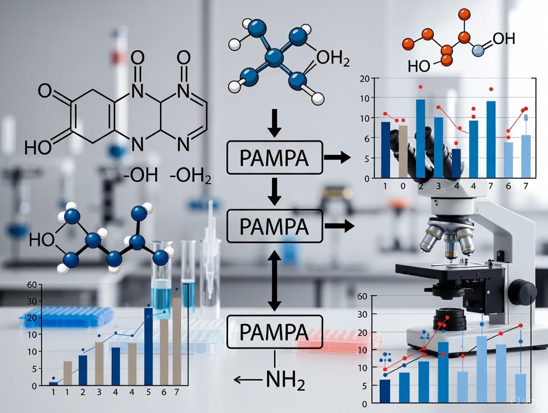
The standard PAMPA procedure can be visualized as a sequential workflow, ensuring consistent and reproducible results. The diagram below illustrates the key stages from membrane preparation to data analysis.
Detailed Methodological Steps
Step 1: Membrane Preparation A microtiter filter plate (often 96-well format with a hydrophobic PVDF membrane) serves as the donor plate. A small volume (e.g., 5 µL) of a lipid solution is pipetted into each well to form the artificial membrane. A common lipid solution is 1-2% (w/v) lecithin (phosphatidylcholine) in an organic solvent like n-dodecane. The solvent is allowed to evaporate, leaving a lipid-impregnated membrane [3] [5].
Step 2: Assembly The donor plate is filled with a buffer solution (e.g., PBS) containing the test compound at a standard concentration of 10-500 µM. The acceptor plate is filled with a blank buffer solution. The donor plate is then carefully positioned on top of the acceptor plate, ensuring the membrane contacts the acceptor buffer without introducing air bubbles, thus forming the "sandwich" [3] [5].
Step 3: Incubation The assembled plate sandwich is incubated at room temperature with constant, gentle shaking for a defined period, typically ranging from 4 to 16 hours. This facilitates the diffusion of compounds across the membrane [3].
Step 4: Disassembly and Analysis After incubation, the sandwich is carefully disassembled. The concentration of the test compound that has permeated into the acceptor compartment is quantified. This is commonly done using UV-Vis spectrophotometry for compounds with sufficient chromophores or, more universally, by LC-MS/MS, which offers higher sensitivity and specificity [3] [1] [5].
Step 5: Data Evaluation The effective permeability (Pe) is calculated for each compound using the following established equation, which accounts for the concentrations in both compartments and the system's geometry [1]:
[ Pe = C \times \ln \left(1 - \frac{[drug]{acceptor}}{[drug]{equilibrium}} \right) ] where [ C = \frac{VD \times VA}{(VD + VA) \times Area \times Time} ]
[drug]acceptor= Concentration in acceptor compartment[drug]equilibrium= Theoretical equilibrium concentrationVD= Donor compartment volumeVA= Acceptor compartment volumeArea= Membrane surface areaTime= Incubation time
Each compound is typically tested in multiple replicates (e.g., n=3) to ensure accuracy and reliability [3].
The Scientist's Toolkit: Essential Reagents and Materials
Successful execution of a PAMPA screen requires specific reagents and materials. The table below catalogs the key components and their functions.
Table 1: Essential Research Reagents and Materials for PAMPA
| Item | Function and Specification |
|---|---|
| PAMPA Donor/Filter Plate | A 96-well microtiter plate with a hydrophobic microporous filter (e.g., PVDF) that serves as the support for the artificial membrane [5]. |
| Acceptor Plate | A 96-well plate, ideally made of PTFE or other low-binding plastic, to hold the acceptor buffer and minimize compound adsorption [5]. |
| Phospholipid | The membrane-forming agent (e.g., Lecithin, DOPC, or porcine brain lipid extract). Its composition determines the barrier being modeled (GI, BBB, etc.) [3] [5] [4]. |
| Organic Solvent | A solvent like n-dodecane used to dissolve the lipid and facilitate its uniform application onto the filter membrane [5]. |
| Buffer Solution | An aqueous buffer (e.g., Phosphate Buffered Saline) to maintain pH, typically 7.4, though other pH values can be explored to simulate GI gradients [1] [5]. |
| Analysis Instrumentation | UV-Vis Plate Reader or LC-MS/MS system for sensitive and accurate quantification of compound concentration in the acceptor well [3] [5]. |
| Control Compounds | High-permeability (e.g., Propranolol) and low-permeability (e.g., Atenolol) controls to validate each assay run and ensure system performance [3]. |
Data Interpretation and Integration into Screening Strategies
Interpreting Permeability Results
The primary output of a PAMPA assay is the effective permeability coefficient (Pe). Compounds are generally classified based on this value, allowing for rapid prioritization during early-stage screening.
Table 2: PAMPA Permeability Classification
| Permeability Class | Effective Permeability (Pe) | Interpretation and Implication |
|---|---|---|
| Low Permeability | < 1.5 x 10⁻⁶ cm/s | The compound has poor passive diffusion characteristics, which may lead to low oral absorption or inability to cross the BBB. May require structural modification [3] [1]. |
| High Permeability | > 1.5 x 10⁻⁶ cm/s | The compound exhibits favorable passive diffusion, suggesting a higher potential for good absorption via the transcellular route [3] [1]. |
PAMPA in the Context of a Broader Screening Strategy
While PAMPA is excellent for assessing passive diffusion, it is crucial to understand its role relative to other permeability models. The cell-based Caco-2 assay is a gold standard that provides more physiologically relevant data by incorporating active transport, efflux transporters (e.g., P-gp), and paracellular pathways [3] [1].
A powerful strategy is to use PAMPA and Caco-2 in conjunction. The relationship between their results can help diagnose the mechanism of permeation for a given compound [1]:
- Good correlation between PAMPA and Caco-2: Suggests the compound permeates primarily via passive transcellular diffusion.
- Caco-2 permeability << PAMPA permeability: May indicate the compound is a substrate for active efflux transporters, which are present in Caco-2 cells but absent in PAMPA.
- Caco-2 permeability >> PAMPA permeability: Suggests the compound may benefit from active uptake transporters or the paracellular route, mechanisms not captured by PAMPA [1].
Therefore, a tiered screening approach—using high-throughput PAMPA as a primary screen to rank compounds on passive diffusion, followed by a more resource-intensive Caco-2 assay as a secondary screen to elucidate transport mechanisms—provides an efficient and informative permeability assessment strategy [3] [1].
Advanced Concepts and Recent Developments
Overcoming Solubility Limitations
A significant challenge in permeability screening is the low aqueous solubility of many drug candidates. To address this, cosolvent methods have been developed. For instance, using 20% (v/v) acetonitrile in the buffer system allows for the measurement of permeability for very sparingly soluble molecules, such as amiodarone, thereby expanding the applicability domain of PAMPA [4].
Real-Time PAMPA (RT-PAMPA)
A recent innovation involves modifying the traditional end-point assay into a real-time format (RT-PAMPA). This method employs a fluorescent artificial receptor (FAR) in the acceptor well. When a permeating analyte binds to the FAR, it causes a fluorescence change (quenching or enhancement), enabling direct, continuous monitoring of permeation kinetics without disassembling the plate. This allows for the differentiation between fast and slow diffusion events and can provide more detailed mechanistic insights [6].
PAMPA is a robust, high-throughput tool that provides critical early-stage data on the passive permeability of drug candidates. Its simplicity, cost-effectiveness, and flexibility to model various biological barriers make it an indispensable component of the modern drug discovery toolkit. By integrating PAMPA into a broader ADME screening strategy, particularly in combination with cell-based models like Caco-2, researchers can efficiently prioritize lead compounds, diagnose absorption issues, and make informed decisions to advance the most promising candidates through the development pipeline.
Within the framework of permeability screening research, the Parallel Artificial Membrane Permeability Assay (PAMPA) has established itself as a robust, high-throughput in vitro tool for predicting the passive transcellular absorption of potential drug candidates [3]. The assay's simplicity, cost-effectiveness, and excellent reproducibility stem from its core structural components, which work in concert to mimic key biological barriers [3] [7]. This document details the essential components and standard protocols for PAMPA, providing researchers with a definitive guide for implementing this critical technique in early drug development. The fundamental principle involves a "sandwich" assembly where a donor plate, containing the test compound, and an acceptor plate, separated by an artificial lipid-infused membrane, facilitate the measurement of passive diffusion [3].
Key Component Analysis
The functionality of the PAMPA model is governed by three primary physical components: the donor and acceptor plates, which house the aqueous compartments, and the artificial lipid membrane that serves as the permeation barrier.
Donor and Acceptor Plates
The assay is typically configured in a 96-well microplate format, enabling high-throughput screening [3]. The system is composed of two distinct plates:
- Donor Plate: This lower plate is characterized by a microporous filter bottom (e.g., polyvinylidene fluoride, PVDF) and is loaded with the test compound solution [6]. The porosity of this filter is a critical parameter in the permeability calculation [1].
- Acceptor Plate: This upper plate serves as a reservoir for a buffer solution that receives compounds that have successfully permeated the membrane [3]. The entire assembly is designed to create a closed system between the donor and acceptor compartments.
The volumes for these compartments are standardized; a typical configuration uses 300 µL of analyte solution in the donor well and the acceptor well is pre-filled with buffer solution [6]. For more physiologically relevant sink conditions, some advanced systems, such as the Double-Sink PAMPA, incorporate additives in the acceptor compartment to maintain a concentration gradient, thereby improving the correlation with in vivo permeability [7].
Artificial Lipid Membranes
The artificial membrane is the cornerstone of PAMPA's predictive capability. It is formed by impregnating the hydrophobic filter of the donor plate with a specific lipid solution, creating a tortuous path that mimics the lipid bilayer of a cell membrane [3] [6]. The composition of this lipid solution can be tailored to simulate different biological barriers:
- Gastrointestinal Tract (GIT) Models: Often use phospholipids like 1,2-dioleoyl-sn-glycero-3-phosphocholine (DOPC) [6].
- Blood-Brain Barrier (BBB) Models: Utilize more complex lipid mixtures, such as porcine polar brain lipid (PBL), to better represent the unique properties of this barrier [8].
The lipid is typically dissolved in an organic solvent like dodecane, with a standard concentration being 2% (w/w) DOPC in dodecane or a commercial pre-blended solution [6]. A volume of 5 µL of this lipid solution is used to coat each well's filter [6].
Buffer Systems
The aqueous buffer system serves as the solvent for the test compound and fills the acceptor compartment. Its composition and pH are critical for maintaining compound stability and simulating the physiological environment.
- Standard Buffer: Physiological phosphate buffer at pH 7.4 is most commonly used to simulate blood or intestinal pH [8] [1].
- pH Variation: A key advantage of PAMPA is the ability to perform assays over a wide pH range. For instance, the donor compartment can be acidified to simulate the stomach environment, while the acceptor is maintained at pH 7.4 to simulate blood, providing insight into pH-dependent permeability [3] [1].
- Additives: Co-solvents like dimethyl sulfoxide (DMSO) are often used to prepare stock solutions of test compounds, while surfactants like Tween-80 may be added to the buffer to ensure the solubility of poorly soluble compounds during the assay [8].
Table 1: Core Components of a Standard PAMPA System
| Component | Typical Composition / Description | Function in the Assay |
|---|---|---|
| Donor Plate | 96-well plate with microporous filter (e.g., PVDF, 0.45 µm) [6] | Houses the initial solution of the test compound. |
| Acceptor Plate | 96-well plate without a filter | Receives the compound that has permeated the artificial membrane. |
| Artificial Membrane | Lipid solution (e.g., 2% DOPC in dodecane or PBL in dodecane) infused into the filter [8] [6] | Serves as the physical barrier for passive diffusion, mimicking a biological membrane. |
| Buffer System | Physiological phosphate buffer (pH 7.4); can be adjusted [8] [1] | Provides the aqueous environment for compound dissolution and transport. |
Standard PAMPA Protocol and Workflow
The following section outlines a detailed, step-by-step protocol for conducting a PAMPA experiment, from membrane preparation to data analysis.
The assay follows a sequential workflow that can be automated for high-throughput screening. The key stages are membrane preparation, system assembly, incubation, disassembly, and quantitative analysis [3].
Figure 1: A sequential workflow diagram of the standard PAMPA protocol, from membrane preparation to data reporting.
Step-by-Step Experimental Methodology
Step 1: Membrane Preparation Coat each well of the 96-well microtiter filter plate (donor plate) with an artificial membrane. This is achieved by pipetting a small volume of lipid solution (e.g., 5 µL of 2% DOPC in dodecane or a commercial Avanti PAMPA lipid blend) onto the filter surface. The lipid is allowed to evenly impregnate the microporous structure, forming a consistent barrier [3] [6].
Step 2: System Assembly Position the prepared donor plate onto a pre-filled acceptor plate containing the buffer solution (e.g., physiological phosphate buffer, pH 7.4), forming a "sandwich" [3]. The acceptor plate is typically filled with a volume of buffer that matches or complements the donor volume to ensure proper hydrodynamics. For the Double-Sink PAMPA method, the acceptor buffer may contain additives to create sink conditions [7].
Step 3: Incubation The assembled sandwich is maintained under constant shaking at room temperature for a defined incubation period. Standard incubation times range from 4 to 16 hours, with 4-5 hours being common for many commercial services [3] [1]. This agitation reduces the thickness of the aqueous boundary layer, ensuring that membrane permeation is the rate-limiting step [7].
Step 4: Disassembly and Sample Analysis After the incubation, carefully separate the donor and acceptor plates. The concentration of the test compound in both the acceptor and donor compartments is then quantified. While UV spectrophotometry can be used for compounds with sufficient chromophores, liquid chromatography coupled with tandem mass spectrometry (LC-MS/MS) is the preferred method for its sensitivity and specificity, allowing for the accurate quantification of a wide range of compounds [3] [1].
Step 5: Data Evaluation and Permeability Calculation The effective permeability (Pe) is calculated for each compound, typically in multiple replicates (e.g., n=3) to ensure accuracy [3]. The permeability coefficient (Pe, in cm/s) is derived using the following equation, which accounts for the compound's movement across the membrane [1]:
Where:
[drug]acceptor= Concentration of test article in the acceptor compartment[drug]equilibrium= Theoretical concentration at equilibrium in the total volume of donor and acceptor compartmentsC= A constant based on the volumes of donor (VD) and acceptor (VA) compartments, the membrane surface area, porosity, and incubation time [1]
Table 2: Standard Experimental Conditions and Data Output for PAMPA
| Parameter | Typical Specification | Notes |
|---|---|---|
| Test Article Concentration | 10 µM [3] | |
| Number of Replicates | 3 [3] | Ensures data reliability. |
| Incubation Time | 4 - 5 hours [3] [1] | Can be extended up to 16 hours. |
| Incubation Temperature | Room Temperature [3] [1] | |
| Analysis Method | LC-MS/MS quantification [3] [1] | Preferred for sensitivity and accuracy. |
| Positive Control | Propranolol (high permeability) [3] | Validates assay performance. |
| Negative Control | Atenolol (low permeability) [3] | Validates assay performance. |
| Data Delivery | Permeability (Pe in x10⁻⁶ cm/s) and full study report [3] | Pe < 1.5 = low permeability; Pe > 1.5 = high permeability [3]. |
The Scientist's Toolkit: Essential Research Reagent Solutions
Successful execution of a PAMPA study requires carefully selected reagents and materials. The following table details key solutions and their specific functions within the assay.
Table 3: Essential Research Reagents and Materials for PAMPA
| Reagent / Material | Function / Role in the Assay | Example Specifications |
|---|---|---|
| Phospholipids | Forms the core artificial membrane barrier that mimics biological bilayers [3] [8]. | 1,2-dioleoyl-sn-glycero-3-phosphocholine (DOPC) for GIT; Porcine Polar Brain Lipid (PBL) for BBB [8] [6]. |
| Organic Solvent | Dissolves the lipid for uniform application and impregnation of the filter [8]. | Dodecane [8] [6]. |
| Buffer Salts | Provides a stable, physiologically relevant aqueous environment for drug dissolution and transport [8]. | Physiological phosphate buffer (pH 7.4) [8]. |
| Permeability Controls | Benchmarks for validating assay performance and data integrity [3]. | Propranolol (high-Pe control); Atenolol (low-Pe control) [3]. |
| LC-MS/MS Solvents | Used for the quantitative bioanalysis of samples from the acceptor and donor wells [3] [1]. | High-purity acetonitrile and water, often with volatile buffers [8]. |
| Membrane Integrity Marker | Verifies the integrity and uniformity of the artificial membrane post-assay. | Lucifer yellow [1]. |
Within drug discovery, the ability of a compound to cross biological membranes via passive diffusion is a critical determinant of its potential as an effective therapeutic agent. This process fundamentally influences intestinal absorption and access to target tissues, particularly through barriers like the blood-brain barrier (BBB). The Parallel Artificial Membrane Permeability Assay (PAMPA) has emerged as a powerful, high-throughput in vitro technique designed specifically to predict this passive diffusion component. This application note details the science of PAMPA, providing established protocols and contextual data to support its use in permeability screening research. By employing a non-cell-based artificial membrane, PAMPA offers a robust, cost-effective method for ranking compound permeability, enabling researchers to prioritize lead compounds with favorable absorption characteristics early in the drug discovery pipeline [9] [10].
The Scientific Principle of PAMPA
Core Mechanism
The fundamental principle of PAMPA is the simulation of passive transcellular diffusion. The assay measures the ability of test compounds to diffuse from a donor compartment, through a lipid-infused artificial membrane, into an acceptor compartment [10]. This membrane typically consists of a mixture of phospholipids (such as lecithin) dissolved in an organic solvent like dodecane, which is immobilized on a hydrophobic filter support [11] [12]. The entire system is incubated for a set period, after which the concentration of the compound in the acceptor compartment is quantified, allowing for the calculation of its effective permeability ((P_e)) [13].
Key Differentiators from Cell-Based Assays
A significant advantage of PAMPA is its focus on pure passive diffusion. Unlike cell-based models such as Caco-2, which contain a variety of active transporters and efflux mechanisms, the artificial membrane in PAMPA lacks these biological components [9]. This provides a clean, uncomplicated measurement of a compound's intrinsic passive permeability. Consequently, if a compound is a substrate for active efflux, its permeability may be overestimated by PAMPA; conversely, permeability may be underestimated for compounds that undergo active uptake or paracellular transport [10]. The assay is also noted for its cost-effectiveness, tolerance to a wider pH range and higher DMSO content, and its amenability to high-throughput screening [9].
The following diagram illustrates the logical relationship between the assay design and its application in predicting in vivo absorption:
Standard PAMPA Protocol
This section provides a detailed, step-by-step methodology for performing a standard PAMPA, based on established protocols [13] [12].
Materials and Reagents
- Donor Plate: A 96-well filter plate with a hydrophobic PVDF membrane (e.g., MultiScreen-IP PAMPA plate) [12].
- Acceptor Plate: A 96-well PTFE or low-binding plastic acceptor plate to ensure minimal compound loss [12].
- Lipid Solution: A solution of 1-2% (w/v) L-∂-Phosphatidylcholine ("lecithin") in a solvent such as n-dodecane [12]. Other aliphatic alkanes like hexadecane may also be used [11] [10].
- Buffer Solution: Phosphate Buffered Saline (PBS), typically with a pH of 7.4, often containing a low percentage (e.g., 0.5-5%) of DMSO to maintain compound solubility [9] [12].
- Test Compounds: Compounds of interest, usually prepared as 10 mM stock solutions in DMSO and further diluted in buffer for the assay [9].
- Instrumentation: A UV/Vis spectrophotometer capable of reading 96-well plates or an LC-MS/MS system for compound quantification [9] [12].
Step-by-Step Procedure
The experimental workflow for a standard PAMPA is methodically outlined below:
Detailed Protocol Steps:
- Lipid Solution Preparation: Prepare a 1% (w/v) solution of lecithin in dodecane. Sonicate the mixture to ensure complete dissolution of the lipid [12].
- Membrane Coating: Using a pipette, carefully apply a precise volume of the lipid solution (e.g., 5 µL) to the filter membrane of each well in the donor plate. Avoid any contact between the pipette tip and the membrane itself [12].
- Solvent Evaporation: Allow the organic solvent to evaporate completely at room temperature, leaving a thin, immobilized artificial lipid membrane on the filter. This typically takes about 20-30 minutes [10] [12].
- Donor Plate Loading: After solvent evaporation, immediately add a measured volume (e.g., 150-200 µL) of the test compound dissolved in buffer (e.g., PBS with 5% DMSO) to each well of the donor plate [13] [12].
- Acceptor Plate Preparation: Fill each well of the acceptor plate with a buffer solution (e.g., 280-300 µL of PBS with 5% DMSO) [10] [12].
- Sandwich Assembly: Carefully place the donor plate on top of the acceptor plate, ensuring that the underside of the filter membrane is in full contact with the buffer solution in the acceptor wells, without introducing air bubbles [12].
- Incubation: Replace the lid and incubate the assembled "sandwich" at room temperature for a predetermined time. Incubation times can vary from 4 to 16 hours, often with constant, gentle shaking (e.g., 150 rpm) to reduce the unstirred water layer effect. To prevent evaporation, the entire assembly can be placed in a sealed container with moistened paper towels [9] [12].
- Post-Incubation Sampling: After incubation, carefully disassemble the plates. The solutions in both the donor and acceptor compartments are then collected for analysis [12].
- Concentration Quantification: Determine the concentration of the test compound in both the donor and acceptor solutions. This is commonly achieved via UV/Vis spectroscopy (comparing spectra to reference standards) or, for greater sensitivity and specificity, LC-MS/MS [10] [12]. The integrity of the artificial membrane can be verified using a marker compound like Lucifer yellow [10].
Data Analysis and Interpretation
The standard parameter derived from PAMPA is the effective permeability ((P_e)). This is calculated using the following equation, which accounts for the assay conditions [13]:
[Pe = - \frac{218.3}{t} \times \log\left(1 - \frac{2 \times CA(t)}{CD(t0)}\right) \times 10^{-6} \text{cm/s}]
Where:
- ( t ) is the incubation time in hours.
- ( C_A(t) ) is the concentration of the compound in the acceptor well at time ( t ).
- ( CD(t0) ) is the concentration in the donor well at time zero (( t_0 = 0 ) h).
Transport percentage can also be calculated as: ( \text{Transport (\%)} = (CA(t) / CD(t_0)) \times 100 ) [13].
Permeability values are typically interpreted using a classification system, as shown in the table below.
Table 1: Interpretation of PAMPA Permeability ((P_e)) Values
| Permeability Category | (P_e) Value (×10⁻⁶ cm/s) | Interpretation |
|---|---|---|
| Excellent / High | > 4.0 | High potential for passive absorption |
| Uncertain / Intermediate | 2.0 – 4.0 | Permeability is borderline |
| Poor / Low | < 2.0 | Low potential for passive absorption [13] |
Research Reagent Solutions
The successful execution of a PAMPA relies on a set of key materials. The following table catalogs essential reagent solutions and their critical functions within the assay.
Table 2: Essential Reagents for PAMPA
| Reagent / Material | Function in the Assay | Exemplary Specifications |
|---|---|---|
| Phosphatidylcholine (Lecithin) | Key lipid component of the artificial membrane, mimicking the composition of biological cell membranes [12]. | 1-2% (w/v) in dodecane [12]. |
| n-Dodecane / Hexadecane | Organic solvent used to dissolve lipids and create the artificial membrane upon evaporation [11] [12]. | Serves as the liquid membrane matrix; e.g., 5 µL applied per well [12]. |
| PVDF Filter Plate | Hydrophobic filter support that immobilizes the lipid solution to form the artificial membrane [12]. | 96-well MultiScreen-IP PAMPA plate (e.g., MAIPNTR10) [12]. |
| Acceptor Plate | Houses the buffer solution that receives compounds diffusing through the membrane; must be low-binding [12]. | 96-well PTFE plate (e.g., MSSACCEPT0R) [12]. |
| Buffer Solution (PBS) | Aqueous medium for dissolving test compounds, maintaining physiological pH and ionic strength [12]. | Phosphate Buffered Saline, pH 7.4, with 0.5-5% DMSO [9] [12]. |
| Lucifer Yellow | Fluorescent marker used to assess the integrity of the artificial membrane post-incubation [10]. | Integrity control to validate assay performance. |
Representative Data and Reproducibility
Benchmark Compound Permeability
To ensure assay validity, it is standard practice to run a set of reference compounds with known permeability properties. The following table presents effective permeability ((P_e)) data for a selection of such drugs, demonstrating the assay's ability to distinguish between high and low permeability compounds.
Table 3: Experimentally Determined PAMPA Permeability ((P_e)) of Reference Compounds
| Compound | Average Log (P_e) (cm/s) | Standard Deviation | Permeability Classification |
|---|---|---|---|
| Testosterone | -4.57 | 0.06 | High |
| Propranolol | -4.92 | 0.08 | High |
| Carbamazepine | -4.95 | 0.07 | High |
| Warfarin | -5.59 | 0.11 | Intermediate |
| Furosemide | -6.20 | 0.13 | Low |
| Methotrexate | -6.46 | 0.21 | Low [12] |
Assay Reproducibility
A critical aspect of any screening assay is its robustness and reproducibility. Data from replicate experiments demonstrate that PAMPA generates highly consistent results. The table below shows the day-to-day reproducibility of log (P_e) measurements for the same set of reference compounds.
Table 4: Day-to-Day Reproducibility of PAMPA Measurements
| Compound | Day 1 Log (P_e) | Day 2 Log (P_e) | Day 3 Log (P_e) | Overall Avg. Log (P_e) |
|---|---|---|---|---|
| Testosterone | -4.55 ± 0.09 | -4.60 ± 0.08 | -4.56 ± 0.10 | -4.57 |
| Propranolol | -4.91 ± 0.11 | -4.95 ± 0.09 | -4.89 ± 0.12 | -4.92 |
| Carbamazepine | -4.93 ± 0.10 | -4.98 ± 0.08 | -4.94 ± 0.11 | -4.95 |
| Warfarin | -5.57 ± 0.14 | -5.62 ± 0.12 | -5.58 ± 0.15 | -5.59 |
| Furosemide | -6.18 ± 0.16 | -6.23 ± 0.18 | -6.19 ± 0.17 | -6.20 |
| Methotrexate | -6.44 ± 0.25 | -6.49 ± 0.22 | -6.45 ± 0.24 | -6.46 [12] |
The low standard deviations associated with the mean log (P_e) values, both within a single plate and across different days and plate lots, confirm that the PAMPA method is a robust and reliable tool for permeability screening [12].
Advanced Applications: PAMPA in Modern Drug Discovery
Predicting Blood-Brain Barrier (BBB) Penetration
A specialized application of PAMPA, known as PAMPA-BBB, utilizes a lipid blend incorporating porcine polar brain lipid to create an artificial membrane that more closely mimics the properties of the blood-brain barrier [8]. This modification allows researchers to predict the passive diffusion of CNS drug candidates into the brain. For example, studies have successfully used PAMPA-BBB to profile the permeability of protein kinase inhibitors, a class of drugs where brain penetration is crucial for treating CNS cancers and neurodegenerative diseases, but must be avoided for peripheral targets to prevent CNS-side effects [8].
Integration with In Silico Models (QSPR and Machine Learning)
The quantitative data generated by PAMPA serves as a high-quality experimental dataset for building in silico predictive models. Quantitative Structure-Permeability Relationship (QSPR) models correlate molecular descriptors of compounds with their measured PAMPA permeability [14] [15]. Modern approaches employ advanced machine learning algorithms such as Support Vector Machines (SVM) and Artificial Neural Networks (ANN).
These models, when trained on large, diverse datasets of PAMPA measurements, can achieve high predictive accuracy for new compounds, with some studies reporting R² values of 0.91 for training sets and 0.84 for external test sets [14]. This integration creates a powerful feedback loop: PAMPA data validates and refines computational models, which in turn can pre-screen virtual compound libraries to guide the synthesis of molecules with optimal permeability properties, significantly accelerating the lead optimization process [9].
Within drug discovery, the Parallel Artificial Membrane Permeability Assay (PAMPA) serves as a robust, high-throughput in vitro tool for predicting the passive, transcellular absorption of potential drug candidates [7] [16]. By utilizing artificial lipid membranes, PAMPA effectively mimics the properties of various biological barriers, minimizing reliance on expensive and time-consuming in vivo studies [7] [3]. This application note details the protocols and applications of three specialized PAMPA models, each engineered to emulate specific biological membranes: the gastrointestinal tract (GIT), the blood-brain barrier (BBB), and the skin.
Model Specifications and Applications
The following table summarizes the key characteristics and applications of the three specialized PAMPA models.
Table 1: Overview of Specialized PAMPA Models
| Model Type | Membrane Composition | Primary Applications | Key Assay Parameters | Data Interpretation |
|---|---|---|---|---|
| GIT-PAMPA | Gastrointestinal tract lipid membranes; often simple phospholipids in n-dodecane or more complex mixtures [3] [17]. | - Prediction of oral absorption and bioavailability [16] [3].- Ranking of drug candidates and formulations based on permeability [7].- Excipient impact assessment [7]. | - Test Concentration: 10 µM [3].- Incubation Time: 4-16 hours [3] [18].- pH Gradient: Often used (e.g., donor pH 5.5-6.5, acceptor pH 7.4) to simulate intestinal conditions [14]. | Permeability (Pe) Classification [3]:- High Permeability: ( Pe > 1.5 \times 10^{-6} ) cm/s- Low Permeability: ( Pe < 1.5 \times 10^{-6} ) cm/s |
| BBB-PAMPA | Porcine brain lipid extract dissolved in an organic solvent like n-dodecane [19] [18]. | - Screening for central nervous system (CNS) drug candidates [19] [18].- Predicting passive blood-brain barrier penetration [19]. | - Test Concentration: 0.05 mM [19].- Incubation Time: 1-18 hours, with modern assays often using 1 hour with stirring [18].- Buffer: Phosphate buffer, pH 7.4 [19]. | Permeability (Pe) Classification [19]:- Moderate/High Permeability: ( Pe > 10 \times 10^{-6} ) cm/s- Low Permeability: ( Pe \leq 10 \times 10^{-6} ) cm/s |
| Skin-PAMPA | Complex mixtures such as certramide, cholesterol, stearic acid, and silicon oil; or 70% silicone/30% isopropyl myristate (IPM) [17]. | - Estimation of skin permeation for transdermal drug delivery [17].- Topical formulation screening. | - Test Concentration: 50 µM [17].- Incubation Time & Temperature: Under controlled temperature (e.g., using a TempPlate) [17].- pH Range: Donor pH 3-7.4 (to ensure neutral form) [17]. | Characterized via Abraham solvation parameter model; compared to experimental skin permeability data [17]. |
Experimental Protocols
Standard PAMPA Workflow
The following diagram illustrates the generalized workflow for a PAMPA experiment, which is common across the different specialized models.
Detailed Methodologies
BBB-PAMPA Protocol
This protocol utilizes the Double-Sink method and is designed for high-throughput screening [19].
- Membrane Preparation: The artificial membrane, composed of a porcine brain lipid extract (e.g., Pion's BBB-1 lipid solution) dissolved in alkane, is immobilized on a PVDF filter plate [19].
- Sample Preparation: Test compounds are typically diluted from 10 mM DMSO stock solutions to a final concentration of 0.05 mM in aqueous phosphate buffer (pH 7.4). The final DMSO concentration should not exceed 0.5% (v/v) [19].
- Assay Assembly:
- Incubation: Place the assembled sandwich on the GutBox stirring system. The assay is run for 60 minutes at room temperature with constant stirring to reduce the aqueous boundary layer to approximately 60 µm [19].
- Analysis and Calculation:
- After incubation, separate the compartments.
- Measure the concentration of the test article in both the donor and acceptor compartments using a UV plate reader.
- Permeability (Pe) calculations are performed using dedicated software (e.g., Pion Software) and expressed in units of ( 10^{-6} ) cm/s [19].
Skin-PAMPA Protocol
This protocol outlines the procedure for assessing skin permeability using a commercial skin-PAMPA model [17].
- Membrane Hydration: Hydrate the skin-PAMPA membrane (e.g., PAMPA-Certramide) overnight with a dedicated hydration solution [17].
- Sample and Buffer Preparation:
- Assay Assembly:
- Add 200 µL of pH 7.4 buffer to the acceptor compartment.
- Add 180 µL of the sample solution to the donor compartment [17].
- Incubation: Assemble the sandwich and incubate under constant stirring using the GutBox, with temperature control (e.g., using a TempPlate) for a specified duration [17].
- Analysis: Quantify the amount of compound in both donor and acceptor compartments using analytical techniques such as UPLC with a diode array detector [17].
The Scientist's Toolkit: Essential Research Reagents and Materials
Successful execution of PAMPA assays requires specific reagents and instrumentation. The following table lists key solutions and their functions.
Table 2: Key Research Reagent Solutions for PAMPA Assays
| Item | Function / Description | Example Application / Note |
|---|---|---|
| BBB-1 Lipid Solution | Porcine brain lipid extract dissolved in alkane. Optimized to mimic the passive permeability properties of the blood-brain barrier [19]. | Used in the BBB-PAMPA model to create the artificial membrane [19]. |
| 96-Well Stirwell Sandwich Plates | Multi-well plates with a filter matrix for membrane immobilization and integrated magnetic stirrers for each well [7]. | Enables high-throughput screening and reduces the aqueous boundary layer via dynamic stirring [7]. |
| Brain Sink Buffer | A specialized buffer for the acceptor compartment in BBB-PAMPA that helps maintain sink conditions [19]. | Critical for achieving accurate permeability measurements by mimicking the sink function of the brain side [19]. |
| GutBox Stirring System | Instrument that provides controlled magnetic stirring to assay plates. Reduces the unstirred water layer (UWL) to a consistent, biologically relevant thickness [7]. | Mimics the hydrodynamics of the intestinal tract or BBB. Ensures more reproducible data by eliminating edge effects common with orbital shakers [7]. |
| PRISMA HT Buffer | A universal buffer solution with constant buffer capacity across a wide pH range (e.g., pH 3-10) [17]. | Useful for performing assays under different pH conditions, such as in GIT-PAMPA or for determining intrinsic permeability in Skin-PAMPA [17]. |
Advanced Applications and Computational Integration
The data generated from PAMPA assays are increasingly integrated with computational models to enhance predictive power and streamline the drug discovery process.
- Machine Learning (ML) and QSAR Models: Permeability data from PAMPA are used to build robust quantitative structure-activity relationship (QSAR) and machine learning models. These models can predict the permeability of new compounds, reducing the need for extensive experimental screening. Algorithms such as Random Forest (RF) and Explainable Boosting Machine (EBM) have demonstrated high accuracy (up to 91% on external test sets) in classifying compounds based on PAMPA permeability [20] [14].
- Blood-Brain Barrier Prediction: In vitro PAMPA-BBB data shows a strong categorical correlation (∼77%) with in vivo brain-to-plasma ratios in rodents. This demonstrates the value of PAMPA as a predictive tool for brain penetration, and models developed from this data can forecast in vivo outcomes [19].
- Cyclic Peptide Permeability: Deep learning models, such as the Molecular Attention Transformer (MAT), are being applied to predict the membrane permeability of challenging cyclic peptides using large PAMPA datasets. These models show promising results, outperforming traditional machine learning methods [21] [22].
Specialized PAMPA models for gastrointestinal, blood-brain barrier, and skin permeability provide critical insights in early drug discovery. Their high-throughput, cost-effective nature allows for efficient screening and rank-ordering of drug candidates based on their potential to cross key biological barriers. When combined with modern computational approaches, PAMPA serves as a powerful tool that significantly de-risks the drug development pipeline and accelerates the progression of viable candidates toward clinical trials.
The Critical Role of PAMPA in Modern ADME Screening and Lead Optimization
In modern drug discovery, absorption, distribution, metabolism, and excretion (ADME) profiling of new chemical entities is crucial for reducing late-stage attrition. Permeability, a key determinant of oral absorption, is routinely assessed using various in vitro models. Among these, the Parallel Artificial Membrane Permeability Assay (PAMPA) has emerged as a foundational tool for high-throughput screening of passive, transcellular permeation. This assay provides a simplistic yet powerful approach to rank compounds based on intrinsic permeability by avoiding the complexities of active transport processes, making it invaluable for early-stage lead optimization [1]. This application note details the critical protocols, data interpretation, and strategic implementation of PAMPA within integrated ADME screening workflows.
PAMPA Fundamentals and Strategic Advantages
PAMPA is a non-cell-based assay that measures a compound's ability to passively diffuse across an artificial phospholipid membrane immobilized on a filter support. Its primary strength lies in its singular focus on passive transcellular permeation, the dominant absorption mechanism for most orally administered drugs [9] [23]. By excluding the confounding variables of active uptake, efflux, and paracellular transport, PAMPA allows medicinal chemists to optimize for a fundamental molecular property early in the discovery process.
The strategic advantages of PAMPA are multifaceted. The assay is highly automated, enabling rapid, cost-effective profiling of large compound libraries with a consistency that can be challenging to achieve with cell-based systems [1]. It is highly adaptable, capable of evaluating permeability across a wide pH range (e.g., pH 5 to 7.4), which provides an early understanding of how a compound might be absorbed throughout the different environments of the gastrointestinal tract [1] [23]. This flexibility, combined with its high throughput and low compound consumption, makes PAMPA an ideal primary permeability screen.
Detailed PAMPA Protocol and Reagent Toolkit
Experimental Workflow
The following diagram illustrates the standardized PAMPA experimental workflow, from plate preparation to data analysis:
The Scientist's Toolkit: Essential Research Reagents and Materials
Table 1: Essential Reagents and Equipment for PAMPA Studies
| Item | Function/Description | Example Source/Model |
|---|---|---|
| GIT-0 Lipid | Proprietary lipid mixture optimized to predict gastrointestinal tract passive permeability. | pION Inc. [23] |
| 96-Well Filter Plates | Plastic matrix for immobilizing the artificial membrane; creates donor and acceptor compartments. | pION Inc. [9] |
| Acceptor Sink Buffer (pH 7.4) | Buffer in acceptor compartment to maintain sink conditions. | pION Inc. [23] |
| PRISMA HT Buffer | Buffer system for donor compartment; can be adjusted to various pH values (e.g., pH 5). | pION Inc. [23] |
| Gutbox | Technology to provide stirring in donor compartment, reducing the unstirred water layer (UWL). | pION Inc. [9] [23] |
| UV Plate Reader | For high-throughput quantification of test article concentration in donor/acceptor wells. | Nano Quant, Infinite 200 PRO (Tecan) [9] |
| LC-MS/MS System | Alternative detection method for compounds with weak UV chromophores. | UPLC-MS [23] |
Core Protocol Steps
- Donor Solution Preparation: Test compounds, typically stocked in DMSO (e.g., 10 mM), are diluted to a final concentration of 0.05 mM in aqueous buffer. The final DMSO concentration should be kept low (e.g., 0.5%) to avoid disrupting the artificial membrane [9] [23].
- Membrane and Acceptor Preparation: An artificial membrane, often a proprietary lipid mixture (e.g., GIT-0 from Pion Inc.) in an organic solvent like dodecane, is applied to a filter plate and allowed to form. The acceptor plate is filled with buffer, typically at pH 7.4 [23].
- Assay Incubation: The donor plate is placed on top of the acceptor plate to create a "sandwich." The assembly is incubated at room temperature for a set period (30 minutes in high-throughput formats [23] up to 5 hours [1]) with constant stirring using a Gutbox to minimize the unstirred water layer.
- Concentration Quantification: After incubation, the concentration of the test compound in both the donor and acceptor compartments is quantified. This is most commonly done via a UV plate reader [9], but LC-MS/MS is used for UV-inactive compounds or for greater specificity [23].
- Permeability Calculation: The effective permeability (Pe) is calculated using software (e.g., from Pion Inc.) based on the compound flux from the donor to the acceptor compartment. The permeability calculation accounts for volumes, membrane surface area, and incubation time [1].
Data Interpretation and Permeability Classification
Quantitative Analysis
The effective permeability (Pe) is calculated from the following equation [1]:
Where:
[drug]acceptor= Concentration of the test article in the acceptor compartment[drug]equilibrium= Concentration of test article in the total volume of the donor and acceptor compartments at equilibriumC = (VD × VA) / ((VD + VA) × Area × Time)VD= Volume of the donor compartment;VA= Volume of the acceptor compartmentArea= Surface area of the membrane multiplied by the porosityTime= Incubation time
Permeability Classification and Strategic Decision-Making
Based on the calculated Pe value, compounds are typically categorized to facilitate rank-ordering and decision-making.
Table 2: PAMPA Permeability Classification and Correlation with Absorption
| Permeability Category | Permeability (Pe) Value (10⁻⁶ cm/s) | Interpretation & Strategic Implications |
|---|---|---|
| Low Permeability | < 1.5 | Poor passive absorption potential. Medicinal chemistry strategy: Focus on structure modifications to improve lipophilicity and reduce hydrogen bonding. |
| High Permeability | > 1.5 | Favorable passive absorption potential. Next step: Progress to secondary, more complex assays (e.g., Caco-2) to investigate transporter effects. |
Studies at the National Center for Advancing Translational Sciences (NCATS) have demonstrated a strong correlation (~85%) between PAMPA permeability (specifically at pH 5) and in vivo oral bioavailability in rodent models, underscoring its predictive value for preclinical candidate selection [23].
Integrated Screening: PAMPA and Caco-2
While PAMPA is excellent for measuring intrinsic passive permeability, it does not model active transport, efflux, or paracellular transport. Therefore, its data is most powerful when used in conjunction with cell-based models like the Caco-2 assay, which incorporates these additional complexities [1] [24]. The relationship between data from these two assays provides invaluable diagnostic insight into a compound's permeation mechanism.
The following diagram illustrates the logical framework for interpreting combined PAMPA and Caco-2 data to diagnose absorption mechanisms:
As illustrated, a significant discrepancy between Caco-2 and PAMPA permeability can signal specific transport mechanisms:
- PAMPA > Caco-2: Suggests the compound may be a substrate for active efflux transporters (e.g., P-gp) [1].
- PAMPA < Caco-2: Suggests potential involvement of active uptake transporters or significant paracellular transport [1].
Advanced Applications and In Silico Modeling
The utility of PAMPA data extends beyond experimental screening. Large, high-quality PAMPA datasets have enabled the development of robust quantitative structure-activity relationship (QSAR) and machine learning models to predict permeability for virtual compounds even before synthesis [9] [23] [25]. These in silico models, built using datasets encompassing thousands of diverse compounds, achieve high prediction accuracy (AUC-ROC > 0.88) and provide medicinal chemists with a powerful tool to prioritize virtual synthetic targets and accelerate lead optimization [9].
Furthermore, the basic PAMPA principle has been successfully adapted to model penetration through other biological barriers, including the blood-brain barrier (BBB-PAMPA) using porcine brain lipid extracts, and the nasal mucosa (Nasal-PAMPA) by incorporating mucin into the system [26].
PAMPA remains a cornerstone of modern ADME screening due to its simplicity, cost-effectiveness, and high-throughput capability. By providing a clean measure of a compound's intrinsic passive permeability, it delivers critical data for early lead optimization. When strategically integrated with cell-based models and advanced in silico predictions, PAMPA forms an essential component of a holistic permeability screening strategy, ultimately enhancing the efficiency of the drug discovery pipeline and increasing the likelihood of identifying successful clinical candidates.
Executing PAMPA Assays: Protocols and Specialized Applications
Within the framework of permeability screening research, the Parallel Artificial Membrane Permeability Assay (PAMPA) serves as a critical, non-cell-based in vitro tool for predicting the passive, transcellular permeation of potential drug candidates [27] [1]. This application note delineates a standardized PAMPA protocol, providing researchers and drug development professionals with a reliable methodology for generating consistent and reproducible data. The assay's ability to evaluate permeability across a wide pH range offers invaluable early insight into how new chemical entities might be absorbed along the gastrointestinal tract, thereby playing a pivotal role in lead optimization during early drug discovery stages [1] [10]. By utilizing an artificial membrane, PAMPA isolates the mechanism of passive diffusion, enabling a clear rank-ordering of compounds based on this fundamental permeability property, free from the complexities introduced by active transport systems present in cellular models [1].
Materials and Reagents
Research Reagent Solutions
The following table catalogues the essential materials and reagents required to execute the standard PAMPA protocol.
Table 1: Essential Research Reagents and Materials for PAMPA
| Item | Function / Description |
|---|---|
| MultiScreen-IP PAMPA Filter Plate (e.g., cat. MAIPNTR10) | Serves as the Donor plate. The PVDF membrane filter acts as the support for the artificial lipid membrane [27]. |
| PTFE Acceptor Plate (e.g., cat. MSSACCEPT0R) | A low-binding Acceptor plate is critical to prevent compound loss. This plate is not supplied with the filter plate and must be procured separately [27]. |
| L-∂-Phosphatidylcholine (Lecithin) | The primary lipid component (e.g., cat. P-3556) used to create the artificial membrane that mimics biological barriers [27]. |
| n-Dodecane | The organic solvent (e.g., cat. D-4259) used to dissolve the lipid for uniform application onto the PVDF membrane [27]. |
| Phosphate Buffered Saline (PBS) | The standard buffer system (e.g., cat. P-3813), often adjusted to pH 7.4, for dissolving test compounds and filling acceptor wells [27]. |
| Dimethyl Sulfoxide (DMSO) | Used as a co-solvent (typically at 5% v/v) to maintain compound solubility in the aqueous buffer solutions [27]. |
| Test Compounds & Standards | Drugs like propranolol, warfarin, and testosterone are used as reference standards to validate assay performance and reproducibility [27]. |
| Lucifer Yellow | A fluorescent marker used to assess membrane integrity and ensure the artificial layer is correctly formed and intact [1] [10]. |
Standard PAMPA Protocol
The following diagram illustrates the logical sequence and key components of the standard PAMPA experimental workflow.
Step-by-Step Procedure
Step 1: Lipid Solution Preparation Prepare a 1% (w/v) solution of lecithin in n-dodecane, requiring approximately 500 µL per plate. To ensure the complete dissolution of the lipid, sonicate the mixture [27].
Step 2: Artificial Membrane Application Using a polypropylene reservoir, carefully pipette 5 µL of the lecithin/dodecane solution into each well of the Donor (filter) plate. It is critical to avoid any contact between the pipette tip and the underlying PVDF membrane [27].
Step 3: Donor and Acceptor Plate Preparation
- Donor Plate: Immediately after applying the lipid, add 150 µL of the donor solution (test compound or reference standard dissolved in buffer, typically 5% DMSO in PBS, pH 7.4) to each well. Concentrations in the range of 100-500 µM are commonly used [27].
- Acceptor Plate: Add 300 µL of buffer (5% DMSO in PBS, pH 7.4) to each well of the PTFE Acceptor plate [27].
Step 4: Plate Assembly and Incubation Place the drug-filled Donor plate directly onto the Acceptor plate, ensuring the underside of the membrane is in full contact with the buffer in the acceptor wells. Replace the lid and incubate the assembled plates at room temperature for a defined period, typically 16 hours. To prevent evaporation, place the entire assembly inside a sealed container with moistened paper towels [27]. Note that some high-throughput protocols use shorter incubation times, such as 5 hours [1].
Step 5: Post-Incubation Sample Analysis After incubation, carefully separate the Donor and Acceptor plates. Transfer samples from both compartments to a UV-compatible microplate. Measure the UV/Vis absorption from 250 to 500 nm. For quantification, it is essential to also prepare and analyze a set of standard solutions at the theoretical equilibrium concentration (the concentration expected if the donor and acceptor solutions were simply mixed) [27].
Step 6: Permeability Calculation The effective permeability ((Pe)) is calculated using the following equation, with the result typically expressed as log (Pe): [ Pe = C \times \ln\left(1 - \frac{[drug]{acceptor}}{[drug]_{equilibrium}}\right) ] Where:
- ( [drug]_{acceptor} ) = Concentration of the test compound in the acceptor compartment.
- ( [drug]_{equilibrium} ) = Theoretical equilibrium concentration.
- ( C = \frac{VD \times VA}{(VD + VA) \times Area \times Time} )
- ( V_D ) = Volume of the donor compartment.
- ( V_A ) = Volume of the acceptor compartment.
- ( Area ) = Surface area of the membrane multiplied by the porosity.
- ( Time ) = Incubation time [27] [1].
Data Quality and Reproducibility
Reproducibility Assessment
The robustness of this standard PAMPA protocol was evaluated through extensive reproducibility studies, measuring the permeability of six model drugs under various conditions. The results demonstrate the assay's high precision and reliability.
Table 2: Reproducibility of PAMPA Measurements (log (P_e)) Across Different Variables
| Compound | Within-Plate (n=16) | Plate-to-Plate (n=6 plates) | Day-to-Day (n=6 days) | Lot-to-Lot (n=3 lots) |
|---|---|---|---|---|
| Propranolol | -4.65 ± 0.08 | -4.68 ± 0.11 | -4.67 ± 0.09 | -4.66 ± 0.10 |
| Carbamazepine | -4.49 ± 0.07 | -4.50 ± 0.10 | -4.49 ± 0.09 | -4.49 ± 0.09 |
| Warfarin | -5.08 ± 0.09 | -5.11 ± 0.11 | -5.11 ± 0.11 | -5.10 ± 0.12 |
| Testosterone | -4.33 ± 0.07 | -4.34 ± 0.09 | -4.33 ± 0.09 | -4.33 ± 0.09 |
| Furosemide | -5.71 ± 0.13 | -5.73 ± 0.17 | -5.74 ± 0.17 | -5.73 ± 0.17 |
| Methotrexate | <-6.00 | <-6.00 | <-6.00 | <-6.00 |
Data adapted from Schmidt & Lynch, demonstrating low variability across multiple experimental parameters [27].
Impact of Protocol Variations
The effect of deliberate, minor alterations to the standard protocol was investigated to simulate the variability that might occur between different operators or laboratories.
Table 3: Effect of Protocol Variations on Apparent Permeability (log (P_e))
| Protocol Variation | Propranolol | Carbamazepine | Warfarin | Testosterone |
|---|---|---|---|---|
| Standard Protocol | -4.65 | -4.49 | -5.08 | -4.33 |
| 2% Lecithin (vs. 1%) | -4.83 | -4.71 | -5.18 | -4.43 |
| 10 µL Lipid (vs. 5 µL) | -4.77 | -4.62 | -5.14 | -4.39 |
| 1-Hr Delay Post-Lipid | -4.67 | -4.50 | -5.09 | -4.34 |
| 4-Hr Delay Post-Lipid | -4.70 | -4.52 | -5.11 | -4.36 |
While minor protocol changes can influence the absolute permeability values for some compounds, the critical rank order of compound permeability remains largely unaffected, underscoring the assay's utility for compound ranking in lead optimization [27]. However, variations in lipid content, such as increasing the lecithin concentration, can more significantly alter the log (P_e) for certain compounds, highlighting the need for strict protocol adherence for comparative studies [27].
This application note provides a detailed, step-by-step guide for the standard PAMPA protocol, from the critical initial step of lipid application through to final UV analysis. The data presented confirms that when executed under controlled conditions, the PAMPA assay is a robust and highly reproducible tool for measuring passive permeability. Its simplicity, cost-effectiveness, and high-throughput capability make it an indispensable primary screen in permeability screening research, enabling effective rank-ordering of drug candidates early in the discovery process.
Within modern drug discovery, the Parallel Artificial Membrane Permeability Assay (PAMPA) has emerged as a high-throughput, cell-free method to predict the passive transcellular absorption of potential drug candidates [28] [9]. Passive diffusion is the primary absorption mechanism for over 80% of commercial drugs, making PAMPA a critical tool for early-stage screening [28] [9]. The core principle of PAMPA involves creating an artificial lipid membrane on a hydrophobic filter support, separating a donor compartment from an acceptor compartment. The test compound's ability to diffuse across this membrane is quantified as an effective permeability (P~e~), providing a key indicator of its absorption potential [28] [1].
The composition of the artificial membrane is the most critical experimental parameter in PAMPA, as it directly dictates the physicochemical properties of the barrier and determines the assay's ability to emulate specific biological membranes, such as those of the gastrointestinal tract (GIT), the blood-brain barrier (BBB), or the skin [28] [29]. This application note details the protocols, data, and key considerations for employing three central lipid components in PAMPA: lecithin, synthetic phospholipids, and hexadecane. By framing this within a broader thesis on permeability screening, we demonstrate how rational selection and customization of membrane composition can yield highly biorelevant and predictive data.
The Scientist's Toolkit: Essential Reagents and Materials
The following table catalogues the key reagents and materials essential for conducting PAMPA studies with lecithin, synthetic phospholipids, and hexadecane.
Table 1: Key Research Reagent Solutions for PAMPA
| Reagent/Material | Function in PAMPA | Examples & Notes |
|---|---|---|
| Lecithin (e.g., L-α-Phosphatidylcholine) | Forms a biomimetic phospholipid membrane to predict gastrointestinal absorption [30]. | Often used as 1-10% (w/v) solution in n-dodecane [30] [31]. |
| Synthetic Phospholipids | Enables study of lipid chemical structure (e.g., hydrophobic chain length) on permeability mechanism [28]. | e.g., Dioleoylphosphatidylcholine (DOPC); custom-synthesized analogs with defined chain lengths (C8, C10, C12) [28]. |
| n-Hexadecane | Serves as a pure, inert solvent for the lipid membrane or as the membrane itself in specific models [32] [31]. | Used in HDM-PAMPA (100% hexadecane) to determine hexadecane/water partition coefficients (K~hex/w~) [32] [31]. |
| n-Dodecane | A common organic solvent used to dissolve phospholipids for membrane formation [28] [30]. | Serves as the base solvent for many lipid formulations, including lecithin and DOPC solutions [28]. |
| PAMPA Plates & Instrumentation | Provides the platform for high-throughput assay; includes donor filter plates and acceptor plates [30] [7]. | Specialized systems (e.g., Pion's PAMPA) include stirring (Gut-Box) to reduce the unstirred water layer [29] [7]. |
| Buffer Solutions (e.g., PRISMA HT) | Maintains pH during assay, critical for compounds that exist in ionizable forms [29] [17]. | Universal buffers with constant buffer capacity across a wide pH range (e.g., 3-10) are available [29]. |
Membrane Compositions and Their Biorelevance
Different lipid formulations are used to model specific biological barriers. The choice of composition directly impacts the hydrogen bonding, hydrophobicity, and overall selectivity of the artificial membrane, which in turn determines its biorelevance [29] [17].
Table 2: Overview of Common PAMPA Membrane Models and Their Applications
| PAMPA Model | Typical Membrane Composition | Primary Biological Target | Key Characteristics |
|---|---|---|---|
| Original PAMPA / Lecithin-Based | 1-10% Lecithin in n-dodecane [30] [31]. | Gastrointestinal Tract (GIT) Absorption [30]. | A robust, cost-effective model for initial passive permeability ranking [30]. |
| DOPC-PAMPA | 2% Dioleoylphosphatidylcholine in n-dodecane [28] [31]. | General Passive Permeability [28]. | Uses a highly purified, synthetic phospholipid for more consistent and defined membrane properties [28]. |
| Biomimetic PAMPA (BM-PAMPA) | Mixture of PC, PE, PS, PI, and cholesterol in organic solvent [31]. | Brush-Border Membrane (GIT) [28]. | A more complex, highly biomimetic mixture intended to better replicate in vivo conditions [28]. |
| Double-Sink PAMPA (DS-PAMPA) | 20% phospholipid mixture in dodecane; acceptor solution contains surfactant [31]. | GIT Absorption [31]. | The "sink" conditions in the acceptor well better mimic in vivo drug removal by circulation [31]. |
| HDM-PAMPA | 100% Hexadecane [32] [31]. | Intrinsic Membrane Permeability; used to predict Caco-2/MDCK permeability [32]. | A simple model that measures hexadecane/water partition (K~hex/w~), highly correlated with passive permeability [32]. |
| Skin-PAMPA | e.g., Certramide, cholesterol, stearic acid, and silicon oil [29] [17]. | Skin Permeation [29] [17]. | Specifically designed to emulate the different hydrophobicity and hydrogen-bonding properties of the skin barrier [29]. |
Quantitative Data and Comparative Analysis
The Impact of Synthetic Phospholipid Chain Length
Research has systematically investigated how the hydrophobic tail length of synthetic phospholipids influences drug permeability. The following table summarizes key findings from a study using custom-synthesized lipids [28].
Table 3: Effect of Phospholipid Hydrophobic Chain Length on Drug Permeability (P~e~) [28]
| Factor Studied | Experimental Conditions | Impact on Effective Permeability (P~e~) |
|---|---|---|
| Hydrophobic Carbon Chain Length | Three phospholipids with chain lengths of C8, C10, and C12. | Longer hydrophobic chains (C12) in the synthetic phospholipid membrane improved the drug permeability for almost all test drugs across nearly all incubation time points [28]. |
| Incubation Time | Time range: 4 to 20 hours. | The P~e~ of each drug increased rapidly with time, then decreased slightly after reaching a maximum value [28]. |
| pH Gradient | Donor pH ranged from 4.6 to 9.32. | The pH gradient changed drug permeability according to the pH-partition hypothesis for drugs with diverse pK~a~ values [28]. |
Interpreting Permeability Data and Correlations
The permeability values obtained from PAMPA require correct interpretation to be meaningful in a drug discovery context.
Table 4: Permeability Data Interpretation and Correlation with Other Models
| Aspect | Description & Interpretation | Notes and Correlations |
|---|---|---|
| Permeability Classification | - High Permeability: P~e~ > 1.5 × 10^-6^ cm/s [1].- Low Permeability: P~e~ < 1.5 × 10^-6^ cm/s [1]. | Used for rank-ordering drug candidates based on passive diffusion potential. |
| Relationship with Caco-2 | PAMPA measures only passive diffusion. Correlation with Caco-2 is strong if passive diffusion is the dominant transport mechanism [9] [1]. | Discrepancies can diagnose other mechanisms: PAMPA overestimates permeability if a compound is a substrate for active efflux in Caco-2; PAMPA underestimates it if there is active uptake or significant paracellular transport [1]. |
| Correlation with Hexadecane/Water Partition (K~hex/w~) | The HDM-PAMPA method determines K~hex/w~, which can be used with the solubility-diffusion model to accurately predict intrinsic permeability in Caco-2 and MDCK cell membranes (RMSE = 0.8) [32]. | Highlights the fundamental role of partitioning into a hydrophobic core as a driver of passive membrane permeability. |
Detailed Experimental Protocols
Standard Lecithin-Based PAMPA Protocol for GIT Permeability Prediction
This protocol is adapted from the robust, high-throughput method described by Schmidt & Lynch [30].
Materials:
- Donor Plate: MultiScreen-IP PAMPA filter plate (e.g., Millipore MAIPNTR10).
- Acceptor Plate: PTFE acceptor plate (e.g., Millipore MSSACCEPT0R).
- Lipid Solution: 1% (w/v) Lecithin (L-α-Phosphatidylcholine, cat. P-3556) in n-dodecane. Sonicate to ensure complete dissolution.
- Drug Solutions: Prepare test compounds in buffer (e.g., PBS) with ≤ 1% DMSO. A typical stock concentration is 10 mM in DMSO, diluted to 100-500 µM in buffer for the assay.
- Buffer: Phosphate Buffered Saline (PBS), pH 7.4.
- Instrumentation: UV/Vis plate reader, sonicator.
Procedure:
- Membrane Formation: Carefully pipette 5 µL of the 1% lecithin/dodecane solution into each well of the donor plate filter, avoiding contact between the pipette tip and the membrane [30].
- Plate Loading:
- Incubation: Place the donor plate into the acceptor plate, ensuring the underside of the membrane is in contact with the buffer. Cover the assembly and incubate at room temperature for 16 hours. To prevent evaporation, place the entire setup in a sealed container with wet paper towels [30].
- Sample Analysis: After incubation, separate the plates. Measure the UV/Vis absorption (250-500 nm) of solutions from both the donor and acceptor compartments. Also, analyze a standard solution at the theoretical equilibrium concentration.
- Data Calculation: Calculate the effective permeability (P~e~) for each compound using the established equation and convert to logP~e~ for analysis [30]:
- ( Pe = \frac{-2.303 \times Vd \times Va}{(Vd + Va) \times A \times (t - t0)} \times \log \left[ 1 - \frac{(Vd + Va) \times Ca(t)}{Vd \times S \times Cd(0)} \right] )
- Where ( S = \frac{Va \times Ca(t)}{Vd \times Cd(0)} + \frac{Cd(t)}{Cd(0)} )
- ( Vd ) = Donor volume (0.150 mL), ( Va ) = Acceptor volume (0.300 mL), ( A ) = Filter area (cm²), ( t ) = Incubation time (s), ( Ca(t) ) = Acceptor concentration at time t, ( Cd(t) ) = Donor concentration at time t, ( Cd(0) ) = Initial donor concentration.
HDM-PAMPA Protocol for Determining K~hex/w~
This protocol focuses on using pure hexadecane to determine a key physicochemical property linked to intrinsic permeability [32] [31].
Materials:
- Lipid Solution: 100% hexadecane [31].
- Buffer & Samples: Universal buffer (e.g., PRISMA HT) adjusted to a pH where the compound is neutral; sample concentration of 50 µM with 0.5% DMSO [29].
- Instrumentation: PAMPA Explorer instrument with Gut-Box and TempPlate for stirring and temperature control (25°C); UPLC-DAD or LC-MS/MS for quantification [29].
Procedure:
- Membrane Formation: Apply a volume of pure hexadecane to the filter of the donor plate as per manufacturer's instructions.
- Plate Loading:
- Donor Plate: Add 180 µL of the neutral-form drug solution.
- Acceptor Plate: Add 200 µL of pH 7.4 buffer.
- Incubation: Assemble the sandwich and incubate on the Gut-Box with stirring for a specified time (e.g., 4-5 hours) at 25°C. Stirring reduces the unstirred water layer, which is critical for accurate measurement [29] [7].
- Sample Analysis: Quantify the amount of compound in both compartments using a highly sensitive method like UPLC-DAD or LC-MS/MS.
- Data Calculation: The hexadecane/water partition coefficient (K~hex/w~) is derived from the assay results. This value can then be used in a calibrated solubility-diffusion model to predict Caco-2 or MDCK intrinsic permeability [32].
Workflow and Property Diagrams
PAMPA Experimental Workflow
The following diagram illustrates the general workflow for a PAMPA experiment, from membrane preparation to data interpretation.
Membrane Composition & Property Relationships
This diagram conceptualizes how the choice of lipid component influences the membrane's properties and its resulting biorelevance.
The strategic selection of membrane composition—from the well-established lecithin in dodecane to the highly specific synthetic phospholipids and pure hexadecane—is paramount for generating meaningful permeability data in PAMPA. As detailed in this note, the lipid component directly controls the membrane's properties, enabling researchers to tailor the assay for everything from high-throughput GI absorption ranking to the prediction of specialized barrier penetration. By understanding and applying the principles and protocols outlined herein, scientists can more effectively leverage PAMPA to accelerate the identification and optimization of promising drug candidates with desirable absorption profiles.
Parallel Artificial Membrane Permeability Assay (PAMPA) has emerged as a robust, high-throughput tool for predicting passive drug transport across biological barriers. Its simplicity, cost-effectiveness, and compatibility with automation make it particularly valuable for early-stage drug discovery screening [31]. While the original PAMPA model was developed to predict gastrointestinal absorption, its principle has been successfully adapted to mimic more complex biological interfaces, notably the blood-brain barrier (BBB) and the skin [33] [19]. These specialized models, BBB-PAMPA and Skin-PAMPA, utilize bespoke lipid compositions to better represent the unique physicochemical properties of their respective target barriers. This application note details the formulations, protocols, and applications of these tailored PAMPA methods within the context of permeability screening research.
Skin-PAMPA: Formulation and Application for Transdermal Penetration Prediction
Rationale and Biomimetic Formulation
The primary barrier to transdermal drug delivery is the stratum corneum (SC), the outermost layer of the skin. Its lipid matrix, composed of approximately 50% ceramides, 35% free fatty acids, and 15% cholesterol, forms a continuous structure through which compounds must diffuse [33]. The goal of Skin-PAMPA is to replicate this rate-limiting barrier using synthetic components.
A key advancement in Skin-PAMPA was the development of a biomimetic membrane that moves beyond simple solvents. Earlier models used silicone oil and isopropyl myristate, which are not natural components of the SC [33] [34]. The more recent, physiologically relevant formulation incorporates:
- Synthetic Certramides: Acting as analogs to natural skin ceramides [33] [34].
- Free Fatty Acid: Typically stearic acid, to mimic the free fatty acid component [33].
- Cholesterol: Completing the core lipid mixture of the stratum corneum [33] [34].
This specific lipid mixture is dissolved in an organic solvent and impregnated into a porous filter, creating an artificial membrane that closely mimics the passive diffusion properties of human skin [33].
Detailed Skin-PAMPA Protocol
The following procedure outlines a standard Skin-PAMPA experiment, from plate preparation to data analysis [33] [35] [34].
Table 1: Key Reagents and Materials for Skin-PAMPA
| Item | Function/Description | Source/Example |
|---|---|---|
| Skin-PAMPA Sandwich | 96-well plate assembly with donor and acceptor compartments and a hydrophobic PVDF filter (0.45 µm pore size) for membrane formation. | Pion Inc. (P/N: 120657) [34] or commercial kits [35]. |
| Lipid Mixture | Certramide, free fatty acid, and cholesterol dissolved in an organic solvent. Forms the biomimetic membrane. | Prepared in-lab [33] or provided in kits. |
| Hydration Solution | Aqueous solution used to hydrate the membrane prior to the assay, improving biomimicry. | Pion Inc. (P/N: 120706) [34]. |
| High/Medium/Low Permeability Controls | Reference compounds for assay validation and quality control. | e.g., Methyl Paraben (High), Propyl Paraben (Medium), Theophylline (Low) [35]. |
| Buffer (PBS, pH 7.4) | Standard acceptor and donor solution. | Commercially available or made in-house [34]. |
| UV-Transparent Microplate | Used for spectrophotometric analysis of compound concentration. | e.g., Greiner Bio-One UV-star microplate [34]. |
Procedure:
- Membrane Preparation: The artificial membrane is created by uniformly impregnating the hydrophobic PVDF filter of the donor plate with the prepared Skin-PAMPA lipid mixture. The integrity of the formed membrane can be confirmed using techniques like Raman microscopy [33].
- Hydration: The membrane is hydrated with a special hydration solution for a recommended 24 hours before the assay [34].
- Sample Preparation: Test compounds are typically dissolved in DMSO and then diluted in PBS (pH 7.4). The final concentration of DMSO in the donor solution should not exceed 0.5-5% to avoid disrupting the membrane integrity [35] [34].
- Assay Execution:
- The acceptor compartment is filled with PBS (pH 7.4).
- The donor solution, containing the test compound, is added to the donor compartment.
- The plate is assembled into a "sandwich" and incubated at room temperature. The incubation time for Skin-PAMPA is typically longer than for other models; protocols report 20 hours [35] or 16-24 hours [33] to allow for the slower diffusion rate across the skin-mimicking membrane.
- Sample Analysis: After incubation, the concentration of the test compound in the acceptor compartment is quantified, typically using a UV plate reader. LC-MS/MS can be used for compounds where UV detection is not suitable [31] [1].
- Data Calculation: The apparent permeability coefficient ((P_{app})) is calculated using the formula below, which accounts for the concentrations and volumes in the system [1].
(P{app} = C \times \ln\left(1 - \frac{[drug]{acceptor}}{[drug]{equilibrium}}\right)) where ( C = \frac{VD \times VA}{(VD + V_A) \times Area \times Time} )
- ( [drug]_{acceptor} ) = Concentration of drug in acceptor well
- ( [drug]_{equilibrium} ) = Theoretical equilibrium concentration
- ( V_D ) = Donor volume
- ( V_A ) = Acceptor volume
- ( Area ) = Membrane surface area (adjusted for porosity)
- ( Time ) = Incubation time
Figure 1: Skin-PAMPA Experimental Workflow
Application and Validation
Skin-PAMPA is designed as a high-throughput research tool for the fast prediction of skin penetration in the early stages of drug discovery and cosmetics research [33] [36]. It has been shown to correlate well with data from human skin permeation studies, offering a cost-effective and easily standardized alternative to more variable methods like the Franz diffusion cell [33] [34]. Research has demonstrated that the rank order of drug diffusion from different formulations (e.g., hydrogel, o/w cream, w/o cream) is consistent between Skin-PAMPA and the Franz cell method, supporting its utility for formulation screening [34].
BBB-PAMPA: Predicting Blood-Brain Barrier Penetration
Rationale and Formulation
The blood-brain barrier (BBB) is a highly selective interface that protects the central nervous system. For drugs targeting the CNS, sufficient passive permeability across the BBB is often a prerequisite for efficacy. BBB-PAMPA models this passive diffusion process.
The critical component of the BBB-PAMPA model is the lipid membrane. Unlike the skin-centric lipids of Skin-PAMPA, the BBB model utilizes:
- Porcine Brain Lipid Extract: A complex lipid mixture dissolved in an alkane, which is optimized to mimic the passive permeability characteristics of the BBB endothelial membrane [19]. This composition is commercially available as the "BBB-1" lipid solution.
The "Double-Sink" PAMPA-BBB method is a common and refined variant. It incorporates a surfactant mixture in the acceptor compartment to create a "sink" condition, mimicking the sink effect of blood flow, which helps maintain a concentration gradient and improves the robustness of the assay for a wider range of compounds [19].
Detailed BBB-PAMPA Protocol
The BBB-PAMPA protocol is designed for higher throughput and shorter duration than the Skin-PAMPA model.
Table 2: Key Reagents and Materials for BBB-PAMPA
| Item | Function/Description | Source/Example |
|---|---|---|
| BBB-1 Lipid Solution | Porcine brain lipid extract in alkane for forming the BBB-mimicking membrane. | Pion Inc. (Catalog #110672) [19]. |
| 96-well Stirwell Sandwich Plate | A specialized plate system with coated magnetic stirrers in the donor wells to reduce the unstirred water layer. | Pion Inc. (Catalog #110243) [19]. |
| Brain Sink Buffer | Acceptor solution with surfactants to create sink conditions. | Pion Inc. (Catalog #110674) [19]. |
| Positive/Negative Controls | Compounds with known BBB permeability for validation. | e.g., Caffeine, Progesterone, Carbamazepine [19]. |
| GutBox Technology | An instrument that controls stirring in the donor compartment to standardize hydrodynamics. | Pion Inc. [19]. |
Procedure:
- Membrane Preparation: The filter on the acceptor plate is impregnated with the BBB-1 lipid solution [19].
- Sample Preparation: Test compounds from 10 mM DMSO stocks are diluted to a final concentration of 0.05 mM in phosphate buffer (pH 7.4), keeping the final DMSO concentration at 0.5% [19].
- Assay Execution:
- The acceptor compartment is filled with the brain sink buffer.
- The donor compartment is filled with the test compound solution.
- The plate is assembled and incubated for 60 minutes at room temperature. During this period, the solutions in the donor compartment are stirred using the GutBox technology to reduce the aqueous boundary layer to a consistent ~60 µm [19].
- Sample Analysis: The concentration of the test compound in both the donor and acceptor compartments is measured using a UV plate reader [19].
- Data Calculation: Permeability ((P_e)) is calculated using the Pion software or a similar algorithm, and results are expressed in units of 10⁻⁶ cm/s. A common cutoff for categorization is 10 × 10⁻⁶ cm/s, with compounds above this value considered to have "moderate to high" permeability [19].
Figure 2: BBB-PAMPA Experimental Workflow
Application, Validation, and QSAR Modeling
BBB-PAMPA serves as a rapid, high-throughput screen to rank-order compounds based on their passive BBB penetration potential during early drug discovery [19]. Its predictive value is significant; a study analyzing ~2,000 compounds found a 77% categorical correlation between in vitro BBB-PAMPA data and in vivo brain/plasma ratios in rodents [19].
The large datasets generated by BBB-PAMPA screens are ideal for building Quantitative Structure-Activity Relationship (QSAR) models. Machine learning techniques, such as random forest and graph convolutional neural networks, can be applied to these datasets to develop computational models that predict BBB permeability based on chemical structure alone, further accelerating the drug discovery process [19].
Comparative Analysis and Strategic Implementation
Key Differences and Classifications
Table 3: Comparative Overview of BBB-PAMPA and Skin-PAMPA
| Parameter | BBB-PAMPA | Skin-PAMPA |
|---|---|---|
| Target Barrier | Blood-Brain Barrier Endothelium | Skin Stratum Corneum |
| Core Membrane Composition | Porcine Brain Lipid Extract [19] | Certramides, Free Fatty Acid, Cholesterol [33] |
| Primary Application | CNS Drug Candidate Screening [19] | Transdermal Drug & Cosmetics Screening [33] |
| Typical Incubation Time | 60 minutes [19] | 16-24 hours [33] [35] |
| Permeability Classification | Low: (Pe) ≤ 10x10⁻⁶ cm/s; Moderate/High: (Pe) > 10x10⁻⁶ cm/s [19] | Varies by model; General PAMPA: Low: (Pe) < 1.5x10⁻⁶ cm/s; High: (Pe) > 1.5x10⁻⁶ cm/s [1] |
| Key Technological Features | Double-Sink buffer, stirring to reduce unstirred water layer [19] | Biomimetic lipid matrix, long hydration period [33] [34] |
Integration into the Drug Discovery Workflow
The strategic application of BBB-PAMPA and Skin-PAMPA within a drug discovery pipeline is crucial for efficient resource allocation. They serve as primary, high-throughput screens to filter out compounds with unfavorable passive permeability properties before advancing to more complex and costly cell-based or in vivo models.
- BBB-PAMPA Strategy: A typical strategy involves screening large compound libraries with BBB-PAMPA to identify leads with promising passive brain penetration. Hits can then be progressed to cell-based models like MDCK-MDR1 or Caco-2 to investigate the potential influence of active transport or efflux mechanisms [19] [1].
- Skin-PAMPA Strategy: In transdermal research, Skin-PAMPA can be used in early formulation development to quickly compare the passive penetration of a drug from different topical formulations (e.g., creams, gels). Results can be correlated with and supplemented by the gold-standard Franz cell method and other techniques like Raman mapping for a more comprehensive understanding [34].
The synergy between PAMPA and cell-based assays is powerful. A significant discrepancy where PAMPA permeability is high but cell-based permeability is low may indicate that the compound is a substrate for active efflux transporters, providing critical mechanistic insight for medicinal chemists [1].
Within the framework of a broader thesis on the utility of the Parallel Artificial Membrane Permeability Assay (PAMPA) for permeability screening, this document addresses the critical experimental parameters that dictate the reliability and predictive power of the assay. PAMPA serves as a high-throughput, non-cellular in vitro model primarily used to evaluate the passive transcellular permeation of drug candidates, a key determinant for oral absorption and blood-brain barrier penetration [1] [9]. The simplicity of PAMPA, which avoids the complexities of active transport, allows for the ranking of compounds based on intrinsic passive permeability [1]. However, the validity of this data is contingent upon the rigorous optimization and control of key physicochemical and operational parameters. This application note provides detailed methodologies and data to guide researchers in addressing three of the most influential factors: pH, incubation time, and stirring.
Core Parameter Optimization
pH Influence
The pH of the donor and acceptor compartments is a critical parameter as it influences the ionization state of the test compound, thereby directly affecting its apparent lipophilicity and passive diffusion rate through the artificial membrane.
- Physiological Rationale: The gastrointestinal tract exhibits a pH gradient, from the acidic stomach (pH ~1-3) to the neutral distal intestine (pH ~7.4). To accurately predict in vivo absorption, PAMPA can be configured to simulate these conditions [1]. Furthermore, for blood-brain barrier (BBB) permeability assessment, a physiologically relevant pH of 7.4 is standard [8] [37].
- Experimental Strategy: While a single-point measurement at pH 7.4 is common, a more comprehensive strategy involves profiling permeability across a pH gradient (e.g., pH 5.0 to 7.4) to understand how ionizable compounds might behave throughout the GI tract [1] [38]. Research indicates that PAMPA permeability data obtained at pH 5 shows a strong correlation (∼85%) with in vivo oral bioavailability in rodent models [38].
Table 1: Summary of pH Conditions and Their Applications in PAMPA
| pH Value | Physiological Relevance | Typical Application | Key Consideration |
|---|---|---|---|
| pH 5.0 | Proximal Small Intestine | Tier I ADME screening for oral bioavailability prediction [38]. | Particularly valuable for compounds with basic ionizable groups. |
| pH 6.5 | Proximal Small Intestine | GI permeability modeling. | Useful for creating a pH gradient with the acceptor compartment. |
| pH 7.4 | Distal Small Intestine, Blood, BBB | Standard condition for systemic and CNS permeability screening [1] [8] [37]. | The most universally applied pH for passive permeability assessment. |
Incubation Time
The incubation time must be sufficient to allow for measurable compound permeation while ensuring the artificial membrane remains stable and intact throughout the experiment.
- Assay Throughput vs. Membrane Integrity: Longer incubation times (e.g., 16-18 hours) are used in some PAMPA-BBB protocols to allow for adequate permeation of slower-diffusing compounds [37]. However, this can increase the risk of membrane instability, particularly at basic pH [39].
- Optimized Conditions: For high-throughput screening, shorter incubation times are desirable. Studies optimizing skin-PAMPA have determined that a 4-hour incubation period is sufficient to generate quality data while guaranteeing membrane stability, especially when combined with stirring [39]. The double-sink PAMPA-BBB method uses an even shorter 60-minute incubation with stirring to reduce the aqueous boundary layer [19].
Table 2: Comparison of Incubation Times Across PAMPA Configurations
| PAMPA Model | Typical Incubation Time | Rationale | Reference |
|---|---|---|---|
| PAMPA-GIT | 4-5 hours | Balances data quality with throughput and membrane stability. [1] [39] | |
| PAMPA-BBB (Double-Sink) | 60 minutes | High-throughput; stirring reduces unstirred water layer, shortening required time. [19] | |
| PAMPA-BBB (Other) | 16-18 hours | Allows for measurable permeation of compounds across the more restrictive BBB-mimicking membrane. [37] |
Stirring
Stirring is employed to minimize the thickness of the unstirred water layer (UWL), a stagnant layer of water adjacent to the membrane that can become the rate-limiting step for permeability, especially for lipophilic compounds.
- Impact of the Unstirred Water Layer: The UWL can act as a significant barrier to diffusion. If not minimized, it can lead to an underestimation of a compound's true intrinsic membrane permeability [9].
- Optimization and Implementation: The use of stirring is a key factor in assay optimization. The implementation of stirring in skin-PAMPA was found to be essential for reducing the UWL and generating reliable permeability data [39]. Commercially available systems, such as the Gutbox technology, are specifically designed to provide standardized stirring in PAMPA assays, typically reducing the UWL to a defined thickness of ~60 µm [9] [19].
Diagram 1: Impact of stirring on the unstirred water layer in PAMPA.
Integrated Experimental Protocol
This section provides a detailed protocol for a standardized PAMPA assay, integrating the optimized parameters discussed above.
Detailed PAMPA Protocol for Permeability Screening
Objective: To determine the effective permeability (Pe) of test compounds across an artificial membrane under optimized conditions of pH, time, and stirring.
Materials:
- PAMPA Plate Assembly: 96-well filter plate (donor) and matching acceptor plate.
- Artificial Membrane Lipid Solution: e.g., 2% DOPC in dodecane or a proprietary lipid blend (e.g., Pion BBB-1 lipid) [19] [6].
- Buffer Systems: Phosphate Buffered Saline (PBS), pH 5.0 and 7.4.
- Test Compound Solutions: 50-100 µM in appropriate buffer (DMSO ≤0.5% v/v).
- Equipment: UV plate reader, microplate stirrer (e.g., Gutbox), and LC-MS/MS system for quantification if needed.
Procedure:
- Membrane Preparation: Pipette 5 µL of the lipid solution onto the filter surface of the donor plate. Allow it to sit at room temperature for a defined period (e.g., 30 minutes) to ensure even impregnation and membrane formation [6].
- Plate Assembly:
- Add 150-300 µL of buffer (pH as required by experimental design) to the acceptor wells.
- Carefully place the donor plate on top of the acceptor plate, ensuring contact between the membrane and the acceptor solution to form a "sandwich".
- Pre-incubation: Allow the plate sandwich to equilibrate for approximately 30-60 minutes at room temperature with or without stirring, depending on the protocol.
- Dosing: Add 150-300 µL of the test compound solution to the donor wells. The pH of the donor compartment can be adjusted to create a gradient (e.g., donor pH 5.0, acceptor pH 7.4) or be uniform.
- Incubation: Incubate the assembled plate sandwich for the predetermined time (e.g., 4 hours with stirring) at room temperature [39].
- Termination and Sampling: After incubation, carefully separate the donor and acceptor plates.
- Quantify the concentration of the test compound in both the acceptor and donor compartments. This is typically done by UV spectroscopy (for chromophores) or LC-MS/MS for higher sensitivity and specificity [1] [9].
- The use of a fluorescent artificial receptor (FAR) in the acceptor well for real-time fluorescence detection (RT-PAMPA) is an emerging alternative that eliminates the need for plate disassembly [6].
Data Analysis: The effective permeability (Pe) is calculated using the following equation, which accounts for the compound concentration in both compartments and the system's geometry [1]: ( Pe = C \times \ln(1 - \frac{[drug]{acceptor}}{[drug]{equilibrium}}) ) Where: ( C = \frac{VD \times VA}{(VD + VA) \times Area \times Time} ) ( VD ) = Volume of the donor compartment, ( V_A ) = Volume of the acceptor compartment, Area = Membrane surface area, Time = Incubation time.
Diagram 2: Standardized PAMPA experimental workflow.
The Scientist's Toolkit: Essential Research Reagents and Materials
The following table lists key materials required to perform a robust PAMPA study.
Table 3: Essential Research Reagent Solutions for PAMPA
| Item Category | Specific Examples | Function & Rationale |
|---|---|---|
| Artificial Membrane Lipids | Porcine Brain Lipid (PBL) [8]; DOPC/Stearic Acid blends [6]; Proprietary GIT or BBB lipid solutions [2] [19]. | Mimics the composition of the biological barrier of interest (e.g., intestinal epithelium, blood-brain barrier) to provide physiologically relevant permeability data. |
| Plate Systems | 96-well "stirwell" sandwich plates with coated magnetic stirrers [19]; Hydrophobic PVDF or PTFE filter plates. | Provides the physical platform for the assay. Specialized plates with integrated stirrers are key for standardizing the reduction of the unstirred water layer. |
| Buffer Systems | Phosphate Buffered Saline (PBS); "Double-Sink" Buffer [19]. | Maintains a stable pH during the assay. Double-sink buffers contain additives to maintain sink conditions by keeping the free concentration of permeating drug in the acceptor compartment low. |
| Quality Control Compounds | Caffeine (high permeability); Carbamazepine (high permeability); Lucifer Yellow (membrane integrity) [1] [19]. | Serves as internal standards to validate assay performance and membrane integrity in each run. |
| Detection Tools | UV-Vis Plate Reader; LC-MS/MS System; Fluorescent Artificial Receptors (FARs) [6]. | Enables quantification of compound concentration in donor and acceptor wells. LC-MS/MS offers broad applicability and sensitivity, while FARs allow for innovative real-time monitoring. |
Within the broader context of PAMPA (Parallel Artificial Membrane Permeation Assay) for permeability screening research, the accurate determination of the effective permeability coefficient (Pe) serves as a critical foundation for predicting the passive absorption of orally administered drug candidates [40] [41]. As a cell-free assay, PAMPA models passive transcellular permeability by measuring the rate at which a compound diffuses from a donor compartment, through an artificial lipid membrane, into an acceptor compartment [31]. This application note details the experimental protocols for conducting a combined solubility and PAMPA workflow, provides the essential equations for calculating Pe, and offers guidance on data interpretation to support robust and reliable permeability ranking in early drug discovery.
Experimental Protocols
Combined Solubility and PAMPA Workflow
Integrating solubility determination with the PAMPA assay into a single workflow conserves sample, increases efficiency, and improves data reliability by ensuring permeability is measured at the compound's limit of aqueous solubility, thereby minimizing issues related to membrane retention and analytical detection limits [40].
Materials and Reagents:
- Compound Stocks: 10 mM stock solutions in DMSO.
- Universal Buffer: Typically pH 7.4, to mimic physiological conditions.
- MultiScreen Solubility Filter Plate: A 96-well filter plate for high-throughput solubility determination.
- PAMPA Plate: A 96-well filter plate designed to create a donor-acceptor system separated by an artificial membrane.
- Lipid Solution: Composition varies by model (e.g., 10% lecithin in dodecane for Original PAMPA, 100% hexadecane for HDM-PAMPA, or phospholipid mixtures for bio-mimetic models) [40] [31].
- Acceptor Plate: A UV-compatible 384-well plate for analytical detection.
- Acetonitrile: For sample dilution prior to analysis.
Procedure:
- Solubility Incubation: Add 285 µL of universal buffer (pH 7.4) to each well of the MultiScreen Solubility plate. Subsequently, add 15 µL of the 10 mM DMSO stock compound solution. The final compound concentration is 500 µM. Incubate the plate while shaking, taking care to avoid spillage [40].
- Filtration: After incubation, filter the solubility plate to separate precipitated solids from the dissolved compound. The resulting filtrate contains the compound at its limit of aqueous solubility [40].
- Sample Allocation for Solubility Determination: Transfer a 60-75 µL aliquot of the solubility filtrate into a UV-compatible 384-well plate. Add 15 µL of acetonitrile to this aliquot. This sample will be used to determine the compound's solubility classification or quantification [40].
- PAMPA Donor Compartment Setup: Transfer a 150 µL aliquot of the solubility filtrate into the donor compartment of the PAMPA plate [40].
- PAMPA Membrane Preparation: Prepare the artificial membrane by applying the appropriate lipid solution to the porous filter of the PAMPA plate [31].
- PAMPA Acceptor Compartment Setup: Fill the acceptor compartment of the PAMPA plate with a buffer solution (without the test compound) [41].
- Permeability Incubation: Assemble the donor and acceptor compartments and incubate for a specified period (e.g., 5 hours at room temperature) [31].
- Equilibrium Concentration Sample: Prepare a sample to determine the equilibrium concentration by diluting 25 µL of the original solubility filtrate into 50 µL of buffer in a separate 384-well plate [40].
- Analysis: After incubation, measure the compound concentration in the acceptor compartment and the equilibrium sample, typically using UV/Vis spectroscopy or LC-MS/MS for more complex analyses [40] [31].
Key PAMPA Models and Configurations
Different lipid compositions can be used to tailor the PAMPA assay for specific permeability predictions.
Table 1: Common PAMPA Model Configurations
| Model Name | Lipid Solution Composition | Primary Application |
|---|---|---|
| Original PAMPA [31] | 10% lecithin in dodecane | Gastrointestinal tract (GIT) absorption |
| HDM-PAMPA [40] [31] | 100% hexadecane | Passive transcellular permeability |
| DOPC-PAMPA [31] | 2% DOPC in dodecane | General passive permeability |
| Bio-mimetic PAMPA (BM-PAMPA) [31] | Mixture of PC, PE, PS, PI, and cholesterol | More physiologically relevant GIT prediction |
| Double-Sink PAMPA (DS-PAMPA) [31] | 20% dodecane solution of a phospholipid mixture | Improved sink conditions for challenging compounds |
Data Calculation
Effective Permeability (Pe) Equation
The effective permeability coefficient (Pe) is calculated using the following equation, which accounts for the compound's diffusion across the membrane [40]:
[ Pe = -\frac{2.303}{A \cdot t \cdot (1/VD + 1/VA)} \cdot \log{10} \left[ -\frac{(1/VD + 1/VA)}{(1/VD)} \cdot \frac{CA(t) - CD(t)}{CD(0)} + 1 \right] ]
Where:
- ( P_e ) = Effective permeability (cm/s)
- ( A ) = Effective filter area (cm²)
- ( t ) = Incubation time (seconds)
- ( V_D ) = Volume of the donor compartment (mL)
- ( V_A ) = Volume of the acceptor compartment (mL)
- ( C_A(t) ) = Compound concentration in acceptor compartment at time ( t )
- ( C_D(t) ) = Compound concentration in donor compartment at time ( t )
- ( C_D(0) ) = Initial compound concentration in donor compartment (often represented by the equilibrium concentration sample) [40]
Workflow and Calculation Relationships
The following diagram illustrates the logical sequence of the combined solubility-PAMPA workflow and the relationship between the key experimental steps and the final Pe calculation.
Data Interpretation and Critical Factors
Impact of Donor Concentration on PeReliability
The concentration of the compound in the donor compartment is a critical factor influencing the quality and reliability of the calculated Pe value. Performing the PAMPA assay using the filtrate from the solubility assay (i.e., at the compound's solubility limit) is highly recommended to avoid the pitfalls associated with using arbitrary concentrations [40].
Table 2: Impact of Donor Concentration on PAMPA Data Quality
| Condition | Impact on Permeability (Pe) | Impact on Data Quality | Recommended Action |
|---|---|---|---|
| Below Solubility Limit [40] | Potential underestimation or inaccurate ranking. | Increased data scatter (%CV), poor reproducibility due to low detector response and high membrane retention. | Use combined workflow to test at solubility limit. |
| At Solubility Limit [40] | Provides most accurate and reproducible Pe. | Less data scatter, improved reliability and accuracy; concentrations are optimized for detection. | This is the ideal condition for measurement. |
| Above Solubility Limit [40] | Artificially elevated Pe due to precipitated compound acting as a reservoir. | Impossible to interpret data correctly as acceptor concentration can exceed equilibrium concentration. | Pre-determine solubility and avoid supersaturated conditions. |
The Scientist's Toolkit: Essential Research Reagent Solutions
The following table details key materials and reagents required to successfully execute the combined solubility and PAMPA protocol.
Table 3: Essential Research Reagent Solutions for Combined Solubility-PAMPA
| Item | Function / Purpose | Example / Specification |
|---|---|---|
| MultiScreen Solubility Filter Plate [40] | To incubate compound solutions and filter out precipitated solids to determine the limit of aqueous solubility. | 96-well format, optimized for high-throughput solubility determination. |
| PAMPA Permeability Plate [40] | To create the donor and acceptor compartments separated by an artificial membrane for permeability measurement. | 96-well filter plate compatible with lipid infusion. |
| Phospholipid / Lipid Solutions [40] [31] | To form the artificial membrane that mimics the passive diffusion properties of a biological barrier. | e.g., 10% Lecithin in dodecane, 100% Hexadecane, or bio-mimetic mixtures. |
| Universal Buffer (pH 7.4) [40] | To provide a physiologically relevant aqueous environment for both solubility and permeability assays. | Buffered solution, typically at pH 7.4. |
| UV-Compatible Microplate [40] | To hold diluted samples for spectroscopic analysis of compound concentration. | 384-well plate compatible with UV/Vis spectrophotometers. |
| Integrity Marker [31] | To verify the integrity of the artificial membrane during the PAMPA assay. | e.g., Lucifer Yellow. |
Advanced Analysis: In Silico Prediction of Pe
The integration of in silico models with experimental PAMPA data is an emerging and powerful approach to further accelerate drug discovery. Machine Learning (ML) and Quantitative Structure-Activity Relationship (QSAR) models can predict Pe for virtual compounds, providing insights before synthesis [41] [20] [14].
Recent studies have demonstrated that models like Hierarchical Support Vector Regression (HSVR) and Random Forest (RF) can achieve high predictive accuracy for PAMPA permeability, with some models reporting R² values up to 0.91 on training sets and 0.84 on external test sets for regression tasks, and classification accuracies between 86% and 91% for external validation [20] [14]. These models can handle the complex, non-linear relationships between molecular descriptors (e.g., log D, polar surface area, molecular weight) and permeability, offering a complementary tool for high-throughput virtual screening [41] [20]. The two-QSAR approach, which combines an interpretable linear model (like PLS) with a predictive non-linear ML model, is particularly useful for both elucidating the underlying passive diffusion mechanism and generating robust predictions [41].
Ensuring Robust PAMPA Results: Troubleshooting and Best Practices
Within the broader context of optimizing Parallel Artificial Membrane Permeability Assay (PAMPA) for permeability screening research, assessing methodological reproducibility is fundamental to ensuring data reliability in drug development. PAMPA serves as a primary high-throughput screen for passive transcellular permeability, the dominant gastrointestinal absorption mechanism for many drugs [42]. The reproducibility and precision of this automation-compatible assay are critical for its application in lead compound ranking and profiling during early discovery stages [43] [23]. This application note systematically evaluates PAMPA variability—intra-plate, plate-to-plate, day-to-day, and lot-to-lot—and provides detailed protocols to generate robust, reproducible permeability data for research scientists and drug development professionals.
Reproducibility Data and Variability Assessment
A comprehensive assessment of PAMPA reproducibility was conducted by measuring the permeability of six model drugs (propranolol, methotrexate, warfarin, carbamazepine, furosemide, and testosterone) across different experimental conditions [43]. The results, summarized in the tables below, demonstrate the assay's performance under various reproducibility metrics.
Table 1: Intra-Plate Variability (16 wells per drug in a single plate)
| Drug | Average Log Pe | Standard Deviation |
|---|---|---|
| Propranolol | -5.20 | 0.12 |
| Methotrexate | -6.58 | 0.17 |
| Warfarin | -5.23 | 0.11 |
| Carbamazepine | -4.69 | 0.09 |
| Furosemide | -6.09 | 0.14 |
| Testosterone | -4.59 | 0.10 |
Table 2: Plate-to-Plate Variability (16 wells per drug across 6 plates over 2 days)
| Drug | Average Log Pe | Standard Deviation |
|---|---|---|
| Propranolol | -5.26 | 0.15 |
| Methotrexate | -6.60 | 0.21 |
| Warfarin | -5.22 | 0.14 |
| Carbamazepine | -4.70 | 0.11 |
| Furosemide | -6.11 | 0.18 |
| Testosterone | -4.60 | 0.12 |
Table 3: Day-to-Day Variability (16 wells per drug per plate, 3 plates per day over 6 days)
| Drug | Average Log Pe | Standard Deviation |
|---|---|---|
| Propranolol | -5.25 | 0.19 |
| Methotrexate | -6.62 | 0.24 |
| Warfarin | -5.24 | 0.16 |
| Carbamazepine | -4.71 | 0.13 |
| Furosemide | -6.10 | 0.21 |
| Testosterone | -4.61 | 0.15 |
Table 4: Lot-to-Lot Variability (96 replicates per lot from 3 membrane lots)
| Drug | Average Log Pe | Standard Deviation |
|---|---|---|
| Propranolol | -5.24 | 0.17 |
| Methotrexate | -6.61 | 0.22 |
| Warfarin | -5.23 | 0.15 |
| Carbamazepine | -4.70 | 0.12 |
| Furosemide | -6.10 | 0.19 |
| Testosterone | -4.60 | 0.14 |
The data demonstrate that PAMPA generates highly reproducible results with relatively low standard deviations across all variability testing conditions. The rank order of drug permeability remained consistent regardless of the source of variability tested, supporting the use of PAMPA for reliable compound ranking in permeability screening [43].
Experimental Protocol for PAMPA Reproducibility Assessment
Materials and Equipment
Essential Materials:
- MultiScreen-IP PAMPA filter plates (Donor plates), cat. MAIPNTR10 or MAIPN4510 [43]
- PTFE Acceptor plates, cat. MSSACCEPT0R or equivalent low-binding plastic [43]
- L-∂-phosphatidylcholine ("lecithin"), cat. P-3556 [43]
- n-dodecane, cat. D-4259 [43]
- Dimethyl sulfoxide (DMSO), cat. D-8779 [43]
- Phosphate buffered saline (PBS), cat. P-3813 [43]
- Reference standard drugs: propranolol (cat. P-0884), methotrexate (cat. A-7019), warfarin (cat. A-2250), carbamazepine (cat. C-8981), furosemide (cat. F-4381), testosterone (cat. T-1500) [43]
Equipment:
- UV/Vis spectrophotometer capable of analyzing 96-well plates [43]
- UV-compatible 96-well quartz plate [43]
- Sonicator/cell disruptor [43]
- Electronic pipettors and polypropylene tips [43]
- Polypropylene reagent reservoirs [43]
Detailed Methodology
Step 1: Lipid Solution Preparation
- Prepare a 1% (w/v) solution of lecithin in dodecane (~500 μL per plate) [43]
- Sonicate the mixture to ensure complete dissolution of the lipid [43]
Step 2: Membrane Formation
- Carefully pipette 5 μL of the lecithin/dodecane mixture into each well of the Donor plate [43]
- Avoid pipette tip contact with the membrane surface [43]
- Use polypropylene reservoirs for consistent lipid delivery [43]
Step 3: Compound Solution Preparation
- Prepare donor solutions of test compounds in 5% DMSO/PBS [43]
- For reproducibility assessment, use reference compounds at specified concentrations:
Step 4: Plate Assembly and Incubation
- Add 150 μL of drug-containing donor solution to each well of the Donor plate immediately after membrane formation [43]
- Add 300 μL of buffer (5% DMSO in PBS, pH 7.4) to each well of the PTFE Acceptor plate [43]
- Carefully place the drug-filled Donor plate into the Acceptor plate, ensuring the underside of the membrane contacts the buffer [43]
- Replace the plate lid and incubate at room temperature for 16 hours [43]
- Place the assembled plate into a sealed container with wet paper towels to minimize evaporation [43]
Step 5: Post-Incubation Analysis
- After incubation, separate the Donor and Acceptor plates [43]
- Measure UV/Vis absorption from 250 to 500 nm for:
- Prepare standard curves at theoretical equilibrium concentration for quantification [43]
Step 6: Permeability Calculation
- Calculate effective permeability (Pe) using the established equation [43]
- Convert to Log Pe for data analysis and comparison
Workflow Visualization
PAMPA Experimental Workflow
Impact of Protocol Variations
Studies evaluating the effect of minor protocol modifications simulate the analytical variability encountered between laboratories and operators [43]. The table below summarizes how specific protocol changes affect apparent permeability.
Table 5: Effects of Protocol Variations on Apparent Permeability
| Protocol Variation | Impact on Log Pe | Key Observations |
|---|---|---|
| Lipid volume increase | Variable by compound | More pronounced effect on some compounds |
| Time delay (lipid to drug addition) | Minimal effect | Up to 4-hour delay showed minimal impact |
| Lecithin concentration variation | Significant for some compounds | Phenazopyridine showed concentration-dependent permeability changes |
These findings demonstrate that while the PAMPA method is generally robust, specific protocol variations—particularly those affecting lipid content—can significantly alter apparent permeability for certain compounds [43]. This supports using PAMPA with different lipid formulations to simulate various membrane types (e.g., blood-brain barrier, intestinal membrane) [43].
The Scientist's Toolkit: Research Reagent Solutions
Table 6: Essential Materials for PAMPA Reproducibility Studies
| Item | Function | Application Note |
|---|---|---|
| MultiScreen-IP PAMPA Plate (MAIPNTR10) | Donor plate with PVDF membrane for lipid immobilization | Hydrophobic PVDF filter material impregnated with lipid solution [43] |
| PTFE Acceptor Plate (MSSACCEPT0R) | Buffer reservoir in acceptor compartment | Low-binding plastic minimizes compound adsorption [43] |
| Lecithin in dodecane (1% w/v) | Artificial membrane formation | Represents gastrointestinal membrane composition; sonication recommended [43] |
| Reference Standard Compounds | Assay performance qualification | Propranolol, testosterone (high permeability); methotrexate, furosemide (low permeability) [43] |
| 5% DMSO/PBS Buffer | Compound solvent and sink condition | Maintains compound solubility while providing physiological conditions [43] |
Advanced Methodological Considerations
Real-Time PAMPA (RT-PAMPA) Innovations
Recent methodological advances include Real-Time PAMPA (RT-PAMPA), which employs fluorescent artificial receptors (FAR) composed of a macrocycle with an encapsulated fluorescent dye in the acceptor chamber [44]. This modification enables direct fluorescence detection without sample transfer, allowing permeation monitoring in real time rather than single endpoint measurement [44]. The RT-PAMPA method significantly reduces the lag time until first read-out, particularly beneficial for fast-permeating drugs, and provides mechanistically relevant information through actual permeation kinetics assessment [44].
Protocol Standardization Recommendations
Based on comprehensive reproducibility assessment, the following practices enhance PAMPA reliability:
- Standardize lipid application: Consistent volume (5 μL) and concentration (1% lecithin in dodecane) with sonication ensure uniform membrane formation [43]
- Control timing: While minor delays between lipid application and drug addition have minimal impact, standardized timing improves reproducibility [43]
- Maintain sink conditions: Use of 5% DMSO/PBS in acceptor compartment supports consistent permeation kinetics [43]
- Implement reference standards: Include high and low permeability compounds in each assay for quality control [43]
- Environmental control: Incubation in sealed containers with humidification prevents evaporation-related variability [43]
These standardized protocols ensure that PAMPA remains a robust, reproducible tool for passive permeability screening in early drug discovery, successfully balancing throughput with reliability for compound ranking and selection [43] [23].
Within the context of a broader thesis on Parallel Artificial Membrane Permeability Assay (PAMPA) for permeability screening, the critical importance of protocol standardization cannot be overstated. PAMPA serves as a vital high-throughput tool for predicting passive, transcellular permeation in drug discovery and development [45] [1]. However, the artificial membrane's composition and preparation represent key sources of methodological variability that can significantly influence permeability measurements [46] [47]. This application note systematically examines the impact of specific lipid-related parameters—volume, concentration, and application timing—on assay performance and data reproducibility. The findings presented herein provide researchers with evidence-based guidance for protocol optimization and standardization, ensuring more reliable permeability screening outcomes.
Quantitative Impact of Protocol Variations
Systematic investigation reveals that controlled variations in lipid protocol parameters directly affect the resulting effective permeability (log Pₑ) values, although the rank order of compound permeability generally remains consistent.
Table 1: Impact of Lecithin Concentration and Volume Variations on Apparent Permeability (Log Pₑ)
| Compound | Standard Protocol (Mean Log Pₑ ± SD) | 0.5% Lecithin (vs 1%) | 2.0% Lecithin (vs 1%) | 2.5 µL Lipid (vs 5 µL) | 7.5 µL Lipid (vs 5 µL) |
|---|---|---|---|---|---|
| Propranolol | -5.15 ± 0.10 | -5.36 | -4.95 | -5.45 | -4.92 |
| Warfarin | -5.36 ± 0.11 | -5.52 | -5.21 | -5.64 | -5.17 |
| Carbamazepine | -4.89 ± 0.11 | -5.11 | -4.72 | -5.22 | -4.65 |
| Testosterone | -4.66 ± 0.10 | -4.85 | -4.48 | -4.95 | -4.42 |
| Furosemide | <-6.00 | <-6.00 | <-6.00 | <-6.00 | <-6.00 |
| Methotrexate | <-6.00 | <-6.00 | <-6.00 | <-6.00 | <-6.00 |
Data adapted from Sigma-Aldrich Technical Note [46]. Standard protocol uses 5 µL of 1% (w/v) lecithin/dodecane solution.
Table 2: Effect of Incubation Delay After Lipid Application on Log Pₑ
| Compound | No Delay (Mean Log Pₑ) | 30-Minute Delay | 60-Minute Delay | 120-Minute Delay | 180-Minute Delay |
|---|---|---|---|---|---|
| Propranolol | -5.15 | -5.17 | -5.18 | -5.26 | -5.31 |
| Warfarin | -5.36 | -5.38 | -5.39 | -5.47 | -5.52 |
| Carbamazepine | -4.89 | -4.91 | -4.92 | -5.01 | -5.07 |
| Testosterone | -4.66 | -4.68 | -4.69 | -4.77 | -4.83 |
Data adapted from Sigma-Aldrich Technical Note [46]. Delays represent time between lipid application and drug solution addition.
The data indicates that increased lipid concentration and volume generally enhance apparent permeability, likely due to the formation of a thicker membrane barrier that reduces the permeability of compounds [46]. Conversely, extended delays between lipid application and assay initiation modestly reduce permeability, potentially due to solvent evaporation or membrane structural changes [46].
Detailed Experimental Protocol
Materials and Reagents
- Artificial Membrane: 1% (w/v) L-α-phosphatidylcholine (lecithin) in n-dodecane [46]
- Membrane Support: MultiScreen-IP PAMPA filter plates (PVDF membrane, 0.45 µm pore size) [46]
- Acceptor Plate: PTFE or low-binding plastic 96-well acceptor plates [46]
- Buffer System: Phosphate buffered saline (PBS), pH 7.4, with 5% DMSO [46]
- Reference Compounds: Propranolol (high permeability), methotrexate (low permeability), and additional validation compounds as specified in Table 1 [46]
- Instrumentation: UV/Vis spectrophotometer capable of reading 96-well plates [46]
Lipid Preparation and Application Protocol
- Lipid Solution Preparation: Dissolve lecithin in n-dodecane to 1% (w/v) concentration. Sonicate the mixture to ensure complete dissolution of phospholipids [46].
- Membrane Impregnation: Precisely pipette 5 µL of lipid solution onto the PVDF membrane of each donor plate well. Avoid contact between pipette tip and membrane surface [46].
- Immediate Processing: After lipid application, immediately proceed to drug solution addition (within 10 minutes) to minimize solvent evaporation effects [46].
- Donor Solution Preparation: Add 150 µL of drug-containing solution (typically 100-500 µM in 5% DMSO/PBS) to each well of the prepared donor plate [46].
- Acceptor Solution Preparation: Add 300 µL of buffer (5% DMSO in PBS, pH 7.4) to each well of the acceptor plate [46].
- Assembly and Incubation: Carefully place the donor plate into the acceptor plate, ensuring contact between membrane underside and acceptor solution. Incubate assembled plates for 16 hours at room temperature in a sealed container with humidified atmosphere to prevent evaporation [46].
Analysis and Data Calculation
- Sample Measurement: Following incubation, measure UV/Vis absorption from 250-500 nm for both donor and acceptor solutions [46].
- Standard Curve Preparation: Prepare standard solutions at theoretical equilibrium concentration for quantification [46].
Permeability Calculation: Calculate effective permeability (Pₑ) using the equation:
Pₑ = { -ln(1 - [Drug]ₐᶜᶜᵉᵖᵗᵒʳ/[Drug]ₑqᵤᵢₗᵢբᵣᵢᵤₘ) } × { VD × VA / (VD + VA) × Area × Time } [1]
Where VD = donor volume, VA = acceptor volume, Area = membrane surface area × porosity, and Time = incubation time.
PAMPA Experimental Workflow and Lipid Impact
Figure 1: PAMPA Experimental Workflow with Critical Lipid Parameters. This diagram illustrates the standard PAMPA protocol with key lipid-related variables (concentration, volume, and timing) that influence membrane properties and ultimately affect apparent permeability measurements.
The Scientist's Toolkit: Essential Research Reagents
Table 3: Key Research Reagent Solutions for PAMPA Assay Development
| Reagent Solution | Composition | Function in Assay | Protocol Considerations |
|---|---|---|---|
| Artificial Membrane Lipid | 1% (w/v) L-α-phosphatidylcholine in n-dodecane [46] | Forms phospholipid barrier simulating biological membranes | Concentration variations (0.5-2.0%) significantly alter permeability [46] |
| Buffer System | PBS, pH 7.4 with 5% DMSO [46] | Maintains physiological pH and compound solubility | DMSO concentration critical for compound solubility without disrupting membrane [46] |
| BBB-Specific Lipid | Porcine brain lipid extract in alkane [48] | Optimized membrane for blood-brain barrier permeability prediction | Specialty formulation for CNS-targeted compounds [48] |
| GIT-Specific Lipid | Proprietary lipid mixture in dodecane [9] | Mimics gastrointestinal tract membrane composition | Alternative to standard lecithin for GI absorption prediction [9] |
| Sink Buffer Solution | Proprietary surfactant-containing buffer [48] | Maintains sink conditions in acceptor compartment | Enhances concentration gradient; requires membrane compatibility [47] |
| Membrane Integrity Probe | Lucifer yellow [1] | Assesses membrane integrity post-assay | Alternative water transport monitoring may be more reliable [47] |
The systematic evaluation of lipid volume, concentration, and timing parameters in PAMPA protocols demonstrates their significant impact on permeability measurements. While the rank order of compounds generally remains consistent across protocol variations, absolute permeability values show notable dependence on these methodological factors. Researchers should adhere to standardized lipid protocols—specifically 5 µL of 1% lecithin solution applied immediately before assay initiation—to ensure inter-laboratory reproducibility. Furthermore, intentional modification of these parameters enables customization of membrane properties for specific applications, such as BBB permeability assessment [48]. This detailed analysis provides a foundation for robust PAMPA implementation in permeability screening pipelines, supporting more reliable predictions of compound absorption in drug discovery research.
Within drug discovery, the Parallel Artificial Membrane Permeability Assay (PAMPA) serves as a critical, high-throughput tool for predicting the passive transcellular absorption of potential drug candidates [9] [10]. The assay's value lies in its simplicity, using an artificial phospholipid membrane to simulate passive diffusion without the complications of active transport systems [1]. However, this simplicity can be deceptive, as the reliability of PAMPA data is highly contingent on stringent experimental control. Errors, if not identified and mitigated, can lead to misleading permeability rankings, potentially derailing the optimization of lead compounds. This application note details common sources of experimental error in PAMPA and provides validated protocols to enhance data quality and reproducibility, framed within the broader context of robust permeability screening.
A systematic approach to PAMPA must account for physicochemical, procedural, and analytical variables. The table below summarizes key challenges and their solutions.
Table 1: Key Sources of Experimental Error and Mitigation Strategies in PAMPA
| Error Category | Specific Source of Error | Impact on Data | Recommended Mitigation Strategy |
|---|---|---|---|
| Compound Properties | High Lipophilicity / Membrane Retention [49] | Overestimation of unstirred water layer permeability; non-steady-state conditions; poor mass balance. | Use LC-MS/MS for detection [1]; increase stirring frequency; verify mass balance. |
| Chemical Instability or Volatility [49] | Apparent loss of compound during assay; inaccurate permeability calculation. | Include stability checks; minimize headspace in wells; shorten incubation time if necessary. | |
| Assay Conditions | Unstirred Water Layer (UWL) Effects [9] [49] | Permeability becomes limited by the UWL rather than the membrane, especially for lipophilic compounds. | Employ consistent, calibrated stirring (e.g., Gutbox technology) [9]. |
| Incubation Time & Temperature [49] | Violation of steady-state assumption for highly permeable/retentive compounds; variable diffusion rates. | Validate linearity of permeation over time; conduct assays at a controlled, constant temperature (e.g., 25°C) [50]. | |
| Membrane Integrity | Inconsistent Membrane Composition & Thickness [50] | High well-to-well and plate-to-plate variability. | Standardize lipid solvent, volume, and evaporation time (e.g., 15 μL, 60 min evaporation) [50]. |
| Membrane Leakage | Inaccurate concentration measurements due to direct mixing between compartments. | Assess integrity with markers like Lucifer Yellow at the end of incubation [10] [1]. | |
| Analytical Workflow | UV-Inactive Compounds [9] | Inability to detect and quantify the compound. | Utilize a more universal detection method like LC-MS/MS [1]. |
| DMSO Concentration & Solubility [9] | Precipitation or altered permeability. | Maintain low, consistent DMSO concentration (e.g., ≤0.5%) [9]. |
The Permeability Calculation and Its Pitfalls
The standard calculation for effective permeability (P~e~) is derived from Fick's law of diffusion [1]:
P_e = C × ln(1 - [drug]_acceptor / [drug]_equilibrium)
Where C is a constant incorporating membrane area, and donor/acceptor volumes.
A critical, often overlooked, source of error is the violation of the steady-state assumption. The model assumes a constant diffusion rate, which may not be achieved for highly hydrophobic chemicals within the standard incubation time, leading to a significant underestimation of true membrane permeability [49]. Furthermore, for compounds with substantial membrane retention, the term [drug]_equilibrium is not representative of the initial donor concentration, invalidating the calculation. Always performing a mass balance check (sum of donor, acceptor, and membrane concentrations) is essential to identify this issue [49].
Detailed Experimental Protocol for Error-Reduced PAMPA
The following protocol is optimized to minimize the errors detailed in Section 2.
Materials and Reagents
Table 2: Essential Research Reagent Solutions for PAMPA
| Item | Function / Rationale | Example & Specification |
|---|---|---|
| Artificial Lipid | Forms the permeability barrier. Composition is critical for predictability. | Porcine Polar Brain Lipid (PBL) for BBB-PAMPA [8]; proprietary GIT lipid mixtures for intestinal prediction [9]. |
| Filter Plates | Support for the artificial membrane. | 96-well MultiScreen-Permeability filter plates (0.45 μm polycarbonate filter, 0.3 cm² surface area) [50]. |
| Inert Solvent | Vehicle for lipid dissolution and membrane formation. | Hexane or Dodecane [50] [9]. |
| Buffer System | Aqueous solvent for compound dissolution. | Phosphate Buffer Saline (PBS, pH 7.4) or MES Buffer (pH 6.5) to model different GI tract regions [50] [8]. |
| Membrane Integrity Marker | Verifies absence of physical leaks in the artificial membrane. | Lucifer Yellow [10] [1]. |
| Detection Instrumentation | Quantifies compound concentration. | LC-MS/MS is preferred for sensitivity and specificity, especially for problematic compounds [1]. |
Step-by-Step Workflow
Step 1: Membrane Preparation
- Pipette a consistent volume (e.g., 15-17 μL) of the lipid solution (e.g., 5% w/v in hexane or dodecane) onto the filter of each donor well [50] [10].
- Allow the organic solvent to evaporate completely under ambient conditions for a standardized time (e.g., 60 minutes) to form a uniform, dry lipid membrane [50].
Step 2: Compound and Plate Preparation
- Prepare test compound solutions in the appropriate buffer. Maintain DMSO concentration at a low, consistent level (e.g., 0.5% v/v) to prevent solubility issues or membrane disruption [9].
- Add the blank buffer (typically 200-280 μL) to the acceptor wells [10] [1].
- Carefully add the compound solution to the donor wells.
Step 3: Assay Incubation and Sampling
- Assemble the "sandwich" by placing the donor plate on top of the acceptor plate, ensuring contact between the membrane and the acceptor solution.
- Incute the assembled plate at room temperature (25°C) with constant, calibrated shaking (e.g., 150 rpm) to minimize the Unstirred Water Layer [50] [9].
- Incubate for a predetermined time (e.g., 2.5 to 5 hours) that has been validated to maintain steady-state for the compound set [50] [1].
- At the end of the incubation, disassemble the sandwich.
Step 4: Sample Analysis and Data Integrity Checks
- Collect samples from both donor and acceptor compartments.
- Analyze samples using a quantitative method (UPLC or LC-MS/MS) [50] [1].
- Critical Step: Perform a mass balance calculation by analyzing the donor, acceptor, and (if possible) the membrane residue. Recovery outside the 90-110% range indicates potential compound retention, instability, or adsorption, and the P~e~ value should be treated with caution [49].
The following workflow diagram visualizes this error-aware protocol.
Diagnostic Framework for Data Interpretation
When PAMPA results are inconsistent or contradict other data (e.g., Caco-2), a systematic diagnostic approach is required. The diagram below outlines a logical decision tree to identify the root cause.
This diagnostic logic aligns with established correlations: a good correlation between PAMPA and Caco-2 is expected for passively diffused compounds. If a compound is an efflux substrate, PAMPA will overestimate its cellular permeability; if it undergoes active uptake or paracellular transport, PAMPA will underestimate it [1].
Strategies for Enhancing Throughput and Automation Compatibility
Parallel Artificial Membrane Permeability Assay (PAMPA) has emerged as a critical high-throughput screening tool in early drug discovery for predicting passive transcellular permeability. This protocol details comprehensive strategies to enhance the throughput and automation compatibility of PAMPA, addressing key bottlenecks in traditional permeability assessment methods. We present optimized experimental workflows, instrumentation configurations, and data processing approaches that collectively enable robust, automated permeability screening. Implementation of these strategies facilitates rapid compound triaging and significantly accelerates lead optimization cycles by providing early absorption, distribution, metabolism, and excretion (ADME) insights with minimal resource expenditure. The integration of machine learning models further extends the utility of PAMPA data for predictive assessment of novel chemical entities.
High-Throughput Automation Strategies
Systematic Workflow Optimization
Liquid Handling Automation: Implement automated liquid handling systems for precise, reproducible transfer of compounds, buffer solutions, and lipid formulations across 96-well or 384-well PAMPA plates. This eliminates manual pipetting variability and enables continuous operation. Configure systems for parallel processing of multiple plates to maximize daily throughput.
Integrated Stirring Systems: Employ Gutbox technology or equivalent magnetic stirring systems to reduce the aqueous boundary layer to approximately 60 μm during the permeation period [9] [48]. This standardized hydrodynamics ensures consistent permeability measurements across batches and minimizes operational variability in high-throughput settings.
Automated UV-Plate Reading: Integrate UV plate readers (e.g., Nano Quant, Infinite 200 PRO) with robotic plate handlers for uninterrupted measurement of compound concentrations in donor and acceptor compartments [9]. Configure automated data export to permeability calculation software to eliminate manual transcription errors.
Assay Miniaturization and Parallelization
Microtiter Plate Formats: Transition from traditional 96-well to 384-well filter plates to quadruple throughput per assay run while reducing reagent consumption. Validate membrane uniformity and sealing integrity across all well positions to maintain data quality.
Batch Processing Capabilities: Design workflows that enable simultaneous processing of multiple PAMPA plates through standardized incubation conditions. Implement barcode tracking for plate identification to maintain chain of custody for large compound libraries.
Automated Data Processing
Software Integration: Utilize proprietary PAMPA calculation software (e.g., Pion Inc.) that automatically derives permeability values (Pe) from UV absorbance data, expressed in units of 10−6 cm/s [9]. Establish direct data pipelines to corporate databases for immediate availability of results to medicinal chemistry teams.
Quality Control Automation: Implement automated flagging systems for data quality assessment based on predefined criteria including membrane integrity, concentration linearity, and reference compound performance.
Experimental Protocol: Automated PAMPA Workflow
Materials and Equipment
Table 1: Essential Research Reagent Solutions
| Item | Specification | Function | Supplier Example |
|---|---|---|---|
| PAMPA Plate System | 96-well or 384-well stirwell sandwich plates with PVDF filter matrix | Artificial membrane support and compound separation | Pion Inc. |
| BBB-1 Lipid Solution | Porcine brain lipid extract in alkane | Mimics blood-brain barrier passive permeability | Pion Inc. |
| Brain Sink Buffer | Proprietary surfactant formulation | Creates sink conditions in acceptor compartment | Pion Inc. |
| Phosphate Buffer | 0.5 M potassium phosphate, pH 7.4 | Maintains physiological pH during assay | Various |
| Reference Compounds | Caffeine, progesterone, carbamazepine | Assay performance qualification | Sigma-Aldrich |
| DMSO | HPLC grade | Compound solubilization | Various |
Automated Workflow Procedure
Day 1: Preparation Phase (30 minutes hands-on time)
Lipid Membrane Preparation:
- Program automated liquid handler to dispense 4-5 μL of BBB-1 lipid solution (for blood-brain barrier assessment) or gastrointestinal-tract specific lipid formulation onto PVDF filter membranes of donor plate [48].
- Incubate plates for 30 minutes to allow uniform membrane formation.
Compound Plate Preparation:
- Dilute 10 mM DMSO stock solutions to 0.05 mM in aqueous phosphate buffer using automated dilution protocols, maintaining final DMSO concentration at 0.5% (v/v) [9].
- Dispense 150-200 μL of diluted compound solutions into donor wells of PAMPA plate.
Acceptor Plate Preparation:
- Fill acceptor compartments with brain sink buffer (for BBB models) or appropriate sink buffer for other membrane types using bulk reagent dispensers.
Day 1: Assay Execution (5 minutes hands-on time)
Plate Assembly and Incubation:
- Automate assembly of donor-acceptor sandwich plates using robotic plate handlers.
- Initiate permeation period of 30-60 minutes at room temperature with continuous stirring at standardized rates [9].
Sample Transfer and Analysis:
- Program robotic systems to separate donor and acceptor compartments post-incubation.
- Transfer solutions from both compartments to UV-compatible plates.
- Initiate automated UV absorbance measurement at predetermined wavelengths for each compound.
Day 1: Data Processing (Fully automated)
- Permeability Calculation:
- Software automatically calculates effective permeability (Pe) using the formula: where A = filter area, VD = donor volume, VA = acceptor volume, and t = incubation time [9].
- Results are automatically uploaded to database with classification (low: ≤10×10⁻⁶ cm/s; moderate to high: >10×10⁻⁶ cm/s) [48].
Quality Control Measures
- Include reference compounds (caffeine, progesterone, carbamazepine) in each plate to monitor assay performance [48].
- Implement automated outlier detection based on reference compound permeability ranges.
- Establish minimum significant ratio (MSR) thresholds for inter-assay reproducibility assessment.
Comparative Analysis of Permeability Assessment Methods
Table 2: Throughput and Automation Compatibility of Permeability Assays
| Method | Theoretical Daily Throughput | Automation Compatibility | Hands-on Time | Key Advantages | Key Limitations |
|---|---|---|---|---|---|
| PAMPA | 1,000-5,000 compounds | High | 1-2 hours | Cost-effective, high tolerance to DMSO and pH range, non-cell based [9] [38] | No transporter activity assessment [9] |
| Caco-2 | 100-500 compounds | Moderate | 4-6 hours | Includes active transport and efflux mechanisms [24] | Extended cultivation time (21 days), labor intensive [24] |
| MDCK-MDR1 | 200-800 compounds | Moderate | 3-5 hours | Faster monolayer formation (7 days), expresses specific transporters [24] | Canine origin may not fully mimic human transporters |
| Organ-on-a-Chip | 10-50 compounds | Low | 6-8 hours | High physiological relevance, incorporates flow dynamics [24] | Very low throughput, high cost, technical complexity |
Workflow Visualization
Automated PAMPA Workflow Diagram
Integration with Predictive Computational Models
Machine Learning for Enhanced Throughput
Complement experimental high-throughput screening with in silico predictions to prioritize compounds for synthesis and testing:
Quantitative Structure-Permeability Relationship (QSPR) Models: Utilize machine learning algorithms including Random Forest, Support Vector Machine, and Graph Convolutional Neural Networks to predict PAMPA permeability from molecular structure [14] [38]. These models achieve accuracies of 71-78% in external validation and can significantly reduce experimental burden [38].
Descriptor-Based Predictions: Employ molecular descriptors (e.g., from RDKit) encompassing topological, electronic, and physicochemical properties as model inputs [48]. Models trained on large datasets (>4,000 compounds) demonstrate excellent predictive power with R² values up to 0.91 for training sets and 0.84 for external test sets [14].
Web-Based Prediction Tools: Implement user-friendly interfaces (e.g., NCATS Open Data ADME portal) for instant permeability predictions of virtual compounds, enabling chemists to screen compounds in silico before synthesis [48] [38].
Troubleshooting and Optimization Guidelines
Throughput Enhancement Strategies
Batch Size Optimization: Determine maximum parallel processing capacity without compromising data quality by monitoring reference compound variability across plate positions.
Scheduling Automation: Program automated systems to run during off-hours by integrating plate storage incubators with liquid handling robots.
Data Pipeline Optimization: Establish direct instrument-to-database connections to eliminate manual data transfer steps and reduce processing time.
Automation Compatibility Considerations
Compound Solubility Management: Implement automated precipitation detection through turbidity measurements prior to assay initiation.
Buffer Compatibility: Ensure all solutions are optimized for automated dispensing systems to prevent clogging or inconsistent delivery.
Maintenance Scheduling: Establish preventive maintenance protocols for automated systems to minimize downtime in high-throughput environments.
The implementation of these comprehensive strategies for enhancing throughput and automation compatibility positions PAMPA as an indispensable tool in modern drug discovery pipelines. The integration of automated experimental workflows with predictive computational models creates a synergistic system that maximizes efficiency in permeability assessment. This approach enables researchers to rapidly triage compound libraries, focus synthetic efforts on promising chemical series, and accelerate the progression of lead compounds toward development candidates. As drug discovery continues to emphasize efficiency, these optimized PAMPA protocols provide a robust framework for generating high-quality permeability data at unprecedented scale.
Within pharmaceutical research, high-throughput screening methods like the Parallel Artificial Membrane Permeability Assay (PAMPA) are vital for early assessment of compound permeability. PAMPA serves as an in vitro model of passive, transcellular permeation, effectively ranking test compounds based on this key property alone [1]. However, a significant challenge persists: experimental variability in biological systems can confound results and hinder accurate cross-study comparisons. This application note details a robust simulation-based methodology for normalizing PAMPA permeability data, enhancing reproducibility and reliability for drug development professionals. By integrating numerical modeling, this technique addresses critical variability sources, strengthening the foundation for lead optimization decisions.
Theoretical Foundation
The simulation model for data normalization is grounded in the principles of passive diffusion, as described by Fick's first law. This law states that the diffusion flux (J) is inversely proportional to the thickness of the membrane barrier [26]. The core relationship is defined by the equation for the apparent permeability coefficient (Papp):
Papp = (dQ/dt) / (A × C₀)
Where:
- dQ/dt is the steady-state flux of the compound across the membrane
- A is the effective membrane surface area
- C₀ is the initial concentration in the donor compartment [26]
For PAMPA specifically, the permeability (Pe) for a test compound is calculated using a derived equation that accounts for the system's geometry:
Pe = C × ln(1 - [drug]acceptor / [drug]equilibrium)
With the factor C being defined as:
C = (VD × VA) / ((VD + VA) × Area × Time)
Where:
- [drug]acceptor = Concentration of the test article in the acceptor compartment
- [drug]equilibrium = Concentration of test article in the total volume of the donor and acceptor compartments
- VD = Volume of the donor compartment
- VA = Volume of the acceptor compartment
- Area = Surface area of the membrane multiplied by the porosity
- Time = Incubation time [1]
The normalization process uses a numerical simulation to adjust the experimentally measured Papp values to a reference membrane thickness, mitigating the confounding effect of this physical variable on permeability comparisons.
Simulation-Based Normalization Protocol
This protocol describes a method to normalize apparent permeability coefficients (Papp) obtained from PAMPA studies to a standardized membrane thickness, thereby reducing data variability and improving cross-comparison. The workflow is as follows:
Materials and Equipment
Table 1: Research Reagent Solutions and Essential Materials
| Item | Function/Description | Example/Specification |
|---|---|---|
| PAMPA Kit | Provides the artificial phospholipid membrane system for permeability measurement. Often includes 96-well plates with pre-loaded stir bars. | Pion Inc. STIRWELL Sandwich Kit [7]. |
| Artificial Lipid Membrane | Mimics the gastrointestinal tract (GIT) or blood-brain barrier (BBB) for passive permeability assessment. | Composition varies (e.g., 2% phosphatidylcholine for BBB-PAMPA) [45] [26]. |
| Test Compounds | Chemical entities whose permeability is being evaluated. | Includes pharmaceuticals and chemicals of environmental concern (CECs) [45]. |
| Buffer Solutions | Maintain pH in donor and acceptor compartments to simulate biological environments. | Typically pH 7.4; can be adjusted for GI tract simulation [1]. |
| LC-MS/MS or UV-Vis Spectrometer | Analytical instrument to quantify compound concentration in acceptor/donor compartments post-incubation. | For high-sensitivity quantification [1]. |
| Integrity Marker | A reference compound to assess membrane integrity and performance. | Lucifer Yellow [1]. |
Step-by-Step Procedure
Perform PAMPA Experiment:
- Conduct the standard PAMPA assay according to established protocols [1]. For the donor compartment, a pH of 7.4 is typical, though other pH values can be investigated to simulate different parts of the gastrointestinal tract.
- Accurately measure the actual thickness of the artificial membrane for each sample or well using appropriate microscopic or calibrated methods.
- Record all critical experimental parameters: donor and receptor chamber volumes, initial loading concentration (C₀), incubation time, and temperature.
Data Collection:
- Quantify the concentration of the test compound in the acceptor compartment over time to establish the permeation profile.
- Calculate the raw Papp value for each sample using the standard formula provided in Section 2.
Simulation Input:
- Input the collected data into the numerical simulation model. Key inputs include:
- Measured raw Papp
- Measured membrane thickness
- Donor and acceptor chamber volumes
- Initial concentration
- Incubation time
- Input the collected data into the numerical simulation model. Key inputs include:
Numerical Simulation and Parameter Estimation:
- Run the numerical model to fit the experimental permeation profile.
- The simulation will output key permeability parameters that are intrinsic to the compound and independent of the specific membrane thickness used in the experiment.
Thickness Normalization:
- Using the parameters derived from the simulation, recalculate the Papp for a reference, standardized membrane thickness. A common reference thickness is 0.80 mm for biological tissues [26], though a standardized artificial membrane thickness should be established for PAMPA.
- The normalization formula is inherently part of the numerical model, which applies Fick's law to adjust for the thickness difference.
Data Analysis:
- Compare the normalized Papp values against the raw Papp values to assess the reduction in data variability.
- Use the normalized values for all subsequent ranking and comparative analyses of compound permeability.
Application and Data Analysis
Impact of Normalization on Data Variability
The primary application of this technique is to reduce intra- and inter-experimental variability caused by inconsistencies in membrane thickness. The table below summarizes the typical impact of normalization, as demonstrated in ex vivo models which share the same core principles with PAMPA.
Table 2: Quantitative Impact of Thickness Normalization on Permeability Data (Based on Ex Vivo Model Data [26])
| Compound (Model) | Tissue Type | Variability Comparison | Key Outcome Post-Normalization |
|---|---|---|---|
| Melatonin (Passive Diffusion Marker) | Porcine Nasal Mucosa | Intra-individual | Thickness normalization substantially reduced variability in Papp, improving statistical power and data reliability. |
| Melatonin (Passive Diffusion Marker) | Porcine Nasal Mucosa | Inter-individual | Mean normalized Papp values became highly consistent between same-pig and different-pig groups. |
| Fluorescein Sodium (Flu-Na) (Paracellular Transport Marker) | Porcine Nasal Mucosa | Intra- and Inter-individual | Relationship between thickness and Papp was weak/inconsistent; normalization had limited effect, highlighting compound-specific limitations. |
Interpreting Normalized Data
The simulation model provides a more accurate representation of a compound's intrinsic permeability by controlling for the confounding variable of membrane thickness. The following diagram illustrates the logical relationship between the experimental setup, the confounding variable, the normalization process, and the final interpreted result.
Integrating simulation models for data normalization represents a significant advancement in the standardization of PAMPA-based permeability screening. This numerical approach directly addresses the critical issue of membrane thickness variability, a key source of experimental noise. By applying this protocol, researchers can generate more reliable, reproducible, and comparable permeability data, thereby de-risking the early stages of drug candidate selection and accelerating the lead optimization process. This technique strengthens the role of PAMPA as a cost-effective, high-throughput tool [45] [1] for predicting a compound's behavior in more complex biological systems.
Validating PAMPA Data: Correlations and Comparative Analysis
Linking In Vitro PAMPA Data to In Vivo Brain Permeation Outcomes
Permeability assessment across the blood-brain barrier (BBB) is a critical determinant in the development of therapeutics targeting the central nervous system (CNS). The BBB comprises endothelial cells forming tight junctions that restrict the passage of many molecules, presenting a major hurdle for CNS drug candidates [19]. While in vivo methods for assessing brain penetration exist, they are resource-intensive, low-throughput, and not feasible for early discovery stages. The Parallel Artificial Membrane Permeability Assay (PAMPA) has emerged as a robust, high-throughput in vitro technique for predicting passive transcellular permeability, which is the primary absorption route for most CNS drugs [19] [1]. This Application Note establishes the scientific and methodological framework for correlating in vitro PAMPA-BBB data with in vivo brain permeation outcomes, providing researchers with validated protocols and interpretive guidelines to enhance drug discovery efficiency.
Scientific Foundation: PAMPA-BBB and In Vivo Correlation
Principles of PAMPA-BBB
PAMPA is a non-cell-based assay designed to predict passive, transcellular permeability of drug candidates. The assay utilizes an artificial membrane impregnated with lipids optimized to mimic the blood-brain barrier environment. Unlike cell-based models such as Caco-2 or MDCK, PAMPA specifically isolates the passive diffusion component of permeability, excluding complexities introduced by active transport, efflux transporters, and paracellular pathways [1] [10]. This focused approach provides a clean mechanistic understanding of a compound's inherent ability to cross lipid membranes via passive diffusion, which represents the dominant permeation mechanism for most CNS drugs [19].
The PAMPA-BBB model employs a specialized lipid composition, typically porcine brain lipid extract, to create a membrane environment that more closely resembles the BBB [19] [37]. During the assay, compounds diffuse from a donor compartment through the artificial membrane into an acceptor compartment. The permeability (Pe) is calculated based on the compound's appearance in the acceptor compartment over time, providing a quantitative measure of passive permeability [1].
Establishing Correlation with In Vivo Outcomes
A critical validation of the PAMPA-BBB assay's predictive value comes from studies correlating its results with in vivo brain penetration data. Research analyzing approximately 2,000 compounds from over 60 drug discovery projects demonstrated a 77% categorical correlation between in vitro PAMPA-BBB permeability and in vivo brain/plasma ratios in rodents [51] [19]. This strong correlation confirms that PAMPA-BBB data can effectively forecast in vivo brain permeability, supporting its use as a reliable screening tool.
Further studies have utilized multivariate analysis to establish quantitative relationships between PAMPA parameters and in vivo LogBB (logarithm of the brain-to-blood concentration ratio). These models incorporate PAMPA permeability values alongside other critical parameters such as plasma protein binding and efflux transporter activity to improve predictions of brain penetration [37]. The integration of these additional factors addresses limitations of relying solely on passive permeability measurements and provides a more comprehensive predictive framework.
Table 1: Key Evidence Supporting PAMPA-BBB and In Vivo Correlation
| Study Evidence | Experimental Details | Correlation Outcome | Reference |
|---|---|---|---|
| Categorical Correlation | ~2,000 compounds screened in PAMPA-BBB vs. in vivo rodent B/P ratios | 77% categorical agreement | [51] [19] |
| QSAR Model Validation | Random Forest and Graph Convolutional Neural Network models | 70-72% balanced accuracy in predicting permeability class | [51] [19] |
| Multiparameter Modeling | PAMPA-BLM combined with plasma protein binding and efflux ratio | Improved prediction of LogBB and classification of BBB± compounds | [37] |
Experimental Protocols
PAMPA-BBB Assay Protocol
The following protocol details the standard procedure for conducting the PAMPA-BBB assay, based on the double-sink method patented by Pion Inc. and implemented across multiple research studies [19] [52].
Materials and Reagents
- Artificial Membrane Lipid: Porcine brain lipid extract dissolved in alkane (commercially available as BBB-1 lipid solution) [19]
- Buffer System: Brain sink buffer or phosphate-buffered saline (PBS), pH 7.4 [19] [52]
- Platform: 96-well stirwell sandwich plates (donor plate) and preloaded support plate (acceptor plate) [19]
- Detection Method: UV-compatible plates for spectrophotometric analysis [19]
- Quality Control: Integrity markers such as Lucifer Yellow [1] [10]
- Reference Compounds: Caffeine, carbamazepine, progesterone for assay standardization [19] [52]
Assay Procedure
Membrane Preparation:
Sample Preparation:
Assay Assembly:
Incubation:
Sample Analysis:
Permeability Calculation:
Calculate effective permeability (Pe) using the following equation [1]:
( Pe = C \times \ln\left(1 - \frac{[drug]{acceptor}}{[drug]{equilibrium}}\right) )
Where: [ C = \frac{VD \times VA}{(VD + VA) \times Area \times Time} ]
( VD ) = Volume of donor compartment ( VA ) = Volume of acceptor compartment Area = Membrane surface area × porosity Time = Incubation time
Express permeability in units of 10⁻⁶ cm/s [19].
Data Interpretation and Classification
- Compounds with Pe > 10 × 10⁻⁶ cm/s are classified as having "moderate to high" permeability [19]
- Compounds with Pe ≤ 10 × 10⁻⁶ cm/s are classified as having "low" permeability [19]
- Some protocols use a lower threshold of 1.5 × 10⁻⁶ cm/s for classifying low permeability compounds [1]
The following workflow diagram illustrates the complete PAMPA-BBB experimental process:
Protocol Variations and Methodological Considerations
Different PAMPA methodologies have been developed to optimize predictability for specific applications:
- PAMPA-BLM (Black Lipid Membrane): Uses a black lipid membrane with 18-hour incubation [37]
- PAMPA-DS (Double Sink): Employs gastrointestinal tract lipid membrane with 4-hour incubation [37]
- PAMPA-BBB-UWL: Incorporates porcine polar brain lipid with optimized unstirred water layer (60 µm) and 1-hour incubation [37]
The selection of specific protocol variants should be guided by the physicochemical properties of the compounds under investigation and the specific objectives of the screening campaign.
Data Interpretation and Translation to In Vivo Outcomes
Integrating PAMPA Data with Other Parameters for In Vivo Prediction
While PAMPA-BBB permeability provides valuable information on passive diffusion, a more comprehensive prediction of in vivo brain penetration requires integration with additional parameters that influence distribution across the BBB.
Table 2: Key Parameters for Predicting In Vivo Brain Penetration from PAMPA Data
| Parameter | Influence on Brain Penetration | Experimental Method | Integration with PAMPA |
|---|---|---|---|
| Passive Permeability | Primary determinant for transcellular diffusion | PAMPA-BBB | Direct measurement |
| Efflux Transport | Active removal from brain tissue; reduces CNS exposure | Caco-2 bidirectional assay, MDR1-MDCK | Identifies compounds where PAMPA may overestimate in vivo penetration [1] [37] |
| Plasma Protein Binding | Reduces free fraction available for brain partitioning | Equilibrium dialysis, ultrafiltration | Explains discrepancies between high permeability and low brain exposure [37] |
| Metabolic Stability | Affects systemic exposure and available compound | Liver microsome stability assay | Impacts overall bioavailability and brain concentration over time |
Research demonstrates that integrating PAMPA permeability with efflux ratio data significantly improves the classification accuracy of brain permeable (BBB+) versus impermeable (BBB-) compounds [37]. Similarly, incorporating plasma protein binding data for 15 compounds resulted in significantly improved prediction of LogBB values compared to PAMPA data alone [37].
The following diagram illustrates the relationship between different permeability assessment methods and their translation to in vivo outcomes:
Computational Models for Enhanced Prediction
Quantitative Structure-Activity Relationship (QSAR) models built on PAMPA-BBB data provide powerful in silico tools for predicting BBB permeability. Studies utilizing state-of-the-art machine learning methods, including random forest algorithms and graph convolutional neural networks, have achieved balanced accuracies of 70-72% in classifying compound permeability [51] [19]. These models leverage molecular descriptors and structural features to predict permeability without additional experimental work, offering valuable tools for virtual screening and compound prioritization.
The most successful computational approaches incorporate:
- RDKit molecular descriptors (197 descriptors after filtering) as features for machine learning models [19]
- Message-passing graph convolutional neural networks that operate directly on molecular structures [51]
- Support vector machine (SVM) models for regression and classification tasks [15] [9]
These computational models have been deployed on publicly accessible platforms such as the NCATS Open Data ADME portal (https://opendata.ncats.nih.gov/adme/) to support the broader drug discovery community [51] [19].
The Scientist's Toolkit: Essential Research Reagents and Materials
Table 3: Key Research Reagent Solutions for PAMPA-BBB Studies
| Reagent/Material | Specification/Function | Application Notes |
|---|---|---|
| Porcine Brain Lipid Extract | Forms artificial membrane mimicking BBB lipid environment | Critical for BBB-specific permeability prediction; concentration typically 1-2% in dodecane [19] [53] |
| PAMPA Sandwich Plates | 96-well filter plates (PVDF membrane, 0.45 µm) | Donor plate houses artificial membrane; acceptor plate collects permeated compound [19] [52] |
| Brain Sink Buffer | Proprietary buffer system (Pion Inc.) | Maintains pH 7.4; contains surfactants to create sink conditions [19] |
| Reference Compounds | Caffeine, carbamazepine, progesterone | Quality control standards for inter-assay comparison and normalization [19] [52] |
| Lucifer Yellow | Membrane integrity marker | Verifies membrane integrity without interfering with test compounds [1] [10] |
| UV-Compatible Plates | Quartz or UV-transparent plastic | Enables direct spectrophotometric analysis of donor and acceptor compartments [19] [52] |
The integration of in vitro PAMPA-BBB data with in vivo brain permeation outcomes represents a validated strategy for accelerating CNS drug discovery. The demonstrated 77% categorical correlation between PAMPA permeability and in vivo brain/plasma ratios provides strong justification for its implementation as a primary screening tool [51] [19]. By following the standardized protocols outlined in this Application Note and adopting an integrated approach that combines PAMPA data with efflux transport and protein binding information, researchers can significantly improve the prediction of brain penetration during early drug discovery. The continued development of computational models based on PAMPA data further enhances the utility of this approach, enabling virtual screening and compound optimization before synthesis. When properly implemented within a comprehensive ADME screening strategy, PAMPA-BBB serves as a powerful tool for reducing attrition in CNS drug development programs.
The Parallel Artificial Membrane Permeability Assay (PAMPA) has emerged as a critical high-throughput screening tool in early drug discovery for predicting passive, transcellular permeation of potential drug candidates [1]. This non-cell-based assay offers significant advantages in cost, time efficiency, and amenability to automation compared to cellular models like Caco-2 [9]. In response to the growing need for pre-synthesis compound prioritization, Quantitative Structure-Activity Relationship (QSAR) models built upon PAMPA data have become invaluable computational frameworks. These models empower medicinal chemists to design novel compounds with optimal permeability properties, thereby accelerating the lead optimization pipeline [9] [48]. This application note details the integration of PAMPA protocols with advanced machine learning methodologies, providing a structured resource for leveraging computational predictions in permeability screening.
PAMPA Experimental Protocol
PAMPA is designed to model passive diffusion across biological membranes [1]. The assay system is a 'sandwich' consisting of a donor compartment, an artificial lipid-infused membrane, and an acceptor compartment, which emulate the gastrointestinal tract, intestinal epithelium, and blood circulation, respectively [54]. Test compounds diffuse from the donor well, through the lipid membrane, into the acceptor well. This process is driven solely by concentration gradients, as the artificial membrane lacks pores, active transport systems, or metabolizing enzymes [54] [1].
Detailed Methodology
The following protocol is adapted from standardized procedures [55]:
Materials:
- MultiScreen-IP PAMPA Filter Plates (Donor plate), (e.g., Millipore cat. MAIPNTR10)
- PTFE Acceptor Plates (e.g., Millipore cat. MSSACCEPT0R)
- Lipid Solution: 1% (w/v) L-∂-phosphatidylcholine ("lecithin") in n-dodecane
- Drug compounds dissolved in DMSO
- Phosphate Buffered Saline (PBS), pH 7.4
- UV/Vis spectrophotometer and quartz microplates
Procedure:
- Membrane Preparation: Prepare a 1% (w/v) lecithin solution in dodecane (approximately 500 µL per 96-well plate) and sonicate to ensure complete dissolution. Using a polypropylene reservoir, carefully pipette 5 µL of the lipid solution onto the PVDF membrane of each well of the Donor plate, avoiding any contact between the pipette tip and the membrane [55].
- Plate Assembly:
- Immediately after lipid application, add 150 µL of the donor solution (test compound dissolved in 5% DMSO/PBS) to each well of the Donor plate [55].
- Add 300 µL of buffer (5% DMSO in PBS, pH 7.4) to each well of the Acceptor plate [55].
- Carefully place the Donor plate into the Acceptor plate, ensuring the underside of the membrane is in full contact with the buffer in the acceptor wells.
- Incubation: Incubate the assembled plate sandwich at room temperature for a specified period (e.g., 5 hours [1] or 16 hours [55]). To prevent evaporation, place the assembly in a sealed container with moistened paper towels [55].
- Sample Analysis: After incubation, separate the plates. Analyze the compound concentration in the acceptor and donor compartments using a suitable method, such as LC-MS/MS [1] [10] or UV/Vis spectroscopy [55].
- Permeability Calculation: The effective permeability (Pe) is calculated using the following equation, which accounts for the flux of the compound from the donor to the acceptor compartment [1]:
( Pe = C \times \ln \left(1 - \frac{[drug]{acceptor}}{[drug]{equilibrium}} \right) )
Where:
( C = \frac{VD \times VA}{(VD + VA) \times \text{Area} \times \text{Time}} )
- ( [drug]{acceptor} ): Concentration in the acceptor well
- ( [drug]_{equilibrium} ): Theoretical equilibrium concentration
- ( VD, VA ): Volumes of donor and acceptor compartments
- Area: Membrane surface area adjusted for porosity
- Time: Incubation time
The result is often expressed as logPe. Compounds are typically categorized as having low permeability (Pe < 1.5 x 10⁻⁶ cm/s) or high permeability (Pe > 1.5 x 10⁻⁶ cm/s) [1].
Workflow Visualization
The following diagram illustrates the key steps in the PAMPA experimental workflow:
Key Research Reagent Solutions
Successful execution of the PAMPA assay relies on specific materials and reagents. The table below catalogues essential components and their functions.
Table 1: Essential Reagents and Materials for PAMPA
| Item | Function / Role | Example Specifications / Notes |
|---|---|---|
| PAMPA Filter Plate | Serves as the donor compartment; its filter supports the artificial lipid membrane [55]. | 96-well filter plate (e.g., MultiScreen-IP MAIPNTR10); PVDF membrane. |
| Acceptor Plate | Acts as the receiver compartment for compounds permeating the membrane [55]. | 96-well PTFE plate (e.g., MSSACCEPT0R) to minimize compound binding. |
| Lipid Components | Forms the artificial membrane that mimics the biological lipid bilayer for passive diffusion studies [55] [48]. | Lecithin (e.g., L-∂-phosphatidylcholine) dissolved in an organic solvent like dodecane. Porcine brain lipid extract is used for BBB-specific models (PAMPA-BBB) [48]. |
| Buffer Solutions | Provides the aqueous environment for drug dissolution and diffusion; pH can be adjusted to model different physiological environments [1] [55]. | Typically Phosphate Buffered Saline (PBS), often with a low percentage of DMSO (e.g., 0.5-5%) to maintain compound solubility [9] [55]. |
| Analytical Instruments | Quantifies the concentration of the test compound in the donor and/or acceptor compartments after incubation. | UV/Vis plate reader [55] or LC-MS/MS systems for higher sensitivity and specificity [1] [10]. |
QSAR and Machine Learning Models for PAMPA Prediction
Model Development and Performance
In-silico QSAR models trained on experimental PAMPA data provide a powerful strategy for predicting the passive permeability of unsynthesized compounds. Recent advances have leveraged large, high-quality datasets and diverse machine learning algorithms to build highly predictive models.
Table 2: Representative QSAR/Machine Learning Models for PAMPA Permeability Prediction
| Model Description | Dataset | Key Features/Descriptors | Reported Performance |
|---|---|---|---|
| Stacked ANN Ensemble [54] | 190 molecules [54] | 61 selected descriptors, including Lipinski-like properties and BCUT descriptors [54]. | Pearson R = 0.97 on the dataset of 190 molecules [54]. |
| Support Vector Regression (SVR) [9] | 4,071 compounds (quantitative) [9] | Customized molecular descriptors. | AUC-ROC = 0.90 for predicting a separate set of 1,364 qualitative samples [9]. |
| Random Forest (PAMPA-BBB) [48] | ~2,000 compounds from >60 projects [48]. | 197 RDKit molecular descriptors after filtering [48]. | Balanced Accuracy: 0.70 (Training), 0.72 (Prospective Validation) [48]. |
| Quantum Machine Learning [56] | Exploration on open QSAR datasets with limited data [56]. | Data embedding followed by feature selection via PCA [56]. | Demonstrated potential quantum advantage with limited features and training samples [56]. |
These models exemplify a trend towards using larger, more consistent datasets and sophisticated algorithms to achieve robust predictability. The selection of molecular descriptors is critical, with many models utilizing a mix of physicochemical properties (e.g., lipophilicity, hydrogen bonding) and structural fingerprints [54] [48].
QSAR Modeling Workflow
The process of developing a computational model for PAMPA permeability prediction follows a structured pipeline, from data collection to model deployment, as visualized below:
The synergy between robust, high-throughput PAMPA assays and advanced computational models marks a significant evolution in permeability screening. The experimental protocol provides a reliable, cost-effective foundation for generating high-quality permeability data. When this data is used to train modern QSAR and machine learning models—such as ensemble neural networks, support vector machines, and random forests—it creates a powerful in-silico tool [54] [9] [48]. This integrated approach enables medicinal chemists to triage compounds computationally before synthesis, prioritize chemical series with favorable permeability profiles, and guide structural optimization. By embedding these predictive frameworks into the early stages of drug discovery, researchers can accelerate the development of orally administered drugs with optimal absorption characteristics.
Comparative Analysis of Different Artificial Membrane Formulations
Parallel Artificial Membrane Permeability Assay (PAMPA) has emerged as a high-throughput, cost-effective technique for predicting the passive diffusion of drug candidates through various biological barriers [3]. The core principle of PAMPA involves simulating passive transport through a lipid-infused artificial membrane, bypassing the complexities of cell-based assays [3]. The composition of the artificial membrane is the most critical variable, as it determines the assay's ability to emulate specific biological barriers like the gastrointestinal tract (GIT), blood-brain barrier (BBB), or skin [17]. This application note provides a detailed comparative analysis of different artificial membrane formulations, offering structured protocols and data to guide researchers in selecting and implementing the appropriate PAMPA model for their permeability screening needs.
Classification and Composition of PAMPA Membranes
The predictive accuracy of a PAMPA model is primarily governed by the composition of its artificial membrane. By altering the lipid components and solvents, researchers can tailor the system to mimic the physicochemical properties of different biological barriers.
Table 1: Classification and Composition of Common PAMPA Membranes
| PAMPA Model | Membrane Composition | Target Biological Barrier | Key Characteristics |
|---|---|---|---|
| Original PAMPA [57] | 10% lecithin in dodecane | General Intestinal Absorption | The first published formulation; simple composition. |
| DOPC-PAMPA [57] | 2% Dioleoylphosphatidylcholine (DOPC) in dodecane | General Passive Membrane Permeation | Uses a single, defined phospholipid for consistency. |
| HDM-PAMPA [17] [57] | 100% hexadecane | Intestinal Absorption (Passive Diffusion) | A simple hydrocarbon membrane; models passive diffusion without phospholipid interactions. |
| Biomimetic PAMPA (BM-PAMPA) [57] | Mixture of PC, PE, PS, PI, and cholesterol in organic solvent | Complex Biomembranes | A more complex, biologically relevant mixture of phospholipids and cholesterol. |
| Double-Sink PAMPA (DS-PAMPA) [9] [57] | 20% dodecane solution of a proprietary phospholipid mixture | Gastrointestinal Tract (GIT) | Acceptor solution contains a surfactant to create a "sink" condition, improving performance for insoluble compounds. |
| PAMPA-BBB [8] [58] | 2% (w/v) Porcine Brain Lipid (PBL) in dodecane | Blood-Brain Barrier (BBB) | Specifically designed with brain lipids to predict CNS penetration. |
| PAMPA-Skin (Certramide) [17] | Certramide, cholesterol, stearic acid, and silicon oil | Skin | Optimized mixture to mimic the complex structure and low permeability of skin. |
| PAMPA-Skin (IPM) [17] | 70% silicone, 30% isopropyl myristate (IPM) | Skin | Alternative skin model using a different lipid/polymer matrix. |
The relationship between these membrane types and the biological processes they emulate can be visualized as a classification tree, which highlights the specialization from general to barrier-specific models.
Comparative Performance of Membrane Formulations
The effectiveness of various PAMPA membranes can be evaluated by their ability to predict human intestinal absorption (HIA) and correlate with other permeability measures. Analysis using the Abraham solvation parameter model allows for a physicochemical comparison of how well each artificial membrane emulates its target biological process.
Table 2: Comparison of PAMPA Membrane Performance and Characteristics
| Membrane Type | Correlation with Biological Process | Key Physicochemical Properties from LFER Analysis | Best Suited For |
|---|---|---|---|
| HDM-PAMPA (Hexadecane) [17] | Good model for Human Intestinal Absorption (HIA) [17] | High hydrophobicity capacity; very low hydrogen bonding [17] | Screening for general passive intestinal absorption. |
| PAMPA-BBB (Porcine Brain Lipid) [8] | Predicts blood-brain barrier penetration [8] | Distinct hydrophobicity and H-bonding vs. GIT membranes [17] | Early-stage CNS drug discovery to assess brain penetration. |
| PAMPA-Skin (Certramide & IPM) [17] | Excellent emulation of skin permeability [17] | Similar to each other; distinct from GIT and BBB membranes [17] | Dermal absorption studies for transdermal drug delivery or toxicology. |
| DS-PAMPA (Double-Sink) [9] | High correlation with Caco-2 permeability [9] | N/A | Robust assay for compounds with solubility issues; high-throughput GIT prediction. |
Experimental Protocols for Key PAMPA Models
Standard Protocol for PAMPA-BBB
The PAMPA-BBB assay is a critical tool for evaluating the potential of compounds to cross the blood-brain barrier via passive diffusion [8].
Materials:
- Lipid Solution: 2% (w/v) Porcine Brain Lipid (PBL) in dodecane [8] [58].
- Buffer: Phosphate Buffered Saline (PBS), pH 7.4 [8].
- Equipment: PAMPA 96-well filter plates (e.g., MultiScreen-HV, 0.45 μm), acceptor 96-well plate, UV plate reader or LC-MS/MS [8] [3].
- Test Compound: Dissolved in DMSO and further diluted in PBS (typical final concentration 50-100 μM, DMSO ≤1%) [8].
Procedure:
- Membrane Preparation: Coat each well of the filter plate with 4-5 μL of the 2% PBL in dodecane solution to form the artificial membrane [8] [58].
- Plate Assembly: Add 150-200 μL of the compound solution to the donor well. Carefully place the lipid-coated filter plate onto an acceptor plate pre-filled with 200-300 μL of PBS buffer (pH 7.4), creating a "sandwich" [8] [3].
- Incubation: Incubate the assembled plate for 30 minutes to 4 hours at room temperature. For PAMPA-BBB, shorter incubation times (e.g., 30 mins) may be used with stirring to reduce the unstirred water layer [8] [58] [3].
- Disassembly and Analysis: After incubation, separate the donor and acceptor plates. Measure the concentration of the test compound in both the donor and acceptor compartments using a suitable analytical method (e.g., UV spectrophotometry or LC-MS/MS) [3].
- Data Calculation: Calculate the effective permeability (Pe × 10-6 cm/s) using the software provided by instrument manufacturers or standard equations [3].
Protocol for Gastrointestinal Permeability (PAMPA-GIT)
The PAMPA-GIT model is widely used for predicting oral absorption.
Materials:
- Lipid Solution: Varies by model (e.g., 2% DOPC in dodecane for DOPC-PAMPA, or proprietary phospholipid mixture for DS-PAMPA) [9] [57].
- Buffer: PBS, pH 7.4 (or a gradient pH may be used to simulate intestinal conditions) [14].
- Equipment & Test Compound: As described in section 4.1.
Procedure: The general procedure is identical to the PAMPA-BBB protocol. The key differences are:
- Membrane Composition: Use the specific GIT lipid formulation (e.g., DOPC or DS-PAMPA mixture) instead of PBL [57].
- Incubation Time: A longer incubation time of 4-18 hours is common for PAMPA-GIT, especially for models not employing stirring [9] [14].
The following workflow diagram illustrates the general steps common to most PAMPA experiments, from sample preparation to data interpretation.
The Scientist's Toolkit: Essential Research Reagents and Materials
Successful execution of PAMPA requires specific reagents and materials. The following table details the key components of a PAMPA research toolkit.
Table 3: Essential Research Reagents and Materials for PAMPA
| Item | Function/Description | Examples/Specifications |
|---|---|---|
| Porcine Brain Lipid (PBL) | Critical for forming the BBB-mimicking membrane in PAMPA-BBB [8]. | 2% (w/v) in dodecane [8]. |
| Lecithin / Phospholipids | Core components of artificial membranes for GIT models [57]. | Lecithin, DOPC, or proprietary mixtures [57]. |
| Dodecane / Hexadecane | Organic solvent used to dissolve lipids and create the artificial membrane's hydrophobic core [8] [57]. | n-dodecane, hexadecane [8] [57]. |
| PAMPA Filter Plates | 96-well plates with a porous filter (e.g., 0.45 μm) that supports the artificial membrane [8]. | Hydrophobic PVDF membrane (e.g., MultiScreen-HV) [8] [58]. |
| Buffer Systems | Aqueous medium to dissolve test compounds and maintain physiological pH [8] [17]. | Phosphate Buffered Saline (PBS), pH 7.4; universal buffers for pH gradients [8] [17]. |
| Analytical Instrumentation | To quantify compound concentration in donor and acceptor compartments after assay [3]. | UV-Vis plate reader, UPLC/HPLC with diode array detector, or LC-MS/MS [8] [3]. |
The selection of an appropriate artificial membrane formulation is paramount to the successful application of PAMPA in permeability screening. This analysis demonstrates that while basic membranes like HDM-PAMPA are sufficient for predicting general passive absorption, specialized membranes like PAMPA-BBB and PAMPA-Skin, with their tailored lipid compositions, are necessary for reliably mimicking specific biological barriers. The provided protocols and comparative data equip researchers with the practical knowledge to implement these assays effectively. Integrating these in vitro results with in silico QSPR models, which often identify key molecular descriptors like hydrogen bonding and polarizability, creates a powerful, high-throughput strategy for accelerating the discovery of drugs targeting the central nervous system and other tissues [8] [45].
Within pharmaceutical research, reliable intestinal membrane permeability data are indispensable for forecasting the oral absorption of drug candidates during early development stages. The high-throughput screening method known as Parallel Artificial Membrane Permeability Assay (PAMPA) offers a robust, cell-free platform for evaluating passive diffusion. This case study focuses on a specific variant, HDM-PAMPA (Hexadecane-Membrane PAMPA), and its application in predicting intrinsic permeability across biological membranes like Caco-2 and MDCK cell lines. Framed within a broader thesis on PAMPA for permeability screening, this document details the underlying principles, experimental protocols, and data analysis that enable this predictive approach, providing a validated strategy for efficient drug candidate prioritization [32].
The core hypothesis is that the solubility-diffusion model can effectively predict passive transcellular permeability if accurate hexadecane/water partition coefficients (Khex/w) are available. This case study demonstrates that HDM-PAMPA serves as an excellent experimental tool for determining this crucial Khex/w parameter, thereby bridging the gap between a simple artificial membrane system and more complex, biologically relevant cell-based assays [32] [59].
Theoretical Background and Rationale
The Solubility-Diffusion Model
The solubility-diffusion model provides the theoretical foundation for predicting passive membrane permeability. It posits that a compound's permeability is a function of its partitioning into, and diffusion through, the lipid membrane. Intrinsic membrane permeability (P0) can be related to the hexadecane/water partition coefficient (Khex/w), a surrogate for the membrane/water partition coefficient, allowing for direct prediction once Khex/w is known [32] [59].
HDM-PAMPA as a Surrogate for Khex/w Determination
The HDM-PAMPA method utilizes a hexadecane-filled artificial membrane to simulate the lipophilic environment of a biological membrane. The permeability measured in this system (PHDM-PAMPA) is directly related to the compound's Khex/w. This relationship allows researchers to use HDM-PAMPA not merely as a permeability assay, but as a high-throughput tool for determining a fundamental physicochemical property that is the key input for the solubility-diffusion model [32].
Table 1: Key Parameters in the Solubility-Diffusion Model for Permeability Prediction
| Parameter | Symbol | Description | Role in Prediction |
|---|---|---|---|
| Hexadecane/Water Partition Coefficient | Khex/w | Equilibrium partition coefficient between hexadecane and water | Primary input; defines membrane partitioning tendency [32] |
| Intrinsic Membrane Permeability | P0 | Permeability of the uncharged species across a biological membrane | Key output to be predicted for Caco-2/MDCK systems [32] [59] |
| HDM-PAMPA Permeability | PHDM-PAMPA | Apparent permeability from HDM-PAMPA assay | Experimental surrogate used to derive Khex/w [32] |
Experimental Protocols
Determination of Hexadecane/Water Partition Coefficients (Khex/w) via HDM-PAMPA
This protocol outlines the procedure for obtaining Khex/w values using the HDM-PAMPA method.
3.1.1 Research Reagent Solutions
Table 2: Essential Materials for HDM-PAMPA
| Item Name | Function / Description | Example Supplier / Specification |
|---|---|---|
| PAMPA Plate (e.g., MultiScreen-HV) | 96-well filter plate serving as the artificial membrane support | Millipore, cat. no. MAHVN4510 [8] |
| n-Hexadecane | Lipid component forming the core of the artificial membrane | High-purity grade (e.g., ≥99%) [32] |
| Universal Buffer (e.g., PRISMA HT) | Maintains consistent pH and buffer capacity during assay | Pion Inc. [17] |
| Dimethyl Sulfoxide (DMSO) | Solvent for preparing compound stock solutions | High-purity, low UV absorbance grade [8] |
| UV Plate Reader or UPLC | Instrumentation for quantifying compound concentration in donor/acceptor compartments | e.g., Waters UPLC with diode array detector [17] |
3.1.2 Procedure
- Membrane Preparation: Carefully pipette a defined volume (e.g., 5 µL) of pure n-hexadecane onto the filter surface of each well in the donor plate to form a consistent artificial membrane [32].
- Solution Preparation: Prepare a stock solution of the test compound in DMSO. Dilute this stock into a universal buffer at a pH that ensures the compound is in its neutral form (typically pH 7.4 for most compounds), ensuring the final DMSO concentration is low (e.g., 0.5-1.0% v/v) to avoid damaging the membrane [17].
- Assay Assembly: Load the donor compartment with the compound solution. Place the donor plate onto an acceptor plate containing the same universal buffer (pH 7.4), creating a "sandwich." Ensure no air bubbles are trapped at the membrane interface.
- Incubation: Incubate the assembled plate at a constant temperature (e.g., 25°C) for a predetermined time (e.g., 4-16 hours) with continuous stirring to minimize the aqueous boundary layer effect [39].
- Termination and Quantification: After incubation, carefully separate the donor and acceptor plates. Quantify the compound concentration in both the donor and acceptor compartments using a suitable analytical method, such as UV spectroscopy or UPLC [17].
- Data Analysis: Calculate the effective permeability (Pe) using the standard PAMPA equation, which considers the initial donor concentration, the acceptor concentration at time t, the membrane area, and the incubation time. Under specific conditions in HDM-PAMPA, this Pe can be directly related to or used to calculate the Khex/w [32].
The following workflow diagram illustrates the key steps in the HDM-PAMPA protocol:
Measurement of Caco-2/MDCK Intrinsic Permeability
This protocol describes the standard method for determining the intrinsic permeability (P0) using cell-based monolayers.
3.2.1 Procedure
- Cell Culture: Grow Caco-2 or MDCK cells to confluence on porous filter supports (e.g., Transwell inserts) under standard culture conditions. Confirm monolayer integrity by measuring transepithelial electrical resistance (TEER) before the experiment [24].
- Compound Dosing: Prepare a solution of the test compound in a suitable buffer (e.g., Hanks' Balanced Salt Solution, HBSS, pH 7.4). Apply this solution to the apical (for intestinal absorption studies) compartment.
- Incubation: Incubate the cells at 37°C with gentle agitation. Collect samples from the basolateral compartment at specific time intervals over a defined period.
- Analytical Measurement: Analyze the samples using a quantitative method such as LC-MS/MS to determine the concentration of the test compound that has been transported.
- Permeability Calculation: Calculate the apparent permeability (Papp) using the standard equation: Papp = (dQ/dt) / (A × C0), where dQ/dt is the transport rate, A is the membrane surface area, and C0 is the initial donor concentration. To derive the intrinsic permeability (P0) for the uncharged species, corrections may be applied to account for the paracellular transport and the unstirred water layer effects [32] [59].
Data Integration and Predictive Modeling
Correlation of HDM-PAMPA Data with Cell-Based Permeability
The core of this approach lies in using the experimentally determined Khex/w values from HDM-PAMPA to predict intrinsic Caco-2/MDCK permeability (P0) via the solubility-diffusion model. The predictive relationship can be expressed as:
Log P0 (Caco-2/MDCK) = f(Log Khex/w)
A study utilizing this methodology with a set of 29 compounds achieved a high level of accuracy, reporting a root mean square error (RMSE) of 0.8 when predicting Caco-2/MDCK permeability using HDM-PAMPA-derived Khex/w values and a pre-calibrated equation [32].
Table 3: Performance Comparison of Khex/w-Based Permeability Prediction
| Prediction Method | Data Source for Khex/w | Prediction Performance (RMSE) | Key Advantage |
|---|---|---|---|
| Experimental HDM-PAMPA | Direct laboratory measurement | 0.8 (n=29) for Caco-2/MDCK [32] | High accuracy and reliability |
| COSMOtherm Prediction | In silico calculation | 1.20 (n=29) for Caco-2/MDCK [32] | High-throughput; no synthesis required |
| LSER Model | In silico with experimental descriptors | 1.63 (n=29) for Caco-2/MDCK [32] | Mechanistic insight into molecular interactions |
Extension to Blood-Brain Barrier (BBB) Permeability
The principle established for intestinal barriers is also applicable to the nervous system. Research has confirmed that intrinsic passive BBB permeability is equivalent to permeabilities measured in Caco-2 or MDCK assays. Consequently, the solubility-diffusion model, with Khex/w as input, can also successfully predict BBB permeability. For a dataset of 84 compounds, predictions based on COSMOtherm-estimated Khex/w showed satisfactory performance (RMSE = 1.73-2.29), which improved notably for small molecules (MW < 500 g/mol; RMSE = 1.32-1.93) [59]. This underscores the broad utility of the HDM-PAMPA parameter.
Advanced Applications and Alternative Approaches
In Silico Prediction of Khex/w
For situations where experimental throughput is still a limitation, in silico tools provide a valuable alternative. The software COSMOtherm can compute Khex/w with performance nearly matching experimental measurements (RMSE = 1.20 vs. 0.8 for experimental Khex/w) [32]. Additionally, Linear Solvation Energy Relationship (LSER) models offer a complementary approach, especially when experimental descriptors are available, providing deeper insight into the molecular interactions governing partitioning [32] [17].
Machine Learning in Permeability Prediction
Beyond the physicochemical model described, machine learning (ML) offers a powerful, data-driven path for predicting permeability. Quantitative Structure-Property Relationship (QSPR) models using algorithms like Support Vector Machine (SVM) and Artificial Neural Networks (ANN) have been developed to predict PAMPA permeability directly from molecular structures. One such model, trained on nearly four hundred diverse molecules, achieved an R² of 0.91 for the training set and 0.84 for an external test set, demonstrating excellent predictive accuracy [14]. A separate study on protein kinase inhibitors also identified an SVM model as the most reliable for predicting BBB permeability [8].
The following diagram illustrates the two complementary pathways for predicting membrane permeability:
This application note demonstrates a validated and efficient strategy for predicting intrinsic Caco-2/MDCK permeability using HDM-PAMPA-derived hexadecane/water partition coefficients. The methodology, grounded in the solubility-diffusion model, provides a robust high-throughput screening tool that can significantly accelerate the prioritization of drug candidates with favorable absorption properties in the early stages of drug discovery. The approach is further strengthened by the availability of accurate in silico tools like COSMOtherm for Khex/w prediction and advanced machine learning models, offering researchers a versatile and powerful toolkit for permeability assessment.
Conclusion
PAMPA remains an indispensable, high-throughput tool for profiling the passive permeability of drug candidates, effectively balancing speed, cost, and predictive power. Its utility is amplified by the development of specialized membranes that closely mimic specific biological barriers like the intestine, blood-brain barrier, and skin. While the assay is highly reproducible under standardized conditions, careful attention to protocol details is crucial for reliable data. The strong correlation of PAMPA results with both cellular assays and in vivo outcomes, supported by emerging AI and QSAR models, solidifies its role in the early drug discovery workflow. Future directions will likely focus on further refining membrane compositions for greater physiological relevance, deeper integration of machine learning for in silico permeability prediction, and the continued automation of assays to keep pace with the expanding scale of compound screening in pharmaceutical R&D.
References
- 1. Parallel Artificial Membrane Permeability Assay (PAMPA) [https://www.evotec.com/solutions/drug-discovery-preclinical-development/cyprotex-adme-tox-solutions/adme-pk/drug-permeability-and-transporters/pampa]
- 2. Parallel Artificial Membrane Permeability Assay (PAMPA) [https://www.pion-inc.com/blog/parallel-artificial-membrane-permeability-assay-pampa-and-its-role-in-drug-development]
- 3. Parallel Artificial Membrane Permeability Assay (PAMPA) [https://www.creative-bioarray.com/Services/pampa-assay.htm]
- 4. Advances in screening for membrane permeability: high ... [https://www.sciencedirect.com/science/article/abs/pii/S1740674904000617]
- 5. Evaluation of the reproducibility of Parallel Artificial ... [https://www.sigmaaldrich.com/US/en/technical-documents/technical-article/research-and-disease-areas/pharmacology-and-drug-discovery-research/evaluation-of-the-reproducibility-of-pampa?srsltid=AfmBOopRly7aHYVY6InocB90VjzaNmDOtY3HmfPWcjKDcjK5s3UjISGF]
- 6. Real-Time Parallel Artificial Membrane Permeability Assay ... [https://www.frontiersin.org/journals/chemistry/articles/10.3389/fchem.2020.597927/full]
- 7. PAMPA Kit - Parallel artificial membrane permeability assay [https://www.pion-inc.com/solutions/products/pampa]
- 8. Application of parallel artificial membrane permeability assay ... [https://pmc.ncbi.nlm.nih.gov/articles/PMC11373572/]
- 9. Highly Predictive and Interpretable Models for PAMPA ... [https://pmc.ncbi.nlm.nih.gov/articles/PMC5291813/]
- 10. Parallel Artificial Membrane Permeability Assay (PAMPA) [https://www.creative-biolabs.com/drug-discovery/therapeutics/parallel-artificial-membrane-permeability-assay-pampa.htm]
- 11. Immobilized multi-layer artificial membrane for permeability ... [https://patents.google.com/patent/US8986781B2/en]
- 12. Evaluation of the reproducibility of Parallel Artificial ... [https://www.sigmaaldrich.com/US/en/technical-documents/technical-article/research-and-disease-areas/pharmacology-and-drug-discovery-research/evaluation-of-the-reproducibility-of-pampa?srsltid=AfmBOoqvBQ7NQVcfUzySJVrQkIW-vhxZQkbv8HPycXk5qhNAY1BEnfem]
- 13. Parallel Artificial Membrane Permeability Assay (PAMPA) [https://bio-protocol.org/exchange/minidetail?id=7911563&type=30]
- 14. Extending the limitations in the prediction of PAMPA ... [https://www.sciencedirect.com/science/article/pii/S0928098723001446]
- 15. Application of in vitro PAMPA technique and in silico ... [https://www.sciencedirect.com/science/article/pii/S0928098721003584]
- 16. How In Vitro DMPK Studies Can Prevent Late-Stage Drug ... [https://blog.crownbio.com/how-in-vitro-dmpk-studies-can-prevent-late-stage-drug-development-failures]
- 17. Evaluation of the Ability of PAMPA Membranes to Emulate ... [https://www.mdpi.com/2077-0375/13/7/640]
- 18. Evaluation of various PAMPA models to identify the most ... [https://www.sciencedirect.com/science/article/abs/pii/S0939641110000056]
- 19. Development and validation of PAMPA-BBB QSAR model to ... [https://pmc.ncbi.nlm.nih.gov/articles/PMC10722238/]
- 20. Interpretable machine learning and graph attention ... [https://www.sciencedirect.com/science/article/abs/pii/S109332632500110X]
- 21. Cyclic peptide membrane permeability prediction using ... [https://www.frontiersin.org/journals/bioinformatics/articles/10.3389/fbinf.2025.1566174/full]
- 22. Systematic benchmarking of 13 AI methods for predicting ... [https://pmc.ncbi.nlm.nih.gov/articles/PMC12392647/]
- 23. Using in vitro ADME Data for Lead Compound Selection [https://pmc.ncbi.nlm.nih.gov/articles/PMC9843724/]
- 24. Advancements in techniques for analyzing cell permeability [https://www.sciencedirect.com/science/article/pii/S2405844025022236]
- 25. In Silico Prediction of PAMPA Effective Permeability Using ... [https://www.mdpi.com/1422-0067/20/13/3170]
- 26. Improving Ex Vivo Nasal Mucosa Experimental Design for ... [https://pmc.ncbi.nlm.nih.gov/articles/PMC12195738/]
- 27. Evaluation of the reproducibility of Parallel Artificial ... [https://www.sigmaaldrich.com/US/en/technical-documents/technical-article/research-and-disease-areas/pharmacology-and-drug-discovery-research/evaluation-of-the-reproducibility-of-pampa?srsltid=AfmBOoppHfuUjohDpz_gnhPLseeRCaNwxdgmNEnbcA8NU5T_WUrUqy6k]
- 28. A New PAMPA Model Proposed on the Basis of a Synthetic ... [https://pmc.ncbi.nlm.nih.gov/articles/PMC4315410/]
- 29. Evaluation of the Ability of PAMPA Membranes to Emulate ... [https://pmc.ncbi.nlm.nih.gov/articles/PMC10385989/]
- 30. Evaluation of the reproducibility of Parallel Artificial ... [https://www.sigmaaldrich.com/US/en/technical-documents/technical-article/research-and-disease-areas/pharmacology-and-drug-discovery-research/evaluation-of-the-reproducibility-of-pampa?srsltid=AfmBOoq-CthiiCbWQKdZHK90vNGRSwSJec9HwYWT0k-HJyr0S1rEzSRH]
- 31. Parallel Artificial Membrane Permeability Assay (PAMPA) ... [https://dda.creative-bioarray.com/parallel-artificial-membrane-permeability-assay-pampa-protocol.html]
- 32. Predicting Caco-2/MDCK Intrinsic Membrane Permeability ... [https://www.sciencedirect.com/science/article/pii/S0928098725002787]
- 33. Skin–PAMPA: A new method for fast prediction of skin ... [https://www.sciencedirect.com/science/article/abs/pii/S0928098712000541]
- 34. Novel In Vitro Investigational Methods for Modeling Skin ... [https://www.mdpi.com/1999-4923/12/9/803]
- 35. Parallel Artificial Membrane Permeability Assay-Skin Kit [https://bioassaysys.com/parallel-artificial-membrane-permeability-assay-skin-kit/]
- 36. Skin-PAMPA: a new method for fast prediction of skin ... [https://pubmed.ncbi.nlm.nih.gov/22326705/]
- 37. Application of PAMPA-models to predict BBB permeability ... [https://www.sciencedirect.com/science/article/abs/pii/S0378517310003996]
- 38. An emphasis on PAMPA pH 5 permeability and oral ... [https://www.sciencedirect.com/science/article/pii/S0968089621005964]
- 39. Optimization of experimental conditions for skin-PAMPA ... [https://pub.iapchem.org/ojs/index.php/admet/article/view/761]
- 40. Permeability & Solubility Analysis Workflow [https://www.sigmaaldrich.com/US/en/technical-documents/technical-article/research-and-disease-areas/pharmacology-and-drug-discovery-research/combining-permeability-and-solubility-analyses-in-a-single-workf?srsltid=AfmBOopvMdhQcgNLnWb3SEViJsKGhj4IEWzN8rQO1c_KedvCeMb7J8QG]
- 41. In Silico Prediction of PAMPA Effective Permeability Using ... [https://pmc.ncbi.nlm.nih.gov/articles/PMC6651837/]
- 42. Advances in the prediction of gastrointestinal absorption [https://www.sciencedirect.com/science/article/abs/pii/S2468111318301051]
- 43. Evaluation of the reproducibility of Parallel Artificial ... [https://www.sigmaaldrich.com/US/en/technical-documents/technical-article/research-and-disease-areas/pharmacology-and-drug-discovery-research/evaluation-of-the-reproducibility-of-pampa?srsltid=AfmBOoq_WYKtY8mrUi8R4mccdE0EgDGAY1tVi5dsIHc4HTDCtEN0Iqy-]
- 44. Real-Time Parallel Artificial Membrane Permeability Assay ... [https://pmc.ncbi.nlm.nih.gov/articles/PMC7673371/]
- 45. Comparison of in vitro membrane permeabilities of diverse ... [https://www.sciencedirect.com/science/article/abs/pii/S0048969724033916]
- 46. Evaluation of the reproducibility of Parallel Artificial ... [https://www.sigmaaldrich.com/US/en/technical-documents/technical-article/research-and-disease-areas/pharmacology-and-drug-discovery-research/evaluation-of-the-reproducibility-of-pampa?srsltid=AfmBOort2CoAzuMPRbtJ7ljP9f8HSWVPc7r9NX3ouK_chMsL0sxzYeH8]
- 47. A Mechanistic Study of Drug Mass Transport ... [https://www.sciencedirect.com/science/article/abs/pii/S0022354921003579]
- 48. Development and validation of PAMPA-BBB QSAR model ... [https://www.frontiersin.org/journals/pharmacology/articles/10.3389/fphar.2023.1291246/full]
- 49. In silico model-based exploration of the applicability ... [https://www.sciencedirect.com/science/article/pii/S0160412022005165]
- 50. In vitro Parallel Artificial Membrane Permeability Assay ... [https://bio-protocol.org/exchange/minidetail?id=214010&type=30]
- 51. Development and validation of PAMPA-BBB QSAR model to ... [https://pubmed.ncbi.nlm.nih.gov/38108064/]
- 52. Evaluation of the reproducibility of Parallel Artificial ... [https://www.sigmaaldrich.com/US/en/technical-documents/technical-article/research-and-disease-areas/pharmacology-and-drug-discovery-research/evaluation-of-the-reproducibility-of-pampa?srsltid=AfmBOoorCeQWpROibJRwxrsu22AzgkgkeRmOVkyzkCIVzZ0Nmh3fA38m]
- 53. Developing a predictive model for blood-brain-barrier ... [https://www.frontiersin.org/journals/toxicology/articles/10.3389/ftox.2025.1535112/full]
- 54. Development of artificial neural network models to predict the PAMPA ... [https://link.springer.com/article/10.1007/s13721-023-00410-9]
- 55. Evaluation of the reproducibility of Parallel Artificial ... [https://www.sigmaaldrich.com/US/en/technical-documents/technical-article/research-and-disease-areas/pharmacology-and-drug-discovery-research/evaluation-of-the-reproducibility-of-pampa?srsltid=AfmBOootzFcSfFmP1y1JuNVjM2HRWNkf6-JuotMH56VD1vKYxaAV9Bbt]
- 56. [2501.13395] Enhancing Drug Discovery: Quantum Machine ... Learning [https://arxiv.org/abs/2501.13395]
- 57. Parallel Artificial Membrane Permeability Assay [https://dda.creative-bioarray.com/parallel-artificial-membrane-permeability-assay.html]
- 58. Developing a predictive model for blood-brain-barrier ... [https://pmc.ncbi.nlm.nih.gov/articles/PMC12044339/]
- 59. Blood-brain barrier permeability revisited: Predicting ... [https://www.sciencedirect.com/science/article/pii/S0928098725003513]