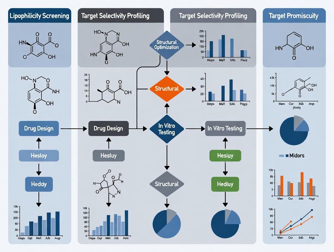
Navigating the Lipophilicity-Promiscuity Nexus: Strategies for Optimizing Drug Safety and Efficacy
This article provides a comprehensive analysis of the critical relationship between high lipophilicity and target promiscuity in small-molecule drug discovery.
Navigating the Lipophilicity-Promiscuity Nexus: Strategies for Optimizing Drug Safety and Efficacy
Abstract
This article provides a comprehensive analysis of the critical relationship between high lipophilicity and target promiscuity in small-molecule drug discovery. It explores the foundational principles of how physicochemical properties influence pharmacokinetics and safety profiles, detailing computational and experimental methodologies for prediction and measurement. The content offers practical strategies for troubleshooting optimization challenges and validates approaches through comparative analysis of successful and discontinued drugs. Aimed at researchers and drug development professionals, this review synthesizes current knowledge to guide the design of compounds with improved therapeutic indices and reduced development attrition.
The Lipophilicity-Promiscuity Link: Understanding Fundamental Mechanisms and Impacts
FAQs on Lipophilicity and Promiscuity
Q1: What is the fundamental difference between LogP and LogD?
- LogP is the logarithm of the partition coefficient (P), which is the ratio of the concentration of a neutral (unionized) compound in an organic phase (typically n-octanol) to its concentration in an aqueous phase (water). It is a constant for a given compound [1] [2] [3].
- LogD is the logarithm of the distribution coefficient (D), which is the ratio of the sum of the concentrations of all species of a compound (both ionized and unionized) in the organic phase to the sum in the aqueous phase at a specified pH. LogD is therefore pH-dependent and provides a more accurate measure of lipophilicity for ionizable compounds under physiological conditions [1] [2] [4].
Q2: Why are LogP and LogD critical parameters in drug discovery?
Lipophilicity is a key physicochemical parameter that influences nearly all aspects of a drug's behavior, including [5] [4] [3]:
- Absorption and Permeability: Impacts a compound's ability to cross biological membranes.
- Distribution: Affects tissue penetration and volume of distribution.
- Metabolism and Clearance: Higher lipophilicity often correlates with increased metabolic clearance.
- Toxicity and Promiscuity: Increased lipophilicity is linked to target promiscuity, off-target effects (e.g., hERG inhibition), and other ADMET (Absorption, Distribution, Metabolism, Excretion, and Toxicity) liabilities.
- Solubility: Higher lipophilicity generally leads to lower aqueous solubility.
Q3: What is molecular promiscuity, and why is it significant?
- Definition: Molecular promiscuity denotes the ability of a ligand to specifically interact with multiple, sometimes distantly related, target proteins [6] [7].
- Significance: While promiscuity can lead to unwanted side effects, it is receiving increasing attention because it can enhance drug efficacy through polypharmacology (simultaneous modulation of multiple targets) and provides a molecular basis for drug repositioning [6] [8]. However, highly promiscuous compounds also carry a higher risk of toxicity [5].
Q4: What are common experimental methods for determining LogP and LogD?
The following table summarizes the key methodologies [4] [3]:
| Method | Description | Key Considerations |
|---|---|---|
| Shake-Flask | The compound is shaken in a mixture of octanol and water (or buffer); concentrations in each phase are measured at equilibrium. | Considered the "gold standard"; can be slow and requires a method for concentration analysis [4] [3]. |
| Chromatographic Methods | Using High-Performance Liquid Chromatography (HPLC) to determine retention time, which is correlated with known LogP values of standard compounds. | A faster, high-throughput alternative to the shake-flask method [3]. |
Q5: How can computational methods for LogP/LogD prediction fail, and how can this be mitigated?
Computational methods, while invaluable, have limitations:
- Fragment-Based Methods: These methods sum contributions from molecular fragments and correction factors. They can be inaccurate for novel scaffolds or functional groups not well-represented in their training data [1] [3].
- Mitigation: It is crucial to use a single, consistent computational tool for a series of compounds and to validate predictions with experimental data whenever possible. Some software allows the training set to be extended with in-house measured values for greater accuracy [3].
Troubleshooting Common Experimental and Data Interpretation Issues
Problem 1: In Vivo Half-Life Does Not Improve Despite Lowering Lipophilicity
- Background: A common strategy to improve pharmacokinetics is to reduce lipophilicity to lower clearance (CL). However, this often fails to extend the in vivo half-life (T~1/2~) [5].
- Root Cause: The volume of distribution (V~d,ss~) and clearance (CL) are often highly correlated and similarly affected by lipophilicity. Reducing lipophilicity can lower both V~d,ss~ and CL, resulting in little to no net improvement in T~1/2~, which is a function of both parameters (T~1/2~ = 0.693 • V~d,ss~ / CL) [5].
- Solution: Focus on identifying and addressing specific metabolic soft-spots in the molecule to improve metabolic stability (lower CL~int~) without necessarily reducing lipophilicity. Matched molecular pair analysis has shown that strategies improving metabolic stability without decreasing lipophilicity have an 82% probability of prolonging half-life, compared to only a 30% probability for strategies that rely solely on lowering lipophilicity [5].
Problem 2: Suspecting Apparent Promiscuity Due to Assay Artifacts
- Background: Apparent multi-target activity (promiscuity) can be a false positive resulting from compound-mediated assay interference [7].
- Root Cause: Compounds can form aggregates, react under assay conditions, or engage in non-specific interactions with proteins, leading to artifactual activity readouts [7].
- Solution: Implement rigorous filtering of screening data to exclude compounds with chemical functionalities prone to artifacts. Use secondary, orthogonal assays to confirm true activity. Analysis of public screening data has shown that after applying such liability filters, a significant proportion of initially "promiscuous" compounds can be eliminated from consideration [7].
Problem 3: Difficulty in Rationalizing or Designing for Desired Promiscuity
- Background: Designing compounds with specific multi-target activity is challenging because the structural basis for promiscuity is often not generalizable [8].
- Root Cause: Machine learning studies reveal that structural features distinguishing promiscuous from non-promiscuous compounds are highly dependent on the specific target combination. Models trained to recognize promiscuity for one target pair typically fail to predict it for a different target pair [8].
- Solution: Instead of seeking universal "promiscuity features," focus on local structure-promiscuity relationships for your target of interest. Analyze structural data (e.g., X-ray complexes) of known promiscuous ligands bound to their targets to understand the specific interaction patterns and binding modes that enable multi-target activity [6] [8].
Diagram: Lipophilicity and Promiscuity in Drug Discovery
Lipophilicity and Promiscuity Relationships
Experimental Protocols & Workflows
Shake-Flask Method for LogD Determination
This is a standard protocol for experimentally measuring the distribution coefficient [4] [3].
Research Reagent Solutions & Materials:
| Reagent/Material | Function |
|---|---|
| n-Octanol | Represents the lipophilic/organic phase. |
| Aqueous Buffer (e.g., pH 7.4) | Represents the aqueous phase at physiological pH. |
| Test Compound | The molecule whose LogD is being characterized. |
| Analytical Instrument (e.g., HPLC-UV, LC-MS) | To accurately quantify the concentration of the compound in each phase after partitioning. |
Detailed Methodology:
- Preparation: Pre-saturate n-octanol with the aqueous buffer and vice-versa by mixing them thoroughly and allowing them to separate. This prevents volume changes in the phases during the experiment.
- Partitioning: Dissolve a known amount of the test compound in a known volume of one of the phases (e.g., the aqueous buffer). Combine this with an equal volume of the other pre-saturated phase (e.g., n-octanol) in a suitable container.
- Equilibration: Shake the mixture vigorously for a predetermined time at a constant temperature to allow the compound to distribute between the two phases.
- Separation: Allow the mixture to stand undisturbed until the two phases are completely separated.
- Analysis: Carefully sample from each phase and measure the concentration of the compound in each using a suitable analytical method (e.g., HPLC-UV).
- Calculation: Calculate the Distribution Coefficient (D) using the formula: ( D = \frac{[Concentration]{octanol}}{[Concentration]{buffer}} ) LogD is then the logarithm (base 10) of this value.
Workflow for Identifying True Multitarget Activity (Promiscuity)
This workflow helps distinguish true promiscuity from false positives [7] [8].
Promiscuity Confirmation Workflow
The Scientist's Toolkit: Essential Research Reagents & Materials
The following table lists key resources used in the experiments and analyses cited in this guide.
| Research Reagent / Resource | Function / Explanation |
|---|---|
| n-Octanol / Water System | The standard solvent system for measuring LogP/LogD, serving as a model for a drug partitioning between lipid bilayers and aqueous body fluids [1] [4] [3]. |
| Rat Hepatocytes (RH CL~int~) | An in vitro system used to measure intrinsic metabolic clearance, helping to predict in vivo metabolic stability and half-life [5]. |
| Matched Molecular Pairs (MMPs) | A computational analysis technique that identifies pairs of compounds differing only by a small, well-defined structural change. Used to quantify the effect of specific chemical transformations on properties like LogD, metabolic stability, and promiscuity [5] [3]. |
| Structural Fingerprints (for ML) | Numerical representations of chemical structure used in machine learning models to diagnose and predict structure-promiscuity relationships for specific target combinations [8]. |
| CYP450 Inhibition Assays | Essential experimental panels to assess a compound's potential for drug-drug interactions, a common liability linked to high lipophilicity and promiscuity [5] [3]. |
Troubleshooting Guide: FAQs on Lipophilicity in Drug Discovery
FAQ 1: Why is my compound showing high membrane permeability in assays but also exhibiting promiscuous behavior and off-target toxicity?
This is a classic consequence of high lipophilicity. While lipophilicity is a key driver for passive diffusion across lipid membranes, it is also a major factor in off-target binding and certain toxicity mechanisms.
- Root Cause: High lipophilicity, often measured by LogP or LogD, increases the likelihood of a compound engaging in non-specific, hydrophobic interactions with a wide range of biological targets beyond its intended one [9]. Furthermore, specific structural features, such as a basic center with a pKa > 6, are strongly associated with promiscuous binding to aminergic GPCRs, ion channels, and transporters [9]. Certain amphiphilic compounds can also induce phospholipidosis, a toxicity driven by their physicochemical properties [10].
- Solution:
- Modify Physicochemical Properties: Aim to reduce lipophilicity by introducing polar groups or replacing lipophilic substituents with more polar bioisosteres [11]. However, this must be done carefully to avoid compromising permeability.
- Evaluate the Necessity of a Basic Center: If a basic amine is not part of the core pharmacophore, consider replacing it with a neutral group to reduce promiscuity [9].
- Adopt "Molecular Chameleonicity": Design larger molecules (e.g., beyond Rule of 5) that can adopt a polar, open conformation in aqueous environments to enhance solubility, and a non-polar, closed conformation in lipid membranes to maintain permeability [11].
FAQ 2: How can I accurately predict passive drug permeability early in the discovery process?
Predicting permeability is essential for estimating oral bioavailability. A combination of computational and experimental methods is recommended.
- Root Cause: Reliance on a single method or insufficient understanding of the relationship between molecular descriptors and permeability can lead to poor predictions.
- Solution:
- Utilize In Silico Tools: Services like the Permeability server can provide theoretical assessments of passive permeability [12].
- Employ High-Throughput Simulations: Physics-based coarse-grained models can efficiently explore chemical space and establish a permeability surface based on key molecular descriptors like bulk partitioning free energy and pKa [13].
- Implement Standardized In Vitro Assays:
FAQ 3: Our lead compound has excellent potency but a high logP (>5). What are the specific risks, and how can we mitigate them?
A high logP is a significant risk factor that requires careful management.
- Root Cause: High lipophilicity is correlated with increased promiscuity, poor aqueous solubility, and a higher risk of off-target toxicities [10] [9].
- Solution:
- Conduct Early Safety Profiling: Screen the compound against a small, representative panel of high-risk off-targets (e.g., aminergic GPCRs, hERG channel) to identify promiscuity early [9].
- Monitor for Phospholipidosis: Be alert for cytoplasmic vacuolation in in vitro or in vivo studies, a common toxicity of cationic amphiphilic drugs [10].
- Balance Properties: Use strategies like Aufheben to simultaneously improve aqueous solubility and membrane permeability through rational molecular design, rather than simply increasing hydrophilicity at the expense of permeability [11].
The following tables consolidate quantitative data and structural alerts related to lipophilicity, permeability, and promiscuity.
Table 1: Permeability and Promiscuity Relationships with Lipophilicity and Charge
| Property / Metric | Impact on Permeability | Impact on Promiscuity / Toxicity | Key Evidence |
|---|---|---|---|
| High Lipophilicity (High LogP) | Increases passive permeability by favoring partitioning into lipid membranes [13] [11]. | Markedly increases promiscuity and risk of off-target effects [9]. | Promiscuity rarely observed for compounds with cLogP < 3 [9]. |
| Basic Center (pKa > 6) | Can enhance permeability in some contexts. | The most important determinant of promiscuity in safety panels; high hit rates at aminergic targets [9]. | Positively charged compounds show ~15% average target hit rate at aminergic GPCRs [9]. |
| Molecular Weight (500-3000 Da, bRo5) | Challenging to achieve high permeability; often requires chameleonic properties [11]. | Can be associated with unique off-target risks, e.g., with oligonucleotides or lipid nanoparticles [10]. | Requires design strategies that go beyond the classic Rule of 5 [11]. |
Table 2: Structural Motifs and Property Alerts
| Structural Motif / Property | Associated Risk or Effect | Recommended Action |
|---|---|---|
| Tricyclic motif with basic amine | High-risk motif for broad promiscuity [9]. | Scrutinize necessity; consider structural modification early. |
| Cationic Amphiphilic Structure | Strong association with phospholipidosis and vacuolation [10]. | Incorporate specific in vitro or in vivo screening for this pathology. |
| High Hydrophobicity + Positive Charge | Increased risk of cytotoxicity and haemolysis [10]. | Monitor in cytotoxicity assays and inspect for haemolytic potential. |
Experimental Protocols for Key Assays
Protocol 1: Determining Apparent Permeability (Papp) using PAMPA
Objective: To measure the passive, artificial membrane permeability of a compound in a high-throughput format [12].
Materials:
- PAMPA plate system (donor and acceptor plates with a membrane filter)
- Artificial lipid solution (e.g., lecithin in dodecane)
- Test compound solution in buffer (e.g., PBS)
- Acceptor sink buffer
- UV plate reader or LC-MS/MS for quantification
Method:
- Membrane Preparation: Coat the filter of the PAMPA donor plate with the artificial lipid solution and allow it to set.
- Plate Assembly: Fill the donor well with the test compound solution. Place the membrane on top and ensure contact. Fill the acceptor well with the sink buffer.
- Incubation: Assemble the plate and incubate at room temperature for a predetermined time (e.g., 4-16 hours) to reach steady state.
- Sampling and Analysis: After incubation, sample from both donor and acceptor compartments. Quantify the drug concentration in each compartment using a suitable analytical method (e.g., UV spectrometry or LC-MS/MS).
- Data Calculation:
- Calculate the drug flux (j): ( j = (dQ/dt) \times (1/A) ), where ( dQ/dt ) is the slope of the accumulated amount in the acceptor compartment over time, and ( A ) is the permeation area.
- Calculate the apparent permeability (Papp): ( P{app} = j / C0 ), where ( C_0 ) is the initial concentration in the donor compartment [12].
- Papp is typically classified as: poor (< 1.0 × 10⁻⁶ cm/s), moderate (1–10 × 10⁻⁶ cm/s), or good (>10 × 10⁻⁶ cm/s) [11].
Protocol 2: Early Safety Profiling with a Focused Target Panel
Objective: To identify pharmacological promiscuity early in drug discovery by screening against a minimal panel of high-risk off-targets.
Materials:
- Cell lines or protein preparations expressing high-risk targets (e.g., hERG, 5-HT2B, α1A-adrenergic receptor, dopamine D2 receptor) [9].
- Relevant assay kits (e.g., binding or functional assays).
- Test compounds and reference controls.
Method:
- Panel Selection: Select a panel of 10-20 targets known to attract high hit rates, particularly for compounds with specific properties (e.g., basic amines). Aminergic GPCRs, the hERG channel, and opioid receptors are prime candidates [9].
- Assay Execution: Run standardized binding or functional assays for each target in the panel according to established protocols.
- Data Analysis: Determine the percentage inhibition or activation at a single concentration of the test compound (e.g., 10 µM). A compound is considered a "hit" for a specific target if it shows significant activity (e.g., >50% inhibition/activation).
- Interpretation: Calculate a "hit rate" for the compound across the panel. A high hit rate indicates a promiscuous compound. This data can be used to prioritize compounds for further development and guide chemical redesign to mitigate off-target interactions [9].
Visualizing Key Concepts and Workflows
Diagram 1: Lipophilicity Impact on Permeability and Promiscuity
Diagram 2: Mechanistic Pathways of Off-Target Toxicity
The Scientist's Toolkit: Research Reagent Solutions
Table 3: Essential Materials for Permeability and Safety Assessment
| Research Reagent / Tool | Function in Research | Key Application Note |
|---|---|---|
| Caco-2 Cell Line | A human intestinal epithelial cell model used to predict drug absorption, incorporating both passive and active transport mechanisms [12]. | The gold standard for in vitro assessment of intestinal permeability; provides data on efflux and transporter effects. |
| PAMPA Plate | A high-throughput artificial membrane system designed to measure passive transcellular permeability [12]. | Ideal for early-stage screening due to its speed, low cost, and suitability for automation. |
| Selected Target Panels (e.g., aminergic GPCRs) | A curated set of recombinant proteins or cell lines for profiling compound activity against known high-risk off-targets [9]. | Enables early detection of pharmacological promiscuity. A panel of ~10 targets can effectively identify most promiscuous compounds. |
| Coarse-Grained (CG) Martini Model | A physics-based computational model that reduces molecular complexity, allowing for high-throughput simulation of membrane permeability across vast chemical spaces [13]. | Used to predict permeability coefficients and map structure-permeability relationships for thousands of compounds in silico. |
Structural and Physicochemical Drivers of Promiscuous Binding
Frequently Asked Questions
What is lipophilicity and why is it critical in drug discovery? Lipophilicity, often measured as LogP, represents the ratio of a compound's concentration in an oil phase versus an aqueous phase at equilibrium [14]. It is a fundamental physicochemical parameter that significantly influences various pharmacokinetic properties, including absorption, distribution, membrane permeability, and routes of clearance [14]. A drug's affinity for biological membranes and its binding ability are heavily influenced by its lipophilicity [15].
How does lipophilicity relate to promiscuous binding and transporter activity? High lipophilicity is a key driver of promiscuous binding, as it can increase a drug's likelihood of interacting with off-target proteins and promiscuous transporters [5]. P-glycoprotein (P-gp), a highly flexible and promiscuous transporter that effluxes over 200 chemically diverse substrates from cells, is a prime example. Its conformational plasticity allows it to bind a wide array of structures, and lipophilicity is a key factor in determining whether a compound will be a substrate [16].
What is the primary experimental method for determining lipophilicity? Reverse-phase thin layer chromatography (RP-TLC) is a widely used, simple, and low-cost method for determining lipophilicity-related parameters like the isocratic retention factor (Rₘ) and chromatographic hydrophobic index (φ₀) [15]. Reverse-phase HPLC (RP-HPLC) is another excellent method valued for its accuracy and on-line detection capabilities [15].
My compound has high lipophilicity and shows high clearance. Will simply reducing lipophilicity always extend its half-life? Not necessarily. While lowering lipophilicity can decrease clearance, it often also reduces the volume of distribution. Since half-life depends on the balance between volume of distribution and clearance, this strategy can fail to improve half-life if it does not specifically address a metabolic soft-spot [5]. Transformations that improve metabolic stability without decreasing lipophilicity are often more successful for half-life extension [5].
What are some common strategies to mitigate high lipophilicity and reduce promiscuity? Common strategies include [5]:
- Introducing metabolically stable polar groups (e.g., replacing a methyl with a fluorine).
- Reducing overall hydrocarbon content and molecular planarity.
- Introducing hydrogen bond donors or acceptors.
- Addressing specific metabolic soft-spots identified in assays, rather than making global changes to lipophilicity.
Troubleshooting Guides
Problem: Poor Aqueous Solubility and High Non-Specific Binding
Background: Compounds with high lipophilicity often suffer from poor aqueous solubility, which can impede absorption and lead to high non-specific binding, confounding assay results [14].
| Investigation | Possible Cause | Suggested Action |
|---|---|---|
| Solubility Check | Poor dissolution in aqueous buffers. | Use solubilizing agents (e.g., DMSO, cyclodextrins); consider salt formation for ionizable compounds. |
| Assay Signal | High background signal due to compound aggregation or adhesion to labware. | Include control wells without biological target; use detergents (e.g., Tween-20) in buffers to reduce non-specific binding [17]. |
| Cellular Uptake | Low intracellular concentration despite good LogP. | Investigate if the compound is a substrate for efflux transporters like P-gp [16]. |
Experimental Protocol: Investigating P-gp Substrate Status
- Cell Line: Use polarized cell lines (e.g., MDCK, Caco-2) overexpressing human P-gp.
- Assay Setup: Seed cells on transwell filters and allow them to form tight monolayers. Check monolayer integrity by measuring transepithelial electrical resistance (TEER).
- Bidirectional Transport: Add your test compound to either the apical (A) or basolateral (B) chamber.
- A-to-B direction: Measures substrate permeation.
- B-to-A direction: Measures active efflux.
- Inhibition: Repeat the experiment in the presence of a known P-gp inhibitor (e.g., Verapamil, Elacridar).
- Data Analysis: Calculate the efflux ratio (B-to-A / A-to-B permeability). An efflux ratio >2 that is reduced by an inhibitor suggests the compound is a P-gp substrate [16].
Problem: Unpredictable Clearance and Short Half-Life
Background: High lipophilicity generally correlates with increased metabolic clearance, leading to a short in vivo half-life, which can be problematic for maintaining target coverage [5].
| Investigation | Possible Cause | Suggested Action |
|---|---|---|
| In Vitro Stability | High intrinsic clearance in hepatocyte or microsomal assays. | Identify metabolic soft-spots using metabolite identification (MetID) studies. |
| In Vivo PK | High in vivo clearance not predicted by in vitro assays. | Investigate extra-hepatic metabolism or other clearance pathways (e.g., biliary excretion). |
| Half-Life | Low volume of distribution (Vd,ss) counteracts reduced clearance. | Focus on structural modifications that lower clearance without drastically reducing Vd,ss, such as targeted blocking of metabolic soft-spots [5]. |
Experimental Protocol: Metabolic Soft-Spot Identification
- Incubation: Incubate the test compound with liver microsomes or hepatocytes (from rat, mouse, or human) in the presence of NADPH cofactor.
- Time Points: Remove aliquots at specified time points (e.g., 0, 15, 30, 60 minutes).
- Termination: Stop the reaction by adding an organic solvent (e.g., acetonitrile).
- Analysis: Centrifuge to precipitate proteins and analyze the supernatant using LC-MS/MS.
- Data Interpretation: Identify metabolites based on mass shifts. The structures of major metabolites reveal the labile parts of the molecule (soft-spots) for further modification [5].
Data Presentation
Table 1: Impact of Lipophilicity on Key Pharmacokinetic Parameters in Neutral Compounds [5]
| LogD₇.₄ Range | Typical Vd,ss,u (L/kg) | Typical CLu (mL/min/kg) | Impact on Half-Life |
|---|---|---|---|
| <1 | Low | Low | Variable; can be short due to renal clearance. |
| 1 - 2.5 | Moderate | Moderate | Most favorable balance for half-life. |
| >2.5 - 4 | High | High | Often short due to very high clearance. |
| >4 | Very High | Very High / Variable | Can be long if clearance is low, but solubility is a major issue. |
Table 2: Effect of Common Molecular Transformations on Half-Life and Lipophilicity [5]
| Transformation | Typical ΔLogD₇.₄ | Impact on Metabolic Stability | Probability of Prolonging Half-Life |
|---|---|---|---|
| H → F | Decrease | Increases | High |
| H → Cl | Increase | Increases | High |
| CH₃ → CF₃ | Increase | Increases | High |
| Lowering LogD without addressing soft-spot | Decrease | No change / slight increase | Low (~30%) |
| Improving metabolic stability without lowering LogD | Variable | Increases | High (~82%) |
The Scientist's Toolkit
Table 3: Key Research Reagent Solutions
| Reagent / Assay | Function in Promiscuity & ADME Research |
|---|---|
| P-glycoprotein (P-gp) Assay Systems | To determine if a new chemical entity is a substrate or inhibitor of this key promiscuous efflux transporter, critically impacting its distribution, particularly to the brain [16]. |
| Rat Hepatocyte (RH) CLint Assay | An in vitro assay to measure intrinsic metabolic clearance, used to predict in vivo hepatic clearance and identify compounds with high metabolic lability [5]. |
| Octanol-Water Partitioning | The gold-standard experimental system for determining the partition coefficient (LogP) or distribution coefficient (LogD) of a compound, defining its lipophilicity [14] [5]. |
| LC-MS/MS Systems | Essential for conducting metabolite identification (MetID) studies to pinpoint metabolic soft-spots and for quantifying drug concentrations in bio-matrices during PK studies. |
Experimental Workflow Diagrams
Diagram 1: Troubleshooting high lipophilicity workflow.
Diagram 2: P-gp transport cycle and promiscuity.
Frequently Asked Questions (FAQs)
Q1: How does lipophilicity fundamentally affect a drug's journey in the body? Lipophilicity, often measured as LogP (partition coefficient) or LogD (distribution coefficient at a specific pH), is a primary physicochemical property that influences every aspect of a drug's pharmacokinetics (PK) [18] [19]. It underlies higher-level properties, affecting passive membrane permeability, solubility, metabolic stability, and the route of clearance [18] [20]. A drug's lipophilicity determines its ability to cross biological membranes for absorption, its distribution into tissues, and how it is ultimately cleared from the body, either via hepatic metabolism or renal excretion [20] [19].
Q2: What is the optimal lipophilicity range for an orally administered drug? While context-dependent, a LogD₇.₄ between 1 and 3 is generally considered desirable for oral drugs [18]. This range often provides a balanced profile:
- LogD₇.₄ < 1: Associated with high solubility but potentially low permeability and poor absorption [18].
- LogD₇.₄ 1–3: Typically offers a good balance of moderate solubility and permeability, with potential for good absorption and bioavailability [18].
- LogD₇.₄ > 3–5: Often leads to low solubility, high metabolism, and variable oral absorption [18].
Q3: I need to increase my drug's brain penetration. Is increasing lipophilicity a reliable strategy? Increasing lipophilicity can enhance blood-brain barrier (BBB) permeation, but it is a double-edged sword [18]. While higher lipophilicity can improve passive diffusion across the compact BBB, it can also increase binding to efflux pumps like P-glycoprotein and raise metabolic clearance [18] [19]. Therefore, simply increasing lipophilicity without considering these other factors may not improve overall brain exposure and could be counterproductive. The parameter Δlog P (a measure of the difference between lipophilicity in two solvent systems) has also been used as an indicator for blood-brain partitioning [18].
Q4: Why did reducing my compound's lipophilicity not extend its half-life as expected? This is a common pitfall. Reducing lipophilicity often decreases clearance (CL), but it can also reduce the volume of distribution (Vd,ss) because the drug is less likely to distribute into tissues [5]. Since half-life (T₁/₂) is proportional to both Vd,ss and CL, if both decrease similarly, the net effect on half-life can be negligible [5]. A more successful strategy is to directly address a metabolic soft-spot to improve metabolic stability, rather than relying solely on global lipophilicity reduction [5].
Q5: How does lipophilicity influence the clearance route of peptide-drug conjugates? For peptide-drug conjugates and other larger modalities, lipophilicity remains a key determinant of clearance route [20]. Higher lipophilicity (higher LogD) favors hepatic clearance and reduces kidney uptake and associated toxicity [20]. Conversely, lower lipophilicity favors renal clearance. Tuning lipophilicity is therefore a viable strategy to shift the clearance route and mitigate organ-specific toxicity, for example, in targeted radiotherapies [20].
Troubleshooting Guides
Issue 1: Poor Oral Absorption
Problem: Your drug candidate shows low oral bioavailability due to poor absorption.
Potential Causes and Solutions:
| Potential Cause | Diagnostic Experiments | Corrective Actions |
|---|---|---|
| Low Permeability (LogD too low) | - Measure apparent permeability in Caco-2 or MDCK cell assays.- Determine experimental LogD₇.₄. | - Increase lipophilicity within the optimal range (e.g., LogD 1-3) [18].- Introduce non-polar groups (e.g., methyl) to enhance membrane penetration [18]. |
| Low Solubility (LogD too high) | - Measure equilibrium solubility in aqueous buffer.- Review in silico LogP/LogD predictions. | - Reduce lipophilicity by introducing polar groups (e.g., amine, hydroxyl) or ionizable moieties [21].- Consider formulation strategies like nanoemulsions or lipid-based drug delivery systems (LBDDS) to enhance solubility [21]. |
| Efflux by P-gp | - Conduct bidirectional permeability assays with and without a P-gp inhibitor. | - Structural modification to reduce the compound's affinity for the P-gp efflux pump, which may involve reducing lipophilicity or specific structural features [19]. |
Issue 2: High Clearance and Short Half-Life
Problem: Your compound is cleared too quickly, leading to a short half-life that may require frequent dosing.
Potential Causes and Solutions:
| Potential Cause | Diagnostic Experiments | Corrective Actions |
|---|---|---|
| High Metabolic Lability | - Assess stability in liver microsomes or hepatocytes.- Identify metabolic soft-spots via metabolite ID studies. | - Targeted metabolism mitigation: Address the specific soft-spot (e.g., replace a labile methyl group with a cyclopropyl or fluorine) [5].- General lipophilicity reduction: Lower LogD to reduce nonspecific binding to CYP450 enzymes, but be aware this may also reduce Vd,ss [18] [5]. |
| High Renal Clearance of Unbound Drug | - Determine fraction unbound in plasma (fᵤ).- Measure renal clearance in vivo. | - Increasing lipophilicity can reduce renal clearance by increasing plasma protein binding and tissue distribution, shifting clearance to hepatic metabolism [18] [20]. |
Issue 3: High Volume of Distribution Leading to Off-Target Tissue Accumulation
Problem: The drug distributes extensively into tissues, leading to a large volume of distribution and potential off-target toxicity.
Potential Causes and Solutions:
| Potential Cause | Diagnostic Experiments | Corrective Actions |
|---|---|---|
| Excessive Lipophilicity | - Determine tissue distribution in vivo.- Measure plasma protein binding and log D₇.₄. | - Reduce overall lipophilicity to decrease tissue partitioning [18].- Introduce polar or ionizable groups (at physiological pH) to increase solubility in plasma and extracellular fluid. |
| Target Promiscuity and Toxicity | - Conduct counter-screening against common off-targets (e.g., hERG).- Use panels like BioMAP for phenotypic toxicity profiling [22]. | - Reduce lipophilicity, as higher LogD is correlated with increased promiscuity and off-target interactions, including hERG inhibition [18] [19]. |
Experimental Protocols for Key Assays
Determination of Lipophilicity (LogD₇.₄) using the Shake-Flask Method
The shake-flask method is considered the gold standard for the direct experimental determination of LogP/LogD [19].
Principle: A compound is partitioned between n-octanol (non-polar phase) and an aqueous buffer (polar phase, typically pH 7.4). After equilibration and phase separation, the concentration of the solute in each phase is quantified, and the LogD is calculated [19].
Materials:
- Research Reagent Solutions:
- n-Octanol (HPLC grade)
- Phosphate Buffered Saline (PBS), pH 7.4
- Test compound solution in a suitable solvent (e.g., DMSO)
- HPLC vials and LC system with UV/Vis or MS detection
Procedure:
- Pre-saturation: Saturate the n-octanol with PBS and the PBS with n-octanol by vortexing the two phases together and allowing them to separate overnight. Use the pre-saturated phases for the experiment.
- Partitioning: Add a known amount of your test compound to a glass vial. Introduce equal volumes (e.g., 1 mL each) of pre-saturated n-octanol and PBS. Seal the vial tightly.
- Equilibration: Agitate the mixture vigorously for 1 hour at constant temperature (e.g., 25°C) using a mechanical shaker.
- Phase Separation: Allow the phases to separate completely for several hours, or use centrifugation to accelerate separation.
- Quantification: Carefully sample from each phase. Dilute the n-octanol phase with a water-miscible solvent (e.g., methanol) if necessary. Analyze the samples using a calibrated LC-UV or LC-MS method to determine the concentration of the compound in each phase.
- Calculation:
- LogD₇.₄ = Log₁₀ (Concentration in n-octanol / Concentration in buffer)
Visual Workflow:
In Vitro Metabolic Stability Assay in Rat Hepatocytes
This assay predicts in vivo metabolic clearance [5].
Principle: The test compound is incubated with metabolically active hepatocytes. The disappearance of the parent compound over time is monitored to calculate an intrinsic clearance (CLᵢₙₜ) value.
Materials:
- Research Reagent Solutions:
- Cryopreserved or fresh rat hepatocytes
- Williams' E Medium (with incubation supplements)
- Test compound (e.g., 1 mM stock in DMSO)
- Stopping solution (e.g., acetonitrile with internal standard)
- LC-MS/MS system for bioanalysis
Procedure:
- Thawing/Preparation: Thaw cryopreserved hepatocytes according to the vendor's protocol and determine viability (should be >80%). Adjust the cell density to a working suspension (e.g., 1 million viable cells/mL).
- Incubation: Pre-warm the hepatocyte suspension in a shaking incubator at 37°C. Initiate the reaction by adding the test compound (final concentration typically 0.5-1 µM, DMSO concentration ≤0.1%).
- Sampling: At predetermined time points (e.g., 0, 5, 15, 30, 45, 60 minutes), remove an aliquot of the incubation mixture and transfer it to a tube containing the stopping solution (acetonitrile) to precipitate proteins and stop the reaction.
- Analysis: Centrifuge the stopped samples to remove precipitated protein. Analyze the supernatant using LC-MS/MS to determine the peak area of the parent compound.
- Calculation: Plot the natural logarithm of the parent compound concentration remaining versus time. The slope of the linear phase is the elimination rate constant (k). Intrinsic clearance (CLᵢₙₜ, mL/min/kg) can be calculated using the formula: CLᵢₙₜ = k / (number of cells per mL * liver weight per kg body weight).
Visual Workflow:
Table 1: Impact of Lipophilicity (LogD₇.₄) on Key Pharmacokinetic Parameters This table synthesizes general relationships observed in drug discovery [18] [20].
| LogD₇.₄ Range | Solubility | Permeability | Primary Clearance Route | Volume of Distribution (Vd,ss) | Common PK Challenges |
|---|---|---|---|---|---|
| < 1 | High | Low | Renal | Low | Low absorption and bioavailability; Potential for renal clearance [18]. |
| 1 - 3 | Moderate | Moderate | Balanced | Moderate | Balanced profile; Potential for good oral absorption [18]. |
| 3 - 5 | Low | High | Hepatic Metabolism | High | Variable oral absorption; Nonlinear PK due to enzyme saturation [18]. |
| > 5 | Poor | High | Hepatic Metabolism | Very High | Poor oral absorption; High metabolic clearance; Promiscuity & toxicity risk [18] [19]. |
The Scientist's Toolkit: Essential Research Reagents and Materials
Table 2: Key Reagents for Lipophilicity and PK Studies
| Reagent / Material | Function/Brief Explanation | Example Use Cases |
|---|---|---|
| n-Octanol & Buffer Systems | The standard solvent system for the direct measurement of partition/distribution coefficients (LogP/LogD) [19]. | Shake-flask LogD determination [19]. |
| Cryopreserved Hepatocytes | Metabolically competent cells used to assess in vitro metabolic stability and predict in vivo hepatic clearance [5]. | Intrinsic clearance (CLᵢₙₜ) assays; Metabolite identification studies. |
| Caco-2 Cell Line | A human colon adenocarcinoma cell line that, upon differentiation, forms a monolayer with properties of intestinal epithelium. Used to model oral drug absorption. | Apparent permeability (Pₐₚₚ) assays; Studies on efflux transport (e.g., P-gp). |
| LC-UV and LC-MS Systems | Essential analytical tools for quantifying compound concentration in complex matrices like buffers, biological fluids, and cell lysates. | LogD analysis; Bioanalysis from in vitro and in vivo samples; Metabolite profiling. |
| Specialized Lipid Excipients | Ionizable lipids, phospholipids, and cationic lipids used in formulations to overcome delivery challenges of highly lipophilic drugs [21]. | Formulating Lipid Nanoparticles (LNPs) for nucleic acids; Creating lipid-based drug delivery systems (LBDDS) for small molecules [21]. |
Frequently Asked Questions (FAQs)
FAQ 1: What is pharmacological promiscuity and why is it a major safety concern? Answer: Pharmacological promiscuity describes the activity of a single compound against multiple, unintended biological targets. This is a significant safety concern because engaging off-target receptors and enzymes can lead to a range of adverse side effects, often causing drug candidates to fail during clinical development or even leading to the withdrawal of approved drugs from the market. Undesired promiscuity is a primary source of safety attrition in the drug discovery process [9].
FAQ 2: Which molecular properties are most strongly associated with increased promiscuity? Answer: The most important molecular properties linked to increased promiscuity are high lipophilicity and the presence of a basic center.
- Lipophilicity: Promiscuity increases with lipophilicity (often measured as LogP or ClogP). Marked promiscuity is rarely observed for compounds with a ClogP < 3 [9].
- Basic Center: A basic center with a calculated pKa (cpKa(B)) greater than 6 is the most significant determinant of promiscuity in typical safety panels. This is because such compounds frequently interact with a small set of targets, such as aminergic GPCRs, which attract surprisingly high hit rates [9].
FAQ 3: How can colloidal aggregation lead to false-positive results in screening? Answer: Highly lipophilic molecules, like cannabidiol (CBD), have very poor aqueous solubility. Above a critical concentration (the Critical Aggregation Concentration, or CAC), these molecules can form colloidal dispersions or aggregates in assay media. These colloids can nonspecifically interfere with proteins and enzymes, leading to false-positive signals in in vitro assays that do not represent true, specific pharmacological activity. This phenomenon can misleadingly suggest a compound is broadly active [23].
FAQ 4: What are some experimental strategies to identify and eliminate false-positive hits? Answer: To prioritize high-quality hits and eliminate artifacts, a cascade of experimental approaches is recommended [24]:
- Counter Screens: Use assays designed solely to detect compound-mediated assay interference (e.g., autofluorescence, signal quenching).
- Orthogonal Assays: Confirm bioactivity using an entirely different readout technology (e.g., follow up a fluorescence-based readout with a luminescence- or absorbance-based assay).
- Biophysical Assays: Employ techniques like Surface Plasmon Resonance (SPR) or Isothermal Titration Calorimetry (ITC) to validate direct binding and measure affinity.
- Use of Detergents: Adding non-ionic detergents like Triton X-100 can disrupt colloidal aggregates and prevent this specific type of false-positive interference [23].
- Cellular Fitness Screens: Test for general cytotoxicity to ensure the observed activity is not simply due to cell death [24].
FAQ 5: Which target families are most frequently hit by promiscuous compounds? Answer: Analysis of large screening datasets reveals that a relatively small set of targets are responsible for the majority of promiscuity. The most frequently hit target classes include [9]:
- Aminergic GPCRs (e.g., serotonin, dopamine, and adrenergic receptors)
- Certain ion channels
- Opioid receptors
- Aminergic transporters (e.g., serotonin transporter)
Troubleshooting Guides
Issue 1: Investigating Promiscuity and Off-Target Activity
Problem: A lead compound shows excellent potency against its primary therapeutic target but demonstrates a high fail rate in a broad safety pharmacology panel, indicating potential promiscuity.
Solution: Systematically investigate the physicochemical drivers and specific off-target interactions.
Step-by-Step Guide:
- Profile Physicochemical Properties: Calculate key properties, especially lipophilicity (LogP/ClogP) and the pKa of any basic centers. Compounds with ClogP > 3 and a basic center (pKa > 6) have a high risk of promiscuity [9].
- Screen Against a Representative Mini-Panel: To enable early recognition of promiscuity, screen the compound against a small, focused panel of high-risk targets. This should include key aminergic GPCRs (e.g., 5-HT2B, 5-HT2C, α1A, α1B, H1, M1, M2) and the hERG channel [9].
- Conduct Binding Assays with Detergents: If promiscuous inhibition is observed in enzymatic or binding assays, repeat the experiments in the presence of a non-ionic detergent like Tocrisolve or Triton X-100 (e.g., at 0.01%). A significant reduction in activity suggests the effect is due to nonspecific colloidal aggregation rather than specific target binding [23].
- Perform Orthogonal Binding Validation: Use a biophysical method like Surface Plasmon Resonance (SPR) to confirm direct binding to the primary target and to characterize binding kinetics (kon, koff). This helps distinguish specific from nonspecific interactions [25] [24].
- Analyze Dose-Response Curves: Examine the shape of the dose-response curve. Steep, shallow, or bell-shaped curves can indicate toxicity, poor solubility, or compound aggregation, signaling a potential false-positive result [24].
Workflow Diagram:
Issue 2: Managing High Lipophilicity in Lead Compounds
Problem: A compound series has high lipophilicity (LogP > 5), leading to poor aqueous solubility, assay promiscuity, and potential long-term toxicity risks.
Solution: Implement strategies to reduce lipophilicity and mitigate its negative effects.
Step-by-Step Guide:
- Quantify the Risk: Calculate the Fraction Lipophilicity Index (FLI). The drug-like FLI range is typically 0-8. Compounds falling outside this range, especially on the high end, present a significant risk for promiscuity and poor absorption [26].
- Apply Structural Modifications:
- Introduce Polar Groups: Add hydrogen bond donors/acceptors or slightly acidic/basic groups to increase solubility and reduce LogP.
- Reduce Aromatic Rings: Lower the number of aromatic rings and heavy atoms to decrease molecular "obesity" [26].
- Remove Halogens: Replace halogen atoms (which greatly increase lipophilicity) with more polar bioisosteres like cyanos, amides, or hydroxys [9].
- Optimize Lipophilic Efficiency (LipE): Use metrics like Lipophilic Ligand Efficiency (LLE), where LLE = pIC50 (or pEC50) - LogP (or LogD). Focus on compound series that maintain high potency while achieving lower lipophilicity [26].
- Utilize Automation for Low-Volume Assays: To overcome solubility limitations in testing, use automated, non-contact liquid handlers that enable miniaturization (e.g., nanoliter-range dispensing). This reduces reagent consumption and allows for more reliable testing of compounds with limited solubility [27].
Relationship Diagram: High Lipophilicity and Clinical Consequences
Data Presentation
Table 1: Key Physicochemical Properties Linked to Promiscuity and Safety Attrition
| Property | High-Risk Threshold | Associated Consequence | Clinical Impact |
|---|---|---|---|
| Lipophilicity (LogP/ClogP) | > 3 - 5 [9] [26] | Increased nonspecific binding, poor solubility, metabolic instability [23] [26] | Higher risk of off-target toxicity, poor pharmacokinetics, and drug-drug interactions [9] |
| Presence of a Basic Center | pKa(B) > 6 [9] | High hit rates on aminergic GPCRs, ion channels, and transporters [9] | Cardiovascular side effects, central nervous system disturbances, and other off-target toxicities [9] |
| Fraction Lipophilicity Index (FLI) | Outside 0 - 8 range [26] | Suboptimal balance of permeability and solubility, predicting poor absorption [26] | Low oral bioavailability and increased risk of development failure [26] |
Table 2: Experimental Strategies to Mitigate Promiscuity-Related Artifacts
| Strategy | Method / Reagent | Function / Purpose | Key Outcome |
|---|---|---|---|
| Counter Assays | Signal-based control assays (e.g., fluorescence quenching test) | Identify technology-specific assay interference [24] | Flags compounds that interfere with the detection method, not the biology |
| Orthogonal Assays | SPR, ITC, MST, or a different readout technology (e.g., luminescence) [24] | Confirm bioactivity and binding via an independent method [24] | Validates true positive hits and provides reliable affinity data |
| Aggregation Control | Addition of Triton X-100 (0.01%) or other detergents [23] | Disrupts colloidal aggregates causing false positives [23] | Distinguishes specific target binding from nonspecific colloidal interference |
| Cellular Fitness Assays | CellTiter-Glo, Caspase assays, High-content imaging [24] | Monitor general cell health and viability [24] | Excludes compounds whose activity is due to general cytotoxicity |
The Scientist's Toolkit: Essential Research Reagents and Materials
| Item | Function / Explanation |
|---|---|
| Triton X-100 | A non-ionic detergent used to disrupt and prevent the formation of colloidal aggregates in biochemical assays. Its inclusion helps confirm that observed inhibitory activity is due to specific target binding and not nonspecific aggregation [23]. |
| Tocrisolve | A commercially available, water-soluble lipid emulsion often used as a vehicle for solubilizing highly lipophilic compounds in aqueous assay buffers, helping to mitigate solubility-related artifacts. |
| Bovine Serum Albumin (BSA) | Often added to assay buffers to reduce nonspecific binding of test compounds to plates and equipment, thereby lowering background signal and false-positive rates [24]. |
| I.DOT Liquid Handler | An automated, non-contact dispenser that enables miniaturization of assays to nanoliter volumes. This reduces reagent consumption and allows for more reliable testing of compounds with limited solubility or availability [27]. |
| SPR Chip (e.g., CM5) | The sensor surface used in Surface Plasmon Resonance instruments. It is coated with a dextran matrix that can be functionalized to immobilize a protein target, allowing for label-free analysis of binding kinetics and affinity [25] [24]. |
Computational and Experimental Tools for Prediction and Characterization
Frequently Asked Questions
Q1: What are the most common reasons for large discrepancies between predicted and experimental log P/log D values?
Large discrepancies often arise from several key issues:
- Inadequate Training Data: The chemical space of your compounds is not well-represented in the model's training set. This is particularly common for novel scaffolds, such as Pt(IV) derivatives in platinum complexes, where models trained on older data showed significantly increased Root Mean Squared Error (RMSE) from 0.62 to 0.86 when applied to newer compounds [28].
- Incorrect Descriptor Selection: Using models with descriptors unsuitable for your compound class. For instance, standard small-molecule models often fail for peptides and peptide mimetics, advocating for bespoke approaches [29].
- Improper Protonation State: log D is pH-dependent, and incorrect assignment of the major microspecies at physiological pH (7.4) is a common source of error [29].
Q2: How can I improve prediction accuracy for complex molecules like peptides or metal complexes?
- Use Specialized Models: Employ models specifically developed for your compound class. For peptides and peptide derivatives, machine learning models using tailored molecular descriptors have demonstrated superior accuracy over general-purpose models [29].
- Leverage Consensus Modeling: Combine predictions from multiple models or algorithms. Consensus approaches have been shown to improve accuracy and robustness [30] [28].
- Expand Chemical Space Coverage: If generating new data, ensure it covers underrepresented chemical regions. Retraining a model with a combined dataset that included novel phenanthroline-containing Pt(IV) complexes drastically reduced RMSE from 1.3 to 0.34 [28].
Q3: What is the best workflow to validate computational lipophilicity predictions in a wet-lab setting?
A robust validation workflow integrates both in silico and experimental methods:
- Computational Triangulation: Obtain predictions from multiple algorithms (e.g., AlogPs, XlogP3, ACD/logP) and establish a consensus [30].
- Experimental Benchmarking: Use Reverse-Phase Thin-Layer Chromatography (RP-TLC) as a rapid, cost-effective experimental method to determine the lipophilicity parameter (RₘW) for a representative subset of your compounds [30].
- Correlation Analysis: Establish a quantitative relationship between your experimental RₘW values and the computational predictions to validate and calibrate the in silico models for your specific compound series [30].
Q4: How can I use lipophilicity predictions to address target promiscuity and toxicity risks in early drug discovery?
- Monitor the Lipophilicity Threshold: Compounds with very high log P/log D often have increased risk of promiscuity and toxicity. Implement a log D threshold (e.g., <4 or <5) as a critical filter during compound selection and lead optimization [30].
- Integrate with Other Predictors: Lipophilicity should not be viewed in isolation. Use it as one input into a broader ADMET profiling workflow that includes predictions for solubility, metabolic stability, and hERG binding to gain a more comprehensive safety assessment [31] [32].
Troubleshooting Guides
Issue 1: Poor Correlation Between Predicted and Chromatographic Lipophilicity (RₘW)
Problem: Values from in silico tools (e.g., ALOGPS, XlogP) do not align with experimental RP-TLC results.
Solution:
- Verify Chromatographic Conditions:
- Stationary Phase: Confirm the use of an appropriate reverse-phase plate (e.g., RP-18F₂₅₄ for standard small molecules).
- Mobile Phase: Ensure a binary solvent system with a well-characterized organic modifier (e.g., acetone, acetonitrile, 1,4-dioxane) and water is used. Prepare fresh mobile phases to avoid composition drift [30].
- Standardize Data Processing: Calculate the RₘW value from the slope of the line when Rₘ is plotted against the volume fraction of the organic modifier. Ensure a sufficient number of data points (≥5 concentrations) for a reliable linear fit [30].
- Audit Computational Inputs:
Issue 2: Machine Learning Model Fails on Novel Chemotypes
Problem: A pre-trained ML model for log D prediction performs poorly on your newly synthesized compounds.
Solution:
- Conformity Check: Analyze the new compounds using Principal Component Analysis (PCA) or similar techniques to visualize their position relative to the model's training set chemical space [29].
- Model Retraining/Fine-Tuning:
- If possible, provide experimental log D data for a few representative new chemotypes to fine-tune the existing model.
- For larger datasets, develop a new model using machine learning techniques like Support Vector Regression (SVR) with descriptors selected via methods like LASSO, which have proven effective for specific compound classes like peptides [29].
- Adopt a Multi-Task Learning Approach: Consider using newer models that predict solubility and lipophilicity simultaneously, as these endpoints are thermodynamically linked and can improve overall prediction robustness [28].
Issue 3: Integrating Lipophilicity Predictions into a PBPK Model
Problem: Translating a calculated log D value into reliable parameters for a Physiologically-Based Pharmacokinetic (PBPK) model.
Solution:
- Parameter Mapping: Understand that lipophilicity (log D) is a key input for estimating tissue-to-plasma partition coefficients (Kp) in PBPK models, often via mechanistic equations like those of Rodgers and Rowland.
- Quantify Uncertainty: Acknowledge the inherent uncertainty in in silico predictions. For critical PBPK applications, use a prediction confidence interval (e.g., log D ± 0.5) to perform sensitivity analysis and understand its impact on key PK outcomes like Cmax and AUC [33].
- Leverage AI-Enhanced Workflows: Explore emerging platforms that integrate AI and ML for improved parameter estimation and uncertainty quantification in PBPK modeling, which can help address challenges posed by complex drug formulations and drug-drug interactions [33].
Comparative Data Tables
Table 1: Performance of Computational Log P/Log D Prediction Tools
| Tool / Algorithm | Typical Application Domain | Key Strengths | Reported Error (RMSE) / Accuracy | Key Considerations |
|---|---|---|---|---|
| Consensus Models [30] [28] | Broad small molecules, Pt complexes | Improved robustness by averaging multiple models | RMSE: ~0.62 (Solubility), ~0.44 (log D) [28] | Performance depends on constituent algorithms |
| SVR with Selected Descriptors(e.g., SVR(Lasso)) [29] | Peptides & peptide mimetics | Handles non-linear relationships; tailored descriptors | RMSE: 0.39-0.47 (LIPOPEP); ~90% within ±0.5 log units [29] | Requires feature selection; performance drops on dissimilar chemotypes (RMSE >1.3 on AZ set) [29] |
| ALOGPS(e.g., ALOGPS 2.1) [30] | Broad small molecules | Widely accessible; extensive training data | Accuracy compared to chromatographic results varies [30] | Part of a suite of algorithms (ilogP, XlogP3) for comparison [30] |
| Linear Models (e.g., LASSO) [29] | Peptides (linear, natural) | Interpretable; identifies key physicochemical descriptors | RMSE: ~0.60 (LIPOPEP); ~75% within ±0.5 log units [29] | Less accurate than non-linear methods like SVR for complex molecules [29] |
Table 2: Experimental vs. Computational Lipophilicity Determination
| Method | Throughput | Key Measured Parameter | Typical Cost | Primary Application in Workflow |
|---|---|---|---|---|
| Shake-Flask (Gold Standard) [29] | Low | log P / log D | High | Validation and calibration of computational methods |
| RP-TLC [30] | Medium to High | RₘW (correlates to log P) | Low | Rapid experimental profiling; validation of in silico predictions for neuroleptics and other compound series [30] |
| In Silico Prediction (e.g., QSPR/ML) [29] [28] | Very High | Calculated log P / log D | Very Low | Virtual screening of large libraries; early-stage lead prioritization |
| Multi-Task AI Models [28] [32] | Very High | Simultaneous prediction of solubility & log D | Very Low | Integrated property prediction for efficient candidate optimization [28] |
Experimental Protocols
Protocol 1: Determining Lipophilicity by Reverse-Phase Thin-Layer Chromatography (RP-TLC)
Methodology: This protocol is adapted from studies determining the lipophilicity of neuroleptics and other active substances [30].
Materials:
- Stationary Phase: Reverse-phase TLC plates (e.g., RP-2F₂₅₄, RP-8F₂₅₄, RP-18F₂₅₄) with different chain lengths.
- Mobile Phase: Binary mixtures of an organic modifier and water. Common modifiers include acetone, acetonitrile, and 1,4-dioxane. Prepare a series of at least 5-6 solutions with varying volume fractions of the organic modifier (e.g., 0.3, 0.4, 0.5, 0.6, 0.7, 0.8).
- Sample Preparation: Dissolve test compounds in a volatile, water-miscible solvent (e.g., methanol) to a concentration of ~1 mg/mL.
Procedure:
- Spot 1-2 µL of each sample solution onto the TLC plate.
- Develop the chromatogram in a pre-saturated twin-trough chamber at room temperature.
- After development, air-dry the plates and visualize the spots under UV light or using an appropriate detection method.
- Measure the retention factor, Rₘ, for each spot using the formula: Rₘ = log(1/Rf - 1).
Data Analysis:
- For each compound, plot the Rₘ values against the volume fraction of the organic modifier in the mobile phase.
- The lipophilicity parameter RₘW is defined as the extrapolated Rₘ value for zero organic modifier (pure water as the mobile phase), which is the intercept of the obtained linear relationship. RₘW is interpreted as an experimental log P value [30].
Protocol 2: Building a Support Vector Regression (SVR) Model for Peptide log D₇.₄
Methodology: This protocol outlines the workflow for developing a bespoke machine learning model for peptides, as described in [29].
Data Curation:
- Collect a dataset of peptides with experimentally determined log D₇.₄ values (e.g., the LIPOPEP set of 243 peptides).
- Standardize structures and curate data to remove duplicates and outliers.
Descriptor Calculation and Selection:
- Calculate a comprehensive set of 1D and 2D molecular descriptors (e.g., using software like MOE or Dragon).
- Apply a feature selection algorithm like LASSO (Least Absolute Shrinkage and Selection Operator) to identify the most relevant descriptors (e.g., 11 out of 120 related to charge and surface polarity) [29].
Model Training and Validation:
- Use the selected descriptors as input for a Support Vector Regression (SVR) model with a Gaussian kernel.
- Optimize the hyperparameters (e.g., C, γ) via cross-validation on the training set.
- Validate the final model on a held-out external test set. The reported model achieved an RMSE of 0.39 and 90.6% of predictions within ±0.5 log units on an external test [29].
The Scientist's Toolkit: Essential Research Reagents & Materials
Table 3: Key Reagents and Software for Lipophilicity Research
| Item | Function / Application | Example Use Case |
|---|---|---|
| RP-TLC Plates (RP-2, RP-8, RP-18) [30] | Experimental determination of chromatographic lipophilicity (RₘW) | Comparing lipophilicity trends across a series of neuroleptic drug analogs [30]. |
| Organic Modifiers(Acetone, Acetonitrile, 1,4-dioxane) [30] | Components of the mobile phase in RP-TLC | Optimizing separation and linearity of Rₘ vs. solvent composition plots [30]. |
| Molecular Descriptor Software(e.g., MOE, Dragon) [29] | Generation of numerical representations of chemical structures for QSPR/ML models | Calculating descriptors for input into a Support Vector Regression (SVR) model for peptide log D [29]. |
| Machine Learning Environments(e.g., Python/scikit-learn, OCHEM) [28] | Platform for building, training, and deploying predictive models | Developing a multi-task consensus model for solubility and lipophilicity of platinum complexes [28]. |
| Online Prediction Platforms(e.g., ALOGPS, OCHEM) [30] [28] | Quick, accessible log P/log D predictions for initial screening | Triangulating predictions from multiple algorithms to form an initial consensus for a new chemical entity [30]. |
Workflow and Relationship Visualizations
In Silico Lipophilicity Prediction Workflow
Evolution of Lipophilicity Prediction Methods
Lipophilicity-Driven Promiscuity and Risk Mitigation
Troubleshooting Guide: Common Experimental Challenges
This guide addresses frequent issues encountered in off-target prediction and validation workflows, providing targeted solutions for researchers.
Table 1: Troubleshooting Common Off-Target Prediction and Analysis Problems
| Problem | Possible Causes | Recommended Solutions |
|---|---|---|
| High false positive rate in computational predictions | Overly permissive similarity thresholds; inadequate training data; model overfitting [34]. | Adjust prediction score cut-off (e.g., use ≥0.6 pseudo-score in OTSA); employ ensemble methods; use updated, comprehensive training datasets [34] [35]. |
| Low editing efficiency (CRISPR-Cas9) | Poor gRNA design; inefficient delivery method; suboptimal Cas9 expression [36]. | Verify gRNA targets a unique sequence; optimize delivery (electroporation, lipofection); use a strong, cell-type-appropriate promoter; codon-optimize Cas9 [36]. |
| Cell toxicity | High concentrations of CRISPR components or small molecules; excessive nuclease activity [36]. | Titrate component concentrations to find balance between efficacy and viability; use high-fidelity Cas9 variants; employ Cas9 protein with nuclear localization signal [36] [37]. |
| Uncertainty in off-target validation | Insensitive detection methods; analyzing wrong candidate sites [37]. | Use orthogonal validation methods (e.g., GUIDE-seq, CIRCLE-seq); base candidate sites on robust in silico prediction tools [35] [38]. |
| Difficulty translating in silico predictions to in vivo results | Model trained only on limited in vitro data; lacking epigenetic or cellular context features [39] [35]. | Use models like CCLMoff that incorporate epigenetic data (e.g., chromatin accessibility, DNA methylation) and are trained on diverse datasets [35]. |
Frequently Asked Questions (FAQs)
What are the core components of an integrated off-target prediction framework like OTSA?
The Off-Target Safety Assessment (OTSA) framework employs a hierarchical, multi-method computational process. It integrates:
- Ligand-Centric Methods: 2-D chemical similarity searches and Quantitative Structure-Activity Relationship (QSAR) models [34].
- Target-Centric Methods: 3-D protein structure-based approaches, including binding site pocket similarity searches and automated molecular docking [34].
- Machine Learning Algorithms: Artificial Neural Networks (aNN), Support Vector Machines (SVM), and Random Forests (RF) to improve prediction accuracy [34].
- Score Normalization: Predictions from orthogonal methods are combined into a normalized pseudo-score, where a value ≥ 0.6 is typically considered significant [34].
This integrated approach, covering over 7,000 targets, allows for the prediction of safety-relevant off-target interactions that might be missed by experimental screens alone [34].
How do the latest deep learning models improve CRISPR off-target prediction?
Newer models like CRISPR-M and CCLMoff address key limitations of earlier tools:
- Novel Encoding Schemes: They move beyond simple one-hot encoding, using more sophisticated representations that expand the feature space and better capture sequence relationships [39] [35].
- Advanced Architectures: They employ complex neural networks, such as multi-view models (combining CNNs and Bidirectional LSTMs) and transformer-based language models, which can learn generalizable patterns from sgRNA and DNA sequences [39] [35].
- Comprehensive Training: They are trained on large, diverse datasets compiled from multiple genome-wide detection technologies (e.g., GUIDE-seq, CIRCLE-seq), improving their generalization across different experimental conditions [35].
- Incorporation of Biological Context: Some models, like CCLMoff-Epi, can integrate epigenetic features such as chromatin state, which influences Cas9 accessibility and activity [35].
What is the connection between lipophilicity, molecular properties, and target promiscuity?
Analysis of approved and discontinued drugs reveals a clear link between physicochemical properties and promiscuity.
Table 2: Molecular Properties Correlation with Off-Target Promiscuity [34]
| Property | High Promiscuity Profile | Low Promiscuity Profile |
|---|---|---|
| Molecular Weight (MW) | 300 - 500 Da | > 700 Da or < 200 Da |
| Topological Polar Surface Area (TPSA) | ~200 Ų | Information Not Specific |
| Calculated logP (clogP) | ≥ 7 | Information Not Specific |
| Key Finding | Compounds within this property band average 9.3 off-target interactions per drug. | Higher MW compounds show "significantly lower promiscuity." |
Furthermore, the nature of the protein binding site itself is a major factor. Promiscuous binding sites are often large, hydrophobic, and have specific residue compositions, which facilitate interactions with a variety of ligands. In contrast, selective binding sites tend to be smaller and more polar, interacting with only one type of ligand [40].
What are the best practices for validating off-target effects in a clinical development setting?
A rigorous, multi-stage approach is recommended for therapeutic development:
- Prediction Phase: Use state-of-the-art in silico tools (e.g., CCLMoff, CRISPR-M) to nominate potential off-target sites during gRNA or compound design [39] [35] [37].
- Pre-Clinical Validation: Employ highly sensitive in vitro and cell-based methods to verify predictions.
- Definitive Analysis: Where necessary and feasible, use Whole Genome Sequencing (WGS) to perform an unbiased assessment of off-target effects, including large chromosomal rearrangements [37]. The FDA now expects characterization of CRISPR off-target editing in preclinical studies to minimize patient safety risks [37].
The Scientist's Toolkit: Essential Research Reagents & Materials
Table 3: Key Reagents for Off-Target Prediction and Analysis
| Reagent / Material | Function / Application | Examples / Notes |
|---|---|---|
| High-Fidelity Cas9 Variants | Engineered nucleases with reduced off-target cleavage activity while maintaining on-target efficiency. | e.g., HiFi Cas9, SpCas9-HF1; crucial for therapeutic applications [36] [37]. |
| Chemically Modified gRNAs | Synthetic guide RNAs with modifications that enhance stability and reduce off-target editing. | Modifications like 2'-O-methyl (2'-O-Me) and phosphorothioate (PS) bonds [37]. |
| Curated Bioactivity Databases | Provide training data for ligand-based in silico prediction models. | MOAD (Mother Of All Databases), ChEMBL; source of >1 million compound SAR data points [34] [40]. |
| NGS-Based Detection Kits | Experimental kits for genome-wide identification of off-target sites. | GUIDE-seq, CIRCLE-seq, Digenome-seq; detect Cas9 binding, cleavage, or repair products [35] [38]. |
| Epigenetic Data Tracks | Information on chromatin state used to improve prediction accuracy in specific cell types. | CTCF binding, H3K4me3, DNA methylation (RRBS); can be integrated into models like CCLMoff [35]. |
Experimental Workflow Diagrams
OSHA Computational Prediction Workflow
CRISPR Off-Target Validation Workflow
FAQ: Core Concepts and Definitions
Q1: What are compound aggregators and why are they a problem in drug discovery? Compound aggregators are small molecules that self-associate in aqueous solution to form colloidal particles. These aggregates can non-specifically inhibit target proteins, leading to false positive results in high-throughput screening (HTS) campaigns. This nonspecific attachment deceptively suggests target engagement, wasting significant resources on follow-up studies for invalid hits [42].
Q2: How does lipophilicity relate to aggregation and promiscuity? High lipophilicity is a key physicochemical property strongly correlated with increased compound promiscuity and aggregation potential. Research shows that promiscuity rarely occurs for compounds with calculated log P (cLogP) < 3 and becomes markedly more frequent as lipophilicity increases. Furthermore, high lipophilicity often decreases solubility, directly promoting the formation of aggregates [43] [9].
Q3: What is the role of Machine Learning in identifying aggregators? Machine Learning (ML) models, such as random forest classifiers, can predict compounds likely to cause assay interference based on their chemical structure. Trained on historical data from artefact or counter-screen assays, these models learn structural descriptors and patterns associated with aggregating behavior, allowing for the early flagging of such compounds before they enter costly experimental phases [44].
Q4: What is the difference between a false positive and a false negative in this context? A false positive occurs when a benign compound is incorrectly flagged as an aggregator, potentially causing a genuine hit to be discarded. A false negative occurs when a true aggregator is not identified, allowing it to proceed and cause interference in subsequent assays. The goal of optimization is to minimize false positives without increasing false negatives [45] [46].
FAQ: Troubleshooting Experimental Issues
Q1: Our ML model has high accuracy but is missing known aggregators (high false negatives). What can we do? A model missing aggregators often suffers from an unrepresentative training set or imbalanced data.
- Solution: Review your training data. Ensure it contains a sufficient number of confirmed, structurally diverse aggregators. Techniques like synthetic minority over-sampling (SMOTE) can help balance the dataset. You can also adjust the classification threshold to favor recall over precision, making the model more sensitive to potential aggregators [46].
Q2: How can we validate a positive result from our ML predictor in the lab? ML predictions should always be confirmed experimentally. Here are key methods:
- Photonic Crystal (PC) Biosensor Assay: A label-free method that quantifies the mass density of material adsorbed to a surface, providing a direct and quantitative measure of aggregation. It is compatible with high-throughput automated systems [42].
- Dynamic Light Scattering (DLS): Measures particle size distribution in solution, but can be inconsistent for non-spherical aggregates [42].
- Enzymatic Counter-Screens (e.g., α-chymotrypsin assay): A colorimetric assay that detects inhibition insensitive to enzyme concentration, a hallmark of aggregation-based inhibition [42].
- Scanning Electron Microscopy (SEM): Provides visual confirmation of aggregate formation [42].
Q3: Our model is flagging too many compounds as potential aggregators (high false positives). How can we refine it? A high false positive rate can stem from overly broad structural alerts or a lack of contextual information.
- Solution: Fine-tune the model's detection rules. Analyze the structural features of the compounds being incorrectly flagged and adjust the model's parameters or feature weights. Incorporate additional filters, such as stringent lipophilicity cutoffs (e.g., cLogP > 5). Implementing a multi-layered approach where ML predictions are combined with results from a rapid, primary experimental screen (like a PC biosensor assay) can effectively triage and validate predictions [47] [45].
Experimental Protocols for Aggregator Identification
Protocol 1: Aggregation Detection Using a Photonic Crystal Biosensor Assay This protocol provides a label-free, quantitative method for identifying small-molecule aggregators [42].
- Preparation: Dilute test compounds in an aqueous assay buffer (e.g., PBS). A known aggregator and non-aggregator should be included as controls.
- Baseline: Using an automated liquid handler, transfer buffer alone to the photonic crystal (PC) biosensor microplate wells and record the baseline signal.
- Sample Addition: Replace the buffer with the compound solutions.
- Incubation: Incubate the microplate to allow aggregates to form and adsorb to the biosensor surface.
- Measurement: Measure the peak wavelength value (PWV) shift of the PC biosensor. This shift is proportional to the mass density of material adsorbed.
- Analysis: A significant PWV shift relative to the negative control indicates compound aggregation. The results can be confirmed visually using Scanning Electron Microscopy (SEM).
The workflow for this experimental protocol is as follows:
Protocol 2: Building an ML Model for Aggregator Prediction This outlines the steps for creating a predictive ML model using historical screening data [44].
- Data Collection: Compile a dataset of compounds with known aggregation status (e.g., from artefact/counter-screen assays). Annotate each compound as a "Compound Interfering with an Assay Technology" (CIAT) or non-CIAT.
- Feature Generation: Calculate molecular descriptors (e.g., 2D fingerprints, molecular weight, logP, number of rotatable bonds) for all compounds.
- Model Training: Split the data into training and test sets. Train a machine learning algorithm, such as a Random Forest classifier, on the training set to distinguish between CIATs and non-CIATs based on their descriptors.
- Model Validation: Evaluate the model's performance on the held-out test set using metrics like ROC AUC, precision, and recall.
- Deployment: Use the trained model to predict the aggregation propensity of new, untested compounds early in the discovery pipeline.
The workflow for developing this machine learning model is as follows:
Table 1: Comparison of Experimental Methods for Aggregator Identification [42]
| Method | Principle | Throughput | Key Advantage | Key Limitation |
|---|---|---|---|---|
| Photonic Crystal Biosensor | Label-free mass density measurement | High | Quantitative, direct measurement of adsorption | Requires specialized equipment |
| Dynamic Light Scattering (DLS) | Measures particle size distribution | Medium | Provides size information | Inconsistent for non-spherical particles |
| α-chymotrypsin Assay | Enzymatic inhibition detection | Medium | Simple, colorimetric readout | Indirect measure of aggregation |
| Scanning Electron Microscopy (SEM) | High-resolution imaging | Low | Provides visual confirmation | Low throughput, qualitative |
Table 2: Key Research Reagent Solutions
| Reagent / Material | Function in Aggregator Identification |
|---|---|
| Photonic Crystal Biosensor Microplates | Transducer surface for label-free, quantitative measurement of aggregate adsorption. |
| Assay Buffer (e.g., PBS) | Aqueous solution for diluting compounds to simulate physiological aggregation conditions. |
| Reference Aggregators (e.g., known promiscuous compounds) | Positive controls to validate the performance of both ML models and experimental assays. |
| Reference Non-Aggregators | Negative controls to establish baseline signals and specificity. |
| Molecular Descriptor Software (e.g., RDKit) | Generates numerical features from chemical structures for training machine learning models. |
| Machine Learning Library (e.g., Scikit-learn) | Provides algorithms (e.g., Random Forest) for building predictive classification models. |
High-Throughput Screening Assays for Secondary Pharmacology Profiling
Core Concepts: Lipophilicity, Promiscuity, and Secondary Pharmacology
Secondary pharmacology profiling investigates a drug candidate's "off-target" interactions—its effects on biological targets other than the primary intended one. This process is crucial for predicting potential adverse drug reactions (ADRs) early in the discovery pipeline [48] [49]. When framed within research on high lipophilicity and target promiscuity, this profiling becomes vital for de-risking compounds. Lipophilicity, often measured as LogD at pH 7.4, is a key physicochemical property that can drive unwanted off-target activity and is a major focus of optimization campaigns [5].
The Lipophilicity-Promiscuity Link: Lipophilic compounds have a higher tendency to interact with multiple, diverse biological targets, a phenomenon known as target promiscuity [5] [48]. This increases the risk of ADRs. Systematic analysis of marketed drugs has shown that compounds with higher overall promiscuity contribute a significant portion of physiologically relevant off-target activities, which can manifest as clinical ADRs [48]. Therefore, a primary goal of secondary pharmacology profiling is to identify these off-target liabilities early, allowing chemists to redesign compounds for improved selectivity and a safer profile.
Assay Development and Validation
A robust, well-validated assay is the foundation of any reliable High-Throughput Screening (HTS) campaign for secondary pharmacology.
Key Assay Formats
HTS assays for secondary pharmacology generally fall into two main categories [50]:
- Biochemical Assays: These measure direct interactions with a purified target protein (e.g., enzyme inhibition or receptor binding). They are typically performed in a cell-free environment and offer high precision and control.
- Cell-Based Assays: These are conducted in live cells and can capture more complex phenotypic responses or pathway activities, providing information on functional effects in a cellular context.
The Assay Development Workflow
The process of developing a reliable HTS assay involves several critical stages, from reagent selection to statistical validation [51]. The following workflow outlines this iterative process:
Critical Performance Metrics
A successful HTS assay must be statistically robust to ensure the data generated is reliable. The table below summarizes the key performance metrics used for validation [50]:
Table 1: Key Statistical Metrics for HTS Assay Validation
| Metric | Definition | Acceptance Criteria | Purpose |
|---|---|---|---|
| Z'-Factor | A statistical parameter reflecting the assay signal window and data variation. | Z' > 0.5 indicates an excellent assay [50]. | Measures overall assay robustness and suitability for HTS. |
| Signal-to-Noise Ratio (S/N) | The ratio of the specific assay signal to the background noise. | A higher ratio is better; specific thresholds depend on the assay type. | Evaluates the strength of the detectable signal. |
| Coefficient of Variation (CV) | The ratio of the standard deviation to the mean, expressed as a percentage. | A low CV (<10-20%) is desirable. | Measures the precision and reproducibility of the assay measurements. |
This section details key reagents and materials essential for developing and running HTS campaigns for secondary pharmacology.
Table 2: Research Reagent Solutions for HTS Assays
| Item / Resource | Function / Description | Key Considerations |
|---|---|---|
| Compound Libraries | Collections of small molecules used to screen for biological activity [50]. | Can be general or focused (e.g., kinase-targeted). Quality is critical to minimize false positives from PAINS (pan-assay interference compounds). |
| Microtiter Plates | Miniaturized assay platforms with multiple wells (96, 384, 1536) [52] [50]. | The trend is towards higher density (e.g., 1536-well) to reduce reagent costs and increase throughput. |
| Cellular Microarrays | Solid supports with arrays of cells or biomolecules for multiplexed interrogation [52]. | Used in cell-based assays to study phenotypic responses and maintain cellular functions on patterned surfaces. |
| Detection Reagents | Chemistries (e.g., fluorescence, luminescence) used to measure biological activity [50]. | Homogeneous, "mix-and-read" assays (e.g., using TR-FRET or FP) are preferred for simplicity and automation compatibility. |
| Secondary Pharmacology Database (SPD) | A curated resource with off-target activity data for nearly 2,000 marketed drugs across ~200 assays [48]. | Serves as a benchmark for interpreting off-target results and investigating mechanisms of ADRs. |
Troubleshooting Guides and FAQs
Interpreting Results and Managing Liabilities
Q1: Our lead compound shows high lipophilicity (LogD > 3) and is highly promiscuous in a broad panel of off-target assays. What is the most efficient strategy to improve its selectivity and safety profile?
A strategy focused solely on reducing lipophilicity is often insufficient and can be counterproductive. Analysis of extensive pharmacokinetic data reveals that simply lowering LogD without addressing specific metabolic soft-spots tends to decrease both clearance and volume of distribution, resulting in no net improvement in half-life [5]. Furthermore, high lipophilicity is a key driver of promiscuity and safety-related liabilities like hERG inhibition [5].
- Recommended Strategy: Instead of broad lipophilicity reduction, focus on targeted structural modifications to block specific metabolic soft-spots identified in stability assays. Transformations that improve metabolic stability (as measured in systems like rat hepatocytes) without decreasing lipophilicity have a significantly higher probability (82%) of prolonging half-life and improving the overall profile [5].
- Data-Driven Guidance: Matched molecular pair analysis suggests effective transformations often involve introducing stable, minimally metabolized groups (e.g., halogens) or specific modifications like replacing a methyl with fluorine, which reduces lipophilicity while drastically improving metabolic stability [5].
Q2: How do I determine if an off-target hit from my secondary pharmacology panel is clinically relevant?
An in vitro off-target activity is not necessarily predictive of an in vivo ADR. Its clinical relevance is determined by calculating a safety margin [48].
- Method: Calculate the safety margin as follows:
Safety Margin = (in vitro AC₅₀) / (free human Cₘₐₓ)Where AC₅₀ is the concentration for 50% activity in the off-target assay, and free Cₘₐₓ is the maximum unbound plasma concentration of the drug at its highest approved clinical dose. - Interpretation: A safety margin ≤ 10 is often considered potentially physiologically relevant and warrants further investigation [48]. A large margin (>50-100) generally indicates a low risk for that particular off-target activity.
Q3: A significant number of our "hits" in the primary screen are later confirmed to be false positives. How can we reduce this rate?
False positives are a common challenge in HTS and can arise from compound interference with the assay detection system (e.g., fluorescence quenching, chemical reactivity) or non-specific binding.
- Best Practices:
- Employ Robust Assay Design: Use homogeneous, "mix-and-read" assay formats that minimize steps and are less prone to interference [50].
- Incorporate Counterscreens: Early in the screening cascade, run detergent-based or other types of interference assays to identify and eliminate promiscuous false positives and PAINS [53] [50].
- Use Orthogonal Assays: Confirm primary "hits" using a secondary assay that operates on a different detection technology (e.g., follow a fluorescence-based screen with a luminescence-based confirmation assay) [50].
Technical and Operational Challenges
Q4: Our cell-based assay shows high well-to-well variability after automation. What are the key parameters to check?
High variability can render an HTS campaign useless. The Z'-factor is the key metric to monitor.
- Troubleshooting Steps:
- Recheck Reagent Stability and Dispensing: Ensure all reagents are stable at the temperature used during the automated run and that liquid handlers are accurately and precisely dispensing small volumes.
- Verify Cell Health and Consistency: For cell-based assays, ensure cells are in a consistent state of health and passage number, and are being dispensed evenly.
- Check Environmental Controls: Fluctuations in temperature and incubation times during automated steps can introduce significant variability. Ensure all instruments are properly calibrated and maintained.
Q5: Which off-targets are considered most critical to screen, and is there a standard panel?
While there is no universally mandated standard panel, regulatory analyses and industry practice have established a core set of high-priority targets. A study of FDA submissions found the most frequently tested target was the histamine H1 receptor (tested 938 times), and the target with the highest hit rate was vesicular monoamine transporter 2 (VMAT2), hit 42.2% of the time [49].
- Common High-Risk Targets: These often include:
- hERG (KCNH2): For QT interval prolongation risk.
- Monoamine Receptors & Transporters: (e.g., 5-HT2B, DRD2, SERT) for neurological and cardiovascular ADRs.
- Adrenergic Receptors: (e.g., ADRA1A) for cardiovascular effects.
- Sodium Channels: (e.g., Site 2) for effects on neuronal and cardiac excitability [48] [49].
The following diagram illustrates the logical decision process for investigating and mitigating off-target activity based on HTS results, integrating concerns of lipophilicity and clinical exposure:
Core Concepts: Frequently Asked Questions (FAQs)
FAQ 1: What is a structural alert, and why is its identification critical in early drug discovery? A structural alert is a specific atom arrangement or functional group within a molecule associated with undesirable properties, such as metabolic instability (leading to rapid clearance) or target promiscuity (leading to off-target effects and toxicity) [54]. Identifying these alerts early allows researchers to rationally design them out of lead compounds, improving the compound's safety and metabolic stability profile and reducing late-stage failure rates [54] [55].
FAQ 2: How does high lipophilicity relate to metabolic soft spots and promiscuity? High lipophilicity, often measured as LogP or LogD, is a key contributor to compound promiscuity and poor metabolic stability [55]. Lipophilic compounds can bind more avidity to metabolic enzymes like Cytochrome P450s (CYP450), leading to rapid clearance [55]. Furthermore, high lipophilicity is recurrently associated with a 6-fold greater risk during preclinical toxicology testing, as it often correlates with promiscuous binding to unintended targets [55].
FAQ 3: What is the "Rule of 3" and how does it guide fragment library design? The "Rule of 3" is a set of guidelines for designing high-quality fragment libraries to ensure compounds have a higher probability of being optimized into successful drugs [54]. Fragments compliant with this rule typically have the following properties:
- Molecular weight < 300 Da
- Hydrogen bond donors ≤ 3
- Hydrogen bond acceptors ≤ 3
- ClogP ≤ 3 [54] Libraries designed with these principles allow for more efficient exploration of chemical space and provide better starting points for optimization [54].
FAQ 4: What computational tools can I use to predict ADME and drug-likeness during structural alert mapping? The SwissADME web tool is a free resource that provides a robust predictive models for key parameters [56]. It can evaluate:
- Physicochemical properties: Molecular weight, topological polar surface area (TPSA), lipophilicity (Log P)
- Pharmacokinetics: Gastrointestinal absorption, blood-brain barrier penetration
- Drug-likeness: Compliance with medicinal chemistry rules (e.g., Lipinski's Rule of 5)
- Medicinal chemistry friendliness: Identification of problematic substructures via PAINS (Pan-Assay Interference Compounds) and other filters [56] Its Bioavailability Radar provides a quick, intuitive visual assessment of a compound's drug-likeness [56].
FAQ 5: What is Lipophilic Metabolic Efficiency (LipMetE) and how is it applied? Lipophilic Metabolic Efficiency (LipMetE) is a design parameter that normalizes the lipophilicity (log D) of a compound with respect to its metabolic stability (expressed as log10 of unbound intrinsic clearance, log10CLint,u) [55]. It helps identify compounds that maintain adequate metabolic stability at a given lipophilicity. Drug-like compounds typically show LipMetE values between -2.0 and 2.0, with values >2.5 indicating greater metabolic stability [55]. It is calculated using the formula: LipMetE = logD - log10CLint,u [55].
Troubleshooting Common Experimental Issues
Problem 1: High In Vitro Clearance in Human Liver Microsomes (HLM)
- Potential Cause: The presence of metabolic soft spots, often highly lipophilic regions or specific functional groups (e.g., unsubstituted aromatic rings, alkyl chains) that are substrates for CYP450 enzymes [55].
- Solution:
- Map Metabolic Soft Spots: Use software like SwissADME to predict sites of metabolism [56].
- Calculate LipMetE: Determine the LipMetE value for your compound. A low value (e.g., <0) suggests clearance is driven by high lipophilicity [55].
- Implement Strategic Design:
- Reduce Lipophilicity: Introduce polar groups or heteroatoms to lower log D. Aim for a log D~7.4 of approximately 2.5 for better metabolic profiles [55].
- Block the Soft Spot: Strategically add steric hindrance (e.g., methyl groups) or introduce electron-withdrawing groups to deactivate the site towards oxidation [54].
Problem 2: Unspecific Binding or Off-Target Activity in Biochemical Assays
- Potential Cause: Compound promiscuity, potentially due to high lipophilicity or the presence of problematic fragments and structural alerts (e.g., some Michael acceptors, quinones) that can act as frequent hitters [54] [56].
- Solution:
- Screen for Structural Alerts: Run compound structures through the SwissADME medicinal chemistry filters or other tools to identify known promiscuous motifs [56].
- Analyze Ligand Efficiency Metrics: Calculate Lipophilic Efficiency (LipE). A high LipE (e.g., >10) can sometimes indicate potential for off-target interactions. Optimize for a balance of high potency and lower lipophilicity [55].
- Conduct Orthogonal Assays: Use biophysical techniques like Surface Plasmon Resonance (SPR) to confirm specific binding and rule out aggregation-based false positives [54].
Problem 3: Low Hit Rate in a Fragment-Based Screening Campaign
- Potential Cause: The fragment library may not comply with the "Rule of 3," or the biophysical screening technique may lack the sensitivity to detect weak binding [54].
- Solution:
- Curate Your Library: Ensure your fragment library is composed of small, simple molecules (MW < 300) with good solubility [54].
- Employ Sensitive Techniques: Use highly sensitive methods like NMR or X-ray crystallography (e.g., via the high-throughput XChem platform) for initial screening, as they are adept at detecting weak fragment-binding events [54].
- Validate Hits Structurally: Always follow up initial hits with X-ray crystallography or NMR to confirm the binding pose and validate the fragment's binding mode before initiating optimization [54].
Key Experimental Protocols
Protocol 1: In Silico Profiling for Structural Alerts and Drug-Likeness
- Objective: To computationally evaluate lead compounds for potential metabolic soft spots, promiscuous fragments, and overall drug-likeness.
- Methodology:
- Input: Draw the 2D chemical structure of your compound or provide its SMILES string.
- Tool: Access the free SwissADME web tool at http://www.swissadme.ch [56].
- Analysis:
- Review the Bioavailability Radar for a quick visual assessment of drug-likeness across six key parameters [56].
- Examine the Physicochemical Properties section for molecular weight, TPSA, and lipophilicity consensus Log Po/w [56].
- Check the Medicinal Chemistry section for any flagged PAINS or other structural alerts [56].
- Output Interpretation: A molecule whose radar plot falls entirely within the pink area of the bioavailability radar is considered to have high probability of oral bioavailability. Any structural alerts will be explicitly listed [56].
Protocol 2: Fragment-Based Hit Identification and Validation (FBDD Workflow)
- Objective: To identify and validate weak-binding fragments as starting points for lead optimization.
- Methodology:
- Library Screening: Screen a "Rule of 3" compliant fragment library (typically 500-1000 compounds) against the purified target protein using a primary biophysical method (e.g., Differential Scanning Fluorimetry - DSF, or SPR) [54].
- Hit Validation: Subject primary hits to orthogonal biophysical techniques (e.g., NMR, Microscale Thermophoresis - MST) to confirm binding and eliminate false positives [54].
- Structural Characterization: Determine the 3D structure of the fragment bound to the target using X-ray crystallography or NMR. This is critical for identifying the binding pose and informing the optimization strategy (growing, linking, merging) [54].
- Fragment Optimization: Use the structural data to guide the synthesis of analogs with improved affinity and properties, typically through iterative cycles of design, synthesis, and testing [54].
The Scientist's Toolkit: Essential Research Reagents & Materials
Table 1: Key Research Reagents and Software for Structural Alert Mapping and FBDD
| Item Name | Function / Application | Key Characteristics |
|---|---|---|
| Diamond-SGC Poised Library (DSPL) | A fragment library designed for high-throughput crystallography, containing ~760 fragments with functional groups amenable to rapid follow-up chemistry [54]. | "Rule of 3" compliant, chemically diverse, poised for synthesis. |
| SwissADME Web Tool | A free online tool for predicting ADME properties, pharmacokinetics, drug-likeness, and medicinal chemistry friendliness of small molecules [56]. | User-friendly interface, provides multiple predictive models (e.g., iLOGP, BOILED-Egg), fast computation. |
| XChem Platform | A high-throughput FBDD platform located at Diamond Light Source (UK) that uses X-ray crystallography to screen fragment libraries by soaking individual crystals [54]. | Enables rapid structural characterization of hundreds of fragments. |
| Human Liver Microsomes (HLM) | An in vitro system used to assess the metabolic stability of compounds and identify metabolic soft spots by measuring intrinsic clearance (CLint) [55]. | Contains a full complement of human CYP450 and other drug-metabolizing enzymes. |
| Rule of 3 Compliant Library | A general-purpose fragment library where members adhere to the "Rule of 3" guidelines, maximizing the probability of identifying optimizable hits [54]. | MW < 300, HBD ≤ 3, HBA ≤ 3, clogP ≤ 3, good solubility. |
Visual Workflows and Diagrams
Experimental FBDD Workflow
Structural Alert Mapping Process
Lipophilicity & Metabolic Stability Relationship
Practical Strategies for Mitigating Risks and Optimizing Drug Properties
In the pursuit of developing safe and effective drugs, medicinal chemists often face the challenge of optimizing a compound's half-life—the critical parameter that determines how long a drug remains active in the body. A common but frequently unsuccessful strategy involves the simplistic reduction of molecular lipophilicity. This technical guide explores why this straightforward approach often fails and provides troubleshooting guidance for researchers navigating the complex relationship between lipophilicity, pharmacokinetics, and half-life extension.
FAQs: Understanding the Lipophilicity-Half-Life Relationship
Why doesn't reducing lipophilicity always extend half-life?
Answer: While reducing lipophilicity can decrease metabolic clearance (CLu), half-life (t~half~) is determined by the ratio of volume of distribution (V~ssu~) to clearance [57]. The relationship is expressed as:
t~half~ ∝ V~ssu~ / CLu
If structural modifications that reduce lipophilicity cause a proportional decrease in both clearance and volume of distribution, the half-life remains unchanged. Half-life extension occurs only when this ratio shifts favorably—either by reducing clearance more than distribution, or by increasing distribution more than clearance [57].
When is half-life optimization more important than clearance optimization?
Answer: The relative importance depends on your current half-life value [57]:
- For short half-lives (<2 hours in rat): Half-life optimization dramatically lowers the predicted human dose. A 4-fold improvement in rat half-life (0.5 to 2 hours) can lower the BID human dose by approximately 30-fold.
- For longer half-lives (>2 hours in rat): Further half-life extension provides diminishing returns. Below this threshold, focus on clearance reduction.
This nonlinear relationship means dose is more sensitive to half-life changes than clearance changes when half-lives are short [57].
How can strategic lipophilicity increases actually extend half-life?
Answer: Controlled increases in lipophilicity can extend half-life when they preferentially enhance tissue binding over plasma protein binding (PPB). Since the body contains more tissue than albumin, carefully increasing lipophilicity can significantly increase volume of distribution with less impact on PPB, thereby extending half-life [57].
Matched molecular pair analyses demonstrate that strategic introduction of halogens (e.g., H→F transformations) can statistically significantly increase half-life, presumably by increasing nonspecific tissue binding [57].
Troubleshooting Guide: Common Experimental Challenges
Problem: Structural modifications reduce lipophilicity but fail to extend half-life
Symptoms: Reduced clearance but proportional reduction in volume of distribution; minimal change in actual half-life despite improved metabolic stability.
Solution: Monitor both CLu and V~ssu~ during optimization campaigns. Aim for structural modifications that decouple the relationship between these parameters. Specifically [57]:
- Identify trends that move molecules away from the CLu-V~ssu~ regression line
- Favor modifications that increase V~ssu~ or lower CLu disproportionately
- Consider targeted lipophilicity increases to enhance tissue partitioning
Problem: Difficulty predicting human half-life from preclinical data
Symptoms: Promising in vitro and animal data failing to translate to acceptable human pharmacokinetics.
Solution: Apply appropriate allometric scaling factors and focus on the most relevant preclinical endpoints [57]:
- For small molecules, assume human t~half~ is approximately 4.3 times longer than in rat
- Prioritize achieving rat t~half~ >2 hours for BID dosing in humans
- Use both AUC-based and C~trough~-based target coverage hypotheses for dose predictions
Table: Relationship Between Rat Half-Life and Projected Human Dose for BID Dosing
| Rat Half-Life (hours) | Projected Human Dose Relative to 0.5h Baseline | Dose Reduction from 15-Minute Extension |
|---|---|---|
| 0.5 | 1.0x | - |
| 0.75 | ~0.25x | ~4-fold reduction |
| 1.0 | ~0.14x | ~7-fold reduction (from 0.5h) |
| 1.5 | ~0.07x | ~2-fold reduction (from 1.0h) |
| 2.0 | ~0.03x | Diminishing returns beyond this point |
Problem: Half-life extension leads to unacceptable promiscuity or toxicity
Symptoms: Extended half-life compounds showing increased off-target activity, particularly hERG inhibition or other toxicity signals.
Solution: Recognize that lipophilicity increases beyond optimal ranges (typically logP >3) often drive promiscuity [18]. Mitigation strategies include:
- Balance lipophilicity increases with targeted polarity introductions
- Monitor ΔlogP as an indicator for blood-brain partitioning and absorption
- Implement early promiscuity screening for compounds with rising lipophilicity
Experimental Protocols for Systematic Half-Life Optimization
Protocol: Matched Molecular Pair Analysis for Half-Life Extension
Purpose: Systematically evaluate the impact of specific structural transformations on half-life.
Methodology:
- Select a core scaffold with known pharmacokinetic limitations
- Design pairs differing by single chemical transformations (e.g., H→F, CH~3~→CF~3~)
- Determine CLu and V~ssu~ for each analog in relevant preclinical species
- Calculate half-life improvements and statistical significance
- Apply promising transformations to other chemical series
Expected Outcomes: Analysis of H→F transformations shows sequential addition of fluorine atoms statistically significantly increases t~half~ [57].
Table: Impact of Halogen Addition on Half-Life
| Transformation | Average Δt~half~ (hours) | Statistical Significance (p-value) | Recommended Applications |
|---|---|---|---|
| H → F (single) | +0.15 | <0.05 | Initial optimization |
| H → F (double) | +0.32 | <0.01 | Further extension needed |
| H → F (triple) | +0.49 | <0.001 | Challenging half-life targets |
Protocol: Strategic Lipophilicity Optimization for Tissue Distribution
Purpose: Increase volume of distribution through controlled lipophilicity increases that favor tissue binding.
Methodology:
- Start with a low-V~ssu~ compound with acceptable potency
- Introduce lipophilic substituents known to enhance tissue binding (e.g., halogens, adamantane) [58]
- Monitor both PPB and tissue binding coefficients
- Confirm V~ssu~ increases outpace PPB increases
- Verify half-life extension in vivo
Key Consideration: Adamantane derivatives provide value beyond simple hydrophobicity—their rigid three-dimensional scaffold allows precise positioning of substituents for optimal target engagement [58].
Visualization: Conceptual Framework for Half-Life Optimization
The Scientist's Toolkit: Essential Research Reagents
Table: Key Reagents for Half-Life Optimization Studies
| Reagent/Category | Primary Function | Application Notes |
|---|---|---|
| Halogenated Building Blocks | Strategic lipophilicity modulation | Fluorine addition increases half-life with minimal metabolic liability [57] |
| Adamantane Derivatives | 3D scaffold incorporation | Provides rigid framework for precise positioning beyond simple hydrophobicity [58] |
| TR-FRET Assay Systems | High-throughput binding assessment | Use ratiometric data analysis (acceptor/donor) for robust results [59] |
| Metabolic Stability Assays | Hepatic clearance prediction | Correlate lipophilicity with CYP450 metabolism rates [18] |
| Tissue Binding Assays | Volume of distribution prediction | Critical for understanding tissue vs. plasma protein binding [57] |
Successfully extending drug half-life requires moving beyond simplistic lipophilicity reduction toward a more nuanced understanding of the interplay between clearance, volume of distribution, and molecular properties. By implementing the systematic approaches outlined in this guide—including strategic lipophilicity modulation, matched molecular pair analyses, and focused optimization of the CLu-V~ssu~ ratio—researchers can overcome common pitfalls and develop compounds with optimized pharmacokinetic profiles.
Frequently Asked Questions
What is the core principle behind Matched Molecular Pair (MMP) Analysis? MMPA is based on the concept that it is easier to predict differences in an activity or property than to predict an actual value. It identifies pairs of compounds that differ only by a small, localized structural change, allowing researchers to analyze the specific effect of that chemical transformation on a property of interest, such as potency, lipophilicity, or melting point [60] [61].
How does MMPA help address high lipophilicity and target promiscuity? By systematically analyzing transformations that reduce lipophilicity (e.g., logP or logD), MMPA can guide medicinal chemists toward structural changes that improve drug-like properties. The Fraction Lipophilicity Index (FLI), a composite metric combining logP and logD, has been established with a drug-like range of 0-8, helping to prioritize compounds with a higher chance of good oral absorption and reduced metabolic promiscuity [26].
My MMP analysis is yielding too many trivial or irrelevant pairs. How can I fix this? This is typically controlled by setting appropriate indexing filters. You can:
The same transformation seems to have different effects in different molecular contexts. How is this handled? The chemical context is crucial. Most MMPA tools allow you to tune the amount of the common core included in the transformation analysis. Using zero-bond context (
OEMatchedPairContext_Bond0) will group all identical transformations together, while increasing the context (e.g., to one or two bonds) will differentiate the same transformation occurring in different chemical environments, providing more precise insights [61].Can MMPA be applied to very large datasets, such as public bioactivity databases? Yes. Recent publications demonstrate that MMPA methods are stable and scale well with large datasets, enabling the analysis of hundreds of thousands of compounds. This makes it a powerful technique for mining large proprietary or public databases to share findings on structure-property relationships [60] [62].
What are the common pitfalls when interpreting MMPA results? A major pitfall is overinterpreting results from a small number of pairs. Always consider the statistical significance of the observed property change. Rely on transformations that are well-supported by multiple examples and calculate standard deviations to understand the variability of the effect [63].
Quantitative Data on Property Changes from MMPA
Table 1: Example Molecular Transformations and Their Typical Impact on Key Properties This table summarizes hypothetical, yet representative, data inspired by published MMPA studies, showing how common transformations can influence properties like lipophilicity and CYP inhibition.
| Transformation | Avg. ΔLogP | Avg. ΔCYP3A4 Inhibition (pCHEMBL) | Minimum Observations for Significance | Key Contextual Consideration |
|---|---|---|---|---|
[c]Br >> [c]F |
Decrease | Varies; use mean and std. dev. from analysis [63] | ≥ 3 pairs [63] | Aromatic ring system |
[c]C#N >> [c]F |
Decrease | Varies; use mean and std. dev. from analysis [63] | ≥ 3 pairs [63] | Electronic effects on aromatic core |
[C]C >> [C]O |
Decrease | Varies; use mean and std. dev. from analysis [63] | ≥ 3 pairs [63] | Aliphatic chain, impacts H-bonding |
Table 2: Drug-like Property Ranges for Prioritization Use these established ranges to assess whether a transformation moves a compound into a more desirable property space.
| Property Metric | Target Drug-like Range | Rationale |
|---|---|---|
| Fraction Lipophilicity Index (FLI) | 0 - 8 [26] | Encompasses 92% of highly/medium absorbed drugs; balances intrinsic lipophilicity (log P) and apparent lipophilicity at physiological pH (log D). |
| Molecular Weight (Mw) | ≤ 500 [26] | Part of Lipinski's Rule of 5; associated with better oral absorption. |
| Hydrogen Bond Donors (HD) | ≤ 5 [26] | Part of Lipinski's Rule of 5; limits polarity for membrane permeability. |
Detailed Experimental Protocol: A CYP3A4 Inhibition MMPA Workflow
This protocol outlines the steps to perform an MMPA to identify transformations that reduce CYP3A4 inhibition, a key goal in mitigating metabolic promiscuity and drug-drug interactions.
Objective: To identify and statistically validate molecular transformations that consistently reduce CYP3A4 inhibition activity using a dataset from a source like ChEMBL.
Materials & Software:
- Dataset: A collection of compounds with curated CYP3A4 inhibition data (e.g., pCHEMBL values from ChEMBL) [63].
- KNIME Analytics Platform: An open-source data analytics platform.
- Vernalis KNIME Nodes: A specialized node collection for cheminformatics [63].
- ChemAxon/Infocom Marvin Extensions: For chemical structure handling [63].
Procedure:
Data Preparation:
- Load your compound dataset (e.g., SDF file or SMILES with pCHEMBL values) into a KNIME workflow.
- Standardize structures (e.g., neutralize charges, remove duplicates) to ensure a consistent analysis.
Molecular Fragmentation:
Generate and Filter Matched Pairs:
- The fragmentation output is processed to generate the final list of matched molecular pairs (MMPs).
- For each unique molecular transformation, calculate the mean and standard deviation of the change in pCHEMBL value (
pCHEMBL_VALUE (R-L)). A negative value indicates reduced inhibition [63]. - Apply statistical filters. For example, retain only transformations where:
- There are at least 3 observed pairs.
- The mean pCHEMBL value is at least 1 standard deviation below 0.0 (indicating a significant, consistent reduction in inhibition) [63].
Apply and Validate Transforms:
- Use the "Apply Transforms" node to virtually apply the top-ranked, statistically significant transformations to your lead compounds.
- This generates a list of proposed new compounds with predicted improved CYP3A4 profiles [63].
- (Optional) For greater precision, you can filter transformations based on the similarity of their attachment point fingerprints to your target molecule's environment [63].
Analysis and Triangulation:
- Cross-reference the promising transformations with their effect on other properties, such as ALogP and Polar Surface Area (PSA), to ensure overall profile improvement [63].
- Visually inspect several examples to confirm the chemical context is reasonable.
MMP Analysis Workflow for CYP3A4 Inhibition: A step-by-step process from data input to proposing new compounds.
The Scientist's Toolkit: Essential Research Reagents & Software
Table 3: Key Resources for Implementing MMPA
| Tool / Resource | Function | Key Features / Notes |
|---|---|---|
| OEMedChem TK (OpenEye) | A specialized toolkit for performing MMPA [61]. | Uses a fragmentation (not MCS) approach for robust performance on large datasets. Allows control over context and substituent size [61]. |
| Vernalis KNIME Nodes | Provides cheminformatics nodes within the KNIME platform, including an "MMP Molecule Fragment" node [63]. | Ideal for building visual, workflow-based analyses. The example workflow uses ChEMBL data to find transforms that reduce CYP3A4 inhibition [63]. |
| MedChem Designer (Simulations Plus) | Calculates physicochemical properties like log P and log D, which are critical for assessing lipophilicity [26]. | Used in studies to calculate the Fraction Lipophilicity Index (FLI), a key metric for oral drug-likeness [26]. |
| Scispot Platform | An end-to-end high-throughput screening (HTS) software platform [64]. | Can be integrated to manage the large-scale experimental data that often feeds into and validates MMPA findings. |
| KNIME Analytics Platform | An open-source platform for data analytics [63]. | Serves as the foundation for integrating various data sources, nodes, and scripting to create a complete MMPA pipeline [63]. |
Metabolic Soft-Spot Remediation Versus Global Property Manipulation
Troubleshooting Guides
FAQ: How do I decide between modifying a metabolic soft spot and adjusting a global property like lipophilicity?
Answer: The decision is based on the specific issue, the stage of your project, and the underlying cause. This table outlines the core differences and applications of each strategy.
| Feature | Metabolic Soft-Spot Remediation | Global Property Manipulation |
|---|---|---|
| Primary Goal | Improve metabolic stability by blocking a specific site of metabolism [65] [66]. | Modulate overall compound behavior, such as reducing pharmacological promiscuity or improving permeability [9]. |
| Approach | Targeted structural change to block a labile site (e.g., replacing a hydrogen with fluorine, modifying a functional group) [67]. | Altering fundamental physicochemical properties, most commonly by reducing lipophilicity (cLogP) [9]. |
| Key Tools | MetaSite prediction; LC-MS/MS metabolite identification; HLM incubations [65] [66]. | Calculated physicochemical parameters (cLogP, pKa); broad pharmacological profiling panels [9]. |
| Ideal Use Case | High microsomal clearance driven by a single, dominant metabolic pathway [66]. | High promiscuity and off-target activity, often linked to high lipophilicity and basic centers [9]. |
| Impact | Directly addresses metabolic clearance with minimal impact on other properties. | Broadly affects multiple parameters including promiscuity, solubility, and metabolic stability [9]. |
The following workflow can help you decide on the best path:
FAQ: My compound has high metabolic clearance. What is the step-by-step protocol for identifying the soft spot?
Answer: Follow this established protocol for metabolic soft-spot identification (MSSID) using liver microsomes and LC-MS/MS analysis [65] [66].
Experimental Protocol: Metabolic Soft-Spot Identification (MSSID)
- Determine Metabolic Stability: First, incubate the test compound (e.g., 1 µM) in human liver microsomes (HLM) to determine the intrinsic clearance and calculate the half-life (t₁/₂) [66].
- Optimize Incubation Conditions: Incubate the compound at a low concentration (3-5 µM) in HLM for a single, variable time point based on its predetermined t₁/₂. The goal is to achieve 20-40% parent compound depletion, generating primary metabolites without significant secondary metabolites [66].
- In Silico Prediction: Use metabolism prediction software (e.g., MetaSite) to computationally rank the most likely sites of metabolism on the compound [65].
- Sample Analysis & Metabolite Profiling: Analyze the incubation samples using LC/UV/high-resolution MS.
- Use the UV chromatogram to estimate the relative abundance of major metabolites.
- Use full-scan MS and data-dependent MS/MS to acquire structural data for all metabolites [66].
- Data Mining and Structural Elucidation: Correlate the experimental MS/MS data with the in silico predictions. Identify the chemical structure of the major primary metabolite(s) to locate the metabolic soft spot [65] [68].
- Design New Analogues: Synthesize new analogues with targeted modifications (e.g., introducing a fluorine atom, blocking a labile hydrogen, or changing a ring system) to block the identified soft spot [67].
FAQ: My compound is potent on its primary target but shows high promiscuity in safety panels. How can I troubleshoot this?
Answer: High promiscuity is often driven by specific molecular properties. Follow this troubleshooting guide to identify and correct the issue [9].
Troubleshooting Guide for Pharmacological Promiscuity
| Step | Action | Expected Outcome & Interpretation |
|---|---|---|
| 1. Profile Properties | Calculate the compound's cLogP and the pKa of any basic centers. | A cLogP > 3 and a basic pKa > 6 are strong indicators of potential promiscuity risk [9]. |
| 2. Check for Structural Motifs | Identify if the compound contains a basic amine connected by a 2-5 atom linker to an aromatic ring. | This is a prototypical "pharmacophore" for many GPCRs and ion channels and is a major source of promiscuity [9]. |
| 3. Analyze Panel Data | Review which off-targets are being hit. Check for activity against aminergic GPCRs, opioid receptors, and certain ion channels. | These targets have a high hit rate for positively charged compounds. Frequent hits here confirm a property-driven issue [9]. |
| 4. Implement Fix | If the basic center is not essential for primary potency, remove or modify it. If it is essential, focus on reducing cLogP by adding polar groups or reducing aromaticity [9]. | This global manipulation should significantly reduce the off-target hit rate while maintaining, or with further optimization, primary potency. |
The relationship between molecular properties and promiscuity is summarized in the following diagram:
The Scientist's Toolkit: Key Research Reagent Solutions
This table details essential materials and tools used in the experiments cited in this guide.
| Reagent / Tool | Function in Experiment |
|---|---|
| Human Liver Microsomes (HLM) | In vitro system containing CYP enzymes and others for evaluating human metabolic stability and identifying metabolites [65] [66]. |
| MetaSite Software | An in silico tool that predicts the most likely sites of metabolism on a molecule based on 3D structure and CYP enzyme features, prioritizing soft spots for experimental validation [65] [69]. |
| High-Resolution Mass Spectrometer (e.g., Q-Exactive, Triple-TOF) | Provides accurate mass measurements for parent compounds and metabolites, enabling confident determination of elemental composition and structural elucidation via MS/MS fragmentation [66] [68]. |
| Liquid Chromatography Tandem Mass Spectrometry (LC-MS/MS) | The core analytical platform for separating, detecting, and characterizing metabolites from complex incubation samples [65] [66]. |
| BioPrint or Similar Safety Panel | A curated panel of in vitro pharmacological assays used to profile compounds for off-target activity and assess promiscuity potential [9]. |
Balancing Permeability, Solubility, and Promiscuity in Lead Optimization
Troubleshooting Guides and FAQs
My lead compound has low oral bioavailability. How can I diagnose if the issue is solubility or permeability?
Use the Biopharmaceutics Classification System (BCS) as a foundational diagnostic framework. The BCS categorizes compounds based on their solubility and permeability characteristics, which directly influence absorption and bioavailability [70].
Diagnostic Table: Biopharmaceutics Classification System (BCS)
| BCS Class | Solubility | Permeability | Root Cause of Low Bioavailability | Example Drugs |
|---|---|---|---|---|
| Class I | High | High | Rarely a permeability or solubility issue. | Acyclovir, Captopril |
| Class II | Low | High | Poor solubility limits dissolution and absorption. | Atorvastatin, Diclofenac |
| Class III | High | Low | Poor permeability across intestinal membranes. | Cimetidine, Atenolol |
| Class IV | Low | Low | Challenging combination of both poor solubility and permeability. | Furosemide, Methotrexate |
Experimental Protocol: Key Assays for Diagnosis
- Thermodynamic Solubility (Shake-Flask Method): A compound is considered highly soluble when the highest therapeutic dose dissolves in 250 mL of aqueous medium across a pH range of 1.0 to 7.5 [70]. Prepare a saturated solution of your compound in a physiologically relevant buffer. Agitate it for a sufficient time to reach equilibrium, then separate the undissolved solid by centrifugation or filtration. Quantify the concentration of the dissolved compound using a validated analytical method like HPLC-UV [71].
- Apparent Permeability (Papp) Assay: Use in vitro models like Caco-2 (human colon adenocarcinoma) cell monolayers to simulate the intestinal barrier [72]. Grow cells on semi-permeable supports until they form a confluent, differentiated monolayer. Add your compound to the donor compartment and measure its rate of appearance in the receiver compartment. A high Papp value correlates with high in vivo permeability [70].
My compound is potent but shows off-target activity in safety panels. What structural features should I investigate?
Pharmacological promiscuity, the activity of a single compound against multiple targets, is a major safety concern. It is often driven by specific molecular properties and structural motifs [9].
Diagnostic Table: Structural Alerts and Mitigation Strategies for Promiscuity
| Risk Factor | High-Risk Threshold | Associated Off-Targets | Mitigation Strategy |
|---|---|---|---|
| High Lipophilicity | cLogP > 3 | Broad range of targets; risk increases with lipophilicity [9]. | Reduce logP by introducing polar groups, decreasing aromatic rings, or using bioisosteres [72]. |
| Basic Center | pKa(B) > 6 | Aminergic GPCRs, Opioid Receptors, certain Ion Channels [9]. | Evaluate necessity; replace with neutral group, lower pKa, or add steric hindrance [9]. |
| Tricyclic Motifs & Ergoline-like Cores | Structural presence | Frequently hits aminergic GPCRs and transporters [9]. | Scaffold hop to structurally distinct chemotypes with different topology [9] [72]. |
| Large Flat Aromatic Surfaces | High aromatic ring count | Kinases and other protein families with planar binding sites [72]. | Introduce steric bulk to disrupt planarity or break large aromatic systems [72]. |
Experimental Protocol: Early Promiscuity Screening
- Focused In Vitro Panel Screening: To prioritize early compounds, screen against a small, representative panel of frequently hit targets. This should include key aminergic GPCRs (e.g., 5-HT2B, α1A), the hERG ion channel (for cardiotoxicity risk), and relevant kinases [9] [72].
- In silico Toxicophore Screening: Use computational tools to screen compound structures for known toxicophores like Michael acceptors, aromatic amines, and epoxides, which are associated with genotoxicity [72].
- Machine Learning Profiling: Employ AI/ML models like MoleculeFormer or other graph neural networks that are trained on large safety datasets. These can predict off-target interactions and toxicities directly from chemical structure [73] [72].
How can I simultaneously improve the solubility of my compound without compromising its permeability?
Enhancing solubility often involves introducing polar groups, which can negatively impact passive permeability. This trade-off requires strategic optimization [70].
Strategy Table: Balancing Solubility-Permeability Trade-Offs
| Strategy | Mechanism of Action | Impact on Solubility | Impact on Permeability | Experimental Consideration |
|---|---|---|---|---|
| Prodrug Design | Temporarily masks polar groups with cleavable promoiety to enhance permeability; active drug is released in vivo [70]. | ↑ (of prodrug form) | ↑ (of prodrug form) | Requires design of a bioreversible linker and confirmation of enzymatic conversion to active drug [70]. |
| Optimize Lipophilicity (LogP) | Fine-tunes the partition coefficient to a balanced range (often LogP ~1-3). | ↑ with lower LogP | ↑ with higher LogP (within limits) | Use metrics like Lipophilic Efficiency (LipE) to track the balance between potency and lipophilicity [72]. |
| AI-Guided Multi-Parameter Optimization (MPO) | Machine learning models (e.g., GNNs, Transformers) predict the combined effect of structural changes on multiple properties [73] [72]. | Predicts overall effect | Predicts overall effect | Models like STELLA can generate novel structures optimized for conflicting parameters like solubility, permeability, and potency [74]. |
Experimental Protocol: Prodrug Approach to Enhance Permeability
- Design: Select a promoiety (e.g., ester, phosphate) to temporarily mask polar functional groups (e.g., -OH, -COOH) on your parent drug. The linkage should be bioreversible [70].
- Synthesis and Characterization: Synthesize the prodrug candidate and confirm its structure using NMR and mass spectrometry.
- In Vitro Permeability Assessment: Measure the apparent permeability (Papp) of the prodrug and the parent compound using the Caco-2 assay. A successful prodrug will show significantly higher Papp [70].
- Stability and Conversion Studies: Incubate the prodrug in simulated gastric and intestinal fluids, as well as with human liver S9 fraction or plasma, to confirm it remains stable during absorption and efficiently converts to the active parent drug at the site of action [70].
What computational tools can help me prioritize compounds with the best balance of properties?
Traditional trial-and-error is being replaced by AI-driven approaches that can navigate vast chemical spaces and optimize multiple parameters simultaneously [72].
Toolkit Table: Computational Resources for Balanced Lead Optimization
| Tool / Resource | Type | Primary Function | Application in Balancing Properties |
|---|---|---|---|
| STELLA [74] | Metaheuristics-based Generative Framework | De novo molecular design using an evolutionary algorithm and clustering. | Excels at extensive fragment-level chemical space exploration and balanced multi-parameter optimization, generating candidates with high docking scores and drug-likeness (QED) [74]. |
| MoleculeFormer [73] | Graph Convolutional Network-Transformer (GCN-Transformer) | Molecular property prediction by integrating atom and bond graphs with 3D structural information. | Provides robust predictions for efficacy, toxicity, and ADME properties, helping to select compounds with a favorable overall profile [73]. |
| REINVENT 4 [74] [72] | Deep Learning (Reinforcement Learning) | Generative molecular design using a transformer model and reinforcement learning. | Optimizes leads based on user-defined reward functions that can include potency, solubility, permeability, and promiscuity metrics [74]. |
| FGBench [75] | Dataset & Benchmark for LLMs | Provides functional group-level reasoning for molecular properties. | Helps uncover hidden relationships between specific functional groups and properties, enabling more interpretable and structure-aware molecular design [75]. |
| Molecular Dynamics (MD) + ML [71] | Simulation & Machine Learning | Predicts solubility using MD-derived properties (e.g., SASA, logP, DGSolv). | Gradient Boosting models trained on MD properties can achieve high predictive accuracy (R² = 0.87) for aqueous solubility, guiding the design of soluble compounds [71]. |
Experimental Protocol: AI-Guided Multi-Parameter Optimization
- Define Objectives and Reward Function: Quantitatively specify your optimization goals (e.g., IC50 < 10 nM, LogS > -4, Caco-2 Papp > 10 * 10⁻⁶ cm/s, hERG IC50 > 30 µM) and combine them into a single scoring function [74] [72].
- Model Training or Setup: For predictive models (QSAR, property prediction), train on historical company data or fine-tune on public benchmarks. For generative models (STELLA, REINVENT 4), initialize the model with your seed compound(s) [73] [74].
- Virtual Compound Generation and Scoring: Use the AI to generate thousands of virtual compounds and score them against your multi-parameter reward function.
- Iterative Design-Make-Test-Analyze (DMTA) Cycle: Synthesize and test the top-ranked virtual compounds. Feed the experimental results back into the AI model to refine its predictions and guide the next design cycle [72] [76].
The Scientist's Toolkit: Key Research Reagent Solutions
| Reagent / Material | Function in Experiment |
|---|---|
| Caco-2 Cell Line | An in vitro model of the human intestinal epithelium used to assess compound permeability (Papp) [72]. |
| Human Liver Microsomes / S9 Fraction | Enzyme-containing subcellular fractions used to study metabolic stability and identify metabolic soft spots [72]. |
| Target-Specific In Vitro Safety Panels | Focused assays against frequently hit targets (e.g., aminergic GPCRs, hERG) for early promiscuity and toxicity risk assessment [9]. |
| DNA-Encoded Libraries (DELs) | Vast collections of small molecules (millions to billions) tagged with DNA barcodes, enabling high-throughput screening for hit discovery against a protein target [77]. |
| PROteolysis TArgeting Chimeras (PROTACs) | Heterobifunctional molecules that recruit a target protein to an E3 ubiquitin ligase, leading to its degradation; a strategy to target undruggable proteins [70] [77]. |
| Click Chemistry Reagents (e.g., Azides, Alkynes) | A set of highly reliable and selective chemical reactions (e.g., CuAAC) used for rapid synthesis and modular assembly of compounds, including PROTACs and library building [77]. |
Pharmacological promiscuity—the activity of a single compound against multiple unintended biological targets—is a major concern in drug discovery, often linked to adverse effects and compound toxicity. A primary driver of this promiscuity is high compound lipophilicity. Research analyzing large datasets consistently shows that promiscuity increases with lipophilicity, with marked promiscuity (hit rates >5%) being rare for compounds with a calculated logP (cLogP) below 3 [9]. This guide provides targeted troubleshooting and design rules to help researchers mitigate these risks for three key target classes: GPCRs, Kinases, and Protein-Protein Interactions (PPIs).
GPCR-Targeted Compound Design
FAQ: GPCR-Focused Troubleshooting
Why is my compound series showing high hit rates against aminergic GPCRs? The most probable cause is the presence of a basic center with a pKa(B) > 6, which is a key determinant for binding to aminergic GPCRs. Analysis of the BioPrint dataset shows that aminergic GPCRs attract the highest average target hit rate (5.6% overall), which increases to over 20% for positively charged compounds with cLogP > 3 [9]. Consider if the basic center is essential for your primary pharmacophore.
How can I design a targeted GPCR library? You can implement a property-based design approach using a scoring scheme to classify molecules as "GPCR-ligand-like" or "non-GPCR-ligand-like." This method involves:
- Descriptor Calculation: Encode molecular structures using a set of chemical descriptors.
- Model Training: Train a neural network classifier on large databases of known GPCR-active and non-GPCR-active molecules (e.g., 5,736 GPCR-active vs. 7,506 non-GPCR-active molecules) [78].
- Library Prioritization: Use the trained model to score and prioritize compounds from large collections for bioscreening, constraining library size and focusing on the most promising candidates [78].
Research Reagent Solutions: GPCR-Focused Profiling
| Research Reagent / Assay | Function in Experiment |
|---|---|
| GPCR-Targeted Neural Network Model | A computational tool to classify compounds as "GPCR-ligand-like" or "non-GPCR-ligand-like" based on molecular descriptors, aiding in library prioritization [78]. |
| BioPrint Database | A large dataset containing pharmacological profiles of compounds; used to analyze target hit rates and identify structural motifs associated with promiscuity [9]. |
| Representative Target Panel (Aminergic GPCRs) | A small, focused panel of frequently hit targets (e.g., aminergic GPCRs) used for early promiscuity detection and series prioritization [9]. |
Kinase-Targeted Compound Design
FAQ: Kinase-Focused Troubleshooting
Why am I getting different IC50 values for the same compound between different labs or assay types? The primary reason for inter-lab differences is often variations in the preparation of compound stock solutions [59]. For differences between cell-based and biochemical assays, potential causes include:
- The compound may be unable to cross the cell membrane or is being actively pumped out.
- The compound may be targeting an inactive form of the kinase in the cell-based assay, whereas the biochemical assay uses the active form. A binding assay (e.g., LanthaScreen Eu Kinase Binding Assay) can be used to study binding to the inactive kinase form [59].
My TR-FRET assay has failed. What is the most common cause? The single most common reason for TR-FRET assay failure is the use of incorrect emission filters. The excitation filter has less impact on the assay window compared to the emission filters, which must be exactly those recommended for your specific instrument [59].
How can I assess the performance of my kinase assay? Use the Z'-factor, a key metric that considers both the assay window (the difference between the maximum and minimum signals) and the data variability (standard deviation). The formula is:
Z' = 1 - [3*(σ_max + σ_min) / |μ_max - μ_min|]Assays with a Z'-factor > 0.5 are considered robust and suitable for screening. A large assay window with high noise can have a worse Z'-factor than a small window with low noise [59].
Experimental Protocol: TR-FRET Kinase Assay Data Analysis
This protocol outlines the best practices for analyzing data from Time-Resolved Fluorescence Resonance Energy Transfer (TR-FRET) assays, such as the LanthaScreen kinase assay [59].
Calculate Emission Ratio: For each well, calculate the emission ratio by dividing the acceptor signal by the donor signal.
- For a Terbium (Tb) donor: Ratio = Acceptor 520 nm / Donor 495 nm.
- For a Europium (Eu) donor: Ratio = Acceptor 665 nm / Donor 615 nm.
- Rationale: This ratio accounts for pipetting variances and lot-to-lot reagent variability, as the donor serves as an internal reference.
Plot Titration Curve: Plot the calculated emission ratio against the logarithm of the compound concentration.
Normalize Data (Optional): To easily visualize the assay window, normalize the data to a response ratio by dividing all emission ratio values in the curve by the average emission ratio from the bottom (minimum response) of the curve. This sets the bottom of the curve to 1.0.
Calculate Z'-factor: Determine the Z'-factor using the means (μ) and standard deviations (σ) of the maximum and minimum control signals to ensure assay robustness before proceeding with screening.
Research Reagent Solutions: Kinase-Focused Profiling
| Research Reagent / Assay | Function in Experiment |
|---|---|
| LanthaScreen Eu Kinase Binding Assay | A binding assay used to study compound interactions with both active and inactive forms of a kinase, which is not possible in standard activity assays [59]. |
| Z'-LYTE Assay Kit | A fluorescence-based kinase activity assay that uses a ratio metric readout (blue/green) to measure peptide substrate phosphorylation and inhibition [59]. |
| TR-FRET Emission Filters | Specific filter sets for a microplate reader that are critical for successfully detecting the FRET signal in TR-FRET assays [59]. |
Property-Based Design Rules Across Target Classes
The following table summarizes critical design rules derived from large dataset analyses to minimize promiscuity and toxicity.
Table: Property-Based Design Rules to Mitigate Promiscuity
| Target Class | Key Risk Factors & Structural Motifs | Design Strategies & Property Guidelines |
|---|---|---|
| GPCRs | • Basic center (pKa(B) > 6) [9]• Tricyclic motifs (e.g., phenothiazine) and ergoline motifs [9]• High Lipophilicity (cLogP) [9] | • Question the necessity of any basic center [9].• For aminergic GPCR targets, a basic center may be part of the desired pharmacophore [9].• Use property-based design and neural network models to prioritize "GPCR-ligand-like" compounds [78]. |
| Kinases | • High Lipophilicity (cLogP) [43]• Compound targeting inactive kinase form in cellular assays [59] | • Control cLogP to reduce promiscuity and in vivo toxicity [43].• Use binding assays (e.g., LanthaScreen) to study inactive kinase conformations [59].• Implement TR-FRET assays with correct filter sets and Z'-factor validation [59]. |
| General / Multi-Target | • High Lipophilicity: Strong correlation with increased promiscuous behavior and in vivo toxicity [43] [9].• Compound series-specific motifs. | • Reduce cLogP even at the potential cost of some potency to improve the overall selectivity and toxicity profile [43] [9].• Screen against a small, representative panel of frequently hit targets (e.g., aminergic GPCRs) for early promiscuity detection [9]. |
Visualizing the Promiscuity Risk Assessment Workflow
The following diagram illustrates a logical workflow for early recognition and mitigation of pharmacological promiscuity during compound design and profiling.
Key Takeaways for the Practicing Scientist
- Lipophilicity is a Key Lever: Controlling cLogP is one of the most effective strategies to reduce general promiscuity and in vivo toxicity across all target classes [43] [9].
- Context Matters for Basic Centers: While a basic center with pKa > 6 is the most important determinant for promiscuity in safety panels, it is often essential for targeting aminergic GPCRs. Its necessity must be critically evaluated on a case-by-case basis [9].
- Implement Early and Focused Screening: Integrating a small, representative target panel into early discovery can efficiently identify promiscuous compounds and series, saving time and resources before significant investment is made [9].
Benchmarking Success: Comparative Analyses and Developability Assessment
Troubleshooting Guides
Guide 1: Addressing High Attrition in Early Discovery
Problem: A high proportion of drug candidates are failing in early development due to toxicity or lack of efficacy.
- Question: What is the typical clinical approval success rate, and what drug features influence it?
- Answer: The overall probability of a drug candidate progressing from clinical trials to marketing approval is approximately 12.8% [79]. The success rate is not uniform and is significantly influenced by specific drug properties [79]:
- Drug Modality: Biologics (excluding monoclonal antibodies) have a higher approval success rate than small molecules [79].
- Drug Action: Compounds with a stimulant mechanism of action show a higher probability of approval [79].
- Therapeutic Application: Success rates are statistically higher for drugs in certain categories, including "B" (blood and blood forming organs), "G" (genito-urinary system and sex), and "J" (anti-infectives for systemic use) [79].
- Recommended Action: Use this data during target selection and candidate prioritization. Favor targets and mechanisms with historically higher success rates when possible.
Problem: Lead compounds are showing promiscuous behavior in off-target pharmacological screens.
- Question: Which molecular properties are most associated with pharmacological promiscuity?
- Answer: Promiscuity is primarily driven by two key factors [43] [9]:
- High Lipophilicity: Promiscuity and in vivo toxicity correlate strongly with increased lipophilicity. One study found marked promiscuity (hit rates >5%) was rarely observed for compounds with a calculated logP (cLogP) < 3 [9].
- Basic Center: A basic center with a pKa (B) > 6 is the most important determinant of promiscuity. Positively charged compounds with this property interact with a small set of frequently hit targets, such as aminergic GPCRs and certain ion channels [9].
- Recommended Action:
- Aim to design lead compounds with cLogP < 3.
- Scrutinize the necessity of any basic center with pKa > 6. If it is not part of the essential pharmacophore, consider replacing it with a neutral or acidic group [9].
- Implement early screening against a panel of high-risk, frequently hit targets (e.g., aminergic GPCRs like 5-HT2B) to identify and eliminate promiscuous compounds early [9].
Guide 2: Troubleshooting Assay and Protocol Issues
Problem: A TR-FRET assay shows no signal or a poor assay window.
- Question: What are the most common reasons for TR-FRET assay failure?
- Answer: [59]
- Incorrect Filter Setup: Unlike other fluorescence assays, TR-FRET requires specific emission filters. Using incorrect filters is a primary cause of failure.
- Improper Instrument Setup: The instrument may not have been configured correctly for TR-FRET detection.
- Reagent Variability: Small lot-to-lot differences in donor/acceptor labeling can affect signal intensity.
- Recommended Action: [59]
- Verify your microplate reader's configuration using the manufacturer's instrument compatibility portal and setup guides.
- Confirm that the exact recommended emission filters for your instrument and assay are installed.
- Use the ratio of the acceptor signal to the donor signal (e.g., 520 nm/495 nm for Tb) for data analysis, as this corrects for pipetting variance and reagent lot variability.
Problem: Different labs report different IC50 values for the same compound.
- Question: What is the most likely source of this discrepancy?
- Answer: The primary reason for differences in EC50/IC50 values between labs is typically differences in the preparation of compound stock solutions [59].
- Recommended Action: Standardize the protocol for compound stock solution preparation across all labs, paying close attention to solvent, concentration, and storage conditions.
Guide 3: Mitigating Attrition in Clinical Trials
Problem: High patient dropout rates in palliative/supportive oncology clinical trials are compromising study power.
- Question: What are the common reasons and predictors for patient attrition in these trials?
- Answer: A study of 18 clinical trials (1,214 patients) found an overall attrition rate of 26% before the primary endpoint and 44% by the end of the study [80].
- Common Reasons for Dropout: Symptom burden (21%), patient preference (15%), hospitalization (10%), and death (6%) [80].
- Predictors of Attrition: Higher baseline intensity of fatigue and dyspnea, longer study duration, and outpatient setting were significantly associated with higher dropout rates [80].
- Recommended Action:
- Incorporate baseline symptom burden (e.g., fatigue and dyspnea scores) into eligibility criteria and stratification models.
- Design trials with the shortest feasible duration to answer the primary question.
- Implement robust patient support and monitoring strategies, especially in outpatient settings, to mitigate dropout.
Frequently Asked Questions (FAQs)
FAQ 1: What is the current clinical trial success rate for the entire industry? Recent dynamic analyses show that the clinical trial success rate (ClinSR) had been declining since the early 21st century but has recently hit a plateau and begun to show signs of increase. There is significant variation (ranging from 7% to 20%) in reported success rates due to differences in data sources, time frames, and calculation methods [81].
FAQ 2: Besides lipophilicity, what other structural motifs are linked to promiscuity? Basic compounds containing a tricyclic motif or an ergoline motif are particularly prone to promiscuity. These structural motifs are present in a high percentage of promiscuous, positively charged compounds and are the core of many known promiscuous ligands [9].
FAQ 3: How can we assess the quality of a high-throughput screening assay? The Z'-factor is a key metric used to assess the robustness and quality of an assay for screening. It takes into account both the assay window (the difference between the maximum and minimum signals) and the data variation (standard deviation) [59]. An assay with a Z'-factor > 0.5 is considered excellent and suitable for screening.
FAQ 4: Are there specific therapeutic areas with particularly low success rates? Yes, great variations in success rates exist among different diseases. Historically, oncology and neurology drugs have relatively low approval success rates, while anti-infectives and drugs for hematology and ophthalmology have higher rates [79] [81].
Data Presentation
| Parameter | Category | Approval Success Rate | Notes |
|---|---|---|---|
| Overall | All Compounds | 12.8% | Based on 3,999 compounds from 2000-2010 |
| Drug Action | Stimulant | 34.1% | Statistically significant factor |
| Drug Modality | Biologics (excl. mAb) | 31.3% | Higher than small molecules |
| Drug Target | Enzyme + Biologics (excl. mAb) | 31.3% | Example of a high-performing combination |
| Therapeutic Application | B (Blood), G (Genitourinary), J (Anti-infectives) | High | Statistically associated with high success |
| Predictor | Impact on Attrition | Context |
|---|---|---|
| High Baseline Fatigue | Increased (OR=1.08-1.10 per point) | Associated with both primary endpoint and end-of-study dropout |
| High Baseline Dyspnea | Increased (OR=1.06 per point) | Associated with end-of-study dropout |
| Longer Study Duration | Increased | Significant for both primary endpoint and end-of-study dropout |
| Outpatient Setting | Increased | Compared to inpatient studies |
| Hispanic Race | Increased (OR=1.87) | Associated with end-of-study dropout |
Experimental Protocols
Protocol 1: Early Promiscuity and Toxicity Risk Assessment
Objective: To evaluate the potential for off-target pharmacology and in vivo toxicity related to high lipophilicity in a lead series. Methodology: [43]
- Compound Profiling: Synthesize a matrix of analogs with systematic variations in lipophilicity.
- In Vitro Profiling: Screen compounds in a broad pharmacological panel (e.g., 40-100 targets) focusing on high-risk target classes like aminergic GPCRs.
- Data Analysis: Calculate promiscuity hit rates and correlate with calculated/logged measured lipophilicity for each compound.
- In Vivo Validation: Advance compounds with reduced lipophilicity and improved selectivity profiles into rodent toxicity studies to confirm the improved safety profile.
Protocol 2: TR-FRET Binding Assay Optimization
Objective: To establish a robust TR-FRET-based binding assay for screening inhibitors against a kinase target. Methodology: [59]
- Instrument Setup: Verify proper configuration of the microplate reader using manufacturer guides. Confirm the correct excitation and emission filters are installed (e.g., 340 nm excitation, 495 nm & 520 nm emission for Tb donors).
- Assay Validation:
- Perform a development reaction with controls to establish a signal window.
- Test a known inhibitor to generate a titration curve and calculate an IC50 value.
- Data Analysis:
- Collect donor and acceptor relative fluorescence units (RFUs).
- Calculate the emission ratio (Acceptor RFU / Donor RFU).
- Plot the emission ratio against the log of the compound concentration.
- Calculate the Z'-factor using the formula below to validate assay robustness:
Z' = 1 - [ (3σ_positive_control + 3σ_negative_control) / |μ_positive_control - μ_negative_control| ][59]
Visualizations
Diagram 1: The Lipophilicity-Promiscuity-Toxicity Relationship
Diagram 2: Strategy for Mitigating Promiscuity in Drug Design
The Scientist's Toolkit: Research Reagent Solutions
Table 3: Essential Tools for Profiling and Assay Development
| Research Tool | Function / Application | Key Consideration |
|---|---|---|
| TR-FRET Assay Kits (e.g., LanthaScreen) | Measure molecular interactions (e.g., kinase binding); used for inhibitor screening. | Requires specific microplate reader filters; use emission ratio for robust data. |
| In Vitro Safety Pharmacology Panels | Profile compound activity against a wide range of off-targets (GPCRs, ion channels, etc.). | Focus on targets with high hit rates (e.g., aminergic GPCRs) for early triage. |
| Z'-LYTE Assay Kits | Biochemically measure kinase activity and inhibition using a fluorescence ratio. | The output is a blue/green ratio; requires validation of development reaction. |
| Lipophilicity Prediction Software | Calculate cLogP/logD to guide compound design toward lower lipophilic space. | Aim for cLogP < 3 to reduce promiscuity and toxicity risk. |
Modern drug discovery faces two persistent and interconnected challenges: high lipophilicity and target promiscuity. As therapeutic targets become more complex, researchers increasingly encounter candidates with unfavorable physicochemical properties that compromise bioavailability and therapeutic potential. High lipophilicity often leads to poor aqueous solubility, limited absorption, and increased metabolic clearance, while target promiscuity can result in unexpected off-target effects and toxicity concerns. This technical support center provides practical guidance for researchers navigating these challenges, offering evidence-based troubleshooting approaches and experimental protocols to optimize drug properties while maintaining therapeutic efficacy. The following sections present key insights from successful modern drugs, detailed methodologies for addressing common issues, and practical solutions for daily laboratory work.
Core Concepts: Lipophilicity and Promiscuity in Modern Drug Development
What are the key property trends in successful modern drugs?
Analysis of successful drug development programs reveals several important trends in physicochemical properties. While Lipinski's Rule of Five (molecular weight ≤500, logP ≤5, hydrogen bond donors ≤5, hydrogen bond acceptors ≤10) provides initial guidance, successful modern drugs often demonstrate more optimized characteristics [82]. The relationship between lipophilicity and promiscuity is particularly important - increasing lipophilicity correlates strongly with increased promiscuity across multiple target classes [9]. Basic compounds with pKa >6.0 show particularly high promiscuity risks, especially at aminergic GPCR targets where hit rates can exceed 20% for positively charged compounds [9].
Table 1: Optimal Property Ranges for Modern Drug Candidates
| Property | Traditional Guideline | Modern Optimal Range | Rationale |
|---|---|---|---|
| logP/logD | ≤5 | 1-3 | Balances membrane permeability with aqueous solubility [82] |
| Molecular Weight | ≤500 Da | 300-350 Da | Lower MW correlates with improved bioavailability [82] |
| Basic Centers (pKa) | Not specified | Avoid pKa >6 unless necessary | Greatly reduces promiscuity risk [9] |
| Ligand-Lipophilicity Efficiency | Not considered | Maximize LLE | Combines potency and lipophilicity [82] |
How do successful drugs achieve efficacy despite multi-target activity?
Target promiscuity is not universally undesirable - many successful drugs derive therapeutic benefits from polypharmacology. However, the pattern of promiscuity matters significantly. Analysis of promiscuity patterns reveals that most multi-target activities occur within related target families rather than across unrelated target classes [83]. Successful kinase inhibitors in oncology, for example, often deliberately target multiple kinases within relevant pathways [9]. The key is distinguishing beneficial polypharmacology from problematic off-target effects, particularly against targets with known safety concerns.
Experimental Protocols & Methodologies
Protocol 1: Assessing and Mitigating Lipophilicity Issues
Principle: High lipophilicity (logP >3) correlates with poor solubility, increased metabolic clearance, and higher promiscuity risk [9] [82]. This protocol provides a systematic approach to identification and resolution.
Materials:
- HPLC system with C18 column (logP determination)
- Shake-flask or potentiometric titration apparatus (solubility measurement)
- Parallel Artificial Membrane Permeability Assay (PAMPA) kit
- Liver microsomes or hepatocytes (metabolic stability)
- Required buffers at physiological pH range
Procedure:
- Determine logP/logD using validated HPLC methods or shake-flask techniques
- Measure kinetic solubility in biologically relevant buffers (pH 1.2-7.4)
- Evaluate membrane permeability using PAMPA or Caco-2 models
- Assess metabolic stability in liver microsomes (especially CYP450 metabolism)
- Calculate ligand-lipophilicity efficiency: LLE = pIC50 - logP (or logD)
Troubleshooting Guide:
- Poor solubility despite moderate logP: Consider crystal form modification (salt formation, cocrystals) or amorphous solid dispersions [82]
- High permeability but poor absorption: Evaluate for efflux transporter substrates (P-gp, BCRP)
- Rapid metabolic clearance: Identify metabolic soft spots and implement blocking strategies
Protocol 2: Systematic Promiscuity Assessment
Principle: Early identification of promiscuity patterns prevents late-stage attrition due to off-target effects [9] [83].
Materials:
- Focused safety pharmacology panel (minimum 10-15 targets)
- Secondary pharmacology screening panel (40-50 targets)
- Radioligand binding or functional assay reagents
- High-throughput screening infrastructure
Procedure:
- Primary screening at 10 μM against focused safety panel (aminergic GPCRs, ion channels, transporters)
- Dose-response confirmation for hits (IC50/Ki determination)
- Structural alert analysis for known promiscuity-inducing motifs
- Selectivity profiling against therapeutically relevant target classes
- Counter-screening against unrelated targets to assess specificity
Critical Interpretation Guidelines:
- Differentiate assay interference from true pharmacology (confirm activity in multiple assay formats)
- Prioritize activities at targets with known safety concerns (hERG, 5-HT2B, etc.)
- Consider therapeutic context - some polypharmacology may be beneficial
Diagram 1: Lipophilicity and Promiscuity Optimization Workflow (76 characters)
Technical FAQs: Addressing Common Laboratory Challenges
How can we distinguish true polypharmacology from assay artifacts?
Artifact identification requires orthogonal assay approaches:
- Confirm activity using different detection technologies (binding vs. functional assays)
- Evaluate concentration-response relationships across multiple assays
- Test for assay interference mechanisms (aggregation, fluorescence, reactivity)
- Use counter-screens specifically designed to detect common artifacts [84] [83]
Artifact rates can exceed 5% in HTS campaigns, primarily due to chemical reactivity, assay technology limitations, autofluorescence, and colloidal aggregation [84]. Implement stringent triage protocols including pan-assay interferent substructure filters and statistical QC methods for outlier detection [84].
What formulation strategies effectively address high lipophilicity?
Table 2: Formulation Solutions for Lipophilic Compounds
| Formulation Approach | Mechanism | Best Use Cases | Limitations |
|---|---|---|---|
| Lipid-Based Drug Delivery Systems | Enhance solubility via lipid solubilization; promote lymphatic transport [85] | BCS Class II/IV compounds; significant food effects [85] | Complex manufacturing; stability challenges; not universal [85] |
| Amorphous Solid Dispersions | Increase apparent solubility through amorphous state stabilization [82] | Moderate to high lipophilicity; reasonable potency | Potential for re-crystallization; polymer-dependent performance |
| Nanonization | Increase surface area through particle size reduction [82] | High crystalline energy; chemical stability | Ostwald ripening; physical stability concerns |
| Salt Formation | Improve solubility through ionization [82] | Ionizable compounds; need for rapid exposure | pH-dependent precipitation; potential hygroscopicity |
Which structural modifications reduce promiscuity while maintaining potency?
- Reduce lipophilicity - Even small logP reductions (0.5-1 unit) can significantly decrease promiscuity [9]
- Eliminate or modify basic centers - Especially secondary and tertiary amines with pKa >6.0 [9]
- Introduce steric hindrance around recognition elements for off-targets
- Increase target-directed interactions to improve selectivity over related targets
- Utilize matched molecular pairs analysis to identify structural changes that reduce off-target activities [86]
The most significant reductions in promiscuity come from addressing basic centers, which are the dominant source of promiscuity in typical safety panels [9].
Diagram 2: Structural Risk Factors for Promiscuity (76 characters)
The Scientist's Toolkit: Essential Research Reagents & Solutions
Table 3: Key Reagents for Lipophilicity and Promiscuity Assessment
| Reagent/Category | Function | Application Notes |
|---|---|---|
| PAMPA Kit | Predicts passive membrane permeability | Use with biologically relevant lipid compositions; validate with reference compounds |
| Liver Microsomes | Assess metabolic stability and identify metabolites | Include multiple species for translational assessment; use with co-factors |
| Focused Safety Panel | Early promiscuity assessment | Must include aminergic GPCRs, hERG, major transporters [9] |
| Lipid Excipients | Formulation screening | Medium-chain triglycerides, mono/diglycerides, surfactants [85] |
| Polymer Carriers | Amorphous dispersion screening | HPMC, PVP, copovidone, enteric polymers |
| Cryoprotectants | Lyophilization of nano-formulations | Sucrose, trehalose, mannitol for stability |
Emerging Technologies & Future Directions
Novel approaches are transforming how researchers address lipophilicity and promiscuity challenges. Artificial intelligence and machine learning now enable more accurate prediction of ADME properties and promiscuity risks during early design stages [87] [82]. Pharmacotranscriptomics-based screening represents a third paradigm alongside target-based and phenotype-based screening, allowing comprehensive assessment of a compound's impact on cellular pathways [87]. High-throughput formulation screening platforms enable rapid identification of optimal delivery systems for challenging compounds.
The most promising trend involves integrated design approaches that simultaneously optimize potency, physicochemical properties, and selectivity profiles. By addressing lipophilicity and promiscuity concerns early in discovery, researchers can significantly improve compound viability and reduce late-stage attrition rates.
For additional technical support regarding specific experimental challenges, consult your institutional drug discovery core facility or contact the corresponding author for specialized guidance.
Troubleshooting Guides
Guide 1: Addressing Poor Aqueous Solubility
Problem: Your drug candidate demonstrates unacceptably low aqueous solubility during early development, risking poor oral bioavailability.
Solution: Systematically diagnose the root cause and apply target-class-specific formulation strategies.
| Investigation Step | Methodology | Interpretation & Action |
|---|---|---|
| Diagnose Solubility Limitation | Use the General Solubility Equation; experimentally determine melting point (Tm) and LogP/D. [88] | • High Tm + High LogP: Solid-state limited (high lattice energy). Consider amorphization (e.g., amorphous solid dispersions).• Low Tm + High LogP: Solvation-limited (high hydrophobicity). Consider lipid-based delivery systems. [88] |
| Select Formulation Strategy | Match the strategy to the target class and solubility limitation. [88] | • Kinase Inhibitors: Often require amorphous solid dispersions due to high lattice energy and hydrophobicity. [88]• Nuclear Hormone Receptors: Often amenable to lipid-based formulations. [88] |
| Mitigate Lipophilicity | Evaluate cLogP/D and Fsp³ during lead optimization. [88] | A high cLogP (e.g., >3) and low Fsp³ correlate with poor solubility. Introduce polar groups or sp³-hybridized carbons to improve solvation and lower melting point. [88] |
Guide 2: Managing Unwanted Target Promiscuity
Problem: Your compound shows off-target activity, leading to potential toxicity or side effects.
Solution: Understand the drivers of promiscuity and re-engineer the molecule for higher specificity.
| Investigation Step | Methodology | Interpretation & Action |
|---|---|---|
| Profile Against Target Families | Use broad panel in vitro assays (e.g., kinase panels, GPCR screens). Analyze data from public repositories like ChEMBL. [89] | • Promiscuity rates are target-family dependent. [89]• If off-targets share a conserved binding site (e.g., ATP-site of kinases), specificity is challenging. |
| Analyze Molecular Drivers | Calculate physicochemical properties and analyze crystal structures. [89] [90] | • High Lipophilicity: A major driver of promiscuous, non-specific binding. Reduce cLogP. [88] [90]• Molecular Size: Small, low-complexity molecules are more easily accommodated in diverse binding sites. [89] [90] |
| Design for Specificity | Incorporate polarity and charged groups; optimize molecular shape. [90] | • Polar/Charged Groups: Can confer specificity via strong, directional interactions like salt bridges and H-bonds, but must be placed to avoid conserved motifs. [90]• Shape Complementarity: Design molecules to perfectly fit the unique topology of the target's binding site. [90] |
Guide 3: Overcoming High Clinical Attrition
Problem: Compounds are failing in late-stage development due to efficacy or safety issues linked to molecular properties.
Solution: Implement early developability assessment using property guidelines to de-risk candidates.
| Investigation Step | Methodology | Interpretation & Action |
|---|---|---|
| Conduct Developability Assessment (DevMA) | Early-stage profiling of solubility, permeability, metabolic stability, and physicochemical properties. [88] | This interfacial capability bridges discovery and development, accelerating decisions and facilitating risk assessment during candidate selection. [88] |
| Adhere to Property Guidelines | Monitor key physicochemical properties against historical trends and attrition data. [88] | • Keep H-Bond Donors (HBD) low: This is the most conserved property in FDA-approved oral drugs. [88]• Manage cLogP/D, HBA, PSA: These are significant indicators of compound attrition. Avoid excessive lipophilicity (cLogP >3) and overly high polarity. [88] |
| Contextualize for Target Class | Understand the typical property landscape for your target class. [88] | For example, kinase inhibitors are inherently lipophilic and aromatic. Development must account for this by planning for advanced formulations from the outset. [88] |
Frequently Asked Questions (FAQs)
FAQ 1: What are the most critical physicochemical properties to monitor for reducing attrition in oral drugs? The most critical properties are Lipophilicity (cLogP/cLogD), Hydrogen Bond Donor (HBD) count, Polar Surface Area (PSA), and Fraction of sp³ carbons (Fsp³). HBD count is the most conserved property in approved drugs, highlighting its importance for permeability. High lipophilicity is a major driver of attrition due to poor solubility and increased risk of toxicity. [88]
FAQ 2: How does the choice of target class influence the ideal physicochemical property landscape? The target class heavily influences the chemical space. For instance:
- Kinase Inhibitors: Tend towards high molecular weight, high lipophilicity, and low Fsp³, leading to poor solubility driven by both hydrophobicity and high lattice energy. [88]
- Nuclear Hormone Receptors: Often have higher lipophilicity but are more amenable to formulation with lipid-based drug delivery systems. [88] Understanding these trends helps set realistic property expectations and plan appropriate formulation strategies.
FAQ 3: What is the relationship between a compound's size, lipophilicity, and its tendency for promiscuity? Data mining reveals that smaller compounds and molecular fragments have a general tendency to be more promiscuous than larger, more complex molecules. This is likely because smaller molecules can be more easily accommodated in differently shaped binding sites. However, high lipophilicity in molecules of any size can also drive promiscuity through non-specific hydrophobic interactions. [89] [90]
FAQ 4: Are approved drugs generally more or less promiscuous than typical bioactive compounds from early discovery? Approved drugs are significantly more promiscuous. On average, an approved drug interacts with about 6.9 targets, compared to 2.7-3.7 targets for typical bioactive compounds. This could be because promiscuous drug candidates are preferentially selected during development, or that the target activities of drugs are much more thoroughly characterized. [89]
FAQ 5: Can conformational flexibility in a drug molecule be used to improve its specificity? Yes, interestingly, conformational flexibility can increase the specificity of polar and charged ligands. While flexibility is often associated with promiscuity, it can allow a molecule to achieve a perfect, strong interaction with its primary target that is not possible with other potential off-targets, thereby lowering the binding free energy for the desired interaction relative to others. [90]
Experimental Protocols & Data
Key Property Trends in FDA-Approved Oral Molecules (1900-2020)
This table summarizes the temporal analysis of molecular properties, highlighting which have remained constant and which have evolved, providing a historical context for design. [88]
| Property | Trend Over Time | Implication for Development |
|---|---|---|
| H-Bond Donor (HBD) Count | Constant | Critical for cell permeability and lattice energy; this is the most preserved property. |
| Molecular Weight (Mw) | Increased | Contributes to higher melting points and potential solubility challenges. |
| Polar Surface Area (PSA) | Increased | Can negatively impact permeability; must be balanced. |
| H-Bond Acceptor (HBA) Count | Increased | Correlates with higher lattice energy, challenging solubility. |
| cLogP | Increased (last decade) | Rising lipophilicity directly impacts solubility and increases toxicity risk. |
Promiscuity Analysis Across Compound Types
This table compares the average number of targets per compound for different classes of molecules, showing how promiscuity evolves along the development path. [89]
| Compound Type | Data Source | Average Number of Targets (for Promiscuous Compounds) |
|---|---|---|
| Screening Hits | PubChem BioAssay | 3.7 |
| Bioactive Compounds (Ki subset) | ChEMBL | 2.9 |
| Bioactive Compounds (IC50 subset) | ChEMBL | 2.7 |
| Experimental Drugs | DrugBank | 4.7 |
| Approved Drugs | DrugBank | 6.9 |
Protocol: Developability Molecule Assessment (DevMA) Workflow
Purpose: To systematically evaluate and de-risk lead compounds based on their physicochemical and biopharmaceutical properties before committing to costly clinical development. [88]
Diagram Title: Developability Molecule Assessment Workflow
Protocol: Diagnosing Solubility Limitations
Purpose: To determine whether poor solubility is driven by high crystallinity (lattice energy) or high hydrophobicity, guiding the correct formulation approach. [88]
Procedure:
- Measure Thermodynamic Solubility: Determine the equilibrium solubility of the most stable crystalline form in aqueous buffer at physiologically relevant pH (e.g., 1.2, 6.5).
- Determine Melting Point (Tm): Use differential scanning calorimetry (DSC) to obtain the melting point of the crystalline compound.
- Calculate/Measure LogP or LogD: Obtain the partition coefficient (LogP) or distribution coefficient (LogD at pH 6.5).
- Apply General Solubility Equation (GSE): Use the GSE to analyze the contributions. Poor solubility due to a high melting point indicates solid-state-limited solubility (high lattice energy). Poor solubility with a low melting point but high LogP indicates solvation-limited solubility (high hydrophobicity). [88]
The Scientist's Toolkit: Research Reagent Solutions
| Item | Function & Application |
|---|---|
| ChEMBL Database | A public repository of bioactive molecules with drug-like properties, used for mining target annotation data and analyzing promiscuity patterns across target classes. [89] |
| DrugBank Database | A comprehensive resource containing drug and drug candidate data, used for analyzing target annotations and promiscuity rates of approved and experimental drugs. [89] |
| PubChem BioAssay | A public database of chemical biology screening assays and results, used for gathering data on the activity and promiscuity of screening hits. [89] |
| AdisInsight Platform | A pharmaceutical pharmacology and clinical trial database, used for curating and updating the development status of molecules (e.g., preclinical, Phase I, II, III, launched). [88] |
| High-Throughput Assay Panels | Broad panels (e.g., for kinases, GPCRs, ion channels) used for experimental profiling of compound activity across multiple targets to assess selectivity and promiscuity. [89] |
Troubleshooting Guides and FAQs
This technical support resource addresses common challenges in developability assessment, providing targeted guidance for researchers and scientists to de-risk candidate selection in drug development.
Troubleshooting Guide: Common Experimental Challenges
Table: Troubleshooting Common Developability Assessment Issues
| Problem | Potential Cause | Recommended Solution |
|---|---|---|
| High Aggregation Propensity | Hydrophobic patches, unstable frameworks, or formulation stress [91] [92]. | Implement early screening via Dynamic Light Scattering (DLS) or Size-Exclusion Chromatography with Multi-Angle Light Scattering (SEC-MALS). Consider sequence engineering to reduce surface hydrophobicity [91] [92]. |
| Poor Solubility | High lipophilicity, strong non-specific self-interaction [93] [91]. | Use cross-interaction chromatography (CIC) or self-interaction chromatography (SIC) as surrogates. For direct measurement, employ gentle static solvent absorption concentrators over pressure-inducing ultrafiltration [91]. |
| Rapid In Vivo Clearance | Charge heterogeneity, target-independent tissue uptake, or instability in serum [91] [92]. | Characterize charge variants via capillary electrophoresis. Assess serum stability by incubating candidates in relevant serum at 37°C and analyzing binding properties and integrity [91] [92]. |
| Undesired Pharmacological Promiscuity | High lipophilicity, presence of a basic center with pKa > 6, "sticky" structural motifs [43] [9] [94]. | Lower cLogP during lead optimization. Scrutinize the necessity of strongly basic centers. Employ small, representative in vitro safety panels (e.g., aminergic GPCRs) for early detection [9]. |
| Chemical Instability | Deamidation, oxidation, or isomerization of the molecule during production or storage [92]. | Identify and engineer out modification-prone residues (e.g., asparagine in deamidation hotspots). Use accelerated stability studies in relevant solvents and formulations [93] [92]. |
Frequently Asked Questions (FAQs)
Q1: What are the most critical physicochemical properties to calculate first for a small molecule candidate, especially concerning promiscuity? For an initial developability profile, focus on molecular weight (MW), calculated lipophilicity (clogP), topological polar surface area (TPSA), and aromatic ring count [93]. Lipophilicity is particularly critical; high clogP is consistently correlated with increased off-target promiscuity and in vivo toxicity [43] [9] [94]. For oral drugs, marked promiscuity (hit rates >5%) is rarely observed for compounds with clogP < 3 [9].
Q2: How can I assess promiscuity risk early when comprehensive safety panels are too costly or require large compound quantities? Focus on a limited set of high-risk, frequently hit targets. Research indicates that a small number of targets, particularly aminergic G protein-coupled receptors (GPCRs), certain ion channels, and transporters, are responsible for a majority of promiscuity issues, especially for compounds with basic centers [9]. Screening against a curated panel of these targets can efficiently identify problematic compounds early.
Q3: Our lead antibody candidate has strong binding but shows viscosity issues at high concentration. What could be the cause? High viscosity is often linked to unfavorable non-specific protein-protein interactions [91]. This "stickiness" can be assessed using techniques like cross-interaction chromatography (CIC) or affinity-capture self-interaction nanoparticle spectroscopy (AC-SINS) during candidate screening. Candidates with lower self-interaction scores are less likely to present viscosity and solubility challenges at high concentrations required for subcutaneous formulation [91].
Q4: Why did our candidate show clean results in early assays but failed in a later-stage rodent toxicity study due to ER-stress? Endoplasmic reticulum (ER) stress is often linked to compound promiscuity and specific physicochemical properties. Studies show that compounds inducing ER-stress are often promiscuous and possess high lipophilicity, low polar surface area, and low passive permeability [94]. Incorporating early, high-throughput ER-stress assays, such as those detecting spliced XBP1, can help flag these liabilities before advancing to costly in vivo studies [94].
Experimental Protocols & Data Presentation
Key Experimental Methodologies for Developability Assessment
Protocol 1: Accelerated Chemical Stability Assessment for Small Molecules
Purpose: To rapidly identify chemical instability (degradation, isomerization) in topical-relevant excipients or formulations [93]. Methodology:
- Prepare solutions of the candidate molecule in selected solvents/excipients.
- Subject the solutions to stressed conditions (e.g., elevated temperature such as 40°C or 60°C) for a defined period (e.g., 2 days to 2 weeks).
- Analyze samples at pre-determined time points using HPLC/LC-MS.
- Quantify the percentage of intact parent compound remaining and identify major degradation products. Interpretation: A significant drop in parent compound or the rise of specific degradants indicates poor chemical stability, de-risking formulation development [93].
Protocol 2: Thermal Shift Assay (Differential Scanning Fluorimetry) for Biologics
Purpose: To assess the conformational stability and unfolding temperature (Tm) of therapeutic antibodies with low sample consumption [91]. Methodology:
- Purify the antibody candidate.
- Mix the protein with a fluorescent dye (e.g., Sypro Orange) that fluoresces strongly in hydrophobic environments.
- Gradually increase the temperature (e.g., from 25°C to 95°C) in a real-time PCR instrument while monitoring fluorescence.
- Plot the fluorescence signal against temperature. Interpretation: The midpoint of the protein unfolding transition is the Tm. A higher Tm generally indicates a more conformationally stable molecule, which is less prone to aggregation and degradation [91].
Quantitative Data for Candidate Profiling
Table: Key Physicochemical Property Ranges for Developability
| Parameter | Small Molecules (Topical Dermatology) [93] | Small Molecules (General Safety) [9] [95] | Therapeutic Antibodies [91] |
|---|---|---|---|
| Molecular Weight | Framework-defined stringent range [93] | Promiscuity higher in MW 300-500 range [95] | ~150 kDa (IgG) |
| clogP / Lipophilicity | Framework-defined stringent range [93] | clogP ≥ 3 correlates with increased promiscuity risk [9] | - |
| Topological Polar Surface Area | Framework-defined stringent range [93] | Low TPSA contributes to ER-stress [94] | - |
| Aromatic Ring Count | Framework-defined stringent range [93] | - | - |
| Isoelectric Point (pI) | - | - | Most marketed antibodies have pI ≥ 8.0 [91] |
| Aggregation | - | - | Assessed by SEC-MALS/DLS; minimal aggregates desired. |
Visualization of Workflows and Relationships
Developability Assessment Workflow
Lipophilicity-Promiscuity-Toxicity Relationship
The Scientist's Toolkit: Essential Research Reagents & Solutions
Table: Key Reagents and Materials for Developability Assessment
| Item / Solution | Function / Application | Key Consideration |
|---|---|---|
| Relevant Solvents & Excipients | To assess solubility and chemical stability under realistic conditions [93]. | Use excipients relevant to your final formulation (e.g., for topical, oral, subcutaneous). |
| Sypro Orange Dye | Fluorescent dye for Thermal Shift Assays (DSF) to measure protein thermal stability [91]. | Signal increases as protein unfolds and exposes hydrophobic cores. |
| Size-Exclusion Chromatography (SEC) Columns | To separate and quantify monomer, fragments, and aggregate species in biopharmaceutical samples [91]. | Coupling with MALS detectors allows for absolute molecular weight determination. |
| Cross-Interaction Chromatography (CIC) Resin | To assess non-specific interaction propensity of antibodies, predicting solubility and viscosity [91]. | "Sticky" antibodies will have delayed retention times. |
| Cellular ER-Stress Reporter Assays | High-throughput screening for induction of the Unfolded Protein Response, a toxicity marker [94]. | Detects nuclear translocation of spliced XBP1 (XBP1s). |
| Curated In Vitro Safety Panel | A limited set of high-risk targets (e.g., aminergic GPCRs) for early promiscuity screening [9]. | More cost-effective than comprehensive panels for early triage. |
A central challenge in modern medicinal chemistry is designing potent compounds that do not interact with undesirable biological targets, a phenomenon known as pharmacological promiscuity. This promiscuity is a major safety concern and is closely linked to a molecule's physicochemical properties, with lipophilicity being a key determinant. [9] High lipophilicity often leads to non-specific, hydrophobic-driven interactions with multiple targets, increasing the risk of adverse effects. [9] This technical resource provides troubleshooting guidance and case studies for researchers aiming to optimize this critical balance, directly supporting thesis research on mitigating high lipophilicity and target promiscuity.
Foundational Concepts and Key Metrics
The Molecular Basis of Promiscuity
Analysis of large pharmacological datasets reveals that certain molecular features predispose compounds to promiscuity. [9] The most significant of these is a basic center with a pKa > 6. [9] Positively charged compounds containing such basic amines, particularly when connected by a 2-5 atom linker to an aromatic ring, form a prototypical pharmacophore for many G-protein coupled receptors (GPCRs) and ion channels, making them particularly prone to off-target activity. [9] Lipophilicity further amplifies this effect; marked promiscuity (hit rates >5%) is rarely observed for compounds with cLogP < 3 but becomes increasingly common at higher lipophilicity levels. [9]
Essential Metrics for Optimization
The table below summarizes key metrics used to differentiate successful drugs from typical research compounds in lead optimization campaigns.
Table 1: Key Efficiency Metrics for Balancing Potency, Lipophilicity, and Selectivity
| Metric Name | Calculation Formula | Interpretation & Ideal Range |
|---|---|---|
| Ligand Efficiency (LE) [96] | p(Activity) × 1.37 / Heavy Atom Count | Measures binding energy per atom. Higher values indicate more efficient use of molecular size. |
| Lipophilic Ligand Efficiency (LLE) [96] | p(Activity) – LogP (or LogD) | Balances potency against lipophilicity. Higher values indicate potent, non-lipophilic compounds. A 2012 analysis noted marked promiscuity is rare for compounds with cLogP < 3. [9] |
| Lipophilic Ligand Efficiency Adjusted for HA (LLEAT) [96] | 0.111 + (1.37 × LLE) / Heavy Atom Count | A size-adjusted version of LLE. |
| LLE Price (LELP) [96] | ALogP / LE | Assesses the "price paid" in lipophilicity for binding energy. Lower values are preferred. |
A large-scale study comparing 643 marketed drugs to their target comparator compounds found that 96% of drugs had either LE or LLE values, or both, greater than the median values of the other reported molecules acting at the same targets. [96] This underscores the critical importance of these metrics in guiding successful optimization.
Troubleshooting Guide: FAQs on Lipophilicity and Selectivity
FAQ 1: My lead compound is highly potent but also highly lipophilic (cLogP >5). How can I reduce its lipophilicity without losing potency?
Answer: This is a common challenge. Focus on strategic molecular modifications to improve LLE.
- Strategy 1: Bioisosteric Replacement. Replace lipophilic groups (e.g., unsubstituted phenyl) with polar, isosteric groups (e.g., pyridyl, pyrazine, or pyridone). These groups maintain similar spatial and electronic properties while reducing overall LogP. [97]
- Strategy 2: Incorporate Polarity. Introduce small, polar substituents like hydroxyl, amine, or amide groups at metabolically stable positions. This can disrupt nonspecific hydrophobic interactions and improve solubility.
- Strategy 3: Reduce Aromaticity. High carboaromaticity is another feature that differentiates typical compounds from successful drugs. [96] Saturated or partially saturated rings (increasing Fsp3) can improve physicochemical properties and selectivity.
Table 2: Troubleshooting High Lipophilicity
| Observed Problem | Potential Root Cause | Recommended Experimental Action |
|---|---|---|
| High lipophilicity (cLogP >5) and low LLE | Excessive aromatic rings, aliphatic chains, or halogenated groups. | 1. Perform systematic SAR using calculated LogP.2. Synthesize analogues with bioisosteric replacements.3. Measure chromatographic LogD (e.g., RP-TLC/HPLC) to validate computational predictions. [98] |
| Good potency but poor selectivity in broad panels | Presence of a strong basic amine (pKa >6) combined with high lipophilicity. [9] | 1. Determine pKa of basic centers.2. If not critical for target engagement, replace with neutral groups or weaker bases.3. If essential, rigidify the structure to disallow conformations fitting off-target binding sites. |
FAQ 2: My compound shows "pharmacological promiscuity" in safety panels. Which off-targets are most likely involved, and how can I investigate this?
Answer: Promiscuity is often not random. Research indicates a small set of targets are frequently hit, especially by basic compounds. [9]
- Primary Suspects: The primary off-targets to investigate are aminergic GPCRs (e.g., 5-HT2B, α1A, α2A adrenergic receptors, histamine H1), certain transporters, and ion channels like the hERG channel. [9] One analysis found aminerigic GPCRs have an average target hit rate of 15% from positively charged compounds. [9]
- Experimental Protocol:
- Run a Focused Counter-Screen: Instead of a full, expensive safety panel early on, screen against a small panel of 5-10 representative, frequently hit targets (e.g., 5-HT2B, H1, hERG). [9] Activity here is a strong indicator of broader promiscuity.
- Analyze Structural Motifs: Check if your compound contains a "tricyclic motif" or an "ergoline-like motif," which are known to be highly promiscuous cores. [9]
- Profile Kinase Selectivity: If working on kinase targets, use small kinase assay panels to measure selectivity across the kinome and identify off-target kinase activity. [9]
The diagram below illustrates the decision-making workflow for diagnosing and addressing pharmacological promiscuity.
Case Study: Optimization of a Kinase Inhibitor Scaffold
Background and Challenge
The development of Vemurafenib, a BRAF kinase inhibitor for melanoma, serves as a classic example of a successful optimization campaign. [97] The initial lead compound was identified via high-throughput in silico screening targeting the BRAF (V600E)-mutant kinase. While potent, early leads often face challenges regarding selectivity over other kinases and overall drug-like properties. [97]
Optimization Strategy and Experimental Validation
The medicinal chemistry team employed an iterative structure-activity relationship (SAR) process guided by efficiency metrics. [97]
- Structural Hypothesis: The core "informacophore" – the minimal structural features essential for BRAF (V600E) inhibition – was identified. Modifications were then made to this scaffold to improve selectivity and properties. [97]
- Experimental Validation: The promise of computational predictions and synthetic analogues was rigorously confirmed through cellular assays. These assays measured downstream effects like ERK phosphorylation and tumor cell proliferation, confirming the compound's functional activity in a biologically relevant context. [97]
- Data-Driven Design: The team focused on improving both Ligand Efficiency (LE) and Lipophilic Ligand Efficiency (LLE). This involved strategic changes to reduce molecular weight and lipophilicity while maintaining or enhancing potency, ultimately guiding the SAR efforts to enhance potency and reduce off-target effects. [97]
Key Takeaways
The Vemurafenib case demonstrates that even for targets where a potent lead is readily found, a disciplined focus on ligand efficiency metrics and early functional validation in cells is critical for developing a selective and efficacious drug. [97]
The Scientist's Toolkit: Essential Research Reagents and Materials
Table 3: Key Research Reagent Solutions for Lipophilicity and Selectivity Studies
| Reagent / Material | Function in Experiments | Specific Example & Notes |
|---|---|---|
| Ultra-Large Virtual Libraries | Provides billions of "make-on-demand" compounds for virtual screening to identify novel leads with desirable properties. [97] | Enamine (65B compounds) & OTAVA (55B compounds). [97] |
| TR-FRET Binding Assays | Label-free technique to measure binding kinetics (kon, koff) and affinity (KD). Critical for profiling selectivity. [25] | LanthaScreen Eu Kinase Binding Assay format. Filter setup is critical for success. [59] |
| Cellular Functional Assays | Validates computational predictions in a biologically relevant system, providing data on potency, mechanism, and cytotoxicity. [97] | Assays measuring enzyme inhibition, cell viability, or pathway-specific readouts (e.g., ERK phosphorylation). [97] |
| RP-TLC / HPLC Systems | Determines experimental lipophilicity (RM0, logPTLC) for validation of computational LogP predictions. [98] | Uses silica gel 60 RP-18F254 plates with acetone/TRIS buffer mobile phases. [98] |
| Focused Counter-Screening Panels | Cost-effective early assessment of promiscuity potential against high-risk off-targets. [9] | Custom panels of 5-10 targets (e.g., aminergic GPCRs, hERG). [9] |
| ADME Prediction Platforms | In silico prediction of key pharmacokinetic and physicochemical parameters early in design. [98] | SwissADME, pkCSM. Use multiple algorithms (iLOGP, XLOGP3, WLOGP) for consensus. [98] |
Advanced Protocol: Determining Experimental Lipophilicity by RP-TLC
Methodology
This protocol is adapted from a 2025 study on tetracyclic anticancer azaphenothiazines to determine a key experimental physicochemical parameter. [98]
- Stationary Phase: Use TLC plates pre-coated with Silica Gel 60 RP-18F254. [98]
- Mobile Phase: Prepare a series of mixtures containing acetone and TRIS buffer at varying ratios (e.g., 40:60, 50:50, 60:40, 70:30 v/v). [98]
- Chromatography: Spot test compounds and run the plates in the pre-saturated chambers.
- Data Analysis: Measure the retention factor (Rf) for each compound in each mobile phase. Calculate the RM value using the formula: RM = log (1/Rf – 1).
- Extrapolation: Plot RM values against the volume fraction of the organic modifier (acetone). The chromatographic lipophilicity parameter (RM0) is the intercept value (extrapolated to 0% organic modifier). This RM0 value can be converted to an apparent logPTLC value for direct comparison with computational estimates. [98]
Application
This method allows for the rapid, simultaneous evaluation of lipophilicity for multiple compounds, providing an experimental check on computational LogP values, which can vary significantly between different algorithms (e.g., iLOGP, XLOGP3, ClogP). [98]
Conclusion
Successfully navigating the lipophilicity-promiscuity nexus requires moving beyond oversimplified reductionist approaches to embrace integrated strategies. The evidence confirms that while high lipophilicity remains a significant driver of promiscuity and toxicity, simply lowering LogP without addressing specific metabolic soft spots often fails to improve overall pharmacokinetic profiles. Future success depends on leveraging advanced computational frameworks early in discovery, applying target-class-specific design principles, and implementing robust developability assessment that balances multiple physicochemical parameters. The field is moving toward smarter lead optimization that considers the complex interplay between properties rather than isolated parameter manipulation, ultimately enabling the development of safer, more effective therapeutics with reduced clinical attrition rates.
References
- 1. LogP and logD calculations [https://docs.chemaxon.com/display/docs/logp-and-logd-calculations.md]
- 2. logP and logD calculation [https://www.uwosh.edu/faculty_staff/gutow/marvin.1/chemaxon/marvin/help/logPlogD.html]
- 3. LogD [https://www.cambridgemedchemconsulting.com/resources/physiochem/logD.html]
- 4. Lipophilicity - an overview [https://www.sciencedirect.com/topics/biochemistry-genetics-and-molecular-biology/lipophilicity]
- 5. Why Decreasing Lipophilicity Alone Is Often Not a Reliable ... [https://pmc.ncbi.nlm.nih.gov/articles/PMC6004569/]
- 6. Promiscuity in drug discovery on the verge of the structural ... [https://pubmed.ncbi.nlm.nih.gov/37489516/]
- 7. Identifying Promiscuous Compounds with Activity against ... [https://pmc.ncbi.nlm.nih.gov/articles/PMC6891533/]
- 8. Machine learning reveals that structural features ... [https://www.nature.com/articles/s41598-021-87042-z]
- 9. Can we discover pharmacological promiscuity early in the ... [https://www.sciencedirect.com/science/article/abs/pii/S1359644612000037]
- 10. Off target toxicities and links with physicochemical ... [https://www.sciencedirect.com/science/article/abs/pii/S0378427423002242]
- 11. Challenges of Aufheben to Promote Druglikeness [https://pmc.ncbi.nlm.nih.gov/articles/PMC12557401/]
- 12. Drug permeability [https://en.wikipedia.org/wiki/Drug_permeability]
- 13. Drug–Membrane Permeability across Chemical Space - PMC [https://pmc.ncbi.nlm.nih.gov/articles/PMC6396385/]
- 14. Lipophilicity - an overview [https://www.sciencedirect.com/topics/pharmacology-toxicology-and-pharmaceutical-science/lipophilicity]
- 15. Lipophilicity as a Central Component of Drug-Like Properties ... [https://pmc.ncbi.nlm.nih.gov/articles/PMC6515054/]
- 16. Structural and dynamic perspectives on the promiscuous ... [https://www.sciencedirect.com/science/article/abs/pii/S0197018616300882]
- 17. Troubleshooting Protocols - Biological Sciences: Summer ... [https://guides.library.cmu.edu/biologicalsciencesSRI/troubleshooting]
- 18. Lipophilicity - an overview | ScienceDirect Topics [https://www.sciencedirect.com/topics/chemistry/lipophilicity]
- 19. Liquid Chromatography on the Different Methods for ... [https://www.mdpi.com/2227-9040/10/8/340]
- 20. Lipophilicity Determines Routes of Uptake and Clearance ... [https://pmc.ncbi.nlm.nih.gov/articles/PMC8033749/]
- 21. Lipophilicity of Drug [https://www.bocsci.com/blog/lipophilicity-of-drug/?srsltid=AfmBOoqoVfGxoJMGZZ3Fga0UG8GqPCqs-e_YgFRrSGpQTC0saGz4JI3R]
- 22. Eurofins BioPharma Services News - Summer 2025 [https://eurofinscentrallaboratory.com/eurofins-biopharma-services-news-summer-2025/]
- 23. Regarding the Mechanisms of Promiscuous Cannabinoid ... [https://pmc.ncbi.nlm.nih.gov/articles/PMC8713260/]
- 24. Brief Guide: Experimental Strategies for High-Quality Hit ... [https://pmc.ncbi.nlm.nih.gov/articles/PMC8293735/]
- 25. Are Drugs Becoming More Lipophilic Over Time? [https://drughunter.com/articles/are-drugs-becoming-more-lipophilic-over-time]
- 26. Drug-like Properties and Fraction Lipophilicity Index as a ... [https://pmc.ncbi.nlm.nih.gov/articles/PMC8920096/]
- 27. How Automation Overcomes Variability & Limitations in ... [https://dispendix.com/blog/how-automation-overcomes-variability-limitations-in-hts-troubleshooting]
- 28. Online OCHEM multi-task model for solubility and ... [https://www.sciencedirect.com/science/article/pii/S0162013425000704]
- 29. Lipophilicity prediction of peptides and peptide derivatives by ... [https://pmc.ncbi.nlm.nih.gov/articles/PMC6151477/]
- 30. Use of TLC and Computational Methods to Determine ... [https://pmc.ncbi.nlm.nih.gov/articles/PMC12472637/]
- 31. Computational/in silico methods in drug target and lead ... [https://pmc.ncbi.nlm.nih.gov/articles/PMC7673338/]
- 32. Revolutionizing pharmacology: AI-powered approaches in ... [https://www.sciencedirect.com/science/article/pii/S259009862500020X]
- 33. Opportunities for machine learning and artificial ... [https://pmc.ncbi.nlm.nih.gov/articles/PMC12573771/]
- 34. Novel Computational Approach to Predict Off-Target ... [https://pmc.ncbi.nlm.nih.gov/articles/PMC7931946/]
- 35. A versatile CRISPR/Cas9 system off-target prediction tool ... [https://www.nature.com/articles/s42003-025-08275-6]
- 36. Troubleshooting Guide for Common CRISPR-Cas9 Editing ... [https://synapse.patsnap.com/article/troubleshooting-guide-for-common-crispr-cas9-editing-problems]
- 37. CRISPR Off-Target Editing: Prediction, Analysis, and More [https://www.synthego.com/blog/crispr-off-target-editing/]
- 38. Identifying CRISPR editing off-target sites | IDT [https://www.idtdna.com/pages/support/faqs/how-do-i-identify-crispr-editing-off-target-sites]
- 39. CRISPR-M: Predicting sgRNA off-target effect using a multi ... [https://pmc.ncbi.nlm.nih.gov/articles/PMC10965074/]
- 40. High Impact: The Role of Promiscuous Binding Sites in ... [https://pmc.ncbi.nlm.nih.gov/articles/PMC6680532/]
- 41. Screening Strategies and Methods for Better Off-Target ... [https://www.sciencedirect.com/science/article/pii/S247255522212558X]
- 42. A Method for Identifying Small-Molecule Aggregators Using ... [https://www.sciencedirect.com/science/article/pii/S1535553509001439]
- 43. Impact of lipophilicity on promiscuity and in vivo toxicity [https://pubmed.ncbi.nlm.nih.gov/25599836/]
- 44. Identification of Compounds That Interfere with High‐ ... [https://pmc.ncbi.nlm.nih.gov/articles/PMC6856845/]
- 45. Strategies to Reduce False Positives in Fraud Prevention [https://trustdecision.com/articles/strategies-reduce-false-positives-fraud-prevention]
- 46. [2204.02526] Emphasis on the Minimization of False Negatives or False Positives in Binary Classification [https://arxiv.org/abs/2204.02526]
- 47. False Positive Elimination: Enhancing Cybersecurity ... [https://brandefense.io/blog/drps/false-positive-elimination-enhancing-cybersecurity-efficiency/]
- 48. A preclinical secondary pharmacology resource illuminates ... [https://www.nature.com/articles/s41467-023-40064-9]
- 49. Analysis of secondary pharmacology assays received by ... [https://www.sciencedirect.com/science/article/pii/S1056871922000521]
- 50. High Throughput Screening Assays for Drug Discovery [https://bellbrooklabs.com/high-throughput-screening-assays-drug-discovery/?srsltid=AfmBOop3jws3OYZQyUM8Z7o-zz3jSZqk6QZoyCVWZev1hrS0ak6q3sdL]
- 51. Resources for Assay Development and High Throughput ... [https://drugdiscovery.msu.edu/facilities/addrc/resources-for-assay-development-and-high-throughput-screening.aspx]
- 52. Adaptation of High-Throughput Screening in Drug Discovery ... [https://pmc.ncbi.nlm.nih.gov/articles/PMC3269696/]
- 53. High-Throughput Screening (HTS): A Simple Guide ... [https://www.chemcopilot.com/blog/hts-material-discovery]
- 54. In silico Strategies to Support Fragment-to-Lead ... [https://pmc.ncbi.nlm.nih.gov/articles/PMC7040036/]
- 55. Lipophilic Metabolic Efficiency (LipMetE) and Drug Efficiency ... [https://pmc.ncbi.nlm.nih.gov/articles/PMC6963890/]
- 56. SwissADME: a free web tool to evaluate pharmacokinetics ... [https://www.nature.com/articles/srep42717]
- 57. Strategy for Extending Half-life in Drug Design and Its ... [https://pmc.ncbi.nlm.nih.gov/articles/PMC6004573/]
- 58. The adamantane scaffold: Beyond a lipophilic moiety [https://www.sciencedirect.com/science/article/pii/S0223523425003575]
- 59. Drug Discovery Assays Support—Troubleshooting [https://www.thermofisher.com/us/en/home/technical-resources/technical-reference-library/drug-discovery-development-support-center/drug-discovery-assays-support/drug-discovery-assays-support-troubleshooting.html]
- 60. Matched Molecular Pair Analysis in Short: Algorithms, ... [https://www.sciencedirect.com/science/article/pii/S2001037016300848]
- 61. Matched Molecule Pair Analysis — Toolkits -- Python [https://docs.eyesopen.com/toolkits/python/medchemtk/matchedpairanalysis.html]
- 62. Matched Molecular Pair Analysis on Large Melting Point ... [https://pubmed.ncbi.nlm.nih.gov/28650584/]
- 63. Simple Matched Molecular Pairs (MMP) Example [https://hub.knime.com/knime/spaces/Examples/99_Community/04_Vernalis/03_Simple_MMP_Example~FXA7Bm8GVh2xtHwC/current-state]
- 64. Top High Throughput Screening Software for Labs | Trends [https://www.scispot.com/blog/top-high-throughput-screening-software-for-labs]
- 65. Metabolic soft spot identification and compound ... [https://pubmed.ncbi.nlm.nih.gov/19108654/]
- 66. High-Throughput Metabolic Soft-Spot Identification in Liver ... [https://pmc.ncbi.nlm.nih.gov/articles/PMC9693510/]
- 67. Utilizing structure based drug design and metabolic soft ... [https://www.sciencedirect.com/science/article/abs/pii/S0968089621002832]
- 68. Metabolite characterisation to accelerate drug discovery [https://www.sygnaturediscovery.com/news-and-events/blog/metabolite-characterisation-to-accelerate-drug-discovery/]
- 69. Seeking the soft spots | Nature Reviews Drug Discovery [https://www.nature.com/articles/nrd2836]
- 70. Prodrug Approach as a Strategy to Enhance Drug Permeability [https://pmc.ncbi.nlm.nih.gov/articles/PMC11946379/]
- 71. Machine learning analysis of molecular dynamics ... [https://www.nature.com/articles/s41598-025-11392-1]
- 72. AI in lead optimization: From Trial-and-Error to First-Try ... [https://www.linkedin.com/pulse/ai-lead-optimization-from-trial-and-error-first-try-eskici-ph-d--lpncc]
- 73. MoleculeFormer is a GCN-transformer architecture for ... [https://www.nature.com/articles/s42003-025-09064-x]
- 74. STELLA provides a drug design framework enabling ... [https://www.nature.com/articles/s41598-025-12685-1]
- 75. FGBench: A Dataset and Benchmark for Molecular ... [https://arxiv.org/html/2508.01055v1]
- 76. the Future of Lead Optimization: Benchmarking Compound ... [https://chemrxiv.org/engage/chemrxiv/article-details/686b04ffc1cb1ecda0741680]
- 77. New strategies to enhance the efficiency and precision of ... [https://www.frontiersin.org/journals/pharmacology/articles/10.3389/fphar.2025.1550158/full]
- 78. Property-based design of GPCR-targeted library [https://pubmed.ncbi.nlm.nih.gov/12444729/]
- 79. Approval success rates of drug candidates based on target, ... [https://pmc.ncbi.nlm.nih.gov/articles/PMC8212735/]
- 80. Attrition rates, reasons and predictive factors in supportive ... [https://pmc.ncbi.nlm.nih.gov/articles/PMC3568443/]
- 81. Dynamic clinical trial success rates for drugs in the 21st ... [https://www.nature.com/articles/s41467-025-64552-2]
- 82. Overcoming Challenges in Small-Molecule Drug ... [https://www.mdpi.com/1422-0067/25/23/13121]
- 83. Compound promiscuity: what can we learn from current data? [https://www.sciencedirect.com/science/article/abs/pii/S1359644613000706]
- 84. High-Throughput Screening - Drug Discovery [https://www.technologynetworks.com/drug-discovery/articles/high-throughput-screening-principles-applications-and-advancements-405121]
- 85. Exploring LIPIDs for their potential to improves ... [https://pmc.ncbi.nlm.nih.gov/articles/PMC10694332/]
- 86. Promiscuity of in Vitro Secondary Pharmacology Assays ... [https://pubmed.ncbi.nlm.nih.gov/31714773/]
- 87. Advances in high-throughput drug screening based on ... [https://www.sciencedirect.com/science/article/pii/S2090123225006897]
- 88. Trends in small molecule drug properties: A developability ... [https://www.sciencedirect.com/science/article/abs/pii/S1359644622003592]
- 89. Exploring Compound Promiscuity Patterns and Multi-Target ... [https://pmc.ncbi.nlm.nih.gov/articles/PMC3962225/]
- 90. Specificity in Molecular Design: A Physical Framework for ... [https://pmc.ncbi.nlm.nih.gov/articles/PMC3465720/]
- 91. Developability assessment of therapeutic antibodies [https://www.drugtargetreview.com/article/32916/developability-assessment-of-therapeutic-antibodies/]
- 92. Why Promising Antibody Candidates Fail Before Clinical ... [https://www.evitria.com/therapy-development/why-promising-antibody-candidates-fail-clinical-trials/]
- 93. Developability profile framework for lead candidate ... [https://pubmed.ncbi.nlm.nih.gov/34051321/]
- 94. Target promiscuity and physicochemical properties ... [https://www.sciencedirect.com/science/article/abs/pii/S0887233312002809]
- 95. Novel Computational Approach to Predict Off-Target ... [https://www.frontiersin.org/journals/big-data/articles/10.3389/fdata.2019.00025/full]
- 96. Target-based evaluation of 'drug-like' properties and ligand ... [https://pmc.ncbi.nlm.nih.gov/articles/PMC7610969/]
- 97. From Patterns to Pills: How Informatics Is Shaping Medicinal ... [https://pmc.ncbi.nlm.nih.gov/articles/PMC12114992/]
- 98. Study of the Lipophilicity of Tetracyclic Anticancer ... [https://www.mdpi.com/2218-273X/15/8/1194]