Molecular Docking in Virtual Screening: A Comprehensive Guide from Theory to Validation for Drug Discovery
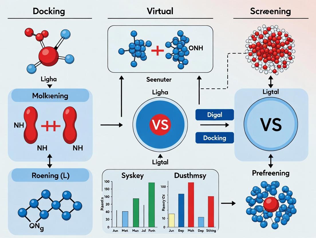
This article provides a comprehensive guide to the critical role of molecular docking within virtual screening (VS) pipelines for drug discovery.
Molecular Docking in Virtual Screening: A Comprehensive Guide from Theory to Validation for Drug Discovery
Abstract
This article provides a comprehensive guide to the critical role of molecular docking within virtual screening (VS) pipelines for drug discovery. Aimed at researchers and drug development professionals, it explores the foundational principles that underpin these computational techniques, detailing their strategic advantages in cost and time reduction over traditional high-throughput screening [citation:1][citation:2]. The article systematically walks through established methodological workflows, from target selection and library preparation to the execution of docking simulations using common software tools [citation:2][citation:5][citation:10]. It addresses key challenges and optimization strategies, including handling protein flexibility and the limitations of scoring functions [citation:3][citation:8][citation:9]. Finally, the guide emphasizes robust validation protocols, covering the use of benchmarking sets, enrichment analysis, and the essential integration of computational hits with experimental assays to translate virtual discoveries into viable therapeutic candidates [citation:4][citation:7][citation:9].
Virtual Screening and Molecular Docking: Foundational Principles and Strategic Advantages in Drug Discovery
Within the continuum of modern drug discovery, computational methods have become indispensable for accelerating the identification and optimization of lead compounds. This whitepaper frames the core concepts of virtual screening (VS) and molecular docking within the broader thesis that molecular docking serves as the central, enabling engine of structure-based virtual screening campaigns. While VS encompasses a wide array of ligand- and structure-based techniques, the precision of docking—simulating the atomic-level interaction between a small molecule and a target protein—provides the critical predictive power that drives hit identification and optimization in contemporary VS research.
Core Concepts and Definitions
- Virtual Screening (VS): A computational methodology used to evaluate very large libraries of chemical compounds (virtual databases) to identify those structures most likely to bind to a drug target and elicit a desired biological effect. It acts as a funnel, prioritizing a manageable number of candidates for experimental testing.
- Molecular Docking: A computational technique that predicts the preferred orientation (posing) and binding affinity (scoring) of a small molecule (ligand) when bound to a macromolecular target (e.g., protein). It is a core component of structure-based virtual screening (SBVS).
Their relationship is hierarchical: Molecular docking is a specific, mechanistic task; virtual screening is a broader strategy that often employs docking as its primary evaluative step.
The Virtual Screening Workflow and Docking's Pivotal Role
A standard SBVS workflow, where docking is central, involves sequential steps:
Diagram: Central Role of Docking in SBVS Workflow (81 chars)
3.1. Detailed Methodological Protocols
A. Target Preparation (Pre-Docking):
- Source: Obtain a 3D protein structure from experimental methods (X-ray crystallography, cryo-EM) or homology modeling.
- Processing: Using software like Schrödinger's Protein Preparation Wizard or UCSF Chimera:
- Add missing hydrogen atoms and correct protonation states (e.g., for His, Asp, Glu).
- Optimize hydrogen-bonding networks.
- Remove water molecules, except those structurally integral to binding.
- Assign partial charges and energy minimize the structure to relieve steric clashes.
- Define Binding Site: Identify the pocket using co-crystallized ligands or computational prediction tools (e.g., FTMap, SiteMap).
B. Ligand Library Preparation:
- Source Libraries: Use public (ZINC, ChEMBL) or proprietary databases.
- Standardization: Filter by drug-like properties (Lipinski's Rule of Five). Generate plausible tautomers and protonation states at physiological pH (e.g., using Epik or MOE).
- Energy Minimization: Apply a force field (e.g., OPLS4, MMFF94s) to generate low-energy 3D conformations.
C. Molecular Docking Protocol (Example using AutoDock Vina):
- Grid Box Definition: Configure a search space encompassing the binding site. Typical box dimensions are 20x20x20 Å with 1 Å grid spacing.
- Command example:
--center_x 10.5 --center_y 12.3 --center_z 15.8 --size_x 20 --size_y 20 --size_z 20
- Command example:
- Docking Execution: Run the Vina algorithm, which performs conformational sampling and scoring.
- Command:
vina --receptor protein.pdbqt --ligand ligand.pdbqt --config config.txt --out docked_ligand.pdbqt --log log.txt
- Command:
- Output: Generates multiple pose-ranked output files (e.g.,
docked_ligand.pdbqt) with estimated binding affinities in kcal/mol.
D. Post-Docking Analysis:
- Pose Clustering: Group similar ligand poses (e.g., by RMSD < 2.0 Å).
- Visual Inspection: Manually assess top-ranked poses for key interactions (H-bonds, pi-stacking, hydrophobic contacts).
- Rescoring & MM/GBSA: Apply more rigorous, computationally expensive methods like Molecular Mechanics/Generalized Born Surface Area (MM/GBSA) to refine affinity predictions on a subset of top hits.
Quantitative Performance Metrics
The success of a VS/docking campaign is measured by its ability to enrich true hits. Standard retrospective validation metrics are summarized below.
Table 1: Key Metrics for Evaluating Virtual Screening Performance
| Metric | Formula | Interpretation |
|---|---|---|
| Enrichment Factor (EF) | EFX% = (Hitssel / Nsel) / (Hitstotal / Ntotal) |
Measures how much better the selection is than random at a given fraction (X%) of the screened library. EF > 1 indicates enrichment. |
| Area Under the ROC Curve (AUC-ROC) | Area under the plot of True Positive Rate vs. False Positive Rate. | Overall classifier performance. AUC = 0.5 is random; AUC = 1.0 is perfect. |
| True Positive Rate (TPR/Sensitivity) | TPR = True Positives / (True Positives + False Negatives) | Proportion of actual hits correctly identified. |
| False Positive Rate (FPR) | FPR = False Positives / (False Positives + True Negatives) | Proportion of inactive compounds incorrectly identified as hits. |
| Hit Rate | Hit Rate = (True Positives) / (Selected Compounds Tested) | The empirical success rate from experimental validation. |
The Scientist's Toolkit: Essential Research Reagent Solutions
Table 2: Key Reagents and Software in Molecular Docking & Virtual Screening
| Item / Solution | Function / Role | Examples |
|---|---|---|
| Protein Structure Database | Source of experimentally determined 3D target structures. | Protein Data Bank (PDB), AlphaFold Protein Structure Database. |
| Small Molecule Database | Source of compounds for screening libraries. | ZINC, ChEMBL, PubChem, Enamine REAL, internal corporate libraries. |
| Molecular Docking Software | Performs ligand sampling and scoring. | AutoDock Vina, Glide (Schrödinger), GOLD (CCDC), MOE (CCG). |
| Force Field | Provides the energy functions for scoring and minimization. | OPLS4, CHARMM36, AMBER, MMFF94s. |
| Visualization & Analysis Software | For inspecting protein-ligand interactions and analyzing results. | PyMOL, UCSF Chimera, Maestro (Schrödinger), BIOVIA Discovery Studio. |
| High-Throughput Assay Kits | For experimental validation of computational hits (e.g., binding or activity assays). | Fluorescence Polarization (FP) kits, Time-Resolved Fluorescence Energy Transfer (TR-FRET) kits, enzymatic activity kits (e.g., from Cisbio, Thermo Fisher). |
Virtual screening represents a paradigm shift in early drug discovery, enabling the intelligent prioritization of chemical matter from vast virtual spaces. Molecular docking is not merely a component within this paradigm; it is the foundational computational experiment that imbues SBVS with predictive, mechanistic insight. The continued evolution of docking algorithms—through improved scoring functions, incorporation of machine learning, and better handling of protein flexibility—directly strengthens the central thesis of its irreplaceable role in driving efficient and successful virtual screening research. The integration of robust experimental protocols, rigorous quantitative validation, and specialized research tools, as outlined, is critical for translating computational predictions into tangible therapeutic leads.
Within the broader thesis on the role of molecular docking in virtual screening (VS) research, the strategic choice between VS and HTS is pivotal. Molecular docking, as a core computational methodology, is not merely a low-cost precursor to HTS but a complementary and often prerequisite strategy that fundamentally alters the economics and logic of early drug discovery. This whitepaper provides a technical and economic comparison, framing VS powered by molecular docking as a strategic filter that enriches the quality and probability of success of subsequent HTS campaigns or, in some cases, replaces them entirely.
Core Principles and Methodologies
2.1 High-Throughput Screening (HTS): Experimental Protocol A standard HTS campaign for a novel enzyme target involves the following key steps:
- Assay Development & Validation: A biochemical assay (e.g., fluorescence resonance energy transfer, FRET) is developed to measure target activity. Key parameters: Z'-factor >0.5, signal-to-noise ratio >10.
- Library Management: A chemical library (e.g., 500,000 compounds) is formatted into 384- or 1536-well plates using liquid handling robots.
- Primary Screening: Compounds are dispensed into assay plates, followed by addition of enzyme and substrate. Plates are read by a plate reader. A hit threshold is set (e.g., >50% inhibition at 10 µM).
- Hit Confirmation: Primary hits are retested in dose-response (IC50 determination) and counterscreened for assay interference (e.g., fluorescence quenching, aggregation).
- Hit-to-Lead: Confirmed hits undergo medicinal chemistry optimization.
2.2 Virtual Screening (VS) via Molecular Docking: Experimental Protocol A structure-based VS protocol leveraging molecular docking involves:
- Target Preparation: A 3D protein structure (from X-ray crystallography or cryo-EM, PDB ID) is prepared: adding hydrogen atoms, correcting protonation states, and defining binding site coordinates.
- Ligand Library Preparation: A virtual compound library (e.g., 1-10 million molecules from ZINC or Enamine) is prepared: generating 3D conformers, assigning correct tautomers, and calculating partial charges.
- Molecular Docking: Using software (AutoDock Vina, Glide, GOLD), each compound is computationally "docked" into the binding site. A scoring function ranks poses based on estimated binding affinity.
- Post-Docking Analysis: Top-ranked compounds (e.g., top 1,000) are visually inspected for sensible binding interactions (e.g., hydrogen bonds, hydrophobic packing). Further filtering by drug-likeness (Lipinski's Rule of Five) and synthetic accessibility is applied.
- Purchasing & Testing: A final, prioritized list of 20-100 compounds is acquired and tested experimentally in a low- to medium-throughput assay.
Strategic and Economic Comparison: Data Tables
Table 1: Operational and Economic Parameters (Representative 2024 Data)
| Parameter | High-Throughput Screening (HTS) | Virtual Screening (VS) |
|---|---|---|
| Initial Library Size | 100,000 – 2,000,000 compounds | 1,000,000 – 10,000,000+ compounds |
| Typical Compounds Tested | 100,000 – 500,000 | 50 – 500 (post-prioritization) |
| Time per Campaign | 3 – 12 months | 1 – 4 weeks (computational phase) |
| Direct Cost per Campaign | $50,000 – $500,000+ | $5,000 – $50,000 (compute + compounds) |
| Hit Rate (Average) | 0.01% – 0.3% | 5% – 20% (enrichment over random) |
| Primary Resource | Physical compound library, robotics, assay reagents | High-performance computing (HPC), software, protein structure |
| Key Bottleneck | Assay robustness, false positives from interference | Availability & quality of target structure, scoring function accuracy |
Table 2: Strategic Advantages and Limitations
| Aspect | HTS Advantages | HTS Limitations | VS Advantages | VS Limitations |
|---|---|---|---|---|
| Coverage | Tests real compounds with confirmed activity; identifies unexpected chemotypes. | Limited to physical library; diverse but finite. | Can screen ultra-large, virtual chemical space; includes hypothetical molecules. | Purely predictive; requires experimental validation. |
| Information | Provides direct experimental readout (activity, cytotoxicity). | Little initial structural insight; mechanism of action often unknown. | Provides structural binding hypotheses (pose, interactions) for design. | Accuracy hinges on force fields & scoring functions; may miss allosteric sites. |
| Flexibility | Can screen phenotypic or complex targets without a defined structure. | Difficult for membrane proteins or unstable targets. | Target agnostic if a structure exists; can be rapidly adapted to new variants. | Absolutely requires a high-quality 3D structure of the target. |
| Lead Quality | Hits are readily available for follow-up. | High false-positive rate; hits may have poor drug-likeness. | Can pre-filter for drug-likeness, ADMET properties, and synthetic accessibility. | May eliminate promising but non-canonical binders due to scoring bias. |
Integrated Workflow and Pathways
Diagram 1: VS and HTS Strategic Pathways in Drug Discovery
Diagram 2: Molecular Docking Virtual Screening Core Workflow
The Scientist's Toolkit: Key Research Reagent Solutions
Table 3: Essential Materials and Tools for Featured Experiments
| Item/Category | Function in HTS | Function in VS (Molecular Docking) |
|---|---|---|
| Compound Library | Physical collection (e.g., 500K diversity set) in DMSO, stored in plate formats. Source of chemical matter for screening. | Digital collection (e.g., ZINC, Enamine REAL) in SDF or SMILES format. The search space for computational prediction. |
| Assay Reagents | Purified target protein, fluorescent/ luminescent substrate, buffer components. Enables biochemical activity measurement. | Not applicable in the computational phase. Critical for subsequent experimental validation of VS hits. |
| Detection Instrument | Microplate reader (fluorescence, luminescence, absorbance). Measures assay signal across thousands of wells. | High-Performance Computing (HPC) cluster or cloud computing (AWS, Azure). Provides CPU/GPU power for docking millions of compounds. |
| Liquid Handling Robot | Automates dispensing of nanoliter volumes of compounds and reagents into microplates. Enables speed and precision. | Not applicable. |
| Docking Software | Not applicable. | Core engine (e.g., AutoDock Vina, Glide, GOLD). Performs conformational search and scoring of protein-ligand interactions. |
| Protein Structure | Not always required, but beneficial. A 3D structure (PDB) aids in understanding HTS hits. | Absolute prerequisite. The input model (from PDB or homology modeling) defines the binding site for docking. |
| Visualization Software | Used for data analysis (e.g., ActivityBase, Spotfire). | Critical for post-docking analysis (e.g., PyMOL, Chimera). Used to visually inspect predicted binding poses and interactions. |
Within the paradigm of modern drug discovery, virtual screening (VS) via molecular docking has become a cornerstone methodology. Its core value proposition is tripartite: it significantly accelerates the identification of novel bioactive compounds, drastically reduces the costs associated with early-stage experimental screening, and facilitates the exploration of vast, previously inaccessible regions of chemical space. This whitepaper provides an in-depth technical analysis of these advantages, supported by contemporary data, detailed experimental protocols, and essential resource guidance for the practicing researcher.
Quantitative Impact: Data-Driven Advantages
The efficacy of molecular docking in VS is quantifiable across key performance indicators. The following tables consolidate recent findings from the literature and industry reports.
Table 1: Comparative Efficiency of HTS vs. Structure-Based VS
| Metric | High-Throughput Screening (HTS) | Structure-Based Virtual Screening (VS) | Notes |
|---|---|---|---|
| Library Size | 10⁵ – 10⁶ compounds | 10⁶ – 10⁹ compounds (commercial + in silico) | VS accesses virtual, enumerable libraries. |
| Primary Screen Cost | $0.10 – $1.00 per compound | ~$0.001 – $0.01 per compound (compute cost) | VS cost is primarily computational infrastructure. |
| Time per Screen | Weeks to months | Days to weeks | Dependent on library size and computing cluster scale. |
| Typical Hit Rate | 0.01% – 0.1% | 1% – 20% (post-filtering, enrichment) | VS hit rate is after application of filters/scoring. |
| Lead Optimization Entry | 12-24 months | Can be reduced to 6-12 months | Acceleration due to earlier structural insights. |
Table 2: Key Performance Metrics from Recent VS Campaigns (2020-2024)
| Target Class | Initial VS Library | Experimental Hits Identified | Hit Rate | Reported Cost Saving vs. HTS | Reference Context |
|---|---|---|---|---|---|
| Kinase (Oncology) | 2.5 million | 127 nM – 2.1 μM inhibitors | ~5% (of tested) | ~85% | J. Med. Chem. (2023) |
| GPCR (CNS) | 4.1 million | 18 novel antagonists (IC50 < 10μM) | ~15% (of tested) | ~75% | Nat. Commun. (2022) |
| Viral Protease | 1.7 million | 9 non-covalent inhibitors (Ki < 5μM) | ~8% (of tested) | >90% | Cell Rep. (2024) |
| Protein-Protein Interaction | 890,000 | 3 disruptors (sub-μM) | ~2% (of tested) | ~70% | Sci. Adv. (2023) |
Experimental Protocols for a Standard VS Workflow
The following protocol details a robust, tiered structure-based VS methodology.
Protocol: Tiered Structure-Based Virtual Screening for Lead Identification
A. Preparation Phase
- Target Preparation:
- Obtain a 3D protein structure from PDB or via homology modeling.
- Process the structure: add missing hydrogen atoms, assign protonation states (e.g., using
propkaat pH 7.4), and optimize side-chain conformations of ambiguous residues. - Define the binding site using co-crystallized ligands or site prediction tools (e.g., FTMap, SiteMap).
- Ligand Library Preparation:
- Source a compound library (e.g., ZINC20, Enamine REAL, MCULE).
- Generate plausible 3D conformers for each molecule.
- Apply standard force fields (e.g., OPLS4, GAFF2) to assign partial charges and atom types.
- Filter libraries using drug-like rules (e.g., Lipinski's Rule of Five, PAINS filters).
B. Docking and Screening Phase
- High-Throughput Docking:
- Employ a fast, rigid or semi-flexible docking algorithm (e.g., FRED, HYBRID) to screen the entire prepared library.
- Use a grid-based scoring function for rapid pose evaluation.
- Output: Rank-ordered list of top ~50,000 – 100,000 compounds.
- Standard-Precision (SP) Docking:
- Re-dock the top compounds from Step 3 using a more sophisticated, flexible-ligand docking program (e.g., Glide SP, AutoDock Vina).
- Allow for rotational flexibility in key protein side chains if protocol supports it.
- Output: Refined ranking of top ~5,000 – 10,000 compounds.
- High-Accuracy Refinement:
- Subject the top 500-1,000 compounds from Step 4 to high-accuracy docking (e.g., Glide XP, induced-fit docking).
- Apply more rigorous scoring functions, including terms for solvation and entropy.
- Output: Final prioritized list of 50-200 compounds for visual inspection.
C. Post-Docking Analysis
- Visual Inspection & Clustering:
- Manually inspect top-scoring diverse poses for key interactions (H-bonds, pi-stacking, hydrophobic complementarity).
- Cluster remaining compounds by scaffold to prioritize chemotypes.
- Experimental Validation:
- Procure or synthesize the top 20-50 prioritized compounds.
- Perform primary biochemical assay (e.g., fluorescence polarization, enzyme inhibition) to confirm activity.
- Progress confirmed hits to dose-response analysis (IC50/Ki determination).
Visualizing the VS Workflow and Logic
Title: Tiered Virtual Screening Workflow for Hit Identification
Title: The Core Advantages of Docking in Virtual Screening
The Scientist's Toolkit: Key Research Reagent Solutions
Table 3: Essential Materials and Tools for a VS Campaign
| Item / Solution | Function / Purpose | Example Providers/Tools |
|---|---|---|
| Protein Structure | Provides the 3D target for docking. | RCSB PDB, AlphaFold DB, SWISS-MODEL |
| Compound Libraries | Source of small molecules for screening. | ZINC, Enamine REAL, MCULE, ChemDiv |
| Docking Software | Computationally predicts ligand pose & affinity. | Schrodinger Suite, AutoDock Vina, DOCK 3, GOLD, FRED (OpenEye) |
| Molecular Dynamics (MD) Suite | Refines docked poses and assesses stability. | Desmond (Schrodinger), GROMACS, AMBER, NAMD |
| Force Field Parameters | Defines energy terms for atoms and bonds. | OPLS4, CHARMM36, GAFF2 |
| Visualization Software | Critical for pose inspection and analysis. | PyMOL, Maestro, ChimeraX |
| High-Performance Computing (HPC) | Provides necessary computational power. | Local clusters, Cloud (AWS, Azure, GCP), SLURM schedulers |
| Biochemical Assay Kits | Experimental validation of predicted hits. | Target-specific kits from Cayman Chem, BPS Bioscience, Thermo Fisher |
Within the continuum of virtual screening (VS) research, molecular docking serves as a pivotal computational technique that bridges predictive modeling and experimental validation. This whitepaper delineates the two principal VS paradigms: Structure-Based Drug Design (SBDD) and Ligand-Based Drug Design (LBDD). SBDD leverages the three-dimensional structure of a biological target, while LBDD utilizes known active ligands to infer new candidates. Both approaches are integral to modern drug discovery, often used complementarily to maximize hit identification and optimization efficiency.
Structure-Based Drug Design (SBDD)
Core Principle
SBDD requires prior knowledge of the target's 3D atomic structure, typically obtained via X-ray crystallography, cryo-electron microscopy (cryo-EM), or NMR spectroscopy. The central premise is to predict the binding mode and affinity of small molecules within a defined binding site using molecular docking and scoring functions.
Key Methodologies & Protocols
Molecular Docking Protocol
A standard molecular docking workflow for VS involves:
- Target Preparation: The protein structure (from PDB) is processed by adding hydrogen atoms, assigning protonation states, and optimizing side-chain conformations. Tools: Schrödinger's Protein Preparation Wizard, UCSF Chimera.
- Binding Site Definition: The active site is identified, often using coordinates from a co-crystallized ligand or computational prediction (e.g., FTMap, SiteMap).
- Ligand Library Preparation: Small molecules are converted to 3D, energy-minimized, and assigned correct tautomeric and stereochemical states. Tools: LigPrep, OMEGA.
- Docking Execution: Ligands are computationally posed in the binding site. Popular algorithms include Glide (SP, XP modes), AutoDock Vina, and GOLD.
- Scoring & Ranking: A scoring function (e.g., GlideScore, ChemScore) estimates binding free energy for each pose. The top-ranked compounds are selected for in vitro testing.
Molecular Dynamics (MD) Simulation Protocol
To refine and validate docking poses:
- System Setup: The protein-ligand complex is solvated in an explicit water box (e.g., TIP3P) and neutralized with ions.
- Energy Minimization: Steepest descent/conjugate gradient minimization removes steric clashes.
- Equilibration: NVT and NPT ensembles are used to equilibrate temperature (300K) and pressure (1 bar).
- Production Run: An unrestrained MD simulation (50-200 ns) is performed using AMBER, GROMACS, or NAMD.
- Analysis: Trajectories are analyzed for stability (RMSD), binding interactions (H-bonds, hydrophobic contacts), and binding free energy estimates (MM/PBSA, MM/GBSA).
Table 1: Performance Metrics of Common Docking Software (Representative)
| Software | Scoring Function | Avg. RMSD (Å)¹ | Enrichment Factor (EF₁%²) | Computational Speed (ligands/day)³ |
|---|---|---|---|---|
| AutoDock Vina | Vina | 1.5 - 2.5 | 15 - 25 | ~50,000 (CPU) |
| Glide (SP) | GlideScore | 1.0 - 2.0 | 20 - 35 | ~10,000 (CPU) |
| GOLD | ChemPLP | 1.2 - 2.2 | 18 - 30 | ~5,000 (CPU) |
| LeDock | LeDock SF | 1.5 - 2.5 | 10 - 20 | ~100,000 (CPU) |
| GNINA | CNN Score | 1.3 - 2.3 | 25 - 40 | ~20,000 (GPU) |
¹ Root-mean-square deviation of heavy atoms for re-docked cognate ligands. ² Enrichment factor at 1% of the screened database. ³ Approximate throughput on a standard 24-core server; GPU implementations vary.
Ligand-Based Drug Design (LBDD)
Core Principle
LBDD is employed when the 3D target structure is unknown. It operates on the "similar property principle," assuming structurally similar molecules exhibit similar biological activity. Methods include Quantitative Structure-Activity Relationship (QSAR) modeling, pharmacophore mapping, and similarity searching.
Key Methodologies & Protocols
3D-QSAR Modeling Protocol (e.g., CoMFA)
- Data Set Curation: A set of molecules with measured activity (pIC₅₀) is assembled and divided into training and test sets.
- Molecular Alignment: All molecules are aligned to a common scaffold or pharmacophore using least-squares fitting.
- Field Calculation: Steric (Lennard-Jones) and electrostatic (Coulombic) interaction fields are calculated at grid points around the molecules.
- PLS Regression: Partial Least Squares regression correlates field values with biological activity.
- Model Validation: Predictive power is assessed via cross-validation (q²) and external test set prediction (r²ₚᵣₑd).
Pharmacophore Model Generation Protocol
- Feature Selection: Common chemical features (H-bond donor/acceptor, hydrophobic, aromatic, charged groups) are defined.
- Conformational Analysis: Multiple conformers are generated for each active ligand.
- Model Construction: Software (e.g., Phase, MOE) identifies common feature arrangements among active molecules. Inactive compounds can be used to exclude features.
- Model Validation: The model's ability to retrieve actives from a decoy database is evaluated (e.g., using Güner-Henry score).
Table 2: Benchmarking of LBDD Methods on DUD-E Datasets
| Method | Type | Avg. AUC⁴ | Avg. EF₁%⁵ | Key Descriptor/Feature |
|---|---|---|---|---|
| ROCS (Shape+Color) | Similarity Search | 0.71 | 22.1 | TanimotoCombo (Shape & Chemistry) |
| EON (Electrostatics) | Similarity Search | 0.65 | 18.5 | ET_Combo (Electrostatic & Shape) |
| Phase Pharmacophore | Pharmacophore | 0.75 | 28.5 | 4-5 feature hypothesis |
| Machine Learning (RF) | QSAR | 0.82 | 32.0 | ECFP4 fingerprints |
| Deep Learning (GraphNet) | QSAR | 0.85 | 35.5 | Molecular graph representation |
⁴ Area Under the Receiver Operating Characteristic Curve. ⁵ Enrichment Factor at 1% of the screened database.
Integrated VS Workflows and Visualization
The contemporary VS pipeline often integrates SBDD and LBDD to leverage their respective strengths.
Title: Decision Flowchart for VS Approach Selection
Title: Integrated SBDD and LBDD Virtual Screening Workflow
The Scientist's Toolkit: Essential Research Reagents & Solutions
Table 3: Key Reagents, Software, and Materials for VS Experiments
| Item Name | Category | Function / Purpose | Example Vendor/Software |
|---|---|---|---|
| Purified Target Protein | Biological Reagent | Required for biochemical assay validation of VS hits. | Sigma-Aldrich, custom expression. |
| FRET/FP Assay Kit | Biochemical Assay | High-throughput kinetic or endpoint binding assay. | Thermo Fisher, Cisbio. |
| SPR Chip (CM5) | Biophysical Assay | Surface Plasmon Resonance for measuring binding kinetics (ka, kd). | Cytiva. |
| Compound Library (10^5-10^6) | Chemical Library | Large collection of diverse, drug-like molecules for screening. | Enamine, ChemDiv, ZINC. |
| Schrödinger Suite | Software | Integrated platform for protein prep (Maestro), docking (Glide), and MD (Desmond). | Schrödinger LLC. |
| OpenEye Toolkits | Software | Provides ROCS, OMEGA, and FRED for LBDD and high-performance cheminformatics. | OpenEye Scientific. |
| AMBER/GAFF | Software | Force fields for MD simulations and binding free energy calculations. | University of California. |
| RDKit | Software | Open-source cheminformatics toolkit for descriptor calculation and QSAR. | Open Source. |
| GPU Computing Cluster | Hardware | Accelerates docking (GNINA) and MD simulations by orders of magnitude. | NVIDIA, cloud providers. |
SBDD and LBDD represent the twin pillars of virtual screening. SBDD offers a mechanistic, target-centric approach grounded in structural biology, while LBDD provides a powerful, knowledge-driven strategy when structural data is absent. The integration of both methods, underpinned by robust molecular docking and simulation protocols, consensus scoring, and rigorous experimental validation, constitutes the state-of-the-art in computational drug discovery. This synergistic paradigm continues to enhance the efficiency and success rate of identifying novel lead compounds.
Building an Effective Virtual Screening Workflow: From Library Preparation to Hit Identification
Abstract: Within the framework of virtual screening (VS) for drug discovery, the preliminary stages of target analysis, data collection, and binding site definition are critical determinants of success. This guide details the technical protocols and strategic considerations for these foundational steps, ensuring robust and reproducible molecular docking campaigns.
Target Analysis and Selection
The initial phase involves the rigorous bioinformatic and structural evaluation of the target protein.
Target Druggability Assessment
Druggability predicts the likelihood of a protein binding small molecules with high affinity. Key metrics include:
- Pocket Properties: Volume, depth, and hydrophobicity.
- Sequence & Structural Analysis: Presence of known binding motifs (e.g., kinase ATP pocket).
- Conservation: Evolutionary conservation of the putative site.
Table 1: Quantitative Metrics for Druggability Prediction
| Metric | High Druggability Range | Low Druggability Indicator | Common Tool for Analysis |
|---|---|---|---|
| Pocket Volume (ų) | 500-1000 | <350 | FPocket, DoGSiteScorer |
| Surface Complexity (PSA)*) | 100-250 Ų | >350 Ų | MOE, Schrodinger |
| Hydrophobicity (%) | 40-70% | <25% | CASTp, PyMOL |
| Conservation Score | >0.7 (highly conserved) | <0.3 | ConSurf |
*Polar Surface Area.
Protocol: In-silico Druggability Assessment with FPocket
- Input Preparation: Obtain the target's 3D structure (PDB format). Remove water molecules and heteroatoms except crucial co-factors.
- Pocket Detection: Execute FPocket via command line:
fpocket -f target.pdb. - Output Analysis: The tool outputs predicted pockets ranked by a druggability score (DScore). Analyze the top-ranked pocket(s) for volume, amino acid composition, and ligandability.
- Validation: Cross-reference with known ligands from homologous structures in the PDB.
Data Curation and Ligand Library Preparation
The quality of the screening library directly impacts hit rates.
Compound Sourcing and Filtering
Libraries are assembled from public (ZINC, ChEMBL) and commercial databases. Standard filtering rules adhere to Lipinski's Rule of Five and variants like Veber's rules for improved bioavailability.
Table 2: Standard Pre-processing Filters for VS Libraries
| Filter | Typical Cutoff | Purpose |
|---|---|---|
| Molecular Weight | ≤ 500 Da | Oral bioavailability |
| LogP | ≤ 5 | Solubility and permeability |
| Hydrogen Bond Donors | ≤ 5 | Membrane permeability |
| Hydrogen Bond Acceptors | ≤ 10 | Membrane permeability |
| Rotatable Bonds | ≤ 10 | Oral bioavailability |
| PAINS Filter | Remove matches | Elimination of promiscuous compounds |
| Reactive Functional Groups | Remove matches | Elimination of unstable/ toxic compounds |
Protocol: Library Preparation with OpenBabel and RDKit
- Format Conversion: Convert vendor SDF files to a common format:
obabel input.sdf -O output.sdf --gen3D. - Standardization: Tautomer and protonation state standardization at pH 7.4 ± 0.5 using RDKit's
MolStandardizemodule. - Descriptor Calculation & Filtering: Use RDKit to compute descriptors (MW, LogP, HBD, HBA) and apply filters from Table 2.
- Energy Minimization: Perform a coarse geometry optimization using the MMFF94 force field to resolve steric clashes.
Binding Site Definition and Grid Generation
Accurate spatial and energetic characterization of the binding site is essential for docking scoring.
Methods for Binding Site Delineation
- Ligand-based: Defined from the coordinates of a co-crystallized ligand.
- Structure-based: Using pocket detection algorithms (See 1.2).
- Functional/Consensus-based: Integrating mutagenesis data to identify critical residues.
Protocol: Grid Generation with AutoDockTools
- Protein Preparation: Add polar hydrogens, assign Gasteiger charges, and merge non-polar hydrogens.
- Set the Grid Box: Center the box on the centroid of the binding site residues or a reference ligand.
- Define Box Dimensions: Size must encompass the entire binding site and allow ligand flexibility. A typical margin is 10Å beyond any known ligand atom.
- Example Command (AutoDock Vina):
vina --receptor protein.pdbqt --config config.txt - The
config.txtfile specifiescenter_x, center_y, center_z, size_x, size_y, size_z.
- Example Command (AutoDock Vina):
The Scientist's Toolkit: Research Reagent Solutions
Table 3: Essential Tools and Databases for Preparatory Steps
| Item | Function & Description | Example/Source |
|---|---|---|
| RCSB Protein Data Bank (PDB) | Primary repository for 3D structural data of proteins and nucleic acids. | https://www.rcsb.org |
| PDBsum | Provides schematic diagrams and analyses of PDB entries, including binding site residues. | https://www.ebi.ac.uk/pdbsum |
| UniProt | Comprehensive resource for protein sequence and functional information. | https://www.uniprot.org |
| ChEMBL | Manually curated database of bioactive molecules with drug-like properties and assay data. | https://www.ebi.ac.uk/chembl |
| ZINC Database | Free database of commercially-available compounds for virtual screening, with pre-prepared 3D formats. | https://zinc.docking.org |
| RDKit | Open-source cheminformatics toolkit for descriptor calculation, filtering, and molecule manipulation. | https://www.rdkit.org |
| OpenBabel | Open chemical toolbox for file format conversion and cheminformatics. | http://openbabel.org |
| AutoDockTools / MGLTools | GUI and scripting tools for preparing files and setting grids for AutoDock/Vina. | https://ccsb.scripps.edu/mgltools |
| PyMOL / ChimeraX | Molecular visualization systems for structural analysis and binding site inspection. | https://pymol.org, https://www.cgl.ucsf.edu/chimerax |
Visualizations
Diagram 1: VS Preparatory Phase Workflow (83 chars)
Diagram 2: Ligand Library Curation Process (73 chars)
Molecular docking, a cornerstone of structure-based virtual screening (VS), is only as effective as the chemical library it screens. This guide details the critical preparatory steps of compound sourcing, structural standardization, and conformer generation, which collectively form the foundation of a robust, computationally-ready screening library. The quality and preparation of this library directly determine the success rate of downstream docking campaigns by minimizing false positives stemming from erroneous representations and maximizing the probability of identifying true bioactive molecules.
Compound Sourcing and Curation
The initial step involves aggregating a diverse, drug-like compound collection from reliable sources. Key public and commercial databases are primary sources.
Table 1: Primary Sources for Compound Libraries
| Source | Type | Approximate Size (Compounds) | Key Characteristics | Typical Format |
|---|---|---|---|---|
| PubChem | Public | 110+ Million | Bioactivity data, diverse sources | SDF, SMILES |
| ChEMBL | Public | 2+ Million | Curated bioactive molecules, targets | SDF, SMILES |
| ZINC | Public | 230+ Million (subsets) | Commercially available, purchasable | SDF, SMILES |
| CAS | Commercial | 200+ Million | Authoritative, well-curated | Proprietary |
| Enamine REAL | Commercial | 1.3+ Billion | Make-on-demand, synthesizable | SDF, SMILES |
Experimental Protocol: Initial Data Acquisition and Cleaning
- Download: Acquire compounds in SDF or SMILES format from chosen databases.
- Descriptor Filtering: Apply calculated property filters (e.g., using RDKit or OpenBabel) to retain molecules within a "drug-like" chemical space.
- Common filters: 150 ≤ Molecular Weight ≤ 600 g/mol, -2 ≤ LogP ≤ 6, Rotatable Bonds ≤ 10, Hydrogen Bond Donors ≤ 5, Hydrogen Bond Acceptors ≤ 10.
- Structural Inspection: Remove salts, solvents, and counterions. Standardize metal coordination representations.
- Duplicate Removal: Perform canonical SMILES generation and identify unique structures using tools like
rdkit.Chem.rdmolfiles.MolToSmiles(mol, canonical=True).
Molecular Standardization
Inconsistent molecular representations introduce significant noise. Standardization ensures all molecules adhere to a uniform set of chemical rules.
Table 2: Common Standardization Rules and Actions
| Rule Category | Problem | Standardization Action |
|---|---|---|
| Valence & Bonding | Hypervalent nitrogen, incorrect aromaticity | Re-perceive aromaticity (Kekulization), fix nitro groups, correct sulfoxide/sulfone. |
| Tautomers | Multiple possible protonation states | Choose a representative canonical tautomer (e.g., using the MolVS toolkit). |
| Stereochemistry | Missing or ambiguous chiral centers | Remove undefined stereochemistry or flag for manual inspection. |
| Protonation State | Non-physiological charges at target pH | Generate major microspecies at pH 7.4 ± 0.5 (e.g., using ChemAxon or Epik). |
| Functional Groups | Varied representations (e.g., nitro groups) | Transform to a consistent representation (e.g., [N+](=O)[O-]). |
Experimental Protocol: Standardization Pipeline
- Neutralization: Use a rule-based approach (e.g., RDKit's
rdkit.Chem.rdmolops.Cleanup) to neutralize non-physiological charges while preserving zwitterions. - Aromaticity: Apply
rdkit.Chem.rdmolops.Kekulize(mol, clearAromaticFlags=True)followed byrdkit.Chem.rdmolops.SanitizeMol(mol). - Tautomer Canonicalization: Employ the MolVS
TautomerCanonicalizerto select a consistent representative structure. - Stereo Processing: Use
rdkit.Chem.rdmolops.AssignStereochemistry(mol, cleanIt=True, force=True)to assign/validate stereochemistry. - Output: Write the standardized molecules to a new clean SDF file.
Diagram 1: Compound Library Standardization Workflow
Conformer Generation for Docking
Docking requires 3D conformers. The goal is to generate a representative, energy-accessible ensemble that likely contains the bioactive pose.
Table 3: Conformer Generation Methods and Software
| Method/Software | Algorithm | Key Parameters | Output Conformers | Best For |
|---|---|---|---|---|
| RDKit ETKDG | Distance Geometry + MMFF94 Optimization | pruneRmsThresh, numConfs, useExpTorsionAnglePrefs |
10-50 per molecule | High-throughput, large libraries. |
| OMEGA (OpenEye) | Rule-based + Torsion Driving | MaxConfs, EnergyWindow, RMSThreshold |
10-200+ per molecule | Production docking, high accuracy. |
| CONFGEN | Systematic search + minimization | max_confs, energy_window |
10-100 per molecule | Robust, commercial-grade. |
| MacroModel | Monte Carlo Multiple Minimum (MCMM) | steps, energy_window |
10-1000 per molecule | Complex, flexible molecules. |
Experimental Protocol: High-Throughput Conformer Generation with RDKit
- Input: Read standardized SMILES.
- 3D Generation: Use ETKDGv3 to generate an initial conformer set.
Energy Minimization: Optimize each conformer with the MMFF94 force field.
Clustering and Selection: Cluster conformers by heavy-atom RMSD (e.g., 1.0 Å cutoff) and select the lowest-energy conformer from each cluster to create a diverse, minimal ensemble.
- Output Format: Save final conformers in a multi-conformer SDF or dockable format (e.g., .mol2 with proper charges).
Diagram 2: Workflow for Conformer Generation and Selection
The Scientist's Toolkit: Research Reagent Solutions
Table 4: Essential Tools for Library Preparation
| Tool/Software | Category | Primary Function in Library Prep |
|---|---|---|
| RDKit | Open-Source Cheminformatics | Core toolkit for SMILES parsing, standardization, filtering, and basic conformer generation. |
| OpenEye Toolkit | Commercial Cheminformatics | Industry-standard for high-quality, fast conformer generation (OMEGA) and charge assignment. |
| Schrödinger Suites | Commercial Drug Discovery | Integrated platform for advanced library preparation, property calculation, and LigPrep. |
| Molinspiration / DataWarrior | Property Calculation | Rapid calculation of molecular descriptors and property-based filtering. |
| MolVS | Open-Source Library | Specialized toolkit for molecular standardization (tautomers, normalization). |
| Knime / Pipeline Pilot | Workflow Automation | Visual design of automated, reproducible preparation pipelines. |
| PyMOL / Maestro | Visualization | Manual inspection and validation of 3D conformers and structures. |
| High-Performance Computing Cluster | Infrastructure | Essential for processing large libraries (>1M compounds) in parallel. |
Meticulous library preparation is a non-negotiable prerequisite for successful virtual screening. The processes of sourcing relevant compounds, enforcing rigorous chemical standardization, and generating biologically relevant 3D conformer ensembles directly address critical early-phase vulnerabilities in the VS pipeline. By investing in this foundational stage, researchers ensure that subsequent molecular docking experiments screen a high-fidelity library, thereby increasing the likelihood of identifying novel, potent hits for further experimental validation.
Molecular docking, a pivotal computational technique in structural biology and drug discovery, serves as the core engine for predicting the preferred orientation and binding affinity of a small molecule (ligand) to a target macromolecule (receptor). Within the context of virtual screening (VS), a cornerstone of modern drug development, the docking engine is the workhorse that enables the rapid, in silico evaluation of millions of compounds against a biological target. This technical guide provides an in-depth examination of the core components of the docking engine: its search algorithms, software implementations, and scoring functions, framing their role and optimization within a rigorous VS research pipeline.
Search Algorithms: Navigating Conformational Space
The first challenge for a docking engine is to explore the vast conformational and orientational space of the ligand within the receptor's binding site. This search is governed by key algorithmic strategies.
Detailed Methodology for Key Algorithmic Experiments: A standard protocol for evaluating search algorithms involves docking a set of ligands with known crystallographic poses (e.g., from the PDBbind database) into a prepared receptor structure.
- Receptor & Ligand Preparation: The protein structure is prepared by adding hydrogen atoms, assigning protonation states, and removing water molecules (except critical ones). Ligands are prepared by generating probable 3D conformations and assigning correct bond orders.
- Search Execution: The same set of ligand-receptor complexes is docked using different search algorithms (e.g., Genetic Algorithm, MC, Local Search) within the same software framework, keeping all other parameters constant.
- Pose Prediction Accuracy Assessment: The root-mean-square deviation (RMSD) between the top-scoring docked pose and the experimentally observed crystallographic pose is calculated. A pose with RMSD ≤ 2.0 Å is typically considered successfully docked.
- Analysis: The success rate (percentage of ligands docked within 2.0 Å RMSD) and computational time are recorded and compared across algorithms.
Table 1: Comparison of Core Docking Search Algorithms
| Algorithm | Core Principle | Key Software Implementations | Typical Use Case in VS |
|---|---|---|---|
| Systematic/Incremental | Exhaustively samples torsional angles or places fragments. | DOCK, FRED | When binding site is deeply buried and well-defined. |
| Monte Carlo (MC) | Random moves are accepted or rejected based on a scoring function. | AutoDock, MCDOCK | Exploring broad conformational space; often coupled with minimization. |
| Genetic Algorithm (GA) | Evolves a population of poses via crossover, mutation, and selection. | AutoDock, GOLD | Flexible ligand docking with efficient global search. |
| Molecular Dynamics (MD) | Simulates physical movements based on Newtonian mechanics. | DESMOND, NAMD, Docking-MD hybrids | Refinement of poses and estimation of binding kinetics, not primary VS. |
| Swarm Optimization | Mimics social behavior (e.g., particle swarms) to find optima. | SODOCK, AutoDock Vina (variant) | Efficiently locating global minima in complex energy landscapes. |
Scoring Functions: The Heart of Affinity Prediction
Scoring functions are mathematical models used to predict the binding affinity (ΔG) or to rank potential ligand poses. They are the critical component for prioritizing hits in VS.
Detailed Methodology for Scoring Function Validation: The validation of a scoring function's predictive power is typically performed using a benchmark dataset.
- Dataset Curation: A diverse, high-quality set of protein-ligand complexes with experimentally determined binding constants (Kd, Ki, IC50) is assembled (e.g., PDBbind Core Set).
- Complex Preparation: Each structure is prepared consistently (hydrogen addition, charge assignment).
- Score Calculation: The scoring function is used to compute a score for each complex in the dataset.
- Correlation Analysis: A statistical correlation (e.g., Pearson's r, Spearman's ρ) is calculated between the computed scores and the negative logarithm of the experimental binding affinity (pKd/pKi). A higher correlation indicates better predictive performance.
Table 2: Taxonomy and Performance of Scoring Function Types
| Type | Description | Representative Examples | Typical Correlation (r) with Exp. ΔG* | Computational Cost |
|---|---|---|---|---|
| Force Field-Based | Sums molecular mechanics terms (van der Waals, electrostatics). | AMBER, CHARMM, DOCK | 0.40 - 0.55 | Medium-High |
| Empirical | Fits weighted energy terms to experimental binding data. | ChemScore, PLP, X-Score | 0.50 - 0.65 | Low |
| Knowledge-Based | Derives potentials from statistical analysis of structural databases. | PMF, DrugScore, IT-Score | 0.45 - 0.60 | Low |
| Machine Learning (ML) | Trains models (NN, RF, SVM) on complex structural/feature data. | RF-Score, NNScore, ΔVina RF20 | 0.65 - 0.85 | Varies (Low for inference) |
*Correlation ranges are approximate and dataset-dependent.
Integrated Software Suites
Modern docking engines integrate search algorithms and scoring functions into user-friendly or high-throughput software packages.
Table 3: Prominent Molecular Docking Software Platforms
| Software | Primary Search Algorithm | Scoring Function(s) | Key Feature for VS | License |
|---|---|---|---|---|
| AutoDock Vina | Hybrid of MC and BFGS optimization | Vina (empirical) | Speed, accuracy, open-source. | Open Source (Apache) |
| GOLD | Genetic Algorithm | ChemPLP, GoldScore, ASP | Handling ligand flexibility & water networks. | Commercial |
| Glide | Systematic, hierarchical search | GlideScore (empirical+FF) | High accuracy pose prediction (SP, XP modes). | Commercial (Schrödinger) |
| DOCK | Incremental construction / anchor-and-grow | FF-based, grid scoring | Customizable, long history in academia. | Open Source |
| UCSF Chimera Dock Prep | Integrates external tools (Vina, DOCK) | Varies | Seamless integration with visualization/analysis. | Free for non-commercial |
| HADDOCK | Data-driven, MC sampling | Empirical + desolvation | Specialized for protein-protein/RNA docking. | Web Server / Academic |
The Scientist's Toolkit: Research Reagent Solutions
Table 4: Essential Materials & Tools for a Docking-Based VS Campaign
| Item | Function/Description |
|---|---|
| Protein Data Bank (PDB) Structure | High-resolution 3D structure of the target protein, the foundational input. |
| Chemical Library (e.g., ZINC, Enamine) | A curated, often millions-strong, database of purchasable compounds in a format suitable for docking (e.g., SDF, MOL2). |
| Structure Preparation Software (e.g., Maestro, MOE, UCSF Chimera) | Adds missing atoms/loops, corrects protonation states, and optimizes hydrogen bonding networks. |
| Molecular Docking Software Suite | The core engine (see Table 3) for performing the pose prediction and scoring. |
| High-Performance Computing (HPC) Cluster or Cloud Computing (e.g., AWS, Azure) | Essential computational resource for executing large-scale VS on thousands to millions of compounds. |
| Visualization & Analysis Tool (e.g., PyMOL, UCSF Chimera, Discovery Studio) | For inspecting top-ranked docking poses, analyzing interaction fingerprints (H-bonds, hydrophobic contacts). |
| Benchmarking Dataset (e.g., PDBbind, DUD-E) | A set of known actives and decoys for validating and calibrating the VS protocol before full-screen execution. |
Visualizing the Virtual Screening Workflow
Title: Virtual Screening Pipeline with Docking Core
Visualizing Scoring Function Development & Validation
Title: Scoring Function Development Cycle
Within a comprehensive thesis on the role of molecular docking in virtual screening (VS), docking execution represents a critical, yet intermediate, step. The subsequent, analytical phase—post-docking analysis—is where computational predictions are rigorously evaluated to translate millions of scored poses into a shortlist of viable chemical starting points. This guide details the core technical components of this phase: selecting physiologically relevant poses, analyzing their interaction networks, and triaging compounds for experimental validation. The efficacy of an entire VS campaign hinges on these procedures.
Pose Selection: From Conformational Sampling to Plausible Binding Modes
Pose selection filters the numerous conformations generated by docking algorithms to identify those most likely to represent the true bioactive conformation.
Key Quantitative Metrics for Pose Selection: The following table summarizes primary scoring and consensus metrics used.
Table 1: Key Metrics for Initial Pose Selection and Scoring
| Metric Category | Specific Metric | Typical Optimal Range/Value | Primary Function |
|---|---|---|---|
| Docking Score | Vina Score (kcal/mol) | ≤ -7.0 (context-dependent) | Estimates binding affinity. Lower is better. |
| Consensus Ranking | Rank-by-Rank or Rank-by-Vote | Top 5-10 consensus poses | Identifies poses consistently ranked high across multiple algorithms. |
| Geometric/Internal Strain | RMSD to input ligand geometry | < 2.0 Å | Flags poses with unrealistic ligand conformations. |
| Cluster Population | Size of largest pose cluster | Largest cluster membership | Indicates a stable, low-energy conformation well-sampled by the algorithm. |
| Pose Stability | RMSD during short MD relaxation | < 2.0 Å (backbone-heavy) | Assesses pose robustness using molecular dynamics. |
Experimental Protocol: Consensus Docking and Pose Clustering
- Multiple Algorithm Docking: Dock the same ligand library using 2-3 distinct docking programs (e.g., AutoDock Vina, Glide, rDock).
- Pose Extraction & Alignment: Extract top N poses (e.g., 20) from each program and align them based on the protein's binding site alpha-carbons.
- RMSD-Based Clustering: Perform agglomerative or hierarchical clustering on all poses using a root-mean-square deviation (RMSD) cutoff (typically 2.0 Å).
- Consensus Identification: Select the centroid pose of the largest cluster that contains top-ranked poses from multiple docking programs. This represents the consensus pose.
Interaction Analysis: Decoding the Molecular Dialogue
Beyond affinity scores, detailed interaction analysis reveals the quality of binding, essential for explaining selectivity and guiding medicinal chemistry.
Table 2: Critical Protein-Ligand Interaction Types and Their Implications
| Interaction Type | Functional Group(s) | Optimal Distance (Å) | Energetic Contribution | Role in Drug Design |
|---|---|---|---|---|
| Hydrogen Bond (H-bond) | Donor: O-H, N-HAcceptor: O, N | 2.5 - 3.2 (H-Acceptor) | -1 to -5 kcal/mol each | Provides binding specificity and directionality. |
| Hydrophobic | Aromatic rings, aliphatic chains | 3.3 - 4.0 (C-C) | ~ -0.5 kcal/mol per Ų | Drives desolvation and binding. |
| π-π Stacking | Aromatic ring - aromatic ring | 3.4 - 4.0 (face-to-face) | -1 to -4 kcal/mol | Important for binding aromatic residues. |
| Cation-π | Positively charged group - aromatic ring | 3.5 - 4.5 | -5 to -10 kcal/mol | Strong electrostatic contribution. |
| Salt Bridge | Charged (+) - Charged (-) | 2.7 - 3.3 | -5 to -10 kcal/mol | Very strong, can anchor a ligand. |
| Halogen Bond | C-X---O (X=Cl, Br, I) | 3.0 - 3.5 (X---O) | -1 to -3 kcal/mol | Directional interaction mimicking H-bond. |
Experimental Protocol: Interaction Fingerprinting and Profiling
- Interaction Calculation: Use tools like PLIP, Schrödinger's Pose Analyzer, or RDKit to detect all non-covalent interactions for a selected pose.
- Fingerprint Generation: Encode the presence/absence of specific interactions with key binding site residues into a binary bit string (e.g., "H-bond with Asp93: 1").
- Cluster by Interaction Profile: Cluster ligands based on similarity of their interaction fingerprints (using Tanimoto coefficient).
- Interaction Thermodynamics (Advanced): For key poses, perform WaterMap or MM/GBSA calculations to estimate the free energy contribution of individual interactions and displaced water molecules.
Hit Triaging: Integrating Multi-Filter Criteria
Hit triaging integrates pose quality, interaction data, and drug-like properties to prioritize compounds for purchase or synthesis.
Table 3: Multi-Criteria Hit Triaging Dashboard
| Triage Stage | Criteria | Typical Threshold | Rationale |
|---|---|---|---|
| 1. Pose & Interaction Quality | Docking Score | ≤ -8.0 kcal/mol | Strong predicted affinity. |
| Presence of Key Interaction | e.g., H-bond with catalytic residue | Essential for mechanism/selectivity. | |
| Interaction Fingerprint Similarity | ≥ 0.7 to known active | Validates binding mode hypothesis. | |
| 2. Drug-Likeness & Toxicity | Lipinski's Rule of 5 | ≤ 1 violation | Oral bioavailability potential. |
| PAINS Filters | 0 alerts | Removes promiscuous, assay-interfering motifs. | |
| Synthetic Accessibility Score | ≤ 4.5 (lower is easier) | Feasibility of synthesis/purchase. | |
| 3. Diversity & Novelty | Tanimoto Coefficient (vs. in-house) | < 0.4 (for backbone) | Ensures chemical diversity in the output list. |
| Patent/Literature Search | No close prior art | Identifies novel chemical matter. |
Visualization of Workflows and Pathways
Title: Post-Docking Analysis Workflow
Title: Consensus Docking & Pose Selection Protocol
Title: From Pose to Interaction Profile
The Scientist's Toolkit: Research Reagent Solutions
Table 4: Essential Tools and Resources for Post-Docking Analysis
| Item Name / Software | Category | Primary Function | Key Application in Analysis |
|---|---|---|---|
| Schrödinger Suite (Maestro) | Commercial Software Platform | Integrated computational drug discovery. | Glide docking, Prime MM/GBSA, WaterMap, interaction diagram generation. |
| AutoDock Vina & GNINA | Open-Source Docking Engine | Fast, configurable molecular docking. | Generating initial pose ensembles for consensus analysis. |
| PLIP (Protein-Ligand Interaction Profiler) | Open-Source Web Tool/Server | Automated detection of non-covalent interactions. | Standardized, reproducible interaction analysis from PDB files. |
| RDKit | Open-Source Cheminformatics | Chemical informatics and machine learning. | Processing ligand libraries, calculating molecular descriptors, fingerprint generation. |
| PyMOL / UCSF ChimeraX | Molecular Visualization | 3D visualization and rendering. | Critical for manual inspection of poses, interaction mapping, and creating publication-quality figures. |
| MDAnalysis / PyTraj | Python Library | Analysis of molecular dynamics trajectories. | Calculating RMSD, RMSF, and other metrics for pose stability assessment. |
| KNIME or Python (Pandas) | Data Analytics Platform | Workflow automation and data integration. | Building automated triaging pipelines that merge docking scores, interactions, and physicochemical properties. |
Overcoming Limitations: Troubleshooting Common Pitfalls in Docking and Virtual Screening
Molecular docking is a cornerstone of structure-based virtual screening (VS), a critical methodology for hit identification in modern drug discovery. The central thesis of VS posits that computational prediction of ligand binding modes and affinities can efficiently prioritize compounds for experimental testing, thereby reducing cost and time. For years, the dominant paradigm relied on rigid receptor docking (RRD), treating the target protein as a static structure. While successful for some targets, RRD fails to account for the intrinsic dynamics of biomolecules, a key limitation leading to false negatives and an incomplete exploration of chemical space.
This guide addresses the progression from RRD to methods that explicitly model protein flexibility: Induced Fit Docking (IFD) and Ensemble Docking (ED). These approaches recognize that binding is a mutual adaptation process ("induced fit") and that proteins exist as an ensemble of pre-existing conformational states ("conformational selection").
Quantifying the Challenge: The Impact of Flexibility
The inability to account for side-chain or backbone movements significantly impacts VS performance. The following table summarizes key quantitative findings from recent studies (2020-2024) on the effect of receptor flexibility on docking outcomes.
Table 1: Impact of Protein Flexibility on Virtual Screening Performance
| Metric / Study Focus | Rigid Receptor Docking (RRD) | Induced Fit / Ensemble Docking | Performance Gain & Notes |
|---|---|---|---|
| Enrichment Factor (EF₁%)Kinase targets | 5-15 (varies widely) | 15-35 | 2-3 fold increase in early enrichment. |
| Root-Mean-Square Deviation (RMSD) of PosesCompared to crystal structures | >2.5 Å (for flexible binding sites) | <1.5 Å | IFD/ED yields more accurate binding modes when side-chain adjustments are needed. |
| Hit RateExperimental validation | 1-5% | 5-15% | Improved success rate in identifying true bioactive compounds. |
| Computational CostCPU/GPU hours per 10k compounds | 1-10 units | 50-500 units (IFD)10-100 units (ED) | IFD is significantly more expensive; ED cost scales with ensemble size. |
| Key Failure Mode | Misses ligands requiring >1.5 Å side-chain motion or backbone shift. | Can model local (IFD) and global (ED) changes; may suffer from increased false positives. | The choice between IFD and ED depends on the nature of the expected flexibility. |
Methodological Deep Dive: Protocols and Workflows
Rigid Receptor Docking (RRD): The Baseline
- Core Principle: A single, static protein structure (often the apo or holo form) is used to dock all ligands.
- Standard Protocol:
- Protein Preparation: Obtain a 3D structure (PDB). Remove water molecules, add hydrogens, assign protonation states (e.g., using
PROPKA). Optimize hydrogen bonds. - Binding Site Definition: Define a grid box centered on the known active site (e.g., from a co-crystallized ligand).
- Ligand Preparation: Generate 3D conformers, optimize geometry, assign correct tautomeric and ionization states at physiological pH.
- Docking Execution: Perform search algorithm (e.g., genetic algorithm, Monte Carlo) combined with a scoring function (e.g., Vina, GlideScore, ChemPLP) to rank poses.
- Post-processing: Cluster poses, visualize top-ranked complexes.
- Protein Preparation: Obtain a 3D structure (PDB). Remove water molecules, add hydrogens, assign protonation states (e.g., using
Induced Fit Docking (IFD): Modeling Mutual Adaptation
- Core Principle: Iteratively allows both ligand and binding site residue side-chains (sometimes backbone) to move to achieve complementarity.
- Detailed Protocol (Schrödinger-like workflow):
- Initial RRD: Perform a softened-potential docking (van der Waals radius scaling) of the ligand into the rigid receptor to generate an ensemble of rough poses.
- Protein Refinement: For each top rough pose, perform a constrained energy minimization or short molecular dynamics (MD) simulation on the protein residues within a defined cutoff (e.g., 5-10 Å) of the ligand. This step adjusts side-chains.
- Redocking: Dock the ligand flexibly into each refined protein structure generated in step 2.
- Scoring & Selection: Rescore the final complexes using a more accurate, expensive scoring function (e.g., MM-GBSA). Select the lowest-energy pose(s).
Induced Fit Docking (IFD) Iterative Workflow
Ensemble Docking (ED): Sampling Pre-existing States
- Core Principle: Docks each ligand against a collection of multiple protein conformations, representing the accessible conformational landscape.
- Detailed Protocol:
- Ensemble Generation: Source multiple distinct conformations. Methods include:
- Experimental: Multiple X-ray structures (apo, holo, with different ligands).
- Computational: Molecular Dynamics (MD) simulation snapshots. Normal Mode Analysis (NMA) deformed structures. Structure generation with algorithms like
CONCOORDorFRODA.
- Ensemble Pruning & Alignment: Cluster structures to remove redundancy. Superimpose all structures on a reference (usually by Cα atoms of the protein core).
- Consistent Grid Generation: Define a common docking grid that encompasses the binding site in all ensemble members.
- Docking & Consensus Scoring: Dock the ligand against each member of the ensemble. Apply a consensus ranking strategy:
- Best-Pose Strategy: Select the pose with the absolute best score across all receptors.
- Best-Receptor Strategy: Rank by the score of the best pose from each receptor, then select the best receptor's top pose.
- Average-Rank Strategy: Average the rank of the ligand across all ensemble members.
- Ensemble Generation: Source multiple distinct conformations. Methods include:
Ensemble Docking (ED) Consensus Workflow
The Scientist's Toolkit: Essential Research Reagents & Solutions
Table 2: Key Resources for Advanced Docking Studies
| Item / Solution | Provider/Example | Function in Flexibility Studies |
|---|---|---|
| Protein Conformation Databases | PDB, PDBFlex, MoDEL | Source of experimental or simulated structural ensembles for ED. |
| Molecular Dynamics Software | GROMACS, AMBER, NAMD, Desmond | Generate dynamic conformational ensembles via simulation. |
| Docking Suites with IFD/ED | Schrödinger (Induced Fit), AutoDock Vina/FRED (ED), DOCK 6, rDock | Provide integrated workflows for flexible docking protocols. |
| Scoring & Rescoring Functions | MM-GBSA, MM-PBSA, GlideScore, ChemPLP | Evaluate and rank poses from IFD/ED with higher physical fidelity. |
| Conformational Sampling Tools | CONFLEX, OMEGA, RDKit | Generate diverse, low-energy ligand conformers for input. |
| Analysis & Visualization | PyMOL, Maestro, ChimeraX, MDAnalysis | Analyze pose clusters, protein-ligand interactions, and trajectory data. |
The evolution from RRD to IFD and ED represents a necessary maturation of VS, aligning computational methods with biophysical reality. While IFD is powerful for modeling specific, ligand-induced changes, ED is often more efficient for capturing broader, pre-existing dynamics. The increased computational cost is justified by the substantial improvement in hit rates and pose accuracy. The future lies in hybrid approaches, integrating machine learning for ensemble selection, on-the-fly flexibility in docking algorithms, and the seamless use of enhanced sampling MD simulations to define relevant conformational states. Addressing the protein flexibility challenge is not merely a technical improvement but a fundamental requirement for realizing the full potential of virtual screening in drug discovery.
Molecular docking is a cornerstone computational technique in modern drug discovery, enabling the high-throughput prediction of how small molecule ligands bind to a biological target. Within the virtual screening (VS) pipeline, its primary objectives are affinity prediction (estimating the binding strength, often as a docking score) and rank-ordering (correctly prioritizing active compounds over inactive ones from a large library). The accuracy of these two critical tasks hinges entirely on the scoring function (SF). This guide details the fundamental limitations of current scoring functions that compromise their predictive power, thereby constituting the principal bottleneck in VS efficacy.
Core Limitations of Scoring Functions
Scoring functions are mathematical models used to predict the binding affinity of a ligand-receptor complex. Their limitations can be categorized as follows.
Physical and Energetic Simplifications
Most SFs employ severe approximations of the underlying physical forces.
- Implicit Solvation & Entropy: The treatment of water is often rudimentary. Explicit water-mediated hydrogen bonds, hydrophobic effects, and displacement of key water molecules are poorly modeled. Similarly, the entropic contributions from ligand flexibility, side-chain dynamics, and solvent ordering are approximated with simplistic, often fixed terms.
- Incomplete Electrostatics: Polarization effects, charge transfer, and halogen bonding are frequently absent or crudely parameterized in classical force field-based and empirical SFs.
- Neglect of Quantum Effects: Protonation state changes, covalent binding, and metal coordination chemistry are challenging for standard SFs.
Parametric & Training Set Limitations
- Data Bias: Empirical and machine learning (ML)-based SFs are trained on experimental data (e.g., PDBbind). The quality, diversity, and size of this data limit their generalizability. They perform poorly on target classes or binding modes underrepresented in the training set.
- Overfitting: ML-SFs, particularly deep neural networks, risk overfitting to their training data, leading to spectacular failures on novel chemotypes or scaffolds.
Conformational & Protonation State Dependency
The score is highly sensitive to the precise input conformation and protonation/tautomer state. Small errors in the pre-docking preparation of the ligand or protein can lead to large errors in the predicted score, confounding rank-ordering.
The "Scoring vs. Ranking" Paradox
A SF may successfully rank-order compounds (identify actives) for a specific target without accurately predicting absolute binding affinities (in kcal/mol). This is because rank-ordering requires only a consistent, monotonic relationship between score and affinity, not a physically correct absolute value. This paradox often masks the fundamental inaccuracy of the SF.
Quantitative Comparison of Scoring Function Performance
The following tables summarize key performance metrics from recent benchmark studies, illustrating the core limitations.
Table 1: Performance of SF Classes on Generalized Benchmark Sets (e.g., CASF-2016)
| Scoring Function Class | Example(s) | Avg. Pearson R (Affinity Prediction) | Success Rate (Pose Prediction ≤ 2.0Å) | Enrichment Factor (EF1%) | Key Limitation Demonstrated |
|---|---|---|---|---|---|
| Force Field-Based | AMBER/CHARMM w/ GB/SA | 0.45 - 0.60 | 70-80% | 10-15 | Sensitive to parameterization; slow. |
| Empirical | X-Score, ChemScore | 0.55 - 0.65 | 75-85% | 12-18 | Trained on limited data; poor transferability. |
| Knowledge-Based | IT-Score, DFIRE | 0.50 - 0.62 | 70-80% | 10-16 | Statistical potentials lack physical basis. |
| Machine Learning | RF-Score, CNN-based SFs | 0.70 - 0.85 | 80-90% | 20-30 | Risk of overfitting; requires large data. |
Table 2: Failure Modes in Specific Scenarios
| Challenge Scenario | SF Class Most Affected | Typical Performance Drop (vs. Baseline) | Root Cause |
|---|---|---|---|
| Metal-Binding Sites | Empirical, Knowledge-Based | R drops by ~0.3 | Improper modeling of coordination geometry/energetics. |
| Covalent Inhibitors | All non-specialized SFs | Failure to rank actives | Lack of terms for covalent bond formation/energy. |
| Highly Flexible Loops | Force Field, ML | Pose success rate < 50% | Inability to model induced fit accurately. |
| Novel Target (Not in Training Set) | ML, Empirical | EF1% drop > 50% | Extrapolation beyond training data distribution. |
Experimental Protocols for Evaluating Scoring Functions
To rigorously assess SF limitations, standardized benchmarking protocols are essential.
Protocol 1: The CASF Benchmark
The Community Structure-Activity Resource (CASF) benchmark is the gold standard.
- Dataset Curation: A high-quality, non-redundant set of protein-ligand complexes with experimentally determined binding affinities (Kd/Ki) is compiled (e.g., PDBbind core set).
- Three Test Metrics:
- Pose Prediction: Re-dock the native ligand. Success is measured by RMSD of the top-scored pose to the crystal structure (≤ 2.0 Å).
- Scoring Power: Calculate the correlation (Pearson R) between the computed scores and experimental binding affinities for the native poses.
- Ranking Power: For multiple ligands bound to the same protein, calculate the Spearman correlation between the ranked list based on scores and the ranked list based on experimental affinities.
- Execution: Run multiple SFs against the same prepared dataset. Compare results across all three metrics.
Protocol 2: Virtual Screening Enrichment Assessment
This evaluates SFs in a more practical, rank-ordering context.
- Dataset Preparation: For a target protein, create a compound library containing a small set of known active ligands (decoys) and a large set of presumed inactive molecules (decoys, e.g., from DUD-E or DEKOIS).
- Docking & Scoring: Dock the entire library. Rank compounds based on the docking score from best (most negative) to worst.
- Analysis: Calculate enrichment metrics:
- Enrichment Factor at x% (EFx): (Actives found in top x% / Total actives) / (x%).
- Area Under the ROC Curve (AUC-ROC): Measures the overall ability to discriminate actives from inactives.
- Boltzmann-Enhanced Discrimination of ROC (BEDROC): Emphasizes early enrichment.
Visualizing the Docking & Scoring Workflow and Its Pitfalls
Title: Docking Workflow and Scoring Function Pitfalls
Title: Taxonomy and Principles of Scoring Functions
Table 3: Key Research Reagent Solutions for Docking & Scoring Studies
| Item/Category | Specific Example(s) | Function & Relevance |
|---|---|---|
| Protein Structure Database | RCSB Protein Data Bank (PDB) | Source of experimentally determined receptor structures for docking. Quality and resolution are critical. |
| Curated Binding Affinity Data | PDBbind, BindingDB | Provides the essential experimental data (Kd, Ki, IC50) for training empirical/ML SFs and for benchmarking. |
| Benchmarking Suites | CASF (from PDBbind), DUD-E, DEKOIS 2.0 | Standardized datasets and protocols to objectively evaluate and compare the performance of different SFs. |
| Docking & Scoring Software | AutoDock Vina, GOLD, Glide, UCSF DOCK | Platforms that implement various conformational search algorithms and contain multiple built-in SFs for evaluation. |
| Specialized SF Packages | Smina (Vina variant), RF-Score, NNScore | Standalone or integrated tools offering specific, often ML-based, scoring approaches. |
| Decoy Generator | DUD-E website tools, DECOYMAKER | Generates property-matched decoy molecules to create realistic virtual screening libraries for enrichment tests. |
| Molecular Visualization & Analysis | PyMOL, UCSF Chimera, Maestro | Used for preparing structures, analyzing docking poses, and visualizing interactions critical for interpreting SF output. |
| Force Field Parameter Sets | AMBER/GAFF, CHARMM/CGenFF, OPLS | Foundational physical parameters for force field-based scoring and system preparation. |
Within the framework of molecular docking for virtual screening (VS), predictive accuracy is fundamentally limited by the computational representation of the biological environment. This whitepaper provides an in-depth technical guide on three critical, often underrepresented, physicochemical factors: protonation states, solvation, and entropic effects. We detail current methodologies to address these factors, present quantitative data on their impact on VS performance, and provide experimental protocols to enhance the biological relevance of docking campaigns.
Molecular docking is a cornerstone of structure-based virtual screening, enabling the rapid prediction of ligand binding poses and affinities to a target of interest. However, its success in identifying true bioactive hits is frequently hampered by simplifications in the underlying energy functions and system preparation. Neglecting the dynamic, aqueous, and pH-dependent nature of the biological milieu leads to high false-positive rates and missed opportunities. This document examines the technical challenges and solutions for integrating protonation states, solvation, and entropic considerations into VS workflows to bridge the gap between computational prediction and experimental reality.
Protonation States: The pH-Dependent Reality
The ionization state of titratable residues (e.g., Asp, Glu, His, Lys) and ligand functional groups is dictated by local pH. Incorrect assignment can preclude binding or generate unrealistic poses.
Key Methodologies & Protocols
- PROPKA: A widely used algorithm for predicting pKa shifts of protein residues in 3D structures. It calculates the desolvation penalty and background interaction energy.
- Protocol: Input a PDB file into PROPKA3. The software outputs predicted pKa values for each titratable residue. Residues are protonated if their predicted pKa > environmental pH, deprotonated if pKa < pH.
- H++ / PDB2PQR Web Server: An alternative that uses a Poisson-Boltzmann approach to assign protonation states and generate PQR files for subsequent simulations.
- Protocol: Upload a PDB file, specify pH and ionic strength. The server returns a full protonation state assignment and a force-field compatible file.
- Ligand Tautomer/State Enumeration (e.g., using RDKit or MOE): Essential for screening libraries.
- Protocol: Using RDKit's
MolStandardizemodule, generate major tautomers and protonation states for each ligand at physiological pH (7.4) and target-specific pH (e.g., lysosomal pH 4.5). Filter states based on energy penalties.
- Protocol: Using RDKit's
Quantitative Impact on VS
Table 1: Effect of Protonation State Handling on VS Enrichment
| Study (Year) | Target (pH Context) | Method (vs. Naive) | Early Enrichment (EF1%) | Overall Success Rate Improvement |
|---|---|---|---|---|
| Chen et al. (2022) | β-Secretase 1 (Lysosomal) | PROPKA-guided state assignment | 31.2 (vs. 15.4) | +102% |
| Patel & Wang (2023) | Histone Deacetylase (HDAC8) | Explicit multi-state docking | 28.7 (vs. 12.1) | +137% |
| Roberts et al. (2024) | GPCR (His protonation) | Constant-pH MD pre-sampling | 24.5 (vs. 18.9) | +30% |
Solvation: Beyond the Vacuum
Water molecules mediate interactions, form bridging H-bonds, and occupy specific pockets. Treating solvent implicitly or explicitly is crucial.
Methodologies & Protocols
- Explicit Solvation in Docking (WaterMap, SZMAP): Identifies stable, displaceable, and unfavorable hydration sites.
- Protocol: Run molecular dynamics (MD) simulation of the apo protein in explicit water. Use WaterMap analysis to calculate the enthalpy and entropy of hydration sites. In docking, treat high-energy (unfavorable) sites as displaceable, and conserved, low-energy sites as part of the receptor.
- Implicit Solvation Models (GB/SA, PBSA): Approximate solvent as a continuous dielectric.
- Protocol: In docking software (e.g., Glide SP/XP), the Generalized Born/Surface Area (GB/SA) model is typically integrated. For post-docking refinement, run MM/PBSA or MM/GBSA calculations on docked poses from explicit solvent MD snapshots to improve affinity ranking.
- Conserved Crystal Waters: Using experimentally observed waters.
- Protocol: Analyze the electron density of the target's crystal structure. Retain waters with high occupancy and forming >2 H-bonds to the protein as part of the receptor grid for docking.
Quantitative Impact on VS
Table 2: Impact of Solvation Treatment on Docking Accuracy
| Solvent Treatment Method | Typical VS Application Stage | Effect on Pose Prediction RMSD (<2 Å) | Effect on Ranking (Spearman ρ) | Computational Cost Increase |
|---|---|---|---|---|
| Ignoring Conserved Waters | Standard Docking | Baseline | Baseline | Baseline (1x) |
| Including Conserved Waters | Receptor Preparation | +22% improvement | +0.15 | Negligible |
| Hybrid (WaterMap + Docking) | Pre-processing/Pose Filtering | +35% improvement | +0.28 | High (100-1000x) |
| MM/GBSA Rescoring | Post-docking | +15% improvement | +0.20 | Moderate (10-50x) |
Entropic Effects: The Motional Component
Binding free energy (ΔG) has a significant entropic component (TΔS). Rigid docking ignores conformational entropy of ligand and protein, and hydrophobic effects.
Methodologies & Protocols
- Conformational Entropy Estimation (Normal Mode Analysis - NMA): Approximates protein flexibility and vibrational entropy changes.
- Protocol: Using tools like ProDy or CHARMM, perform NMA on the apo and holo protein structures. The change in vibrational entropy can be estimated from the eigenvalues of the Hessian matrix. This can be added as a correction to docking scores.
- Inclusion of Rotational/Translational Entropy: Often constant for similar-sized ligands but can be modeled.
- Protocol: The rigid-rotor/harmonic-oscillator approximation is used in MM/PBSA calculations, providing an entropic term for rescoring.
- Hydrophobic Effect & Cavity Desolvation: The major driver of binding entropy.
- Protocol: Use implicit solvation models (GB/SA) that include non-polar terms (surface area dependent) to account for the entropic gain from releasing ordered waters from hydrophobic pockets.
Integrated Workflow for Biologically Relevant Docking
A practical pipeline to incorporate these factors.
Title: Integrated VS Workflow for Biological Relevance
The Scientist's Toolkit: Essential Research Reagents & Software
Table 3: Key Reagent Solutions and Computational Tools
| Item/Tool Name | Category | Primary Function in Context |
|---|---|---|
| PROPKA 3 | Software | Predicts pKa values of protein residues to determine correct protonation states at a given pH. |
| PDB2PQR / H++ | Web Server | Prepares structures for electrostatics calculations, assigning protonation states and adding missing atoms. |
| WaterMap (Schrödinger) | Software | Identifies and characterizes hydration sites in protein binding pockets using statistical thermodynamics from MD. |
| GROMACS / AMBER | MD Suite | Performs molecular dynamics simulations to generate conformational ensembles and sample explicit solvent. |
| MMPBSA.py (AMBER) | Analysis Tool | Performs end-state MM/PBSA or MM/GBSA calculations to rescore docking poses with implicit solvation. |
| RDKit | Cheminformatics | Enumeration of ligand tautomers and protonation states for library preparation. |
| Glide (Schrödinger) / AutoDock-GPU | Docking Engine | Performs flexible-ligand docking into prepared receptor grids, often integrating GB/SA models. |
Incorporating accurate protonation states, sophisticated solvation models, and entropic considerations is no longer optional for cutting-edge virtual screening. As the quantitative data demonstrates, these factors dramatically improve pose prediction, enrichment, and affinity ranking. While computationally demanding, the protocols and integrated workflow outlined here provide a practical roadmap for researchers to enhance the biological relevance of their molecular docking campaigns, ultimately increasing the translatability of in silico hits to in vitro leads.
In virtual screening (VS) for drug discovery, the predictive power of molecular docking is fundamentally constrained by the quality of input data. This whitepaper delineates the critical pre-processing steps required to circumvent Garbage-In, Garbage-Out (GIGO) scenarios, thereby ensuring the reliability of docking-driven hit identification within a broader VS research thesis. We present current methodologies, quantitative benchmarks, and essential toolkits for researchers.
Molecular docking is a computational linchpin in modern VS campaigns, predicting the binding affinity and pose of small molecules within a target's binding site. However, its outputs are only as meaningful as its inputs. Errors in ligand or protein structure preparation propagate through the computational pipeline, yielding misleading results, wasted resources, and failed experimental validation. Systematic pre-processing is the indispensable safeguard.
Core Pre-Processing Pipelines: Methodologies and Protocols
Ligand Preparation and Curation
Objective: Generate accurate, chemically realistic, and energetically minimized 3D molecular structures.
Detailed Experimental Protocol:
- Source & Standardization: Acquire ligand structures from databases (e.g., ZINC20, ChEMBL). Apply standardized IUPAC naming and SMILES notation.
- Tautomer and Protonation State Assignment: Use tools like
LigPrep(Schrödinger) orMOEto generate relevant tautomers and calculate protonation states at physiological pH (7.4 ± 0.5). - Stereochemistry and Chirality: Define unspecified chiral centers, enumerating likely stereoisomers for evaluation.
- Energy Minimization: Employ a force field (e.g., OPLS4, MMFF94s) to optimize geometry and remove clashes, with a gradient convergence threshold of 0.01 kcal/mol/Å.
- Format Conversion: Output final structures in docking-ready formats (e.g., MOL2, PDBQT) with appropriate partial charges assigned.
Protein Structure Preparation
Objective: Produce a biologically relevant, stable receptor structure for docking.
Detailed Experimental Protocol:
- Structure Selection: Prioritize high-resolution (<2.0 Å) X-ray crystallographic structures from the PDB. Consider binding site completeness and absence of mutations.
- Structural Repair: Add missing side chains using
PDBFixerorMOE. Model missing loops via homology modeling if critical. - Protonation and Hydrogen Assignment: Add hydrogens, assigning protonation states to key residues (e.g., His, Asp, Glu) using
PropKaorH++. Determine the optimal state of catalytic residues. - Water Molecule Handling: Remove non-essential water molecules. Retain only structural waters involved in conserved H-bond networks within the binding site.
- Energy Minimization: Perform restrained minimization on the hydrogen atoms and side chains within 5 Å of the binding site to relieve steric strain.
Binding Site Definition and Grid Generation
Objective: Precisely define the spatial coordinates for docking exploration.
Detailed Protocol:
- Cofactor and Ion Inclusion: Retain essential cofactors (e.g., NADH, heme) and metal ions, assigning correct charge states.
- Site Identification: Use the native ligand's coordinates or a centroid of key residues (e.g., from a catalytic triad) to define the site center.
- Grid Box Parameterization: Set the grid box dimensions to encompass the binding site with a margin of ≥10 Å in each direction. Grid spacing typically set to 0.375 Å for precision.
Quantitative Impact of Pre-Processing: Data Analysis
The following tables summarize recent benchmarking studies on the effect of pre-processing on docking outcomes.
Table 1: Impact of Protein Preparation on Docking Accuracy (PDB Benchmark Set)
| Preparation Step | Avg. RMSD of Posed Ligand (Å) | Successful Pose Prediction (% , RMSD < 2.0 Å) | Enrichment Factor (EF1%) |
|---|---|---|---|
| Raw PDB File | 4.7 | 22% | 5.1 |
| Basic H-Addition | 3.2 | 41% | 8.7 |
| Full Optimization (H, pKa, Minimization) | 1.8 | 78% | 15.3 |
Table 2: Effect of Ligand Tautomer/State Enumeration on Virtual Screen Yield
| Ligand Treatment | Total Compounds Screened | Hit Rate from HTS Validation | False Positive Rate (Docking Active / Biochem Inactive) |
|---|---|---|---|
| Single State | 50,000 | 0.5% | 65% |
| Multi-State Enumeration (3 states avg.) | 150,000* | 2.1% | 28% |
*Library effectively expands due to state enumeration.
Visualizing the Pre-Processing Workflow
Title: GIGO-Avoidance Pipeline for Docking
Key Signaling Pathway in Target-Driven Pre-Processing
Understanding the target's biological pathway informs critical pre-processing decisions, such as which protein conformation or cofactor to include.
Title: Kinase Activation Pathway Informs Docking Prep
The Scientist's Toolkit: Essential Research Reagent Solutions
Table 3: Key Reagents & Software for Docking Pre-Processing
| Item Name | Type (Software/DB/Reagent) | Primary Function in Pre-Processing |
|---|---|---|
| Protein Data Bank (PDB) | Database | Primary source for experimental 3D protein structures. |
| ZINC20 / ChEMBL | Database | Curated libraries of commercially available and bioactive small molecules. |
| Schrödinger Suite (Protein Prep Wizard, LigPrep) | Software Suite | Integrated environment for robust protein & ligand preparation, protonation, and minimization. |
| Open Babel / RDKit | Open-Source Software | Toolkits for format conversion, descriptor calculation, and basic ligand manipulation. |
| AutoDock Tools / MGLTools | Software | Preparation of PDBQT files and grid parameter definition for AutoDock Vina/GPU. |
| PropKa 3.1 | Software | Predicts pKa values of protein residues to inform correct protonation states. |
| PDBFixer | Software | Corrects common PDB file issues (missing atoms, residues, alternates). |
| MOE (Molecular Operating Environment) | Software Suite | Comprehensive platform for structure preparation, modeling, and analysis. |
| TRIPOS Force Field / MMFF94s | Molecular Model | Provides parameters for energy minimization and conformational search of ligands. |
Validating Virtual Screening Results: Benchmarking, Decoys, and the Path to Experimental Confirmation
Molecular docking is a cornerstone computational technique in modern drug discovery, enabling the prediction of how a small molecule (ligand) binds to a target protein. Virtual Screening (VS) leverages docking to computationally prioritize hundreds of thousands to millions of compounds for experimental testing. The critical question is: How do we know if a docking algorithm or screening protocol is actually effective? This is where standardized benchmarking sets and decoy databases become the indispensable "gold standard" for objective, rigorous performance evaluation. They provide the controlled datasets needed to calculate metrics like enrichment, ensuring that methodological advances are real and not artifacts of biased data.
Core Concepts: Active Compounds, Decoys, and the Ideal Benchmark
A benchmarking set consists of two core components:
- Actives: Known ligands that bind to a specific target (e.g., enzyme inhibitors, receptor antagonists). These are the "needles" in the haystack.
- Decoys: Molecules presumed to be non-binders, designed to be chemically similar to actives (in terms of simple physicochemical properties) but topologically distinct to avoid actual binding. They form the "haystack."
The Directory of Useful Decoys (DUD), first published in 2006, was a landmark in this field. Its core philosophy was to create decoys that were "difficult"—similar in molecular weight, LogP, and number of rotatable bonds to actives, but dissimilar in 2D topology, making them a challenging control set for docking.
Key Performance Metrics:
| Metric | Formula/Description | Ideal Value | Purpose |
|---|---|---|---|
| Enrichment Factor (EF) | (Hitssampled / Nsampled) / (Hitstotal / Ntotal) | >1 (Higher is better) | Measures concentration of actives in top-ranked fraction. |
| Area Under the ROC Curve (AUC-ROC) | Area under plot of True Positive Rate vs. False Positive Rate. | 1.0 (Perfect), 0.5 (Random) | Overall ranking ability across all thresholds. |
| BedROC | Weighted AUC, emphasizes early enrichment. | 1.0 (Perfect) | More relevant for VS where only top ranks are tested. |
| LogAUC | AUC with logarithmic scaling of false positive rate. | Context-dependent | Emphasizes very early enrichment. |
Evolution of Benchmarking Sets: From DUD to DUD-E and Beyond
While pioneering, DUD had documented limitations, including analog bias and the presence of false-positive decoys. This led to the development of improved successors.
| Benchmark Set | Release Year | Key Features & Improvements | # of Targets (Typical) | Decoy Generation Strategy |
|---|---|---|---|---|
| DUD | 2006 | Original set; property-matched decoys from ZINC. | 40 | 36 physicochemical property matches. |
| DUD-E | 2012 | "Enhanced DUD"; corrected errors, more targets, better decoys. | 102 | Improved property matching, topology dissimilarity, excludes "too easy" decoys. |
| DEKOIS | 2011/2013 | Focus on critical assessment, includes "optimistic" and "pessimistic" decoy sets. | 81 (2.0) | Property matching + similarity filtering, public & commercial compounds. |
| MUV | 2008 | Designed for VS benchmark, uses PubChem bioactivity data, emphasizes clean negatives. | 17 | Actives are structurally diverse, decoys are "hard" by topology. |
| DEKOIS 2.0 | 2013 | Includes targets with known crystal structures, high-quality decoys. | 81 | Systematic, automated protocol, diverse docking relevant binding sites. |
| LIT-PCBA | 2019 | Focus on high-confidence actives/inactives from large-scale bioassays. | 15 | Uses PubChem confirmatory assay data for reliable inactives. |
Detailed Protocol: Constructing a Benchmarking Set (DUD-E Methodology)
This protocol outlines the key steps in creating a robust set like DUD-E.
Step 1: Active Compound Curation
- Source actives from trusted databases (ChEMBL, BindingDB, literature).
- Apply filters: pKi/pIC50 ≤ 5 (≥10µM activity), molecular weight 100-600 Da, removal of pan-assay interference compounds (PAINS).
- Cluster actives and select diverse representatives to avoid over-representation of chemical series.
Step 2: Decoy Generation
- Source candidate molecules from a large, drug-like database (e.g., ZINC).
- For each active, select ~50 decoy candidates that match 7 physicochemical properties within a threshold: molecular weight ± 5%, LogP ± 0.5, number of H-bond donors/acceptors, rotatable bonds, formal charge, and number of rings.
- Critical Step: Apply 2D topological dissimilarity filter (Tanimoto coefficient ≤ 0.9 using ECFP4 fingerprints) to the matched candidates to ensure decoys are not actives in disguise.
Step 3: Final Curation and Validation
- Remove any decoy that is a known active for the target (cross-reference with bioactivity DBs).
- Ensure each decoy is only used once per target to avoid artificial enrichment.
- Prepare final files: Actives.smi, Decoys.smi, target protein structure (prepared PDB file).
Experimental Protocol for a Benchmarking Study
A standard workflow to evaluate a docking program using DUD-E.
Objective: To assess the enrichment performance of Vina-2.0 against the kinase target CDK2.
Materials & Software:
- Hardware: Linux computing cluster.
- Software: AutoDock Vina-2.0, UCSF Chimera (protein prep), Open Babel (file conversion), RDKit (analysis), Python scripts.
- Data: DUD-E subset for CDK2 (actives: 47, decoys: 2350), protein structure 1H1Q (prepared).
Procedure:
- Protein Preparation:
- Load PDB 1H1Q in Chimera. Remove water molecules, add polar hydrogens, assign Kollman charges.
- Define docking box centered on the ATP-binding site with dimensions 25x25x25 Å.
- Save protein as
cdk2_prepared.pdbqt.
Ligand Preparation:
- Convert actives and decoys from SMILES to 3D format using Open Babel (
obabel -ismi actives_final.smi -osdf -O actives_3d.sdf --gen3D). - Minimize energy using MMFF94 force field.
- Convert to PDBQT format adding Gasteiger charges.
- Convert actives and decoys from SMILES to 3D format using Open Babel (
Batch Docking:
- Write a batch script to run Vina on each ligand against
cdk2_prepared.pdbqtwith the defined search box. - Record the top-scoring pose and its docking score (affinity in kcal/mol) for each compound.
- Write a batch script to run Vina on each ligand against
Performance Analysis:
- Rank all compounds (actives + decoys) by their docking score (best to worst).
- Calculate EF at 1% and 2% of the screened database.
- e.g., Total N=2397, 1% = 24 compounds. If 10 actives are in the top 24, EF = (10/24) / (47/2397) ≈ 21.3.
- Generate a ROC curve and calculate AUC using a script that iterates through the ranked list.
The Scientist's Toolkit: Key Reagents & Resources
| Item | Function/Description | Example/Supplier |
|---|---|---|
| Benchmark Database | Standardized set for algorithm validation. | DUD-E, DEKOIS 2.0 (publicly downloadable). |
| Compound Database | Source for decoy generation or VS library. | ZINC, PubChem, Enamine REAL. |
| Docking Software | Core computational tool for pose prediction and scoring. | AutoDock Vina, Glide, GOLD, rDock. |
| Protein Prep Tool | Prepares protein structure for docking (add H, charges). | UCSF Chimera, Maestro Protein Prep Wizard, pdb4amber. |
| Ligand Prep Tool | Converts, optimizes, and formats ligand structures. | Open Babel, LigPrep (Schrödinger), RDKit. |
| Scripting Language | Automates workflows and data analysis. | Python (with RDKit, pandas), Bash shell scripting. |
| Visualization Suite | Analyzes docking poses and interactions. | PyMOL, Discovery Studio, UCSF ChimeraX. |
| Bioactivity Database | Source for curating active compounds. | ChEMBL, BindingDB, PubChem BioAssay. |
Diagram 1: Workflow for Creating & Using a Benchmark Set (77 chars)
Diagram 2: Performance Evaluation Logic (56 chars)
Benchmarking sets like DUD-E provide the essential foundation for rigorous, comparable validation of virtual screening protocols. Their careful construction—emphasizing property-matched but topologically distinct decoys—is critical for avoiding inflated performance estimates. The field continues to evolve with benchmarks like LIT-PCBA offering higher-confidence inactive data, and new challenges include creating benchmarks for covalent docking, polypharmacology, and ultra-large library screening. Ultimately, the judicious use of these "gold standard" datasets ensures that advances in molecular docking translate into real-world efficiency gains in drug discovery pipelines.
Within the broader thesis on the role of molecular docking in virtual screening (VS) research, the rigorous evaluation of computational methods is paramount. The predictive power of a docking program directly impacts its utility in identifying novel bioactive molecules. This technical guide examines the core performance metrics used to assess docking efficacy across three critical axes: the ability to enrich active molecules over decoys (Enrichment), the early recognition of actives in a ranked list (Early Recognition), and the accuracy of predicted ligand-binding poses (Pose Prediction Accuracy).
Core Metrics in Docking Evaluation
Enrichment Metrics
Enrichment metrics evaluate the global ranking performance of a docking screen by measuring the preferential ranking of known active compounds over inactive decoys in a benchmark dataset.
Key Metrics:
- Enrichment Factor (EF): Measures the concentration of actives found within a top fraction of the ranked database compared to a random distribution.
- Formula:
EF_X% = (Actives_found_in_top_X% / Total_Actives) / (X% / 100)
- Formula:
- Area Under the ROC Curve (AUC-ROC): Plots the True Positive Rate (TPR) against the False Positive Rate (FPR) across all score thresholds. A value of 1.0 indicates perfect separation, while 0.5 indicates random performance.
- LogAUC: A modified AUC that emphasizes early recognition by plotting the ROC curve on a semi-log scale, giving more weight to early, low false-positive regions.
Experimental Protocol for Enrichment Assessment:
- Dataset Curation: Assemble a benchmark library containing known active compounds (from experimental assays, e.g., ChEMBL) and presumed inactive decoys (e.g., from the DUD-E or DEKOIS databases).
- Docking Execution: Dock every molecule in the library against the target protein using a defined protocol (grid generation, search algorithm, scoring function).
- Ranking: Rank all molecules based on their computed docking score (e.g., most negative to least negative).
- Calculation: Calculate EF at various thresholds (EF1%, EF5%, EF10%) and the full AUC-ROC by comparing the ranked list to the known activity labels.
Table 1: Typical Enrichment Metric Values and Interpretation
| Metric | Random Performance | Good Performance | Excellent Performance |
|---|---|---|---|
| EF₁% | ~1.0 | 5 - 20 | > 20 |
| AUC-ROC | 0.5 | 0.7 - 0.8 | > 0.9 |
| LogAUC | ~7.5* | 15 - 25 | > 30 |
LogAUC for random performance depends on the defined early region (e.g., 0.1% - 100% FPR).
Early Recognition Metrics
Early recognition metrics focus specifically on the initial portion of the ranked list, critical for practical VS where only a small fraction of a vast library can be selected for experimental testing.
Key Metrics:
- Robust Initial Enhancement (RIE): Quantifies the early enrichment, sensitive to the parameter α which defines the "early" weighting. Lower α values emphasize earlier ranks.
- Boltzmann-Enhanced Discrimination of ROC (BEDROC): A normalized version of RIE, ranging from 0 to 1, allowing comparison across different datasets and α values. It represents the probability that an active will be ranked before a decoy, with an exponential weighting favoring early ranks.
Experimental Protocol for Early Recognition:
- Follow steps 1-3 from the Enrichment Assessment protocol.
- Parameter Selection: Choose the weighting parameter α (commonly 20, 80, or 160 for datasets of ~1000-1M compounds).
- Calculation: Compute RIE and subsequently BEDROC using the ranked list and known actives.
Table 2: Early Recognition Metrics for a Hypothetical Docking Run (α=80)
| Metric | Formula/Description | Value (Example) |
|---|---|---|
| RIE | RIE = Σ (activesi * exp(-α * ri/N)) / (N_actives * (1 - exp(-α))/(α/N)) | 12.4 |
| BEDROC | BEDROC = RIE * (sinh(α/2) / (cosh(α/2) - cosh(α/2 - α * Ra))) + 1/(1 - exp(α * (1 - Ra))) | 0.47 |
Where r_i is the rank of the i-th active, N is the total compounds, and R_a is the ratio of actives to total compounds.
Pose Prediction Accuracy
This assesses the geometric fidelity of the top-scored docking pose compared to the experimentally determined ligand conformation from a structure like an X-ray crystallography complex.
Key Metrics:
- Root-Mean-Square Deviation (RMSD): The most common metric. Calculates the average distance between the atoms of the predicted pose and the reference pose after optimal superposition of the protein's alpha carbons or the ligand's heavy atoms.
- Interaction-Based Metrics: Measures the recovery of key, energetically critical protein-ligand interactions (e.g., hydrogen bonds, hydrophobic contacts, salt bridges) observed in the experimental structure.
Experimental Protocol for Pose Prediction Assessment (Cross-Docking):
- Structure Set Preparation: Curate a set of high-resolution protein-ligand co-crystal structures for a given target.
- Cross-Docking: For each complex, extract the ligand, re-dock it into its native protein structure (self-docking) or into other apo/holo protein structures of the same target (cross-docking).
- Pose Generation & Selection: Generate multiple poses per ligand and select the top-ranked pose by the scoring function.
- Alignment & Calculation: Superimpose the predicted pose onto the experimental reference pose using the protein's binding site residues. Calculate the heavy-atom RMSD. A pose with RMSD ≤ 2.0 Å is typically considered "correct."
Table 3: Pose Prediction Success Rates Across Common Docking Programs
| Docking Program/Scoring Function | Average Success Rate (RMSD ≤ 2.0 Å) | Key Strength |
|---|---|---|
| Glide (SP) | ~70-80% (Self-Docking) | High accuracy, robust sampling |
| GOLD (ChemPLP) | ~65-75% (Cross-Docking) | Good for diverse ligand sets |
| AutoDock Vina | ~50-70% | Speed and accessibility |
| MOE (London dG) | ~60-70% | Integrated workflow |
Note: Success rates are highly dependent on the target and benchmark set. Data synthesized from recent CASF benchmarks and literature.
Signaling Pathways & Workflows
Virtual Screening Workflow with Performance Assessment
Diagram 1: VS Workflow with Assessment
Relationship Between Core Performance Metrics
Diagram 2: Hierarchy of Docking Metrics
The Scientist's Toolkit: Research Reagent Solutions
Table 4: Essential Tools & Materials for Docking Benchmarking Experiments
| Item/Reagent | Function in Experiment | Example/Source |
|---|---|---|
| High-Quality Protein Structure | Serves as the target for docking. Requires correct protonation states, resolved side chains, and appropriate water molecules. | PDB (RCSB), PDB_REDO for refined structures. |
| Benchmark Compound Library | Contains known actives and validated decoys to test docking protocol discrimination power. | DUD-E, DEKOIS 2.0, LIT-PCBA, MUV. |
| Native Complex Structures | Provide experimental ligand poses for RMSD-based pose prediction accuracy assessment. | PDB binders subset, PDBbind refined set. |
| Molecular Docking Software | Performs conformational sampling and scoring of ligands in the binding site. | Glide (Schrödinger), GOLD (CCDC), AutoDock Vina, rDock. |
| Scoring Function | Ranks poses and compounds based on estimated binding affinity. Can be physics-based, empirical, or knowledge-based. | GlideScore, ChemPLP, Vina, RF-Score-VS. |
| Scripting & Analysis Toolkit | Automates workflow, calculates performance metrics, and visualizes results. | Python (RDKit, MDAnalysis), R, KNIME. |
| Reference Metrics Calculator | Standardized tool for computing EF, AUC, BEDROC, etc., ensuring reproducibility. | vstools (from DUD-E), creening Python library. |
Molecular docking is the cornerstone of structure-based virtual screening (VS), enabling the rapid prediction of ligand binding poses and affinities across vast chemical libraries. However, its utility is constrained by several well-documented approximations: the use of rigid or semi-flexible protein models, simplified scoring functions, and the neglect of explicit solvent and full protein dynamics. These limitations often result in high false-positive rates and pose inaccuracies. This whitepaper positions Molecular Dynamics (MD) simulations as an essential, high-fidelity refinement and validation tool that operates downstream of primary docking screens. MD addresses the static limitations of docking by providing atomic-level insights into binding stability, conformational plasticity, and thermodynamic profiles, thereby transforming crude docking hits into validated, physicochemically robust leads for experimental pursuit.
Core MD Methodologies for Post-Docking Analysis
System Preparation and Equilibration Protocol
A standardized workflow is critical for reproducible results.
- Initial Structure: Start with the top-ranked docking pose(s).
- Solvation: Embed the protein-ligand complex in an explicit solvent box (e.g., TIP3P water) with a minimum 10-12 Å padding.
- Neutralization & Ionization: Add ions (e.g., Na⁺, Cl⁻) to neutralize the system's net charge and mimic physiological salt concentration (e.g., 0.15 M NaCl).
- Energy Minimization: Use steepest descent/conjugate gradient algorithms to remove steric clashes. (~5000 steps).
- Equilibration:
- NVT Ensemble: Heat the system to the target temperature (e.g., 310 K) using a thermostat (e.g., Berendsen, V-rescale) over 100-200 ps.
- NPT Ensemble: Achieve target pressure (e.g., 1 bar) using a barostat (e.g., Parrinello-Rahman) over 100-200 ps.
- Production Run: Perform an unrestrained simulation in the NPT ensemble. The length is system-dependent but should typically exceed 50-100 ns for meaningful sampling of binding events.
Key Trajectory Analysis Techniques
These methods convert raw MD coordinate data into interpretable metrics.
- Root Mean Square Deviation (RMSD): Measures the stability of the ligand pose and protein backbone relative to the initial docked structure.
- Root Mean Square Fluctuation (RMSF): Identifies regions of high protein flexibility (e.g., loop movements) upon ligand binding.
- Radius of Gyration (Rg): Assesses overall protein compactness and folding stability.
- Interaction Analysis:
- Hydrogen Bonds: Counts and occupancy of specific H-bonds.
- Contact Maps/Footprints: Identifies persistent non-covalent interactions (hydrophobic, ionic, π-stacking).
- Binding Free Energy Calculations:
- MM/PBSA or MM/GBSA: (Molecular Mechanics/Poisson-Boltzmann or Generalized Born Surface Area) End-point methods that estimate ΔG_bind using snapshots from the trajectory.
- Alchemical Free Energy Perturbation (FEP): More rigorous, pathway-dependent methods for calculating relative binding affinities between similar ligands.
Quantitative Data Presentation
Table 1: Comparative Performance of Docking vs. Docking+MD Refinement in Virtual Screening Campaigns
| Study (Example) | Primary Docking Method | MD Refinement Protocol | Key Outcome Metric | Improvement with MD |
|---|---|---|---|---|
| Kinase Inhibitor Screening | Glide SP | 100 ns explicit solvent MD | Enrichment Factor (EF1%) | EF increased from 18 to 32 |
| GPCR Ligand Discovery | AutoDock Vina | Gaussian Accelerated MD (GaMD) | Pose Prediction Accuracy | Accuracy improved from 40% to 85% |
| Protein-Protein Inhibitors | HADDOCK | Multi-replica 500 ns MD | False Positive Rate | Reduced by ~60% in experimental validation |
Table 2: Typical Simulation Parameters and Computational Cost
| Parameter | Typical Setting | Notes / Alternatives |
|---|---|---|
| Force Field | CHARMM36, AMBER ff19SB | Protein parameters. |
| Ligand FF | GAFF2, CGenFF | Requires RESP charges from QM. |
| Water Model | TIP3P | TIP4P/2005 for more accuracy. |
| Simulation Time | 50 - 500 ns | System-dependent; µs-scale now feasible. |
| Time Step | 2 fs | Requires constraints on bonds with H. |
| Temperature | 300 or 310 K | Nose-Hoover or Langevin thermostat. |
| Pressure | 1 bar | Parrinello-Rahman barostat. |
| Wall-clock Time | 24-72 hrs per 100 ns | GPU-accelerated (e.g., NVIDIA A100, V100). |
Visualization of Workflows
Workflow: Integrating MD Simulations for Post-Docking Refinement
Core Analysis Pipeline for MD Trajectory Validation
The Scientist's Toolkit: Essential Research Reagents & Software
Table 3: Key Computational Tools and Resources for MD Refinement
| Item/Category | Example(s) | Primary Function |
|---|---|---|
| MD Simulation Engines | GROMACS, AMBER, NAMD, OpenMM, Desmond | Core software to run high-performance MD simulations. |
| System Preparation Suites | CHARMM-GUI, AMBER tleap, Desmond System Builder | GUI or script-based tools for adding solvent, ions, and generating input files. |
| Force Field Parameterizers | ACPYPE (for GAFF), CGenFF, MATCH | Tools to generate missing force field parameters for novel small molecules. |
| Trajectory Analysis Tools | MDAnalysis, VMD, cpptraj (AMBER), GROMACS built-in tools | Process trajectory data to compute RMSD, RMSF, interactions, etc. |
| Binding Free Energy Tools | gmx_MMPBSA (for GROMACS), AMBER MMPBSA.py, FEP+ (Schrödinger) | Calculate binding affinities from simulation snapshots. |
| Specialized Hardware | GPU Clusters (NVIDIA), Cloud Computing (AWS, Azure), HPC Centers | Provide the necessary computational power for ns-µs scale simulations. |
| Visualization Software | PyMOL, VMD, UCSF ChimeraX | Critical for visualizing binding modes, interactions, and conformational changes. |
Molecular docking is the computational engine of modern virtual screening (VS), predicting the binding pose and affinity of small molecules within a biological target. While docking excels at prioritizing in silico hits from million-compound libraries, these hits are merely starting points. This guide details the essential, multi-stage experimental bridge required to transform a computational prediction into a validated, biologically active lead candidate, framed within the thesis that docking's true value is realized only through rigorous experimental confirmation.
The Validation Funnel: A Tiered Strategy
The transition from computational hit to lead follows a funnel strategy, increasing biological complexity and resource investment with each step. Key attrition points are designed to filter out false positives and artifacts early.
Table 1: The Validation Funnel: Stages, Goals, and Attrition Metrics
| Stage | Primary Goal | Key Assays | Typical Attrition Rate | Success Criteria |
|---|---|---|---|---|
| In Silico Hit Selection | Prioritize top-ranking & diverse compounds for purchase/synthesis. | Docking score, interaction analysis, drug-likeness filters (RO5, PAINS). | N/A (Selection) | 50-500 compounds selected for Tier 1 testing. |
| Tier 1: Primary Biochemical Assay | Confirm target binding and functional modulation. | FRET, FP, TR-FRET, SPR, enzymatic activity. | 70-90% | Dose-response confirmation (IC50/Kd < 100 µM, >50% max inhibition). |
| Tier 2: Orthogonal & Selectivity Assays | Validate activity and assess initial specificity. | Counter-screening against related targets/isozymes, thermal shift assay (DSF). | 50-70% | >10x selectivity vs. closest homolog; confirmed binding (ΔTm > 2°C). |
| Tier 3: Cellular Efficacy & Cytotoxicity | Demonstrate activity in a physiological cellular context. | Cell viability (MTT/XTT), reporter gene, pathway analysis (Western, ELISA). | 60-80% | Cellular EC50 < 10 µM, >10x window vs. cytotoxicity (CC50). |
| Tier 4: In Vivo Pharmacokinetics & Efficacy | Establish ADME properties and proof-of-concept in vivo. | Rodent PK studies, murine disease models. | 80-90% | F > 10%, T1/2 > 1h, in vivo efficacy at tolerated dose. |
Diagram Title: The Multi-Stage Experimental Validation Funnel
Detailed Experimental Protocols
Tier 1: Biochemical Binding Validation (SPR)
Objective: Confirm direct, concentration-dependent binding using Surface Plasmon Resonance.
- Reagent Prep: Immobilize purified target protein on a CM5 sensor chip via amine coupling to achieve ~5000-10000 RU response.
- Running Conditions: Use HBS-EP+ (10 mM HEPES, 150 mM NaCl, 3 mM EDTA, 0.05% P20, pH 7.4) as running buffer at 25°C.
- Kinetic Analysis: Inject hits in a 2-fold dilution series (e.g., 0.78 – 100 µM) at 30 µL/min for 120s association, followed by 300s dissociation.
- Data Processing: Reference-subtract and fit sensograms to a 1:1 binding model using Biacore Evaluation Software. Compounds with a measurable Kd < 100 µM progress.
Tier 2: Orthogonal Functional Assay (TR-FRET)
Objective: Measure functional inhibition in a homogenous, miniaturized format.
- Assay Setup: In a 384-well plate, mix 5 nM target protein, 50 nM fluorescently-labeled substrate/tracer, and test compound in DMSO (<1% final) in TR-FRET assay buffer.
- Incubation: Incubate for 60 min at RT protected from light.
- Reading: Measure time-resolved fluorescence emission at 520 nm and 495 nm using a plate reader (e.g., PHERAstar). Calculate ratio (520/495 nm).
- Analysis: Determine % inhibition relative to controls (DMSO = 0%, reference inhibitor = 100%). Generate dose-response curves to calculate IC50.
Tier 3: Cellular Pathway Modulation (Western Blot)
Objective: Confirm target engagement and downstream signaling modulation in cells.
- Cell Treatment: Seed relevant cell line (e.g., cancer lines for kinase targets) in 6-well plates. At 80% confluency, treat with compound or DMSO vehicle for 2-6h.
- Lysis & Quantification: Lyse cells in RIPA buffer with protease/phosphatase inhibitors. Clarify lysate, quantify protein via BCA assay.
- Electrophoresis & Transfer: Load 20-30 µg protein per lane on 4-12% Bis-Tris gel. Run at 120V, transfer to PVDF membrane using iBlot2.
- Immunodetection: Block membrane, incubate with primary antibodies (anti-target phospho-site & total protein) overnight at 4°C. Incubate with HRP-conjugated secondary, develop with ECL reagent, and image. Reduction in phospho-signal indicates cellular activity.
Tier 4: Preliminary Mouse Pharmacokinetics (PK)
Objective: Obtain initial in vivo absorption and exposure data.
- Dosing & Sampling: Administer a single 5 mg/kg IV bolus and 10 mg/kg PO dose (formulated in 5% DMSO, 10% Solutol, 85% saline) to male CD-1 mice (n=3 per route). Collect serial blood samples via tail vein over 24h.
- Sample Analysis: Process plasma via protein precipitation. Analyze compound concentration using LC-MS/MS against a standard curve.
- PK Analysis: Use non-compartmental analysis (Phoenix WinNonlin) to calculate key parameters: AUC, Cmax, T1/2, clearance (IV), and oral bioavailability (%F).
Diagram Title: Logical Flow of Key Validation Experiments
The Scientist's Toolkit: Essential Research Reagent Solutions
Table 2: Key Reagents and Materials for Experimental Validation
| Category | Item/Reagent | Function & Rationale |
|---|---|---|
| Target Protein | Recombinant purified protein (full-length or domain). | Essential for all biochemical assays (SPR, enzymatic). Must be highly pure and functional. |
| Assay Kits | TR-FRET or FP-based kinase/GPCR/binding kits (Cisbio, Thermo). | Homogeneous, robust, miniaturized assays for high-throughput functional screening. |
| Cell Lines | Engineered cell lines (overexpressing target, reporter gene, or disease-relevant). | Provide physiological context for cellular efficacy and cytotoxicity assessment. |
| Validated Antibodies | Phospho-specific & total target antibodies for Western/ELISA. | Critical for detecting pathway modulation and target engagement in cells/tissues. |
| LC-MS/MS System | Triple quadrupole mass spectrometer coupled to UHPLC (e.g., SCIEX, Agilent). | Gold standard for quantifying compound concentration in in vitro and in vivo samples for PK. |
| Animal Models | Immunocompromised (e.g., NSG) or disease-specific transgenic mice. | Required for in vivo efficacy studies to demonstrate proof-of-concept in a whole organism. |
| Formulation Vehicles | Pharmacose DMF, Solutol HS-15, PEG-400, Captisol. | Enable soluble, stable dosing solutions for in vivo administration, critical for accurate PK/PD. |
Conclusion
Molecular docking is an indispensable, though imperfect, pillar of modern virtual screening. When grounded in a solid understanding of its foundational principles, executed through a rigorous and optimized workflow, and critically validated against robust benchmarks and experimental data, it serves as a powerful statistical filter. This process enriches the pool of candidate molecules, dramatically accelerating the early stages of drug discovery [citation:1][citation:10]. The future of the field lies in evolving beyond standalone docking. Promising directions include the integration of artificial intelligence to improve scoring and sampling [citation:8], the routine use of ensemble and hybrid methods to account for dynamic protein landscapes [citation:9], and the seamless coupling of docking with advanced molecular dynamics simulations for superior pose refinement and affinity prediction [citation:7][citation:8]. By embracing these integrative and AI-augmented approaches, virtual screening will continue to enhance its predictive power, ultimately delivering more reliable leads and fulfilling its promise as a cornerstone of efficient therapeutic development.