Mastering the PAMPA Assay: A Complete Protocol Guide for Drug Permeability Prediction in 2024
This comprehensive guide details the Parallel Artificial Membrane Permeation Assay (PAMPA) protocol for predicting passive transcellular drug permeability.
Mastering the PAMPA Assay: A Complete Protocol Guide for Drug Permeability Prediction in 2024
Abstract
This comprehensive guide details the Parallel Artificial Membrane Permeation Assay (PAMPA) protocol for predicting passive transcellular drug permeability. Tailored for researchers and drug development professionals, it covers the foundational principles and evolution of the assay, provides a step-by-step, optimized methodological protocol for high-throughput screening, addresses common troubleshooting and optimization challenges, and validates PAMPA against other permeability models like Caco-2. The article synthesizes best practices for reliable, predictive data to accelerate early-stage drug discovery and candidate selection.
What is PAMPA? Understanding the Science of Passive Permeability Prediction
The Parallel Artificial Membrane Permeation Assay (PAMPA) is a non-cell-based, high-throughput screening technique designed to predict passive transcellular permeability, a critical factor in drug absorption. Its development was driven by the need for a rapid, low-cost, and reproducible alternative to labor-intensive cellular models like Caco-2 assays.
Historical Timeline:
- 1998: The foundational PAMPA concept was introduced by Manfred Kansy et al. at Roche, using a simple phospholipid membrane in an organic solvent to model the intestinal barrier.
- Early 2000s: The method was widely adopted and optimized. Key advancements included the use of different lipid compositions (e.g., brain lipid extract, synthetic lipids) to model various biological barriers (intestinal, blood-brain barrier).
- Mid-2000s-Present: Commercialization of pre-coated PAMPA plates and automation-friendly formats solidified its role as a primary screen in early drug discovery. Its integration into tiered ADME (Absorption, Distribution, Metabolism, Excretion) testing strategies became standard.
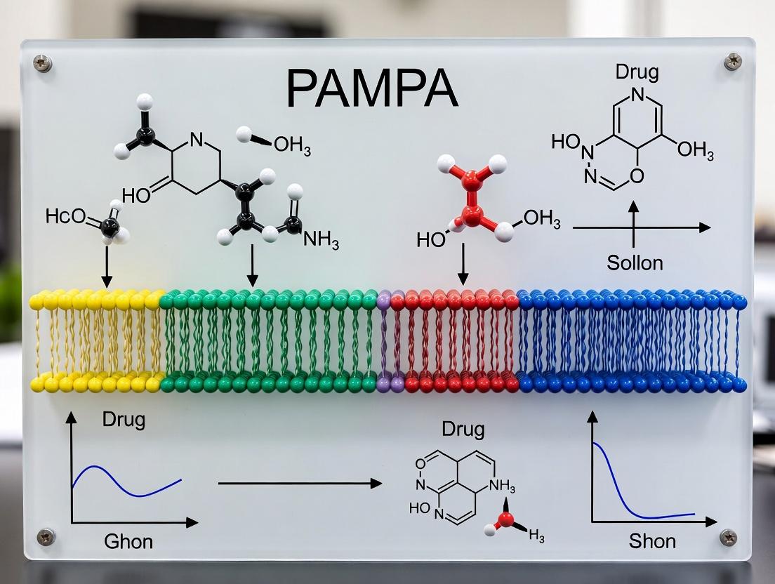
Core Principle: PAMPA models passive diffusion by creating an artificial lipid membrane (often phosphatidylcholine) on a filter support, separating a donor compartment (simulating gastrointestinal tract) from an acceptor compartment (simulating systemic circulation). The test compound is placed in the donor well, and its appearance in the acceptor well over time is measured, typically via UV spectroscopy or LC-MS/MS, to calculate an effective permeability (Pe).
Table 1: Comparison of Permeability Assay Methods
| Assay Type | Throughput | Cost | Complexity | Key Measurement | Primary Use |
|---|---|---|---|---|---|
| PAMPA | Very High (96/384-well) | Low | Low | Passive transcellular permeability (Pe) | Early-stage screening, rank-ordering |
| Caco-2 | Moderate | High | High | Apparent permeability (Papp), includes active transport | Advanced ADME, transporter studies |
| MDCK | Moderate-High | Moderate | Moderate | Apparent permeability (Papp) | Permeability screening, transporter models |
Table 2: Typical PAMPA Permeability Classification
| Effective Permeability (Pe x 10-6 cm/s) | Interpretation | Predicted Human Absorption |
|---|---|---|
| > 3.0 | High Permeability | Well absorbed (>90%) |
| 1.0 – 3.0 | Moderate Permeability | Moderately absorbed (20-90%) |
| < 1.0 | Low Permeability | Poorly absorbed (<20%) |
Experimental Protocols
Protocol 1: Standard PAMPA for Intestinal Permeability Prediction Objective: To determine the passive permeability of drug candidates. Materials: Pre-coated PAMPA plate (e.g., with lecithin in dodecane), 96-well microplate reader, test compounds (100 µM in pH 7.4 buffer), acceptor sink buffer (pH 7.4 with surfactant), and donor buffer (pH 6.5 or 7.4). Procedure:
- Plate Preparation: Add 150-300 µL of acceptor sink buffer to the bottom (acceptor) wells of the PAMPA sandwich.
- Compound Loading: Add 150-300 µL of the donor solution containing the test compound to the top (donor) wells. Include control wells (high permeability reference e.g., metoprolol; low permeability reference e.g., furosemide; and blank buffer).
- Assembly & Incubation: Carefully place the membrane donor plate on top of the acceptor plate to form a sandwich. Cover and incubate for 2-6 hours at 25°C without agitation.
- Termination & Analysis: Disassemble the sandwich. Quantify compound concentration in both donor and acceptor compartments using UV plate reader (direct measurement) or LC-MS/MS.
- Calculations: Calculate effective permeability (Pe) using the equation: Pe = { -ln(1 - [Drug]acceptor / [Drug]equilibrium) } x [VD x VA / (VD + VA) ] / (Area x Time).
Protocol 2: PAMPA for Blood-Brain Barrier (BBB) Penetration Objective: To predict passive diffusion across the blood-brain barrier. Modifications from Protocol 1:
- Membrane Composition: Use a PAMPA plate coated with a specialized porcine brain lipid extract (e.g., 2% PBLE in alkanes).
- Buffer System: Use PBS at pH 7.4 for both donor and acceptor compartments.
- Incubation Time: Typically 3-4 hours.
- Data Interpretation: Compounds with Pe (BBB) > 4.0 x 10⁻⁶ cm/s are considered likely to cross the BBB via passive diffusion.
Visualizations
Title: PAMPA Core Experimental Setup
Title: Standard PAMPA Experimental Workflow
The Scientist's Toolkit: Key Research Reagent Solutions
Table 3: Essential Materials for PAMPA Experiments
| Item | Function & Specification |
|---|---|
| Pre-coated PAMPA Plates | Multiwell plates (96 or 384) with a proprietary artificial lipid membrane immobilized on a filter support. Provides reproducibility and saves preparation time. |
| PAMPA Lipid Solution | For in-house plate coating. Often a 1-2% (w/v) solution of phosphatidylcholine (or brain lipid) in a long-chain alkane (e.g., dodecane, hexadecane). |
| PAMPA Assay Buffer System | Buffered saline solutions at specific pH (e.g., pH 5.0-6.5 for gastric, 6.5-7.4 for intestinal). May contain chemical scavengers to maintain sink conditions. |
| Permeability Control Compounds | A validation set including high (e.g., propranolol, verapamil), medium (e.g., atenolol), and low (e.g., furosemide) permeability markers. |
| UV-Compatible Microplates | Flat-bottom plates for direct concentration measurement of UV-active compounds post-assay. |
| LC-MS/MS System | For quantitative analysis of non-UV active compounds or complex mixtures. Provides high sensitivity and specificity. |
| Plate Sealing Films | Impermeable seals to prevent evaporation during the incubation step. |
Biomimetic membranes are synthetic systems engineered to replicate the core structural and functional properties of biological lipid bilayers. Within pharmaceutical research, particularly in Parallel Artificial Membrane Permeation Assay (PAMPA) protocol development, these membranes serve as indispensable, high-throughput tools for predicting passive, transcellular drug permeability. The fidelity of the biomimetic membrane—its composition, structure, and organization—directly dictates the reliability of permeability data for early-stage drug candidate screening, reducing reliance on more costly and time-consuming cell-based or in vivo models.
Composition and Structure: Key Components
The performance of a PAMPA membrane hinges on the precise reconstitution of a phospholipid bilayer supported on a microfilter. The composition is optimized to mimic the intestinal epithelial barrier for absorption prediction.
Table 1: Common Lipid Components in PAMPA Membranes
| Lipid/Component | Typical Concentration (%) | Role in Membrane Function | Common Source/Formulation |
|---|---|---|---|
| Phosphatidylcholine (PC) | 60-80% | Primary structural lipid, forms bilayer backbone; mimics outer leaflet of plasma membrane. | Egg lecithin, Soy lecithin, synthetic DOPC |
| Cholesterol | 10-20% | Modulates membrane fluidity and packing density; enhances barrier integrity. | Synthetic cholesterol |
| Anionic Phospholipids (e.g., PS, PI) | 5-15% | Introduces negative charge; can influence permeability of charged species. | Brain extract, synthetic |
| Synthetic Additives (e.g., Hexadecane) | 2-5% (v/v in solvent) | Used in some models to create a more hydrophobic interior; standardizes membrane formation. | Liquid alkane |
PAMPA Protocol: Detailed Methodology
Protocol Title: High-Throughput PAMPA for Passive Intestinal Permeability Prediction
Principle: A biomimetic lipid membrane is formed on a hydrophobic filter separating a donor plate (simulating intestinal lumen) and an acceptor plate (simulating bloodstream). The test compound diffuses from the donor compartment through the lipid membrane into the acceptor compartment. Permeability is determined by quantifying compound concentration in the acceptor well over time.
Materials & Reagent Toolkit:
Table 2: Research Reagent Solutions & Essential Materials
| Item | Function/Description | Example Product/Catalog |
|---|---|---|
| PAMPA Plate System | Multi-well plates with donor and acceptor compartments, and a filter support. | Millipore MultiScreen-IP, pION PAMPA Explorer plate |
| Lipid Solution | Dissolved lipids in organic solvent for membrane formation. | 2% (w/v) DOPC in dodecane, or 20 mg/mL egg lecithin in n-hexane. |
| Phosphate Buffered Saline (PBS), pH 7.4 | Aqueous buffer for donor/acceptor compartments; simulates physiological pH. | 10X PBS, diluted to 1X, pH-adjusted. |
| pION Gut-Box (UV plate reader) | Stirred, temperature-controlled UV plate reader for kinetic analysis. | pION μFLUX system |
| Reference Compounds | High & low permeability standards for assay validation. | Propranolol (high Perm), Ranitidine (low Perm), Carbamazepine. |
| Acceptor Sink Solution | Buffer with surfactant (e.g., 5% DMSO or 0.5% Tween) to maintain sink conditions. | PBS pH 7.4 with 5% DMSO. |
| LC-MS/MS System | For quantitative analysis of compound concentration (alternative to UV). | Agilent 6470, Sciex QTRAP. |
Procedure:
Membrane Formation:
- Prepare the lipid solution as specified in Table 2. Vortex thoroughly.
- Pipette 5 µL of the lipid solution directly onto the hydrophobic filter of each donor well/insert.
- Incubate plates for 1 hour at room temperature to allow uniform membrane formation and solvent evaporation/curing.
Plate Assembly:
- Fill the acceptor plate (bottom) with 200-300 µL of Acceptor Sink Solution per well.
- Carefully place the donor plate/insert on top, ensuring no air bubbles are trapped under the filter.
- Add 150-200 µL of the test compound solution (typically 50-100 µM in PBS pH 6.5 or 7.4) to the donor wells.
Incubation and Permeation:
- Seal the assembled plate to prevent evaporation.
- Incubate at 25°C or 37°C with gentle agitation (e.g., 100 rpm orbital shake) for a defined period (typically 2-6 hours).
Sample Collection and Analysis:
- After incubation, carefully separate the donor and acceptor plates.
- Quantify the compound concentration in both donor and acceptor compartments using a validated analytical method (e.g., UV spectrometry at λmax or LC-MS/MS for greater sensitivity and specificity).
- Also analyze the initial donor solution (time=0) for reference.
Data Calculation:
- Calculate the apparent permeability coefficient (Papp) using the formula:
Papp = (V_A / (Area * Time)) * (C_Acceptor / C_Donor_initial)where V_A = volume in acceptor well, Area = membrane area, Time = incubation time, C = concentration. - Classify compounds based on Papp: High permeability (Papp > 1.0 x 10⁻⁶ cm/s), Low permeability (Papp < 1.0 x 10⁻⁶ cm/s).
- Calculate the apparent permeability coefficient (Papp) using the formula:
Key Pathways and Workflow Visualization
Title: PAMPA Experimental Workflow
Title: Drug Permeation Through Biomimetic Barrier
Application Notes Parallel Artificial Membrane Permeation Assay (PAMPA) is a high-throughput, non-cell-based model used to predict passive transcellular permeability, a key determinant of drug absorption. Its use is strategic within a broader thesis on ADME (Absorption, Distribution, Metabolism, Excretion) protocol optimization, prioritizing efficiency and cost-effectiveness in early drug discovery.
Core Applications:
- Lead Optimization Triage: Primary application for screening large chemical libraries or series of analogs. It rapidly ranks compounds based on their passive permeability potential, informing Structure-Activity Relationship (SAR) for absorption.
- BBB Permeability Prediction: Specialized PAMPA models using brain lipid extracts (e.g., Porcine Brain Lipid in Dodecane, PBLD) provide early insight into a compound's potential to cross the Blood-Brain Barrier (BBB) via passive diffusion.
- Differentiating Transport Mechanisms: Used in conjunction with cell-based models (e.g., Caco-2). Low PAMPA permeability but high Caco-2 permeability suggests active transport involvement. Conversely, high PAMPA but low Caco-2 may indicate efflux transporter substrate.
- Formulation Support: Assesses permeability of pre-formulation candidates and can evaluate the permeability-enhancing effects of excipients or prodrugs.
When NOT to Use PAMPA: PAMPA is unsuitable for compounds whose transport is heavily influenced by active transporters, efflux pumps, paracellular pathways (for larger molecules), or metabolism. It should not replace cell-based or in vivo models for late-stage development decisions.
Quantitative Data Summary
Table 1: PAMPA Model Variations and Applications
| Membrane Composition | Common Buffer pH | Primary Application | Typical Permeability (Pe) Range (x10⁻⁶ cm/s) |
|---|---|---|---|
| Phosphatidylcholine in Dodecane | 7.4 (Donor), 7.4 (Acceptor) | Basic GI permeability screening | 0.1 (Low) - 10+ (High) |
| Porcine Brain Lipid in Dodecane (PBLD) | 7.4 / 7.4 | Blood-Brain Barrier (BBB) permeability prediction | < 1.0 (CNS-) > 2.0 (CNS+) |
| Double-Sink PAMPA (with sink additives) | Gradient (e.g., 5.0 / 7.4) | Simulating intestinal pH gradient & sink conditions | Enhances dynamic range for poorly soluble compounds |
Table 2: Interpreting PAMPA Permeability (Pe)
| Pe (10⁻⁶ cm/s) | Predicted Human Fraction Absorbed (%Fa) | Interpretation for Oral Drugs |
|---|---|---|
| < 0.1 | Poor (< 10%) | Likely low passive absorption |
| 0.1 - 1.0 | Moderate (10-50%) | Variable absorption |
| 1.0 - 10.0 | Good (50-90%) | Favorable passive absorption |
| > 10.0 | Excellent (> 90%) | High passive absorption |
Detailed Experimental Protocol: Standard PAMPA for GI Permeability
Principle: A compound diffuses from a donor well, through a lipid-infused filter membrane, into an acceptor well. Permeability is calculated from the concentration in the acceptor compartment after a set incubation time.
I. Materials & Reagent Preparation
- PAMPA Plate: Multi-well filter plate (e.g., 96-well) and matching acceptor plate.
- Artificial Membrane Lipid: 2% (w/v) Phosphatidylcholine (PC) in dodecane.
- Buffer Systems:
- Donor Buffer: Prisma HT Buffer (pH 5.0 or 7.4) or PBS (pH 7.4).
- Acceptor Buffer: PBS (pH 7.4) or Double-Sink Buffer (with surfactant/additives).
- Test Compound: 50-100 µM in donor buffer (from 10 mM DMSO stock). Final DMSO ≤ 1%.
- Analytical Method: UV plate reader or LC-MS/MS for quantification.
- Reference Compounds: Propranolol (high permeability), Ranitidine (low permeability).
II. Procedure
- Acceptor Plate Preparation: Fill each well of the acceptor plate with 200-300 µL of acceptor buffer.
- Membrane Formation: Pipette 4-5 µL of the 2% PC/dodecane solution onto the filter of each donor plate well. Ensure the lipid forms a uniform layer.
- Donor Solution Addition: Carefully place the donor plate on top of the acceptor plate, creating a "sandwich." Add 150-200 µL of the compound solution (or reference/blank buffer) to the donor wells.
- Incubation: Incubate the assembled plate at room temperature (or 37°C) without agitation for 2-6 hours (typically 4 hours) to allow passive diffusion.
- Plate Separation: After incubation, carefully separate the donor and acceptor plates.
- Sample Analysis: Quantify the compound concentration in both donor and acceptor compartments, and in the initial donor solution (C₀), using UV spectrometry (e.g., at 290-310 nm for unspecific detection) or LC-MS/MS.
- Data Analysis: Calculate effective permeability (Pe) using the following equation:
Pe = -{ln(1 - [Drug]acceptor / [Drug]equilibrium)} / {A * (1/V_donor + 1/V_acceptor) * t}Where A = filter area, V = volume, t = incubation time.
III. The Scientist's Toolkit: Essential Research Reagent Solutions Table 3: Key Materials for PAMPA Experiments
| Item | Function & Rationale |
|---|---|
| 96-Well PAMPA Plate System | Provides standardized filter (e.g., PVDF, 0.45µm) and acceptor plates for high-throughput, reproducible sandwich formation. |
| Phosphatidylcholine (e.g., Egg Lecithin) | Forms the core artificial lipid bilayer, mimicking the hydrophobic interior of cell membranes for passive diffusion studies. |
| Porcine Brain Lipid Extract (PBLE) | Specialized lipid mixture for creating a membrane that more closely resembles the lipid composition of the Blood-Brain Barrier. |
| Dodecane | Inert organic solvent used to dissolve lipids and support the formation of a stable artificial membrane on the filter. |
| Prisma HT Buffer | Proprietary buffer system designed to minimize non-specific binding to plates and maintain compound stability during assay. |
| Double-Sink Buffer Additives | Agents (e.g., surfactants, proteins) added to acceptor compartment to create a "sink" condition, improving dynamic range for lipophilic compounds. |
| UV-Compatible Acceptor Plates | Plates with low UV absorbance allow direct concentration measurement of compounds in the acceptor well via UV spectrometry. |
Visualizations
Title: Standard PAMPA Experimental Workflow
Title: Decision Tree for PAMPA Application in ADME
This application note is framed within a broader thesis investigating the standardization and optimization of Parallel Artificial Membrane Permeation Assay (PAMPA) protocols for preclinical drug development. PAMPA serves as a high-throughput, cost-effective model for predicting passive transcellular permeability across biological membranes, such as the gastrointestinal tract, blood-brain barrier (BBB), and skin. Its core value lies in its simplicity and reproducibility, but a critical understanding of its correlation with, and deviations from, complex biological systems is essential for accurate data interpretation.
Core Principles: Model vs. Biology
PAMPA utilizes an artificial membrane, typically composed of a phospholipid mixture (e.g., egg lecletihin, phosphatidylcholine) dissolved in an inert organic solvent (e.g., dodecane, hexadecane), immobilized on a hydrophobic filter. Permeation is driven solely by passive diffusion down a concentration gradient. In contrast, biological membranes are complex, dynamic bilayers containing diverse lipid species, cholesterol, and embedded proteins that facilitate active transport, efflux, and paracellular pathways.
Table 1: Fundamental Comparison: PAMPA vs. Biological Membranes
| Feature | PAMPA (Artificial Model) | Biological Membranes (e.g., GI Tract) |
|---|---|---|
| Membrane Composition | Defined phospholipids in organic solvent. | Complex asymmetric lipid bilayer with cholesterol, glycolipids. |
| Permeation Mechanisms | Passive transcellular diffusion only. | Passive diffusion, carrier-mediated influx/efflux, paracellular, active transport. |
| Membrane Dynamics | Static, non-fluidic. | Fluid, with lateral and transverse mobility. |
| Protein Components | Absent. | Integral and peripheral proteins (transporters, channels, receptors). |
| Electrical Properties | Non-polarized. | Electrically polarized (e.g., tight junction resistance). |
| Throughput & Cost | Very high, low cost per sample. | Low to moderate (e.g., Caco-2), higher cost. |
| Reproducibility | Excellent, minimal variability. | Subject to biological and methodological variability. |
Key Applications and Predictive Performance
PAMPA is primarily used for early-stage screening of passive permeability. Its performance is validated by correlating apparent permeability (Papp) values with established models or human fraction absorbed (Fa%) data.
Table 2: Quantitative Correlation of PAMPA with Biological Data
| PAMPA Variant / Lipid Composition | Reported Correlation (R²) with Human Fa% | Typical Papp Range (x10⁻⁶ cm/s) | Primary Application |
|---|---|---|---|
| Double-Sink PAMPA (DST) | 0.85 - 0.95 | 0.1 - 50 | GI permeability prediction. |
| BBB-PAMPA (Porcine Brain Lipid) | 0.75 - 0.88 (vs. in vivo brain uptake) | 0.01 - 10 | Blood-brain barrier permeability screening. |
| Skin-PAMPA (Ceramide-based) | 0.70 - 0.82 (vs. Franz cell data) | 0.001 - 5 | Transdermal permeability estimation. |
| Caco-2 Cell Model (for reference) | 0.80 - 0.90 | 0.1 - 100 | Includes active transport/efflux. |
Data compiled from recent literature (2022-2024). Correlation strength depends on the specific chemical library and protocol used.
Detailed Experimental Protocols
Protocol 4.1: Standard Double-Sink PAMPA (DST-PAMPA) for GI Permeability Prediction
Objective: To determine the passive permeability (Papp) of test compounds with high correlation to human intestinal absorption.
The Scientist's Toolkit:
| Item / Reagent Solution | Function / Explanation |
|---|---|
| PAMPA Plate Assembly (e.g., 96-well donor/acceptor plate with filter) | Provides the physical structure for the artificial membrane. |
| Phospholipid Solution (2% w/v Lecithin in Dodecane) | Forms the core artificial membrane on the filter support. |
| Donor Solution (pH 5.5 or 6.8 buffer) | Simulates gastrointestinal lumen conditions. |
| Acceptor Sink Solution (pH 7.4 buffer with additives e.g., surfactant) | Mimics blood-side sink conditions, maintaining gradient. |
| Test Compound Solution (100-500 µM in donor buffer) | The drug candidate solution for permeability assessment. |
| UV-compatible Microplate Reader | Enables high-throughput concentration measurement via UV spectrometry. |
| Reference Compounds (e.g., Warfarin [high perme], Furosemide [low perme]) | Validation controls for assay performance and data normalization. |
Methodology:
- Membrane Formation: Pipette 5 µL of the phospholipid solution onto each filter of the donor plate. Allow to sit for 30 minutes to form a uniform, solvent-lipid membrane.
- Plate Assembly: Fill the acceptor plate wells with 300 µL of acceptor sink solution. Carefully place the donor plate on top, ensuring no air bubbles are trapped under the filter.
- Compound Loading: Add 150 µL of the test compound solution (in appropriate pH buffer) to the donor wells. Cover the plate to prevent evaporation.
- Incubation: Incubate the assembled plate at room temperature (or 25°C) without agitation for the determined assay time (typically 3-5 hours for DST).
- Sampling & Analysis: After incubation, disassemble the plate. Quantify the compound concentration in both donor and acceptor compartments (and optionally the membrane) using a validated UV method (e.g., direct UV scan from 250-500 nm).
- Data Calculation: Calculate the apparent permeability (Papp) using the formula: Papp = { -VD * VA / [ (VD + VA) * A * t ] } * ln[ 1 - (CA(t) / Cequilibrium) ] Where V= volume, A= filter area, t= time, C= concentration.
Protocol 4.2: BBB-PAMPA for Blood-Brain Barrier Penetration Screening
Objective: To predict the passive diffusion of compounds across the blood-brain barrier using a porcine brain lipid extract (PBLE) membrane.
Methodology:
- Membrane Preparation: Prepare a 2% (w/v) solution of porcine brain lipid extract in dodecane.
- Assembly & Loading: Follow steps 1-3 of Protocol 4.1, using pH 7.4 buffer in both donor and acceptor compartments to simulate physiological pH.
- Incubation: Incubate for 2-4 hours (BBB-PAMPA typically requires less time than GI-PAMPA due to thinner membrane modeling).
- Analysis & Interpretation: Analyze as in step 5 of Protocol 4.1. Compounds with Papp > ~3.0 x 10⁻⁶ cm/s are considered potentially CNS-permeable via passive diffusion, while those < ~1.5 x 10⁻⁶ cm/s are likely excluded.
Limitations and Strategic Use in Drug Discovery
While invaluable, PAMPA has distinct limitations that mandate complementary assays:
- Lacks Transporters: Cannot identify substrates for efflux (e.g., P-gp) or uptake transporters.
- No Metabolism: Does not account for enzymatic degradation.
- Paracellular Pathway: Poorly models the porosity of epithelial tight junctions for small, polar molecules.
- Protein Binding: Does not incorporate plasma protein binding effects.
Therefore, the optimal strategy is a tiered approach: Use PAMPA for early, high-throughput passive permeability ranking, followed by cell-based models (Caco-2, MDCK) for transporter effects, and finally specialized assays for definitive assessment.
Diagram 1: Permeation Pathways in PAMPA vs Biological Membranes
Diagram 2: Strategic Tiered Use of PAMPA in Drug Discovery
Within the context of advancing PAMPA (Parallel Artificial Membrane Permeation Assay) protocol research, this application note details the evolution from the classic lipid membrane model to sophisticated variants that better predict gastrointestinal (GI) absorption and blood-brain barrier (BBB) penetration. PAMPA serves as a high-throughput, non-cell-based tool for estimating passive transcellular permeability, a critical parameter in early drug discovery. This document provides updated protocols and comparative data for key PAMPA formats.
Classic PAMPA: Principle and Protocol
The classic PAMPA assay utilizes a hydrophobic filter material coated with a phospholipid solution (e.g., 2% Lecithin in dodecane) to create an artificial membrane separating a donor compartment (containing test compound) from an acceptor compartment.
Protocol 1.1: Classic PAMPA for Intestinal Permeability
- Materials: Multi-well PAMPA plate (e.g., 96-well), PVDF filter plate (0.45 µm pore size), acceptor plate, 10 mM compound stock in DMSO, pH 7.4 buffer (e.g., PBS or PRISMA HT), 2% (w/v) Phosphatidylcholine in dodecane.
- Procedure:
- Membrane Formation: Add 5 µL of the lipid solution to each filter well of the donor plate. Incubate for 1 hour to allow solvent evaporation and membrane formation.
- Plate Assembly: Fill the acceptor plate wells with 300 µL of pH 7.4 buffer. Carefully place the lipid-coated donor plate on top.
- Donor Solution: Dilute test compound in pH 6.5 buffer (simulating intestinal pH) to a final concentration of 50-100 µM. Add 300 µL to each donor well.
- Incubation: Assemble the sandwich and incubate at room temperature for 4-6 hours without agitation.
- Analysis: Disassemble the plates. Quantify compound concentration in both donor and acceptor compartments using UV spectroscopy (e.g., 96-well plate reader) or LC-MS/MS. Calculate the effective permeability (Pₑ in cm/s).
Advanced PAMPA Variants: Protocols and Applications
Protocol 2.1: Double-Sink PAMPA Protocol This variant incorporates sink conditions in the acceptor compartment to mimic the continuous drug removal by systemic circulation, enabling the testing of poorly soluble compounds.
- Key Modification: The acceptor buffer contains a chemical sink agent (e.g., 5% w/v Bovine Serum Albumin (BSA) or surfactant micelles) to bind lipophilic compounds.
- Procedure: Follow Protocol 1.1, but prepare the acceptor solution with 5% BSA in pH 7.4 buffer. Due to protein content, analysis typically requires LC-MS/MS. The sink condition allows for a more physiologically relevant gradient for highly permeable, lipophilic drugs.
Protocol 2.2: Bio-Mimetic (BBB) PAMPA Protocol This variant uses a specialized lipid blend to mimic the phospholipid composition of the blood-brain barrier endothelial cell membrane.
- Key Modification: The membrane is formed from a porcine brain lipid extract (e.g., 1% PBL in dodecane) or a tailored mixture of phospholipids and cholesterol.
- Procedure: Follow Protocol 1.1, but use the PBL solution for membrane formation. Use pH 7.4 buffer in both donor and acceptor compartments. This model is specifically calibrated to predict CNS penetration.
Comparative Data Analysis
Table 1: Key Parameters of PAMPA Variants
| Parameter | Classic PAMPA | Double-Sink PAMPA | Bio-Mimetic (BBB) PAMPA |
|---|---|---|---|
| Membrane Composition | 2% PC in dodecane | 2% PC in dodecane | 1% Porcine Brain Lipid in dodecane |
| Donor pH | 5.0-6.5 (GI), 7.4 (BBB) | 6.5 or 7.4 | 7.4 |
| Acceptor Sink | None (pH 7.4 buffer) | Yes (e.g., 5% BSA) | None (pH 7.4 buffer) |
| Primary Application | Basic passive permeability | High, lipophilic compounds; GI | Blood-Brain Barrier prediction |
| Incubation Time | 4-6 hours | 2-4 hours | 3-5 hours |
| Typical Pₑ Range (cm/s) | 10⁻⁶ to 10⁻⁴ | 10⁻⁷ to 10⁻⁴ | 10⁻⁷ to 10⁻⁴ |
Table 2: Example Permeability Classification
| Permeability (Pₑ in cm/s) | Classic/DS-PAMPA Interpretation | BBB-PAMPA Interpretation |
|---|---|---|
| > 2.0 x 10⁻⁶ | High (Well absorbed) | High CNS Penetration (likely) |
| 1.0 - 2.0 x 10⁻⁶ | Moderate | Moderate CNS Penetration |
| < 1.0 x 10⁻⁶ | Low (Poorly absorbed) | Low CNS Penetration (unlikely) |
Experimental Workflow and Pathway Diagrams
Title: Decision Workflow for PAMPA Variant Selection
Title: Core Principle of Passive Diffusion in PAMPA
The Scientist's Toolkit: Research Reagent Solutions
Table 3: Essential Materials for PAMPA Experiments
| Item / Reagent | Function & Application |
|---|---|
| PVDF Filter Plate (0.45 µm pore) | Hydrophobic support for the artificial lipid membrane. Standard for all PAMPA formats. |
| Phosphatidylcholine (PC) | Primary phospholipid for classic GI permeability models (e.g., 2% in dodecane). |
| Porcine Brain Lipid (PBL) Extract | Complex lipid mixture used to create bio-mimetic membranes for BBB permeability studies. |
| Dodecane | Inert organic solvent used to dissolve lipids and facilitate membrane formation on filters. |
| BSA (Bovine Serum Albumin) | Sink agent in Double-Sink PAMPA. Binds lipophilic drugs, maintaining a concentration gradient. |
| PRISMA HT Buffer | A universal buffer system designed to mimic the physicochemical properties of the GI tract. |
| pH 6.5 / 7.4 Buffer | To simulate intestinal lumen pH (6.5) or blood/physiological pH (7.4). |
| UV-Compatible Acceptor Plate | Allows direct quantification of compound concentration via UV spectroscopy in the acceptor well. |
Step-by-Step PAMPA Protocol: From Plate Setup to Data Analysis
The Parallel Artificial Membrane Permeation Assay (PAMPA) is a critical high-throughput screening tool in early drug discovery for predicting passive, transcellular permeability. This application note provides a comprehensive, updated checklist of materials and reagents essential for robust and reproducible PAMPA protocol execution, framed within ongoing research to enhance predictive accuracy for CNS and intestinal absorption.
The Scientist's Toolkit: Essential PAMPA Reagent Solutions
| Item | Function in PAMPA Protocol |
|---|---|
| Phospholipid Membrane Components (e.g., Porcine Brain Lipid Extract, Lecithin) | Forms the artificial lipid bilayer that mimics biological barriers (e.g., intestinal mucosa, blood-brain barrier). |
| Acceptor Sink Buffer (e.g., PBS pH 7.4, Prisma HT Buffer) | Maintains sink conditions in the acceptor well to drive passive diffusion of compounds. |
| Donor Buffer (pH 5.0-6.5 for GI, pH 7.4 for BBB) | Simulates the physiological pH of the donor compartment (gut lumen or systemic circulation). |
| PAMPA Plate (96-well filter plate with PVDF/PE membrane) | Multi-well plate system where the artificial lipid membrane is created and the permeability assay is conducted. |
| pION Gut-Box or BBB-Box Solution System | Commercial surfactant systems for creating consistent, biomimetic lipid membranes. |
| Test Compounds & Reference Standards (e.g., Verapamil, Propranolol, Warfarin) | Compounds of unknown permeability are tested alongside known high/low permeability standards for validation. |
| UV Plate Reader or LC-MS/MS System | For quantitative analysis of compound concentration in donor and acceptor compartments post-assay. |
| Dodecane or Hexadecane (Alkane Solvent) | Used as a support solvent for the lipid solution to form a stable, reproducible artificial membrane. |
Detailed Protocol: Standard PAMPA for Intestinal Permeability
Materials Checklist
- Plate: 96-well Acceptor Plate (flat-bottom); 96-well Donor Plate (filter plate, 0.45µm hydrophobic PVDF membrane).
- Lipid Solution: 2% (w/v) Porcine Brain Lipid Extract in Dodecane.
- Buffers: Donor Buffer (pH 5.5 or 6.5), Acceptor Sink Buffer (pH 7.4). Pre-warm to 37°C.
- Compounds: 10 mM stock in DMSO. Dilute to 50-100 µM in donor buffer (final DMSO ≤1%).
- Equipment: Multichannel pipettes, plate shaker, humidity chamber, UV-Vis plate reader or LC-MS.
Protocol Steps
- Membrane Formation: Pipette 5 µL of lipid solution onto the filter membrane of each donor plate well. Incubate for 5-10 minutes to allow uniform membrane formation.
- Plate Assembly: Fill acceptor plate wells with 300 µL of acceptor sink buffer. Carefully place the donor plate on top, ensuring the lipid-coated filter contacts the buffer in each well to form a "sandwich."
- Compound Addition: Add 150 µL of the diluted test compound or reference standard solution to the donor wells.
- Incubation: Cover the plate to prevent evaporation and incubate at 37°C (without CO₂) for 2-6 hours (optimize based on compound properties) without agitation.
- Termination & Sampling: Carefully separate the donor and acceptor plates. Quantify compound concentration in both the initial donor solution (Cinitial), the final acceptor solution (Cacceptor), and the final donor solution (Cdonor, final) using UV spectrometry (direct measurement) or LC-MS/MS.
Data Analysis: Calculate permeability (Pe) using the following equation:
[ Pe = \frac{-2.303 VD VA}{A(VD + VA)t} \log{10} \left[ 1 - \frac{VA}{VD} \left( \frac{CA(t)}{CD(0)} \right) \right] ] Where: A = filter area, t = incubation time, VD & VA = donor/acceptor volumes, CA(t) = acceptor concentration at time t, CD(0) = initial donor concentration.
| Compound Class | Example Compound | Expected Pe (10⁻⁶ cm/s) | Classification |
|---|---|---|---|
| High Permeability | Propranolol | 15.0 - 25.0 | Well-absorbed |
| Moderate Permeability | Warfarin | 5.0 - 10.0 | Moderately absorbed |
| Low Permeability | Ranitidine | 0.1 - 1.0 | Poorly absorbed |
| CNS High Perm (BBB-PAMPA) | Verapamil | 12.0 - 20.0 | High CNS penetration |
| CNS Low Perm (BBB-PAMPA) | Sucrose | < 0.5 | Low CNS penetration |
Protocol for Blood-Brain Barrier (BBB) Specific PAMPA
- Key Modification: Use a specialized lipid formulation (e.g., pION BBB Lipid). Donor and acceptor buffers are typically both at pH 7.4.
- Incubation: Extend incubation time to up to 18 hours due to slower diffusion kinetics mimicking the BBB.
- Validation: Always include CNS-positive (e.g., Verapamil) and CNS-negative (e.g., Sucrose) controls.
PAMPA Experimental Workflow
Decision Logic for Permeability Classification
1. Introduction Within the broader thesis on optimizing the Parallel Artificial Membrane Permeation Assay (PAMPA), the preparation of reproducible artificial lipid membranes and plates is the foundational step. This protocol details the procedures for creating lipid-infused membranes and preparing the donor and acceptor compartments, critical for generating reliable data on passive, transcellular drug permeability.
2. Key Research Reagent Solutions & Materials
| Item | Function / Rationale |
|---|---|
| Phospholipid Solution | Typically 1-20% (w/v) in alkane (e.g., dodecane). Forms the artificial membrane barrier. Lecithin (e.g., PC, PE, PI mixtures) or pure lipids (e.g., DOPC) are used. |
| Inert Solvent (e.g., Dodecane, Hexadecane) | Dissolves lipids to create the membrane-forming solution. Its viscosity influences membrane stability and permeability. |
| PAMPA Plate (Multi-well filter plate) | Serves as the donor plate. Its microporous filter (0.45 µm, PVDF or similar) supports the lipid membrane. |
| Acceptor Plate (Standard 96-well plate) | Holds the acceptor buffer solution. Must be compatible for creating a "sandwich" with the donor plate. |
| Buffer Solutions (pH 5.0-7.4) | Simulate gastrointestinal (e.g., pH 5.0, 6.5) or blood-brain barrier (pH 7.4) conditions. Include additives to maintain sink conditions. |
| Magnetic Stirrer & Micro-stir Bars | Ensures hydrodynamics in acceptor wells, reducing the unstirred water layer effect. |
| Precision Micro-pipettes & Repeating Dispenser | For accurate, high-throughput dispensing of viscous lipid solutions and buffers. |
3. Protocol: Preparation of Acceptor Plate
- 3.1. Fill each well of a clean 96-well acceptor plate with 200-300 µL of the desired acceptor buffer (e.g., Phosphate Buffered Saline, pH 7.4, with 5% DMSO or surfactant to maintain sink conditions).
- 3.2. Carefully place a compatible micro stir bar (e.g., 2x7 mm) into each acceptor well if a stirred assay format is used.
- 3.3. Seal the plate and set aside until membrane formation is complete.
4. Protocol: Preparation of Artificial Lipid Membranes on Donor Plate
- 4.1. Lipid Solution Preparation: Prepare a fresh phospholipid solution by dissolving the desired lipid (e.g., 2% (w/v) Porcine Brain Lipid Extract or 1% (w/v) DOPC) in dodecane. Vortex until fully dissolved.
- 4.2. Membrane Formation:
- Place a clean, dry multi-well filter plate (donor plate) on a level surface.
- Using a repeating dispenser or multichannel pipette, add 5 µL of the lipid solution directly onto the filter surface of each well.
- Allow the solution to spread spontaneously and completely across the filter for 5-10 minutes. The filter should appear glossy and uniform.
- Critical Note: Do not pipette up and down. The lipid membrane forms by spontaneous distribution.
- 4.3. Donor Solution Addition: After membrane formation, add 150-200 µL of the donor solution (test compound in appropriate buffer, e.g., pH 5.0 or 6.5) to each well on top of the lipid membrane.
5. Summary of Key Quantitative Parameters for PAMPA Membrane Preparation
| Parameter | Typical Range | Optimal Value (Example) | Impact on Assay |
|---|---|---|---|
| Lipid Concentration | 0.5% - 20% (w/v) in alkane | 2% (Porcine Brain) | Higher concentration can reduce permeability, increasing membrane integrity. |
| Membrane Volume | 4 - 10 µL per well | 5 µL | Standardized volume is critical for reproducibility. |
| Acceptor Buffer Volume | 200 - 300 µL | 250 µL | Must be sufficient to maintain sink conditions. |
| Donor Compound Concentration | 10 - 500 µM | 100 µM | Must be within solubility limits and detectable by UV or LC-MS. |
| Incubation Time | 2 - 24 hours | 16-18 hours (unstirred) | Allows for sufficient compound permeation. |
| Assay Temperature | 25°C or 37°C | 25°C (± 2°C) | Controlled temperature is essential for reproducibility. |
6. Experimental Workflow Diagram
PAMPA Plate Preparation Workflow
7. PAMPA Membrane Formation & Permeation Pathway
PAMPA Passive Diffusion Pathway
This document details the critical second phase of the PAMPA (Parallel Artificial Membrane Permeation Assay) protocol, situated within a comprehensive thesis investigating standardized methods for predicting passive, transcellular drug permeability. This section focuses on the precise execution of compound dosing, system incubation, and the analysis of the permeation process, which are fundamental for generating reliable, high-throughput permeability data in early drug development.
Compound Dosing and Incubation Protocol
Preparation of Donor and Acceptor Plates
Materials Required:
- Pre-coated PAMPA plate (lipid membrane in donor well).
- Acceptor plate (typically a 96-well deep well plate).
- Test compound stock solutions (typically 10 mM in DMSO).
- Buffer systems: Commonly used are:
- pH 7.4 PBS: Simulates intestinal blood pH.
- pH 6.5 or 5.5 Buffer: Simulates duodenal/jejunal or gastric pH for gradient assays (e.g., BD-RTM model).
- Prisma HT Buffer: Used in commercial systems for broad pH range.
- Compound diluent buffer.
- Multichannel pipettes and liquid handling robotics.
Procedure:
- Acceptor Sink Preparation: Fill each well of the acceptor plate with 200-300 µL of the appropriate buffer. Ensure no air bubbles are present at the bottom of the wells.
- Donor Solution Preparation: Dilute the test compound from the DMSO stock into the selected donor buffer to a final concentration typically between 50-100 µM. Keep final DMSO concentration ≤ 1% (v/v) to maintain membrane integrity.
- Plate Assembly: Carefully place the donor plate (membrane plate) onto the acceptor plate, ensuring each donor well is aligned with an acceptor well. The lipid membrane forms the interface between the donor and acceptor compartments.
- Dosing: Pipette 150-200 µL of the donor solution into each corresponding donor well. Avoid introducing air bubbles or touching the membrane.
- Sealing and Incubation: Seal the assembled sandwich plate with a lid or adhesive seal to prevent evaporation. Incubate the plate at room temperature (25°C) or 37°C (for physiological relevance) without agitation for a period of 2 to 16 hours, depending on the specific PAMPA model and compound properties. Standard incubation is often 3-5 hours.
Key Incubation Parameters
Table 1: Standard Incubation Conditions for Common PAMPA Models
| PAMPA Model | Typical Donor pH | Acceptor pH | Incubation Time (h) | Temperature (°C) | Key Application |
|---|---|---|---|---|---|
| Classic (PION) | 7.4 | 7.4 | 3-5 | 25 | Basic passive permeability screening. |
| BD-RTM (Biomimetic) | 5.0 - 6.5 | 7.4 | 3-5 | 25 | Simulates pH gradient of GI tract. |
| DS-RTM (Double Sink) | 7.4 | 7.4 | 2-4 | 25 | Enhanced sink conditions for low-solubility compounds. |
| BBB (Blood-Brain Barrier) | 7.4 | 7.4 | 3-4 | 37 | Predicts CNS penetration. |
The Permeation Process: Sampling and Analysis
Post-Incubation Separation and Sampling
- After incubation, carefully separate the donor plate from the acceptor plate.
- Sampling from Acceptor Compartment: Transfer a known volume (e.g., 150 µL) from each acceptor well to a new analysis plate. This sample contains compound that has permeated the membrane.
- Sampling from Donor Compartment (Optional but recommended): Sample from the donor compartment at the end of the experiment to determine mass balance (recovery). This helps identify compound loss due to membrane binding or precipitation.
Quantitative Analysis
- Analyze the concentration of test compound in the acceptor samples (and donor samples, if taken) using a quantitative analytical method. Standard methods include:
- UV-Vis Spectroscopy: Direct measurement if compound has a chromophore.
- LC-MS/MS (Liquid Chromatography with Tandem Mass Spectrometry): Gold standard for sensitivity and specificity, especially for complex matrices.
- Fluorescence Spectroscopy: For fluorescent compounds.
- A calibration curve of the test compound in the acceptor buffer must be run in parallel for accurate quantification.
Data Calculation
The permeation rate is expressed as the Apparent Permeability Coefficient (Papp in cm/s).
Formula:
P_app = (V_D * V_A) / (A * (V_D + V_A) * t) * ln(1 - C_A(t) / C_equilibrium)
Where:
V_D= Volume of donor compartment (cm³).V_A= Volume of acceptor compartment (cm³).A= Effective filter area of the membrane (cm²). (Typically 0.3 cm² for a 96-well plate).t= Incubation time (seconds).C_A(t)= Analyte concentration in acceptor at time t.C_equilibrium= Theoretical concentration at equilibrium (often approximated by initial donor concentration if recovery is high).
Simplified Operational Formula (for sink conditions, <20% permeation):
P_app = (C_A(t) * V_A) / (C_D(0) * A * t)
Where C_D(0) is the initial donor concentration.
Table 2: Permeability Classification Guide
| Papp (x10⁻⁶ cm/s) | Permeability Classification | Typical Oral Absorption |
|---|---|---|
| > 10 | High | Well absorbed (>90%) |
| 2 - 10 | Moderate | Variable absorption |
| < 2 | Low | Poorly absorbed (<20%) |
Experimental Workflow Diagram
PAMPA Permeation Assay Core Workflow
Key Research Reagent Solutions & Materials
Table 3: The Scientist's Toolkit for PAMPA Assay
| Item | Function/Benefit | Example/Notes |
|---|---|---|
| PAMPA Plate | Pre-coated filter plate (PVDF or similar) with immobilized artificial lipid membrane. Provides the barrier for permeation. | MultiScreen IP sterile plates; often coated with lecithin (e.g., 2% w/v in dodecane). |
| Acceptor Plate | Deep-well plate to hold the acceptor buffer sink. Must form a tight seal with donor plate. | 96-well polypropylene deep well plate. |
| Prisma HT Buffer System | Universal buffer designed to maintain consistent pH and ionic strength across a wide range, improving predictability. | Used in commercial PAMPA kits to standardize conditions. |
| BD-RTM Lipid Solution | Proprietary lipid mixture designed to more accurately mimic the composition of the human intestinal brush border membrane. | Enhances correlation of results with human fractional absorption. |
| DS-RTM Sink Enhancer | Additive for the acceptor compartment to create a "double-sink" effect, preventing back-permeation for very lipophilic compounds. | Improves assay dynamic range. |
| Reference Compounds | High, medium, and low permeability controls for assay validation and plate normalization. | e.g., Verapamil (High), Metoprolol (Mid), Ranitidine (Low). |
| LC-MS/MS Compatible Buffers | Buffers formulated with volatile salts (e.g., ammonium acetate) to prevent ion suppression in mass spectrometry analysis. | Essential for direct injection analysis without desalting steps. |
Within the broader thesis on PAMPA protocol optimization, the accurate quantification of analyte concentration in the donor, acceptor, and membrane compartments is paramount for calculating key permeability parameters (e.g., Pe). This protocol details two complementary analytical techniques: high-throughput UV-Plate Reader analysis and selective, sensitive LC-MS/MS analysis.
The choice of analytical method depends on the compound's properties, required sensitivity, and throughput needs.
Table 1: Comparison of UV-Plate Reader and LC-MS/MS for PAMPA Sample Analysis
| Parameter | UV-Plate Reader | LC-MS/MS |
|---|---|---|
| Throughput | Very High (96/384-well plate in minutes) | Moderate to Low (individual sample runs) |
| Sensitivity | Moderate (µM range) | Very High (nM-pM range) |
| Selectivity | Low (interference from UV-absorbing matrix) | Very High (chromatographic separation + MRM) |
| Sample Prep | Minimal (often direct measurement) | Required (often protein precipitation, dilution) |
| Ideal Use Case | Single-compound screening, high purity buffers | Complex matrices, low solubility compounds, cassette dosing (multiple compounds) |
| Key Calculated Metric | Apparent Permeability (Papp) | Effective Permeability (Pe) |
Table 2: Example Permeability Classification from UV/LC-MS Data
| Pe (x 10-6 cm/s) | Permeability Classification | Typical Absorption % (from Acceptor) |
|---|---|---|
| > 2.0 | High | > 70% |
| 0.2 – 2.0 | Moderate | 20% - 70% |
| < 0.2 | Low | < 20% |
Detailed Experimental Protocols
Protocol 3.1: Quantitative Analysis via UV-Plate Reader
Principle: Direct measurement of analyte concentration based on its intrinsic ultraviolet (UV) absorbance at a specific wavelength (λmax).
Materials:
- PAMPA acceptor/donor plate samples.
- Clear-bottom 96-well or 384-well microplate.
- Multi-mode microplate reader with UV-Vis capability.
- Reference buffer blanks (from assay plate).
- Compound standard stock solution.
Procedure:
- Standard Curve Preparation: Serially dilute the compound stock in the appropriate buffer (e.g., PBS pH 7.4) to create 6-8 standard points covering the expected concentration range (e.g., 1 – 100 µM). Include a blank (buffer only).
- Sample Transfer: Aliquot 100-200 µL from each PAMPA donor, acceptor, and reference well into the corresponding wells of the analysis microplate.
- Measurement: Insert the plate into the reader. Perform a wavelength scan (e.g., 200-500 nm) on representative wells to confirm λmax. Then, read the absorbance of all standards and samples at the predetermined λmax.
- Data Analysis:
- Generate a linear standard curve by plotting the absorbance of standards against their known concentration.
- Use the regression equation to calculate the concentration in each sample well.
- Apply the derived concentrations to standard PAMPA permeability equations.
Protocol 3.2: Quantitative Analysis via LC-MS/MS
Principle: Liquid Chromatography (LC) separates the analyte from matrix components, followed by selective and sensitive detection via tandem Mass Spectrometry (MS/MS) using Multiple Reaction Monitoring (MRM).
Materials:
- PAMPA acceptor/donor plate samples.
- LC-MS/MS system (UHPLC coupled to triple quadrupole MS).
- Appropriate LC column (e.g., C18, 50 x 2.1 mm, 1.7-1.8 µm).
- Internal Standard (IS) solution (stable isotope-labeled analog or structural analog).
- Acetonitrile, Methanol, Formic Acid (LC-MS grade).
- Centrifuge and 96-well collection plates.
Procedure:
- Sample Preparation: Add a fixed volume (e.g., 25 µL) of Internal Standard solution to each 100 µL sample. Precipitate proteins by adding 3-4 volumes of ice-cold acetonitrile. Vortex mix vigorously, then centrifuge at 4000 x g for 15 minutes.
- LC-MS/MS Method:
- Chromatography: Inject supernatant onto the column. Use a gradient elution (e.g., Water/Acetonitrile + 0.1% Formic Acid) from 5% to 95% organic over 2-3 minutes. Flow rate: 0.4-0.6 mL/min.
- Mass Spectrometry: Operate in positive/negative electrospray ionization (ESI) mode. Optimize MS parameters (capillary voltage, source temperature) for the analyte. Establish MRM transitions for the analyte and IS (e.g., parent ion > specific product ion).
- Calibration Standards & QC: Prepare calibration standards and quality control (QC) samples in the same matrix as the samples (e.g., PBS). Process and run alongside unknown samples.
- Data Analysis: Use instrument software to integrate peak areas for the analyte and IS. Generate a calibration curve using the analyte/IS peak area ratio. Calculate the concentration in unknown samples from the curve.
The Scientist's Toolkit: Key Research Reagent Solutions
Table 3: Essential Materials for PAMPA Sample Analysis
| Item | Function & Brief Explanation |
|---|---|
| UV-Transparent Microplate | Enables direct photometric measurement without sample transfer loss; typically polystyrene. |
| LC-MS Grade Solvents | High-purity solvents (water, acetonitrile) minimize background noise and ion suppression in MS detection. |
| Stable Isotope-Labeled Internal Standard (SIL-IS) | Corrects for variability in sample prep and MS ionization efficiency; essential for robust LC-MS/MS quantitation. |
| Mobile Phase Additives (e.g., 0.1% Formic Acid) | Enhances analyte ionization in ESI-MS and improves chromatographic peak shape. |
| Protein Precipitation Plates (96-well) | Facilitates high-throughput sample clean-up prior to LC-MS/MS, removing phospholipids and proteins that can foul the system. |
Visualized Workflows
UV-Plate Reader Analysis Workflow
LC-MS/MS Sample Analysis Workflow
Analytical Method Decision Logic
This document provides detailed Application Notes and Protocols for calculating effective permeability (Pe) within the context of Parallel Artificial Membrane Permeation Assay (PAMPA) research. PAMPA is a high-throughput, non-cell-based model used in early drug discovery to predict passive, transcellular permeability, a critical factor for oral bioavailability. Accurate Pe calculation is fundamental for reliable data interpretation in a broader thesis investigating PAMPA protocol optimization for predicting gastrointestinal absorption.
Core Permeability Formulas
The effective permeability coefficient (Pe) is calculated from the rate of compound appearance in the acceptor compartment. The standard equation is derived from Fick's first law of diffusion under sink conditions.
Primary Formula:
Pe = { -ln(1 - [Drug]_acceptor(t) / [Drug]_equilibrium ) } * { V_donor * V_acceptor / (A * t * (V_donor + V_acceptor)) }
Where:
Pe: Effective permeability (cm/s).[Drug]_acceptor(t): Concentration in acceptor well at time t.[Drug]_equilibrium: Theoretical concentration at equilibrium (typically the initial donor concentration in a mass balance corrected assay).V_donor: Volume of donor compartment (cm³).V_acceptor: Volume of acceptor compartment (cm³).A: Effective filter membrane area (cm²).t: Incubation time (seconds).
For assays where the acceptor concentration has not reached a significant fraction of equilibrium, a simplified initial-rate approximation is used:
Pe ≈ { -ln(1 - [Drug]_acceptor(t) / [Drug]_donor(initial) ) } * { V_donor / (A * t) }
Derived Parameters:
- Percent Transport (%T):
%T = 100 * [Drug]_acceptor(t) / [Drug]_donor(initial) - Membrane Retention (R%):
R% = 100 * (1 - ( [Drug]_acceptor(t) + [Drug]_donor(t) ) / [Drug]_donor(initial) )
Table 1: Permeability Classification Based on Calculated Pe
| Pe Value (x 10⁻⁶ cm/s) | Permeability Classification | Predicted Human Fraction Absorbed (Fa%) |
|---|---|---|
| < 0.1 | Very Low / Poor | < 20% |
| 0.1 – 1.0 | Low | 20 – 70% |
| 1.0 – 10 | Moderate | 70 – 90% |
| > 10 | High / Well Absorbed | > 90% |
Note: Classification thresholds can vary based on specific PAMPA model (e.g., BBB, GI).
Detailed PAMPA Protocol for Pe Determination
Materials & Preparation
Research Reagent Solutions & Essential Materials:
Table 2: Key Research Reagent Solutions for PAMPA
| Item | Function & Brief Explanation |
|---|---|
| PAMPA Lipid Membrane | Lecithin (e.g., porcine brain) in dodecane or other organic solvent. Forms the artificial passive diffusion barrier mimicking the intestinal epithelial cell membrane. |
| Buffer (pH-specific) | e.g., pH 5.0-7.4 PBS or universal buffer. Simulates the gastrointestinal tract environment. Acceptor sink conditions are often maintained at pH 7.4. |
| Test Compound Stock | High-concentration DMSO stock solution (typically 10 mM). Allows for consistent dosing across plates. Final DMSO should be ≤1% (v/v). |
| Validation Standards | High (e.g., Verapamil, Propranolol) and low (e.g., Ranitidine, Furosemide) permeability controls. Used to validate each assay run and ensure system performance. |
| PAMPA Plate System | Multi-well plate with donor and acceptor compartments separated by a microfilter. The filter supports the lipid membrane. |
| UV Plate Reader or LC-MS/MS | For quantitative analysis of compound concentration in donor and acceptor compartments. LC-MS/MS is preferred for its specificity and sensitivity. |
Experimental Workflow Protocol
- Acceptor Plate Preparation: Fill the acceptor plate wells with acceptor buffer (e.g., pH 7.4 PBS).
- Membrane Formation: Carefully pipette the lipid solution onto the filter of the donor plate or acceptor plate plate (depending on system design) to form a uniform artificial membrane. Allow to equilibrate.
- Donor Solution Preparation: Dilute test compound and controls from DMSO stock into appropriate donor buffer (e.g., pH 5.5 or 6.5 for GI models).
- Assay Assembly: Place the acceptor plate onto the donor plate (or vice versa), ensuring the lipid-coated filter forms a seal between compartments. This creates a "sandwich."
- Incubation: Incubate the assembled plate at room temperature or 37°C without agitation for a predetermined time (typically 2-6 hours).
- Termination & Sampling: Carefully separate the sandwich. Aliquot samples from both donor and acceptor compartments.
- Quantification: Analyze samples using a validated UV or LC-MS/MS method to determine drug concentrations.
- Data Processing: Input concentrations into the Pe formula. Correct for membrane retention if necessary.
Data Processing and Analysis Workflow
Diagram Title: PAMPA Data Analysis Workflow for Permeability
Interpretation and Integration into Drug Development
Effective Pe values from PAMPA must be interpreted within the assay's limitations (passive diffusion only, no active transport). The primary use is rank-ordering compound libraries. Correlation with human fraction absorbed (Fa%) or Caco-2 permeability is essential for model validation.
Key Interpretation Steps:
- Control Correlation: Ensure control compounds yield expected Pe values.
- Trend Analysis: Check for structure-permeability relationships within a chemical series.
- Integration with Other Data: Combine with solubility and metabolic stability data to form a complete Absorption, Distribution, Metabolism, and Excretion (ADME) profile.
Diagram Title: Integrating PAMPA Pe into Drug Development Decisions
Parallel Artificial Membrane Permeation Assay (PAMPA) is a critical high-throughput, non-cell-based assay for predicting passive, transcellular drug permeability. Within the broader thesis on PAMPA protocol research, the integration of this assay into fully automated robotic platforms represents a pivotal advancement. This synthesis enables the unattended screening of thousands of compounds, dramatically accelerating the early-stage assessment of Absorption, Distribution, Metabolism, and Excretion (ADME) properties in drug discovery pipelines. This document details the application notes and protocols for implementing automated PAMPA on contemporary liquid handling robotic systems, focusing on reproducibility, data integrity, and throughput optimization.
Key Quantitative Data for Automated PAMPA
Table 1: Comparison of Manual vs. Automated PAMPA Workflow Metrics
| Parameter | Manual PAMPA | Automated PAMPA (Integrated Robotic Platform) |
|---|---|---|
| Plates Processed per 8-hour Shift | 4-6 | 40-60 |
| Compound Throughput (wells/day) | 192-288 | 1,920-2,880 |
| Assay Setup Time (per 96-well plate) | ~45 minutes | ~8 minutes |
| Inter-plate Coefficient of Variation (CV) | 10-15% | 5-8% |
| Pipetting Precision (CV for donor addition) | 6-10% | 1-3% |
| Required Researcher Hands-on Time | High | Minimal (for loading consumables) |
Table 2: Typical PAMPA Buffer and Membrane Composition for Automation
| Component | Standard Formulation (for Robotic Preparation) | Function in Assay |
|---|---|---|
| Donor Buffer | pH 5.0-7.4 (e.g., 0.1 M Citrate or PBS) | Simulates GI tract or plasma pH. |
| Acceptor Buffer | pH 7.4 (PBS) | Simulates bloodstream pH. |
| Membrane Lipid | 2% (w/v) Lecithin in Dodecane | Forms the artificial phospholipid barrier. |
| Incubation Time | 2-4 hours (Unattended on platform) | Permeation period. |
| Detection Method | UV Plate Reader (in-line on deck) | Quantifies compound concentration. |
Detailed Automated Protocol: Robotic PAMPA Screening
Protocol: Integrated PAMPA on a Liquid Handling Robot
Objective: To perform high-throughput, unattended PAMPA permeability screening for a library of test compounds.
Materials & Pre-Assay Setup:
- Robotic Platform: Configured with a 96-channel head, plate gripper, in-line orbital shaker, and incubation station.
- PAMPA Plates: 96-well filter plates (e.g., PVDF membrane, 0.45 µm pore size).
- Assay Plates: Deep-well 96-well plates for donor/acceptor buffers, compound library plates.
- Lid Sealing Station: For applying adhesive seals post-membrane creation.
Workflow Steps:
- System Prime and Deck Layout: Prime all fluidic lines with appropriate buffers. Layout the deck with source and destination plates as per the robotic software method.
- Artificial Membrane Formation:
- Using the liquid handler, dispense 5 µL of the 2% lecithin/dodecane solution to the filter of each well of the PAMPA "donor" plate.
- Incubate on-deck for 1 hour (unattended) to allow uniform membrane formation.
- Plate Acceptor Sink Preparation:
- Fill the underside acceptor compartment (a separate receiver plate) with 250 µL/well of acceptor buffer (pH 7.4).
- Carefully place the membrane-coated donor plate on top of the acceptor plate to form the "sandwich."
- Donor Solution Preparation & Assay Start:
- Transfer 150 µL/well of each test compound (typically 100-200 µM in donor buffer, pH 5.0 or 6.5) to the donor plate wells.
- Immediately after compound addition, seal the entire donor-acceptor sandwich assembly with an adhesive lid.
- Initiate the permeation timer (2-4 hours) in the software. The sandwich is moved to the on-deck incubator set to 25°C.
- Post-Incubation Sample Separation & Analysis:
- After incubation, the robotic gripper separates the donor and acceptor plates.
- Transfer 100 µL from both the donor and acceptor compartments to a new 96-well UV-compatible plate.
- The plate is moved to the integrated microplate reader for spectrophotometric analysis at 250-500 nm (or specific λ_max for compounds).
- Data Processing:
- Robotic software or connected LIMS calculates the effective permeability (Pe) using the formula:
Pe = { -ln(1 - [Drug]acceptor / [Drug]equilibrium) } * { V_donor * V_acceptor / (Area * Time * (V_donor + V_acceptor)) }where Area is the membrane area, and Time is the incubation time.
- Robotic software or connected LIMS calculates the effective permeability (Pe) using the formula:
Visualization: Automated PAMPA Workflow
Diagram Title: Automated PAMPA Robotic Workflow Sequence
The Scientist's Toolkit: Essential Research Reagent Solutions
Table 3: Key Materials for Automated PAMPA Screening
| Item | Function in Automated PAMPA | Recommended Specification/Note |
|---|---|---|
| Multi-Channel Liquid Handler | Precise, high-speed dispensing of lipid, buffers, and compounds. | 96- or 384-channel head; positive displacement tips recommended for organic solvents. |
| PAMPA-Compatible Filter Plates | Supports the artificial membrane. | PVDF or hydrophilic PTFE, 0.45 µm pore, compatible with acceptor plates. |
| Phospholipid Solution | Forms the critical permeability barrier. | 1-2% (w/v) Lecithin (e.g., from egg or soy) in dodecane. Pre-filtered (0.2 µm) for robotic use. |
| Buffers (Donor/Acceptor) | Maintain physiological pH gradient. | Pre-mixed, sterile-filtered, and degassed to prevent bubble formation during robotic pipetting. |
| Reference Compounds | Assay validation and QC. | High-Pe (e.g., Verapamil, Pe >10 x 10^-6 cm/s), Low-Pe (e.g., Ranitidine, Pe <1 x 10^-6 cm/s). |
| Adhesive Plate Seals | Prevents evaporation during incubation. | Solvent-resistant, pierceable seals compatible with on-deck piercing. |
| On-Deck Microplate Incubator | Maintains constant assay temperature. | Thermostatically controlled (25°C or 37°C), integrated with robotic scheduler. |
| In-line UV/Vis Spectrophotometer | Quantifies compound concentration in donor/acceptor wells. | Fast scanning (<1 min/plate) with pathlength correction capability. |
Solving Common PAMPA Problems: A Troubleshooting and Optimization Handbook
Application Notes & Protocols
Within the broader thesis on optimizing PAMPA protocols, low or irreproducible permeability (Pe) values present a critical challenge, compromising data reliability for predicting intestinal absorption. These inconsistencies often stem from subtle, overlooked factors in assay execution. This document details root causes and provides corrective protocols.
1.0 Root Cause Analysis & Quantitative Data Summary
Primary causes are categorized into membrane integrity, compound properties, and experimental conditions.
Table 1: Root Causes and Impact on PAMPA Permeability
| Root Cause Category | Specific Factor | Typical Impact on Apparent Pe (x10⁻⁶ cm/s) | Data Source |
|---|---|---|---|
| Membrane Integrity | Lipid Coating Inconsistency | High-variability (e.g., ± 70% CV) vs. optimal (<15% CV) | In-house validation data |
| Plate Sealant Leakage | Pe drop >50% for high-permeability standards | Aksu et al., 2023 | |
| Compound Properties | Non-Sink Conditions (>10% receptor depletion) | Underestimated Pe, non-linear transport | Avdeef, 2012 |
| Micro-precipitation at pH 7.4 | Pe values erratic, often near zero | Bujard et al., 2021 | |
| Binding to Plate Material | Reduced recovery, Pe drop of 20-80% | In-house validation data | |
| Experimental Conditions | Incubation Temperature Variance (±2°C) | Pe change of ~15-25% per °C | Sugano et al., 2023 |
| Inadequate Stirring/Agitation | Pe drop of 30-60% for unstirred vs. stirred | Avdeef, 2012 | |
| Incubation Time Outside Linear Range | Over/underestimation, poor reproducibility | OECD Guideline 428 |
Table 2: QC Compound Acceptance Ranges for PAMPA Validation
| QC Compound | Expected Pe (x10⁻⁶ cm/s) Range | Purpose | Failure Implication |
|---|---|---|---|
| Warfarin (High Perm) | 15.0 - 35.0 | System suitability check | Coating/leakage issue |
| Atenolol (Low Perm) | 0.1 - 1.5 | Low-end sensitivity | Membrane barrier failure |
| Metoprolol (Medium) | 8.0 - 20.0 | Mid-range calibration | Stirring/temperature issue |
2.0 Diagnostic & Corrective Experimental Protocols
Protocol 2.1: Systematic Troubleshooting of Low/Irreproducible Pe Objective: Diagnose the root cause of aberrant permeability values. Materials: See "Scientist's Toolkit" (Section 4.0). Procedure:
- Confirm Assay Linear Range: Run a time course (1-8 hours) with high (Warfarin), medium (Metoprolol), and low (Atenolol) permeability standards. Plot concentration in receptor vs. time. Pe should be calculated only within the linear phase (<10% donor depletion for low Pe, sink conditions maintained).
- Assess Mass Balance: For the problematic compound, quantify mass in donor, receptor, and membrane/well post-assay. Recovery outside 90-110% indicates precipitation, adsorption, or instability.
- Check Membrane Uniformity: Visualize lipid coating under a phase-contrast microscope. Inconsistencies (dry spots, bubbles) necessitate protocol re-standardization.
- Verify Seal Integrity: After assembly, weigh plate. Re-weigh after incubation. A weight change >2% indicates significant evaporation/leakage.
- Test for Compound Adsorption: Pre-incubate compound in buffer in a blank assay plate. Measure concentration over time. A drop indicates binding to plate material.
Protocol 2.2: Optimized PAMPA for Problematic Compounds (e.g., Low-Solubility) Objective: Obtain reliable Pe for compounds prone to precipitation or adsorption. Materials: As in Protocol 2.1, with PREDICTOR PAMPA Plate, and 2% (w/v) Human Serum Albumin (HSA) in receptor buffer. Procedure:
- Donor Preparation: Dilute compound in pre-warmed (37°C) donor buffer (e.g., pH 5.5 for FaSSIF simulation). Use from DMSO stock, keeping final DMSO ≤0.5% (v/v).
- Receptor Preparation: Use pH 7.4 PBS with 2% HSA to create a sink condition and minimize compound adsorption.
- Plate Assembly: Pipette 200 µL of donor solution into donor well. Carefully fill receptor compartment with 300 µL of receptor solution. Apply lipid (4 µL/well) to filter membrane using a positive displacement pipette. Assemble plate and seal with a pre-wetted gas-permeable seal.
- Incubation: Incubate at 37°C in a thermostated orbital shaker (50-100 rpm) for the predetermined linear time (typically 3-5 hours).
- Sample Analysis: Disassemble plate. Quantify compound in donor and receptor compartments via HPLC-UV/LC-MS. Include calibration standards in identical matrix.
3.0 Visualization of Workflow and Relationships
Diagram Title: PAMPA Troubleshooting Decision Tree
4.0 The Scientist's Toolkit: Key Research Reagent Solutions
| Material/Reagent | Function & Rationale | Critical Note |
|---|---|---|
| PREDICTOR PAMPA Plate (e.g., Corning Gentest) | Pre-coated, ready-to-use plates with consistent artificial membrane. | Reduces variability from manual lipid coating. Essential for HTS. |
| GIT-0 or GIT-1 Lipid Solution | Proprietary lipid mixture simulating gastrointestinal tract barriers. | Closer to physiological relevance than simple phospholipids. |
| pION UV Plate (PSA) | UV-transparent acceptor plate for direct concentration measurement. | Enables kinetic reads, reduces sampling error. |
| 2% Human Serum Albumin (HSA) in Receiver Buffer | Maintains sink conditions, reduces compound adsorption to plastic. | Crucial for low-solubility, lipophilic compounds. |
| Gas-Permeable, Pre-Wetted Plate Seal | Allows O2/CO2 exchange while minimizing aqueous evaporation. | Pre-wetting prevents seal adhesion and rupture. |
| Thermostated Orbital Shaker | Provides controlled temperature and consistent, gentle agitation. | Eliminates unstirred water layer (UWL) as a major variable. |
| DMSO, HyPerPure Grade | High-purity solvent for compound stock solutions. | Minimizes interferents and stabilizes compounds. |
| FaSSIF/FeSSIF Powder | For biorelevant media simulating fasted/fed state intestinal fluids. | Provides physiologically relevant solubilization and pH. |
Within the context of Parallel Artificial Membrane Permeation Assay (PAMPA) research, membrane integrity is paramount. The artificial phospholipid membrane is the core functional component that models passive, transcellular permeation. Failures in integrity—such as micelle formation, phase separation, pinholes, or inconsistent lipid deposition—lead to irreproducible permeability coefficients (Pe), false positives/negatives, and invalid data. This document details prevention strategies and QC checks integral to a robust PAMPA thesis protocol.
Common Failure Modes & Preventive Measures
| Failure Mode | Root Cause | Preventive Action |
|---|---|---|
| High Acceptor Baseline | Lipid leaching into buffer; membrane rupture. | Use saturated lipid solutions; optimize solvent evaporation time; use appropriate support filters (e.g., PVDF). |
| Low/Erratic Permeability | Inconsistent lipid deposition; membrane too thick. | Standardize lipid volume (e.g., 5 µL/well); control ambient humidity (<40%); validate lipid solution homogeneity. |
| Negative Apparent Permeability | Acceptor [Compound] > Donor [Compound]; assay artifacts. | Include integrity markers (e.g., high-Pe & low-Pe controls); ensure no donor-to-acceptor leak. |
| Poor Inter-Plate Reproducibility | Variability in lipid batch, solvent quality, or evaporation conditions. | Centralize lipid stock preparation; use anhydrous solvents (Dodecane, Hexadecane); implement environmental controls. |
Essential Quality Control Checks & Protocols
QC Check 1: Membrane Uniformity Assessment (Visual/Microscopic)
Purpose: Detect pinholes, crystalline lipid structures, or non-uniform layers. Protocol:
- Post lipid application and solvent evaporation, examine membrane under 10-40x magnification.
- Illuminate plate at an angle to highlight film interference patterns.
- Acceptance Criterion: Uniform, shimmering film without visible defects or dry spots.
- Document findings for each plate batch.
QC Check 2: Integrity Marker Assay
Purpose: Quantitatively validate barrier function using control compounds. Protocol:
- Marker Selection: Propranolol (High-Pe > 10 x 10⁻⁶ cm/s), Ranitidine (Low-Pe < 1 x 10⁻⁶ cm/s).
- Prepare donor solutions at 50-100 µM in pH 7.4 buffer (or relevant assay pH).
- Run standard PAMPA incubation (e.g., 4-16 hours, unagitated or with gentle shaking).
- Analyze acceptor concentration via UV plate reader (e.g., 290 nm for Propranolol).
- Calculate Pe using the equation:
Pe = -Vd * Va / (A * (Vd + Va) * t) * ln(1 - [Drug]_acceptor / [Drug]_equilibrium) - Acceptance Criteria: Propranolol Pe within 20% of historical plate mean; Ranitidine Pe below threshold (e.g., 2.0 x 10⁻⁶ cm/s).
Table: Expected Permeability Ranges for Integrity Markers (pH 7.4)
| Compound | Log P | Expected Pe (10⁻⁶ cm/s) | Acceptable Range (± SD) |
|---|---|---|---|
| Propranolol | 3.48 | 15.2 | 12.2 - 18.2 |
| Ranitidine | 0.27 | 0.8 | 0.2 - 1.4 |
| Carbamazepine | 2.45 | 12.5 | 10.0 - 15.0 |
QC Check 3: Mass Balance Verification
Purpose: Ensure compound is not irreversibly binding to membrane or plate. Protocol:
- Post-assay, collect all compartments: Donor, Acceptor, and Membrane Wash (with methanol).
- Quantify drug concentration in each fraction via HPLC-UV.
- Calculate recovery:
% Recovery = (Mass_donor + Mass_acceptor + Mass_wash) / Mass_initial * 100 - Acceptance Criterion: Recovery between 85-115%.
The Scientist's Toolkit: Key Research Reagent Solutions
| Item | Function & Specification | Rationale |
|---|---|---|
| Phospholipid Solution | 2% (w/v) Phosphatidylcholine (e.g., Egg Lecithin) in Dodecane. | Forms the artificial lipid bilayer. Batch consistency is critical. |
| Inert Solvent | Anhydrous Dodecane or Hexadecane (>99% purity). | Dissolves lipid without residual water; controls membrane viscosity. |
| Integrity Markers | Propranolol HCl & Ranitidine HCl, USP grade. | High and low permeability controls for barrier function validation. |
| PVDF or IPVH Filter Plate | 0.45 µm pore size, hydrophobic. | Provides structural support for lipid layering without absorption. |
| Universal Buffer System | e.g., Prisma HT or pION’s PBS/RAN buffer. | Minimizes electrostatic interactions; standardizes pH gradients. |
| Sealing Mat (Greiner) | Non-binding, silicone/PTFE. | Prevents evaporation and cross-contamination during incubation. |
Standardized PAMPA Protocol with Integrated QC
Title: PAMPA Protocol with In-Line Integrity Checks
Reagents: As per Toolkit above. Equipment: UV plate reader, liquid handler (optional), humidity-controlled chamber.
Procedure:
- Plate Preparation: Pipette 5 µL of phospholipid solution onto filter of acceptor plate. Allow solvent to evaporate completely (15-20 min, low humidity).
- QC Step 1: Perform visual uniformity check under microscope. Record.
- Assembly: Fill acceptor wells with 200 µL buffer/blank acceptor solution. Carefully place donor plate on top. Fill donor wells with 150 µL of compound solution (or integrity markers).
- Incubation: Seal sandwich assembly. Incubate at 25°C ± 2°C for desired time (e.g., 4h) without agitation.
- Separation & Analysis: Disassemble plates. Quantify compound in donor and acceptor wells via direct UV spectrophotometry or HPLC.
- QC Step 2: Calculate Pe for integrity markers. Compare to acceptance ranges.
- QC Step 3 (Optional): Perform mass balance check on 10% of wells per plate.
Visualizations
Title: PAMPA Workflow with QC Checkpoints
Title: Membrane Integrity: Ideal State vs. Failure Modes
Within the framework of Parallel Artificial Membrane Permeation Assay (PAMPA) protocol research, achieving and maintaining compound solubility is paramount. PAMPA is a high-throughput, non-cell-based model for predicting passive transcellular permeability, a critical parameter in drug development. The assay’s integrity relies on the compound being in a monomolecular, non-aggregated state in the donor compartment. Poorly soluble or "sticky" compounds can adsorb to equipment, form aggregates, or precipitate, leading to artificially low apparent permeability (Pe) values and erroneous structure-permeability relationships. Therefore, the judicious use of solubility enhancers and cosolvents is not merely a convenience but a necessity for generating reliable, reproducible data in permeability screening.
Key Research Reagent Solutions for PAMPA
The following table details essential materials for handling solubility challenges in PAMPA assays.
Table 1: Research Reagent Solutions for Solubility Enhancement in PAMPA
| Reagent/Solution | Primary Function | Key Consideration for PAMPA |
|---|---|---|
| Dimethyl Sulfoxide (DMSO) | Universal solvent for stock compound dissolution. | Final donor concentration should be ≤ 1-5% v/v to avoid membrane disruption. |
| Bovine Serum Albumin (BSA) | Acts as a solubilizing agent and reduces non-specific binding by adsorbing hydrophobic compounds. | Typically added (0.1-1% w/v) to the acceptor compartment to create a "sink" condition and prevent back-diffusion. |
| Hydroxypropyl-β-cyclodextrin (HP-β-CD) | Forms water-soluble inclusion complexes with lipophilic compounds, enhancing aqueous solubility. | Useful at low percentages (e.g., 0.1-1% w/v) in donor buffer. Does not disrupt lipid membranes at these levels. |
| Polysorbate 80 (Tween 80) | Non-ionic surfactant that micellizes insoluble compounds. | Use with extreme caution (<0.01%); surfactants can solubilize the lipid membrane itself, invalidating the assay. |
| Propylene Glycol | Cosolvent that increases solubility of moderately lipophilic compounds. | Often used in combination with DMSO in the stock solution to reduce overall DMSO concentration in the assay. |
| Phosphate Buffered Saline (PBS), pH 7.4 | Standard aqueous buffer system for donor/acceptor compartments. | Maintaining physiological pH is critical for simulating intestinal permeability. |
| Simulated Intestinal Fluid (without enzymes) | Biorelevant buffer (e.g., FaSSIF/FeSSIF) containing bile salts/phospholipids. | Provides a more physiologically accurate solubilization environment for predicting oral absorption. |
Application Notes & Quantitative Data
The selection and concentration of solubility enhancers must balance solubility with membrane integrity. Excessive use of cosolvents or surfactants can compromise the artificial phospholipid membrane, leading to overestimated permeability.
Table 2: Effect of Common Solubility Enhancers on PAMPA Membrane Integrity and Permeability
| Enhancer | Typical Working Concentration in Donor | Effect on Solubility | Risk to PAMPA Membrane Integrity | Recommended Use Case |
|---|---|---|---|---|
| DMSO | 1-5% v/v | High for many APIs | Low at ≤5% | First-line cosolvent for stock solutions. |
| HP-β-CD | 0.1-1% w/v | Moderate to High | Very Low | For highly crystalline, lipophilic compounds prone to precipitation. |
| BSA | 0.5% w/v (Acceptor) | Low (Acts as sink) | None | Standard for reducing compound stickiness and maintaining sink conditions. |
| Tween 80 | 0.001-0.01% v/v | High | Very High | Last resort; requires strict validation with control compounds. |
| Propylene Glycol | 1-3% v/v | Moderate | Low | Co-cosolvent to reduce DMSO load. |
| FaSSIF (pH 6.5) | 100% as buffer | Moderate to High (Biorelevant) | Low | Gold standard for predicting jejunal permeability. |
Detailed Experimental Protocols
Protocol 4.1: Pre-Assay Compound Solubility Assessment
Objective: To determine the maximum viable concentration of a test compound in the selected donor buffer system without precipitation. Materials: Test compound, DMSO, selected donor buffer (e.g., PBS pH 7.4, FaSSIF), solubility enhancers (e.g., HP-β-CD), microplate reader, 96-well UV plate, centrifuge.
- Prepare a 10 mM stock solution of the test compound in 100% DMSO.
- In a 96-well plate, prepare a serial dilution of the target solubility enhancer (e.g., HP-β-CD from 2% to 0% w/v) in donor buffer.
- Spike each well with the DMSO stock to achieve a final target compound concentration (e.g., 50 µM) and a final DMSO concentration of 1%.
- Seal the plate, mix thoroughly, and incubate at 25°C for 1-4 hours.
- Centrifuge the plate at 3000 rpm for 15 minutes to pellet any precipitate.
- Transfer an aliquot of supernatant to a UV plate and measure the absorbance at a suitable wavelength (e.g., 300 nm) against a blank (buffer + enhancer + 1% DMSO).
- The concentration at which absorbance plateaus or begins to drop indicates the maximum stable solubility under those conditions.
Protocol 4.2: PAMPA Permeability Assay with Enhanced Solubility Conditions
Objective: To measure the effective permeability (Pe) of a poorly soluble compound using optimized, membrane-compatible solubility enhancers. Materials: PAMPA plate (filter membrane coated with lecithin in dodecane), donor/acceptor plates, test compound stock, donor buffer (with validated enhancer, e.g., 0.5% HP-β-CD in PBS pH 7.4), acceptor buffer (with sink, e.g., 0.5% BSA in PBS pH 7.4), UV/LC-MS plate reader.
- Pre-Assay: Conduct Protocol 4.1 to define optimal donor buffer composition. Ensure final DMSO ≤ 2%.
- Plate Assembly: Fill the acceptor plate wells with 300 µL of acceptor buffer (with BSA). Carefully place the membrane plate on top. Fill the donor plate (top wells) with 150 µL of the donor solution containing the test compound at the target concentration (e.g., 100 µM) in the optimized buffer.
- Incubation: Invert the donor plate and carefully place it on top of the membrane-acceptor sandwich to form the PAMPA "sandwich." Incubate at 25°C without agitation for the predetermined time (e.g., 4-16 hours).
- Termination: Carefully separate the plates. The donor and acceptor compartments are now separate solutions.
- Analysis: Quantify compound concentration in both the donor (CD, final) and acceptor (CA) compartments via UV spectroscopy or LC-MS/MS.
- Calculation: Calculate Pe using the following equation, accounting for sink conditions if using BSA: [ Pe = \frac{- \ln(1 - \frac{CA(t)}{C{equilibrium}})}{A \times (\frac{1}{VD} + \frac{1}{V_A}) \times t} ] Where A = filter area, VD and VA are donor/acceptor volumes, t = incubation time, and Cequilibrium is the theoretical concentration at equilibrium.
Visualization: Solubility Strategy Workflow for PAMPA
Title: Solubility Enhancement Workflow for PAMPA Assay
Title: Compound States and Membrane Interaction in PAMPA
Within the context of PAMPA (Parallel Artificial Membrane Permeation Assay) protocol research, optimizing the pH of donor and acceptor compartments is a critical determinant for predicting accurate drug permeability. The pH dictates the ionization state of test compounds, influencing their passive diffusion across an artificial phospholipid membrane. This application note details the rationale, experimental data, and protocols for selecting donor and acceptor buffer pH values to model specific biological barriers, such as the gastrointestinal tract (pH 5.0-7.4) and the blood-brain barrier (pH 7.4).
Theoretical Basis and Impact of pH
The fraction of a drug that is un-ionized (Fu) is governed by the Henderson-Hasselbalch equation and the compound's pKa. Passive diffusion is primarily driven by the un-ionized species. Therefore, creating a pH gradient (e.g., pH 5.0 donor / pH 7.4 acceptor) can simulate the in vivo transcellular absorption of ionizable compounds.
The following table summarizes effective permeability (Pe) data for model compounds under different pH conditions, as established in recent literature and validated in-house.
Table 1: Effective Permeability (Pe x 10⁻⁶ cm/s) of Model Compounds in PAMPA Under Various pH Conditions
| Compound (pKa) | Donor pH 5.0 / Acceptor pH 7.4 | Donor pH 6.5 / Acceptor pH 7.4 | Donor pH 7.4 / Acceptor pH 7.4 | Classification |
|---|---|---|---|---|
| Warfarin (5.1) | 15.2 ± 1.8 | 8.5 ± 0.9 | 1.2 ± 0.3 | Acid |
| Propranolol (9.4) | 0.5 ± 0.1 | 12.3 ± 1.5 | 18.7 ± 2.1 | Base |
| Caffeine (~0.5) | 19.8 ± 2.2 | 20.1 ± 2.0 | 19.5 ± 2.3 | Neutral/Weak Base |
| Naproxen (4.2) | 0.8 ± 0.2 | 5.5 ± 0.7 | 10.1 ± 1.2 | Acid |
| Verapamil (8.7) | 1.2 ± 0.3 | 15.6 ± 1.8 | 22.3 ± 2.5 | Base |
Experimental Protocols
Protocol 1: Standard PAMPA Assay with pH Optimization
Objective: To determine the effective permeability (Pe) of a test compound across a specified pH gradient.
Materials & Reagents:
- PAMPA plate system (e.g., 96-well filter plate and matching receiver plate).
- Artificial lipid membrane solution (e.g., 2% Lecithin in dodecane).
- Donor buffers: 50 mM phosphate-citrate (pH 5.0), 50 mM phosphate (pH 6.5), 50 mM phosphate (pH 7.4).
- Acceptor buffer: 50 mM phosphate buffer (pH 7.4) or pH-adjusted to match donor for symmetrical conditions.
- Test compound stock solution (10 mM in DMSO).
- UV-compatible microplate reader.
Procedure:
- Plate Preparation: Coat the filter membrane of the donor plate with 5 µL of the artificial lipid solution and incubate for 30 minutes to allow solvent evaporation and bilayer formation.
- Buffer Addition: Fill the acceptor wells of the receiver plate with 300 µL of the chosen acceptor buffer (e.g., pH 7.4).
- Donor Solution Preparation: Dilute the test compound from stock into the selected donor buffer to a final concentration of 50-100 µM (ensure DMSO ≤1%).
- Assay Assembly: Carefully place the donor plate on top of the acceptor plate, ensuring each filter is in contact with the acceptor buffer without introducing air bubbles.
- Incubation: Incubate the assembled sandwich plate at 25°C for 4-6 hours in a humidity-saturated environment to prevent evaporation.
- Sample Collection: Disassemble the plates. Transfer 150 µL from both donor and acceptor compartments to a new UV plate.
- Analysis: Measure the concentration in donor (Cd), acceptor (Ca), and a reference initial donor solution (C_0) via UV spectrophotometry at the compound's λmax.
- Calculation: Calculate Pe using the following equation: ( Pe = \frac{- \ln(1 - Ca / C{eq})}{A \times (1/Vd + 1/Va) \times t} ) Where A = filter area, Vd and Va are donor/acceptor volumes, t = incubation time, and Ceq is the equilibrium concentration.
Protocol 2: Determination of Sink Condition and pH Stability
Objective: To verify that the pH remains stable during incubation and that acceptor sink conditions are maintained for weak acids/bases under a pH gradient.
- After the incubation period, measure the pH of random donor and acceptor wells using a micro-pH probe.
- Confirm the pH shift is ≤0.2 units. If a larger drift occurs, consider increasing buffer capacity.
- For a compound with known pKa, calculate the theoretical sink condition ratio (acceptor/donor concentration for unionized species) based on the final measured pHs to validate the gradient's integrity.
The Scientist's Toolkit: Essential Research Reagent Solutions
Table 2: Key Reagents for PAMPA pH Optimization Studies
| Item | Function in Experiment |
|---|---|
| PAMPA Plate System | Multi-well plates designed with a hydrophobic filter to support the artificial lipid membrane. |
| Phospholipid Solution (e.g., 2% Lecithin in Dodecane) | Forms the artificial membrane that mimics the lipid bilayer of biological barriers. |
| pH-Stable Buffer Salts (Citrate, Phosphate) | Maintains precise donor and acceptor pH throughout the assay incubation. High buffer capacity (≥50 mM) is critical. |
| UV-Transparent Microplate | Allows for direct concentration measurement of compounds via UV spectrophotometry without sample transfer. |
| Reference Compounds (Warfarin, Propranolol, Caffeine) | Serve as internal controls to validate the performance of the PAMPA system under the selected pH conditions. |
| Micro-pH Meter with Mini-Electrode | Essential for post-assay verification of pH stability in donor and acceptor compartments. |
Visualizing the pH Optimization Workflow and Impact
PAMPA pH Selection and Validation Workflow
How pH Gradient Drives Passive Permeability
Within PAMPA (Parallel Artificial Membrane Permeation Assay) protocol research, maintaining a reliable "sink condition" is a fundamental yet critical experimental parameter. The sink condition is defined as a state where the concentration of a permeated compound in the acceptor compartment is kept sufficiently low—typically below 10% of its concentration in the donor compartment—to ensure that the back-diffusion is negligible. This maintains a constant, pseudo-steady-state concentration gradient across the artificial membrane, which is essential for accurate measurement of passive permeability coefficients (Papp). Failure to maintain sink conditions leads to an underestimation of permeability, compromising data quality in early drug discovery for predicting intestinal absorption or blood-brain barrier penetration. These application notes detail the principles, protocols, and reagent solutions necessary to achieve and validate sink conditions in high-throughput PAMPA workflows.
Core Principles & Quantitative Parameters
The key to sink maintenance is the strategic reduction of acceptor concentration. This is achieved through chemical or biochemical means in the acceptor buffer. The effectiveness of different sink agents is quantified by their ability to bind or sequester the drug molecule, characterized by binding constants or solubility enhancement.
Table 1: Common Sink Agents and Their Effective Parameters
| Sink Agent | Typical Concentration in Acceptor Buffer | Primary Mechanism | Optimal For | Key Binding Constant / Parameter |
|---|---|---|---|---|
| Bovine Serum Albumin (BSA) | 0.5 - 5.0 % w/v | Non-specific protein binding | Lipophilic, acidic, neutral compounds | High binding capacity (n moles/mg BSA); binding affinity (Ka) |
| Ion-Exchange Resins | 1 - 3 mg/mL | Ionic binding to charged resin beads | Ionizable compounds (acids, bases) | Binding capacity dependent on resin type & pH |
| Chemical Scavengers (e.g., Cyclodextrins) | 0.5 - 2.0 % w/v | Inclusion complex formation | Poorly soluble, lipophilic compounds | Stability constant (K1:1) of the complex |
| pH Adjustment | Acceptor pH varies | Ion-trapping of ionizable compounds | Basic (use low pH acceptor) or Acidic (use high pH acceptor) compounds | pKa of drug and ΔpH between compartments |
| Surfactants (e.g., SDS) | 0.1 - 0.5 % w/v | Micelle encapsulation | Highly lipophilic compounds | Critical micelle concentration (CMC) |
Table 2: Impact of Sink Condition on Calculated Papp
| Acceptor Condition (for a model lipophilic drug) | % Donor Depleted at 4h | % Acceptor of Donor Conc. at 4h | Calculated Papp (x10-6 cm/s) | Sink Condition Maintained? (Acceptor <10%) |
|---|---|---|---|---|
| Buffer only (No Sink) | 25% | 23% | 15.2 | No |
| 3% BSA in Buffer | 45% | 5% | 31.8 | Yes |
| 1.5% HP-β-Cyclodextrin | 52% | 3% | 37.1 | Yes |
Detailed Experimental Protocols
Protocol 1: Validating Sink Conditions in a Standard PAMPA Assay
Objective: To experimentally confirm that sink conditions are maintained throughout the incubation period.
Materials:
- PAMPA plate system (e.g., 96-well filter plate with PVDF membrane)
- Donor plate (compatible receiver plate)
- Test compound stock solution (e.g., 10 mM in DMSO)
- Assay buffers: Donor buffer (e.g., pH 6.5), Acceptor buffer with and without sink agent.
- UV plate reader or LC-MS/MS system.
Method:
- Pre-coat Membrane: If required, coat the filter membrane with the desired lipid solution (e.g., 2% lecithin in dodecane) and allow solvent to evaporate.
- Prepare Acceptor Plate: Fill the acceptor wells (typically the bottom plate) with 300 µL of acceptor buffer containing the selected sink agent (e.g., 3% BSA). For the control, use buffer without sink agent.
- Prepare Donor Solution: Dilute test compound in donor buffer to a final concentration of 50-100 µM (ensure final DMSO ≤1%).
- Assay Initiation: Add 150-200 µL of donor solution to the donor plate (filter plate). Carefully place the donor plate onto the acceptor plate to form a "sandwich." Ensure no air bubbles are trapped at the membrane interface.
- Incubate: Cover the plate and incubate at room temperature or 37°C with gentle agitation for the desired period (typically 2-6 hours).
- Termination & Sampling: Carefully separate the sandwich. Quantify the compound concentration in both donor and acceptor compartments at the end time point (t), and in the donor compartment at time zero (t0), using UV spectroscopy or LC-MS/MS.
- Data Analysis:
- Calculate % remaining in donor:
C<sub>donor(t)</sub> / C<sub>donor(t0)</sub> * 100. - Calculate % transported to acceptor:
C<sub>acceptor(t)</sub> / C<sub>donor(t0)</sub> * 100. - Sink Validation: If
C<sub>acceptor(t)</sub> / C<sub>donor(t0)</sub> * 100is <10%, sink condition is maintained. - Calculate Papp:
P<sub>app</sub> = (V<sub>A</sub> * C<sub>A</sub>) / (Area * Time * C<sub>D,avg</sub>), where VA is acceptor volume, Area is membrane area, Time is incubation time, and CD,avg is the average donor concentration [(Cdonor(t0) + Cdonor(t))/2].
- Calculate % remaining in donor:
Protocol 2: Optimization of Sink Agent Concentration via Binding Capacity Test
Objective: To determine the minimum effective concentration of a sink agent (e.g., BSA) required for a specific compound library.
Method:
- Prepare a fixed concentration of test compound (e.g., 50 µM) in a standard buffer.
- Prepare a dilution series of the sink agent in the acceptor buffer (e.g., BSA at 0.1%, 0.5%, 1%, 3%, 5% w/v).
- Mix equal volumes of the compound solution with each sink agent solution. Incubate for 15 minutes.
- Centrifuge or filter (ultrafiltration) to separate bound from unbound compound.
- Analyze the concentration of free (unbound) compound in the filtrate.
- Plot % free compound vs. sink agent concentration. The optimal concentration for the assay is the point where >90% of the compound is bound, ensuring maximal sink capacity.
Diagrams
Diagram 1: Sink Condition Principle in PAMPA
Diagram 2: PAMPA Sink Validation Workflow
The Scientist's Toolkit: Key Research Reagent Solutions
Table 3: Essential Materials for Sink Condition PAMPA
| Item | Function & Rationale | Example Product/Specification |
|---|---|---|
| High-Binding Capacity BSA | The most common universal sink agent. Binds a wide range of lipophilic and acidic compounds non-specifically, reducing free concentration in the acceptor. | Fatty acid-free BSA, ≥96% purity. |
| Hydroxypropyl-β-Cyclodextrin (HP-β-CD) | A chemical scavenger that forms water-soluble inclusion complexes, ideal for highly lipophilic, poorly soluble drugs. Increases apparent solubility. | Pharmacopeia grade, low absorbance in UV. |
| Ion-Exchange Bead Suspension | Cationic or anionic resins actively bind ionized species, providing a powerful sink for strong acids/bases. Used as a suspension in acceptor buffer. | Sephadex SP or Q-type beads, fine grade. |
| pH-Adjusted Buffers | Creates an ion gradient (ion-trapping). For a base, use a low pH acceptor (e.g., pH 5.0) to protonate and "trap" permeated molecules. | Phosphate or acetate buffers for precise pH control. |
| UV-Transparent Plates with Coated Membranes | The physical platform. The acceptor plate must be compatible with the detection method and allow for easy formation of the donor-acceptor sandwich. | 96-well MultiScreen PAMPA plate systems. |
| Membrane Lipid Solution | Forms the artificial barrier. Consistency in preparation (e.g., 2% Phosphatidylcholine in alkanes) is critical for reproducible permeability. | High-purity egg lecithin or synthetic lipids in dodecane. |
Within the broader research on optimizing Parallel Artificial Membrane Permeation Assay (PAMPA) protocols, rigorous assay validation is the cornerstone of generating reliable, predictive permeability data. A standardized validation suite using well-characterized reference compounds establishes assay performance, ensures inter-laboratory reproducibility, and benchmarks against established models. This document details application notes and protocols for validating PAMPA assays using the classic validation set of metoprolol (high permeability), warfarin (intermediate permeability), and ranitidine (low permeability).
Application Notes: Reference Compound Selection and Data Interpretation
- Metoprolol: A β1-selective adrenergic receptor blocker. It serves as a high-permeability standard (Biopharmaceutics Classification System (BCS) Class I). Its high permeability is driven by passive transcellular diffusion. Expected PAMPA effective permeability (Pₑ) should be > 10 × 10⁻⁶ cm/s.
- Warfarin: An anticoagulant. It is the canonical intermediate-permeability marker (BCS Class II). Its permeability is pH-dependent and can be influenced by its charge state. Expected Pₑ typically falls between 1.0 and 10 × 10⁻⁶ cm/s.
- Ranitidine: An H₂ receptor antagonist. It is a standard low-permeability compound (BCS Class III), primarily due to its polar surface area and existence as a cation at physiological pH. Expected Pₑ is typically < 1.0 × 10⁻⁶ cm/s.
Table 1: Reference Compound Properties and Target PAMPA Permeability Ranges
| Compound | BCS Class | Primary Permeation Mechanism | Key Physicochemical Property (at pH 7.4) | Target Pₑ Range (10⁻⁶ cm/s)* | Acceptable Assay Range (10⁻⁶ cm/s)* |
|---|---|---|---|---|---|
| Metoprolol | I | Passive Transcellular | Log P ~1.7, Mostly unionized | > 10.0 | 15.0 ± 5.0 |
| Warfarin | II | Passive Transcellular | Log P ~2.7, >99% unionized | 1.0 - 10.0 | 5.0 ± 2.0 |
| Ranitidine | III | Paracellular/Limited Transcellular | Log P ~0.3, Predominantly cationic | < 1.0 | 0.5 ± 0.3 |
*Values are illustrative and based on a standard PAMPA model (e.g., 2% Phosphatidylcholine in dodecane). Exact ranges must be established historically in each laboratory.
Detailed Experimental Protocol for PAMPA Validation
A. Preparation of Solutions and Plates
- Buffer: Prepare a Prisma HT buffer (pH 7.4) or a suitable alternative (e.g., PBS with pH adjustment). Degas using sonication or vacuum filtration.
- Donor Solution: Prepare reference compounds individually in buffer at a concentration of 100 µM. For a cocktail approach, prepare a mixture where each compound is at 100 µM.
- Acceptor Solution: Prepare plain buffer (pH 7.4). For sink conditions, a pH 7.4 buffer with a quencher (e.g., 0.5% BSA for warfarin) may be used.
- Membrane Lipid: Prepare a 2% (w/v) solution of phosphatidylcholine (e.g., from egg lecithin) in dodecane. Sonicate until clear.
- PAMPA Plate: Use a multi-well PAMPA plate (e.g., 96-well format with a donor plate and an acceptor plate).
B. Assay Procedure
- Membrane Formation: Piper 5 µL of the lipid solution into each well of the filter plate (acceptor plate). Ensure the lipid forms an even layer over the filter.
- Plate Assembly: Carefully place the acceptor plate onto the donor plate.
- Loading: Add 200 µL of the acceptor solution (buffer) to the acceptor well (bottom). Add 300 µL of the donor solution (containing reference compound) to the donor well (top).
- Incubation: Cover the plate to prevent evaporation and incubate at room temperature (25°C) without agitation for 4-6 hours. Note: Incubation time is model-dependent.
- Termination & Sampling: After incubation, carefully separate the donor and acceptor plates. Sample 150 µL from both the donor and acceptor compartments.
- Analysis: Quantify compound concentrations in the initial donor solution (Cinitial), final donor (Cdonor), and acceptor (C_acceptor) compartments using a validated analytical method (e.g., UV plate reader, LC-MS/MS).
C. Data Analysis Calculate the effective permeability (Pₑ) using the following equation:
[ Pe = \frac{- \ln\left(1 - \frac{C{acceptor}}{C{equilibrium}}\right)}{A \times \left(\frac{1}{VD} + \frac{1}{V_A}\right) \times t} ]
Where:
- ( C_{acceptor} ) = Concentration in acceptor well
- ( C{equilibrium} ) = Theoretical concentration at equilibrium = ( (C{initial} \times VD) / (VD + V_A) )
- ( A ) = Filter area (cm²)
- ( V_D ) = Donor volume (mL)
- ( V_A ) = Acceptor volume (mL)
- ( t ) = Incubation time (seconds)
Table 2: Sample Validation Run Data Analysis
| Compound | C_initial (µM) | C_donor (µM) | C_acceptor (µM) | Mass Balance (%) | Calculated Pₑ (10⁻⁶ cm/s) | Pass/Fail vs. Target |
|---|---|---|---|---|---|---|
| Metoprolol | 100.0 | 65.2 | 32.1 | 97.3 | 17.2 | Pass |
| Warfarin | 100.0 | 85.4 | 12.5 | 97.9 | 4.8 | Pass |
| Ranitidine | 100.0 | 97.8 | 1.1 | 98.9 | 0.4 | Pass |
The Scientist's Toolkit: Key Research Reagent Solutions
Table 3: Essential Materials for PAMPA Validation
| Item | Function/Description | Example Product/Catalog # |
|---|---|---|
| PAMPA Plate System | Multi-well plate with donor/acceptor compartments and a PVDF or PTFE filter for lipid support. | Corning Gentest PAMPA Plate System |
| Phosphatidylcholine (PC) | Primary lipid for forming the artificial membrane. Defines the hydrophobicity and character of the barrier. | Avanti Polar Lipids Egg PC (840051) |
| Dodecane | Organic solvent used to dissolve the lipid and create the liquid membrane on the filter. | Sigma-Aldrich Dodecane (D221104) |
| Prisma HT Buffer | Optimized buffer system for PAMPA assays, maintaining pH and reducing compound binding. | pION Inc. Prisma HT Buffer (110669) |
| Reference Standard Set | Certified pure compounds for validation. | Metoprolol tartrate (M5391), Warfarin (A2250), Ranitidine HCl (R1012) from Sigma-Aldrich. |
| UV-Compatible Plate | Used for direct concentration measurement if using UV spectroscopy. | Greiner 96-well UV-Star microplate |
| LC-MS/MS System | For highly sensitive and specific quantification of compounds, especially in cocktail experiments. | Agilent 6470 Triple Quadrupole LC/MS |
Visualizations
PAMPA Validation Workflow
Role of Reference Compounds in PAMPA Validation
PAMPA Validation: Comparing Performance to Caco-2, MDCK, and In Vivo Data
Application Notes
Within the broader thesis on PAMPA protocol optimization, benchmarking against gold-standard cell-based models is critical for validation. The Parallel Artificial Membrane Permeation Assay (PAMPA) is a high-throughput, non-cell-based model used to predict passive transcellular permeability. Its correlation with established cell models like Caco-2 (human colon adenocarcinoma) and MDCK (Madin-Darby Canine Kidney) is essential to establish its predictive value in drug discovery and development.
Key Correlative Findings
PAMPA, which utilizes an artificial lipid membrane, primarily measures passive diffusivity. Cell-based models like Caco-2 incorporate active transport, efflux mechanisms, and paracellular pathways. High correlation is typically observed for compounds that permeate via passive transcellular routes. Discrepancies often highlight compounds affected by active transporters (e.g., P-gp substrates) or paracellular diffusion.
Table 1: Summary of Published Correlation Data (PAMPA vs. Caco-2/MDCK)
| Compound Class / Study | PAMPA Model (Lipid Composition) | Cell Model | Correlation (R²) | Key Insight |
|---|---|---|---|---|
| Diverse Drug Set (n=35) | Double-Sink PAMPA (pH 6.5/7.4) | Caco-2 | 0.78 | Good correlation for passively absorbed compounds. |
| Proprietary Discovery Compounds (n=100) | Hexadecane Membrane | MDCK | 0.65 | Underprediction for compounds with significant paracellular component. |
| Commercial Drugs (n=22) | Biopredict PAMPA (20% Phospholipid) | Caco-2 | 0.89 | Excellent correlation when membrane mimics biological lipid more closely. |
| P-gp Substrates (e.g., Digoxin) | Standard PAMPA (2% Dodecane) | MDCK-MDR1 | < 0.30 | Poor correlation; PAMPA cannot model active efflux. |
Table 2: Advantages and Limitations in Correlation Context
| Model | Key Advantages in Benchmarking | Key Limitations in Benchmarking |
|---|---|---|
| PAMPA | High-throughput, low-cost, reproducible, specific for passive diffusion. | No active transport/efflux, no metabolism, no paracellular pathway. |
| Caco-2 | Includes all intestinal permeability pathways (active, passive, paracellular). | Long culture time (21 days), variable expression of transporters, low-throughput. |
| MDCK | Faster (3-5 days culture), more reproducible than Caco-2, good for transcellular prediction. | Canine origin, lower expression of some human transporters unless transfected. |
Detailed Protocols
Protocol 1: Double-Sink PAMPA for Correlation Studies
Objective: To measure apparent permeability (Papp) of test compounds and compare with Caco-2/MDCK historical or parallel data.
I. Materials & Reagent Preparation
- PAMPA Plate System: Multi-well acceptor and donor plates, filter plate with PVDF membrane.
- Artificial Membrane Lipid Solution: Lecithin in dodecane (e.g., 2% w/v). For biomimetic membranes, use 20% phosphatidylcholine in alkane.
- Buffer Solutions:
- Donor Solution: pH 5.5 or 6.5 (simulating gut lumen). 0.01M buffer (e.g., citrate-phosphate).
- Acceptor Solution: pH 7.4 (simulating blood). Phosphate Buffered Saline (PBS) or Prisma HT buffer.
- Double-Sink Additive: Add a surfactant (e.g., 0.5% w/v sodium cholate) or protein (e.g., 0.1% BSA) to the acceptor sink to create a chemical sink condition.
- Test Compounds: 10 mM stock in DMSO. Final donor concentration typically 50-100 µM (<1% DMSO).
- Analysis: UV plate reader or LC-MS/MS.
II. Experimental Procedure
- Plate Coating: Inject 5 µL of lipid solution onto each filter of the PVDF membrane plate. Ensure uniform coating without air bubbles.
- Plate Assembly: Place the acceptor plate on the workstation. Fill each well with 200 µL of acceptor sink buffer. Carefully place the lipid-coated filter plate on top. Avoid introducing air between the membrane and acceptor buffer.
- Donor Addition: Add 150 µL of compound solution in donor buffer to each corresponding well of the donor (top) plate/insert.
- Incubation: Assemble the sandwich (donor plate/filter plate/acceptor plate). Cover and incubate at 25°C (room temperature) without agitation for 4-6 hours (optimize based on permeability range).
- Sample Collection: Disassemble the sandwich. Transfer aliquots (e.g., 100 µL) from both donor and acceptor compartments to a new analysis plate. For mass balance, sample the donor plate before and after incubation.
- Analysis: Quantify compound concentration in donor and acceptor samples via UV spectrometry (if chromophore present) or LC-MS/MS. Use a reference well containing only buffer for background subtraction.
III. Data Calculation
- Calculate apparent permeability, Papp (in cm/s):
Papp = -V_D * V_A / (A * (V_D + V_A) * t) * ln(1 - [Drug]_A / [Drug]_eq)Where: V_D = Donor volume, V_A = Acceptor volume, A = Filter area (e.g., 0.3 cm²), t = incubation time (s), [Drug]_A = concentration in acceptor, [Drug]_eq = equilibrium concentration. - Under "double-sink" conditions, a simplified equation is often valid:
Papp = (2.303 * V_D) / (A * t) * (C_Acceptor / C_Donor_initial).
Protocol 2: Parallel Caco-2 Assay for Benchmarking
Objective: To generate comparative Papp data from the cell-based model.
I. Cell Culture & Seeding
- Culture Caco-2 cells in high-glucose DMEM with 10% FBS, 1% NEAA, 1% L-Glutamine.
- At ~80% confluence, harvest and seed onto collagen-coated transwell inserts (e.g., 0.4 µm pore, 1.12 cm²) at high density (e.g., 60,000 cells/cm²).
- Culture for 21 days, changing media every 2-3 days. Monitor Transepithelial Electrical Resistance (TEER) to confirm monolayer integrity (TEER > 300 Ω*cm²).
II. Transport Assay
- Pre-incubation: Wash cell monolayers with transport buffer (e.g., HBSS-HEPES, pH 7.4). Incubate at 37°C for 20 min.
- Bidirectional Permeability:
- A-to-B (Apical to Basolateral): Add test compound in donor buffer (pH 6.5) to apical chamber. Add fresh buffer (pH 7.4) to basolateral chamber.
- B-to-A (Basolateral to Apical): Add test compound to basolateral chamber (pH 7.4), fresh buffer (pH 6.5) to apical chamber.
- Incubation: Place plate in orbital shaker (37°C, 5% CO2). Sample (e.g., 100 µL) from the receiver compartment at 30, 60, 90, 120 min. Replace with fresh buffer.
- Analysis: Quantify samples via LC-MS/MS. Include integrity markers (e.g., Lucifer Yellow) and controls (e.g., high-permeability metoprolol, low-permeability atenolol).
III. Data Calculation & Comparison
- Calculate Papp (A-to-B and B-to-A) for each time point where flux is linear.
- Calculate Efflux Ratio (ER) = Papp (B-to-A) / Papp (A-to-B). ER > 2 suggests active efflux.
- Plot Papp(PAMPA) vs. Papp(Caco-2, A-to-B). Perform linear regression analysis. Note outliers with high ER, indicating PAMPA's limitation.
Visualizations
Diagram 1: Benchmarking PAMPA vs. Cell Models Workflow
Diagram 2: Permeability Pathways: PAMPA vs. Cell Models
The Scientist's Toolkit: Key Reagent Solutions
Table 3: Essential Materials for PAMPA Benchmarking Studies
| Item | Function & Relevance in Benchmarking |
|---|---|
| PVDF Filter Plates (Multi-well) | Serves as the support for the artificial lipid membrane. Critical for reproducible membrane formation and high-throughput compatibility. |
| Phospholipid Solutions (e.g., Lecithin, Phosphatidylcholine) | The core component of the artificial membrane. Lipid composition (e.g., 2% vs. 20%) is a key variable affecting correlation with biomembranes. |
| Double-Sink Buffer Systems (e.g., Prisma HT) | Acceptor phase additives that maintain sink conditions, enabling the measurement of low-solubility compounds and improving correlation with in vivo absorption. |
| Caco-2 Cell Line (e.g., HTB-37) | The gold-standard human intestinal model. Provides benchmark Papp data incorporating all relevant permeability pathways. |
| MDCK or MDCK-MDR1 Cell Line | Faster, canine kidney model. MDCK-MDR1 is transfected with human P-gp, crucial for identifying PAMPA's limitation regarding efflux. |
| Transwell Permeable Supports | Used for culturing cell monolayers for Caco-2/MDCK assays. Standardizes growth surface area and allows for compartmental sampling. |
| LC-MS/MS System | The preferred analytical method for quantifying compound concentrations in permeability samples. Provides high sensitivity and specificity essential for accurate Papp calculation across diverse chemotypes. |
Within the broader thesis on refining PAMPA (Parallel Artificial Membrane Permeation Assay) protocols, a central question is its predictive validity for Human Intestinal Absorption (HIA). This application note evaluates PAMPA's performance by comparing its permeability data with established human absorption data, providing detailed protocols for the assay and subsequent data analysis.
Recent literature and meta-analyses provide quantitative insights into the correlation between PAMPA permeability and fraction absorbed (Fa%) in humans.
Table 1: Correlation of PAMPA Permeability with Human Intestinal Absorption
| PAMPA Membrane Type | Number of Compounds (n) | Correlation Coefficient (R²) | Prediction Accuracy (Fa% ±10%) | Key Reference |
|---|---|---|---|---|
| Lecithin in Dodecane | 35 | 0.72 | ~75% | Sugano et al., 2001 |
| Biomimetic (Double-Sink) | 22 | 0.85 | ~86% | Wohnsland & Faller, 2001 |
| Hexadecane Membrane | 17 | 0.45 | ~65% | Avdeef et al., 2005 |
| Commercial (e.g., Pion) | 50+ | 0.68 - 0.81 | 70-80% | Recent vendor data |
Table 2: Classification of Permeability vs. Absorption
| PAMPA Apparent Permeability (Papp) x10⁻⁶ cm/s | Predicted HIA (Fa%) | Classification |
|---|---|---|
| > 2.0 | > 90% | High (Well-Absorbed) |
| 0.5 - 2.0 | 20-90% | Moderate/Variable |
| < 0.5 | < 20% | Low (Poorly Absorbed) |
Detailed Experimental Protocols
Protocol 3.1: Standard PAMPA for HIA Prediction
Objective: To determine the apparent permeability (Papp) of test compounds and correlate with HIA. Materials: See "The Scientist's Toolkit" below. Procedure:
- Membrane Preparation: Coat the hydrophobic filter of the acceptor plate with 5 µL of phospholipid solution (e.g., 2% w/v lecithin in dodecane). Allow solvent to evaporate for 30 minutes.
- Buffer Preparation: Prepare a physiologically relevant acceptor sink buffer (e.g., pH 7.4 phosphate buffer with surfactants) and donor buffer (pH 6.5 or 5.5 to simulate intestinal pH).
- Sample Loading: Add 150 µL of test compound solution (50-100 µM in donor buffer) to the donor plate. Fill the acceptor plate with 300 µL of acceptor buffer.
- Assembly: Carefully place the acceptor plate on top of the donor plate to form a "sandwich" with the artificial membrane at the interface. Seal to prevent evaporation.
- Incubation: Incubate the assembled plate at 25°C (or 37°C) for 4-6 hours without agitation in a humidity-controlled environment.
- Sample Analysis: Disassemble the plate. Quantify compound concentration in both donor and acceptor compartments using a validated analytical method (e.g., UV plate reader, HPLC, LC-MS/MS).
- Data Calculation:
- Calculate Papp (cm/s) using the equation:
Papp = -V_D * V_A / (A * (V_D + V_A) * t) * ln(1 - C_A / C_equilibrium) - Where VD and VA are donor and acceptor volumes, A is membrane area, t is time, and C are concentrations.
- Plot Papp vs. known human Fa% to generate a correlation model.
- Calculate Papp (cm/s) using the equation:
Protocol 3.2: High-Throughput (HT) PAMPA Screening Protocol
Objective: To rapidly rank-order compound permeability for early-stage absorption screening. Procedure:
- Use pre-coated 96-well or 384-well PAMPA plates from commercial sources.
- Prepare compound solutions in DMSO stocks and dilute in appropriate buffer (final DMSO < 1%).
- Use robotic liquid handling to load donor and acceptor compartments.
- Incubate for 2-4 hours using a controlled environment stacker.
- Analyze via direct UV spectroscopy or mass spectrometry.
- Use software (e.g., Pion's pION) for automated Papp calculation and prediction of Fa%.
Visualizations
Diagram 1: PAMPA-HIA Prediction Workflow (65 chars)
Diagram 2: PAMPA Experimental Protocol Steps (55 chars)
The Scientist's Toolkit: Key Research Reagent Solutions
Table 3: Essential Materials for PAMPA-HIA Studies
| Item | Function & Rationale | Example/Note |
|---|---|---|
| PAMPA Plate | Multi-well plate system with donor, acceptor, and filter membrane. Provides the physical platform for the assay. | 96-well or 384-well format, often from suppliers like Corning, Millipore, or Pion. |
| Phospholipid Solution | Forms the artificial membrane that mimics the intestinal epithelial barrier. Critical for biomimicry. | 2% (w/v) Phosphatidylcholine (Lecithin) in dodecane or hexadecane. |
| Buffer System (pH-specific) | Maintains physiological pH gradient to simulate gastrointestinal conditions (stomach/intestine). | Donor: PBS pH 5.5 or 6.5; Acceptor: PBS pH 7.4, potentially with sink enhancers. |
| Sink Condition Enhancer | Increases acceptor sink capacity to maintain a concentration gradient, improving dynamic range. | Surfactants (e.g., Brij-35) or cyclodextrins in acceptor buffer. |
| UV-Transparent Plate | Allows direct UV measurement of compound concentration, enabling high-throughput analysis. | Required for direct UV-Vis methods; quartz or special polymer plates. |
| LC-MS/MS System | Provides gold-standard quantification for low-concentration or structurally complex analytes. | Essential for validating UV-based results and working with low-permeability compounds. |
| Reference Compounds | Validate assay performance and enable plate-to-plate normalization. | High Permeability: Propranolol, Metoprolol. Low Permeability: Atenolol, Ranitidine. |
Application Notes
PAMPA (Parallel Artificial Membrane Permeation Assay) is a cornerstone high-throughput screening tool for predicting passive transcellular permeability. Its simplicity, cost-effectiveness, and reproducibility make it invaluable for early-stage drug discovery. However, its predictive scope is inherently limited to passive diffusion mechanisms. This document details the critical ADME processes that PAMPA cannot model, necessitating complementary assays for a complete absorption profile.
The core PAMPA model consists of a donor compartment, an artificial lipid membrane (often phospholipid-infused), and an acceptor compartment. Compound permeation is measured over time, yielding an effective permeability ((P_e)) value. This setup explicitly excludes biological components like transporters or metabolizing enzymes.
Key Unaddressed Mechanisms:
- Active Transport: PAMPA cannot model carrier-mediated influx (e.g., via peptide transporters like PEPT1) which can enhance the absorption of compounds with low passive permeability.
- Efflux Transport: PAMPA cannot model active efflux by transporters such as P-glycoprotein (P-gp), Breast Cancer Resistance Protein (BCRP), or Multidrug Resistance-Associated Proteins (MRPs). These can significantly reduce net cellular uptake and absorption.
- Metabolism: PAMPA contains no enzymatic systems. It cannot predict pre-systemic or intestinal metabolism by enzymes like Cytochrome P450s (CYPs) or UDP-glucuronosyltransferases (UGTs).
Quantitative Impact of Unmodeled Processes: The following table summarizes data from comparative studies between PAMPA and cell-based or in vivo models, highlighting discrepancies attributable to non-passive processes.
Table 1: Discrepancies Between PAMPA Predictions and Biological Systems Due to Unmodeled Mechanisms
| Compound Example | PAMPA (P_e) (x10⁻⁶ cm/s) | Cell/In Vivo Result | Discrepancy Factor | Attributed Primary Mechanism | Supporting Model (e.g., Caco-2, in vivo Fa%) |
|---|---|---|---|---|---|
| Digoxin | Low (< 1.0) | High Absorption (~90%) | >10x Underprediction | Active Influx (Uptake Transport) | Caco-2 (A-B >> B-A); In vivo Fa~90% |
| Loperamide | High (> 20) | Low Oral Bioavailability (<1%) | >20x Overprediction | P-gp Efflux & Metabolism | Caco-2 (B-A >> A-B); MDCK-MDR1 |
| Ranitidine | Low (< 1.0) | Moderate Absorption (~50%) | Underprediction | Paracellular & Active Influx | Caco-2 (pH-dependent); In vivo Fa~50% |
| Adefovir | Very Low (< 0.1) | Adequate Absorption (~30-40%) | Severe Underprediction | Active Influx (OATP, etc.) | In vivo Fa data; Transfected cell models |
| Vinblastine | Moderate (~5) | Low Bioavailability | Overprediction | Strong P-gp/BCRP Efflux | Caco-2; BCRP knockout animal models |
Experimental Protocols
Protocol 1: Standard PAMPA for Passive Permeability Benchmarking
Purpose: To determine the intrinsic passive transcellular permeability ((P_e)) of test compounds. Materials:
- PAMPA plate (e.g., 96-well filter plate)
- Phospholipid solution (e.g., 2% w/v Lecithin in dodecane)
- PAMPA Buffer (e.g., pH 5.5, 6.5, 7.4)
- Compound stock solutions in DMSO
- UV-compatible acceptor plate
- Plate reader (UV-Vis or LC-MS/MS) Procedure:
- Membrane Formation: Add 5 µL of phospholipid solution to each well of the filter plate. Incubate for 1 hour to allow membrane formation.
- Plate Assembly: Fill acceptor plate with 300 µL of PAMPA buffer (pH 7.4). Carefully place the filter plate on top.
- Donor Solution: Dilute test compound to 50-100 µM in donor buffer (pH 5.5 or 6.5 for simulated intestinal conditions). Add 150 µL to each donor well.
- Incubation: Assemble sandwich and incubate at room temperature for 4-6 hours without agitation.
- Sampling & Analysis: Disassemble plates. Quantify compound concentration in donor and acceptor compartments via UV (if applicable) or LC-MS/MS.
- Calculation: Calculate (Pe) using the equation: (Pe = \frac{-VD \cdot VA}{(VD + VA) \cdot A \cdot t} \cdot \ln(1 - \frac{CA(t)}{C{eq}})), where (V)=volume, (A)=membrane area, (t)=time, (C)=concentration.
Protocol 2: Complementary Caco-2 Assay for Active Transport & Efflux
Purpose: To identify and characterize compounds subject to active influx or efflux transport. Materials:
- Caco-2 cells (passage 30-50)
- Transwell inserts (12-well, 1.12 cm², 0.4 µm pore)
- DMEM culture medium with 20% FBS
- Hanks' Balanced Salt Solution (HBSS) with 10 mM HEPES
- Test compound and control substrates (e.g., Digoxin for P-gp, Atenolol for low permeability control)
- LC-MS/MS for quantification Procedure:
- Cell Culture: Seed Caco-2 cells at high density (~100,000 cells/cm²) on Transwell inserts. Culture for 21-28 days, changing medium every 2-3 days, until transepithelial electrical resistance (TEER) > 500 Ω·cm².
- Experiment Setup: Wash monolayers with HBSS. Add test compound (e.g., 10 µM) to either the apical (A, for A→B) or basolateral (B, for B→A) compartment. Fill the opposite compartment with blank HBSS.
- Incubation: Incubate at 37°C with gentle shaking. Sample 100 µL from the receiver compartment at 30, 60, 90, and 120 minutes, replacing with fresh HBSS.
- Inhibition Studies (Optional): Co-incubate with a known efflux inhibitor (e.g., 20 µM GF120918 for P-gp/BCRP) to confirm transporter involvement.
- Analysis & Calculation: Quantify samples via LC-MS/MS. Calculate Apparent Permeability ((P{app})) and the Efflux Ratio: (ER = P{app}(B→A) / P_{app}(A→B)). An ER > 2 suggests active efflux; ER < 0.5 may suggest active influx.
Protocol 3: PAMPA-Metabolism Hybrid Screen (Indirect Assessment)
Purpose: To flag compounds whose permeability may be overestimated by PAMPA due to potential first-pass metabolism. Materials:
- Standard PAMPA kit
- Liver microsomes (human, S9 fraction)
- NADPH regenerating system
- LC-MS/MS system Procedure:
- Permeability Phase: Perform standard PAMPA assay (Protocol 1).
- Metabolic Phase: In parallel, incubate the test compound (5 µM) with liver microsomes (0.5 mg/mL) and NADPH system at 37°C for 60 min.
- Analysis: Quench both PAMPA and metabolism reactions. Analyze parent compound depletion using LC-MS/MS.
- Data Integration: Compare the measured PAMPA (Pe) with the *in vitro* intrinsic clearance (CLint) from the metabolic stability assay. A compound with high PAMPA (Pe) but high CLint is flagged for potential bioavailability limitations due to metabolism.
Diagrams
PAMPA Gaps & Complementary Assays Workflow
PAMPA vs. Biological Membrane Composition
The Scientist's Toolkit: Research Reagent Solutions
Table 2: Essential Materials for PAMPA and Complementary ADME Studies
| Item | Function in Context | Example Product/Catalog |
|---|---|---|
| PAMPA Plate System | Multi-well filter plate and acceptor plate designed for the sandwich format. Enables high-throughput permeability screening. | Corning Gentest Pre-Coated PAMPA Plate |
| Artificial Lipid | Forms the passive diffusion barrier. Composition (e.g., lecithin in dodecane) mimics the core of a cell membrane bilayer. | Avanti Polar Lipids: Egg Lecithin (840051) |
| pH-Graded Buffers | Simulate different gastrointestinal pH environments (e.g., gastric pH 1-3, intestinal pH 5.5-7.4). | Biorelevant.com: FaSSIF/FeSSIF Powder |
| Caco-2 Cell Line | Human colon adenocarcinoma cell line that differentiates into enterocyte-like monolayers. Gold standard for in vitro absorption/transport studies. | ATCC HTB-37 |
| Transwell Inserts | Permeable supports for culturing polarized cell monolayers, allowing separate access to apical and basolateral compartments. | Corning Transwell polycarbonate inserts |
| MDCK-MDR1 Cells | Madin-Darby Canine Kidney cells transfected with human MDR1 (P-gp) gene. Specific model for P-glycoprotein efflux studies. | NIH Resource: MDCKII-MDR1 |
| Pooled Human Liver Microsomes | Contains a representative mix of human cytochrome P450 enzymes. Used for in vitro metabolic stability and clearance assays. | Xenotech: H0610 (Pooled 150-donor) |
| NADPH Regenerating System | Provides essential cofactors (NADPH) to drive oxidative metabolism by cytochrome P450 enzymes. | Corning Gentest NADPH Regenerating System |
| Specific Transporter Inhibitors | Pharmacological tools to confirm transporter involvement (e.g., Ko143 for BCRP, Verapamil for P-gp). | Sigma-Aldrich: Ko143 (SML1406), Verapamil (V4629) |
| LC-MS/MS System | Gold-standard analytical platform for sensitive and specific quantification of drugs and metabolites in complex biological matrices. | Sciex Triple Quad, Agilent 6495C |
Within drug discovery, assessing intestinal permeability is a critical step in predicting oral bioavailability. The Parallel Artificial Membrane Permeation Assay (PAMPA) is a high-throughput, non-cell-based model that predicts passive transcellular permeability. Its strategic value lies in its role within a tiered screening cascade, where it acts as a rapid, cost-effective primary filter for large compound libraries, prioritizing compounds for more complex, resource-intensive models like Caco-2 or MDCK cell assays. This application note details the protocols for PAMPA integration and its data interpretation within a sequential permeability screening strategy.
Key Concepts and Rationale for Tiered Screening
A tiered cascade optimizes resource allocation. PAMPA, with its simplicity and high-throughput capacity, efficiently eliminates compounds with very poor passive permeability early in the discovery process. This allows secondary assays (e.g., cell-based) to focus on fewer, more promising compounds, providing richer data on active transport and efflux mechanisms.
Table 1: Comparison of Common Permeability Assays
| Assay Model | Throughput | Cost per Sample | Biological Complexity | Primary Output | Key Limitations |
|---|---|---|---|---|---|
| PAMPA | Very High (96/384-well) | Very Low | Artificial lipid membrane | Apparent Permeability (Papp) | Passive diffusion only; no transporters/metabolism. |
| Caco-2 | Medium (24-well) | High | Human intestinal cell monolayer | Papp (A→B, B→A), Efflux Ratio | Long culture time; variable expression of transporters. |
| MDCK | Medium-High (96-well) | Medium | Canine kidney cell monolayer | Papp, Efflux Ratio | Non-human; different transporter profile than human intestine. |
| In Situ Perfusion | Very Low | Very High | Whole animal (rat) | Effective Permeability (Peff) | Low throughput; significant animal use. |
Detailed PAMPA Protocol
Research Reagent Solutions & Materials
Table 2: Essential Materials for PAMPA
| Item | Function & Specification |
|---|---|
| Multiwell Permeation Plate | Acceptor and donor plate system, typically 96-well format. |
| Artificial Lipid Membrane | Lecithin (e.g., 2% w/v in dodecane) or proprietary lipid mixtures (e.g., GIT-0 for GI tract modeling). |
| Buffer Solution (pH 6.5 & 7.4) | Phosphate Buffered Saline (PBS) or similar, with pH adjustment to simulate intestinal (donor) and plasma (acceptor) conditions. |
| Compound Stock Solution | Test compounds dissolved in DMSO (typically ≤1% final concentration). |
| UV-Compatible Microplate | For final sample analysis via UV spectrometry. |
| UV Plate Reader | Spectrophotometer capable of reading 96- or 384-well plates. |
| Positive/Negative Controls | High permeability control (e.g., Propranolol, Papp > 10 x 10⁻⁶ cm/s). Low permeability control (e.g., Ranitidine, Papp < 1 x 10⁻⁶ cm/s). |
Experimental Workflow Protocol
Step 1: Plate Preparation and Membrane Formation
- Fill the acceptor plate wells with Acceptor Sink Buffer (pH 7.4, 200-300 µL).
- Carefully place the filter plate (donor plate) on top of the acceptor plate.
- Add 4-5 µL of the lipid solution (e.g., 2% Phosphatidylcholine in dodecane) to each well of the filter membrane.
- Incubate for 30-60 minutes to allow for uniform membrane formation.
Step 2: Donor Solution Preparation
- Prepare a Donor Solution (pH 6.5) to mimic intestinal pH.
- Dilute test compounds and controls from DMSO stock into the donor buffer. Ensure final DMSO concentration is ≤1% (v/v).
Step 3: Assay Execution
- After membrane formation, remove any excess lipid if necessary.
- Add 150-200 µL of the donor solution (with compound) to the donor (filter) wells.
- Carefully reassemble the sandwich (donor plate on acceptor plate).
- Cover the plate to prevent evaporation and incubate at room temperature or 25°C for 2-6 hours (determined during method validation).
Step 4: Sample Collection and Analysis
- Disassemble the sandwich after the incubation period.
- Transfer samples from both donor and acceptor compartments to a UV plate or directly analyze.
- Measure compound concentration in both compartments via UV absorbance at λmax or using LC-MS for greater specificity.
- Include a reference plate with known donor concentration (C0) for mass balance calculation.
Step 5: Data Calculation Calculate the apparent permeability (Papp) using the formula: [ P{app} = \frac{VA \times CA}{A \times CD \times t} ] Where:
- ( V_A ) = Volume in the acceptor well (cm³)
- ( C_A ) = Analyte concentration in acceptor well at time t (mol/cm³)
- ( A ) = Effective filter area (cm²)
- ( C_D ) = Initial concentration in the donor well (mol/cm³) or the average donor concentration over time.
- ( t ) = Incubation time (seconds)
Table 3: Typical PAMPA Data Interpretation Guide
| Papp (10⁻⁶ cm/s) | Permeability Prediction | Recommended Action in Cascade |
|---|---|---|
| > 10 | High | Proceed to cell-based assays for efflux/transport evaluation. |
| 2 - 10 | Moderate | Likely adequate passive permeability. Prioritize for cell-based assays. |
| < 2 | Low | May have poor oral absorption. Consider structural modification or deprioritize. |
Integration into a Tiered Screening Cascade
The strategic workflow involves sequential decision points based on PAMPA data and subsequent assay results.
Diagram Title: Tiered Permeability Screening Cascade
Advanced Applications and Protocol Variants
- BBB-PAMPA: Utilizes a specialized lipid blend (e.g., Porcine Brain Lipid in Alkane) to predict blood-brain barrier penetration.
- Double-Sink PAMPA: Incorporates a surfactant in the acceptor compartment to maintain sink conditions for highly lipophilic compounds, improving assay dynamics.
- Permeability-pH Profile: Running PAMPA at multiple donor pH values (e.g., 5.0, 6.5, 7.4) to assess the influence of ionization on passive diffusion.
Diagram Title: PAMPA Model Selection Workflow
Application Notes
This case study details the successful integration of a Parallel Artificial Membrane Permeation Assay (PAMPA) to triage compounds for intestinal permeability during the lead optimization phase of a novel Cathepsin K inhibitor program for osteoporosis. The primary objective was to improve the poor oral bioavailability (<5% in rat) observed for the initial lead series (Cmpd-A).
Challenge: The high molecular weight (>500 Da) and polar surface area (>100 Ų) of the initial leads suggested poor passive transcellular permeability as a key limiting factor for oral absorption.
PAMPA Application: A high-throughput, 96-well PAMPA model utilizing a lecithin-based artificial membrane (2% w/v phosphatidylcholine in dodecane) was employed to predict effective human jejunal permeability (Pₑ). The assay was validated with a set of 22 commercial drugs with known human fraction absorbed (Fᵃ).
Outcome: PAMPA data (see Table 1) enabled a structure-permeability relationship (SPR) analysis. This guided the synthesis of analogs with reduced hydrogen bond donor count and optimized lipophilicity (clogP 2-4). The optimized candidate, Cmpd-D, demonstrated high PAMPA Pₑ (15.2 x 10⁻⁶ cm/s), correlating with excellent oral bioavailability (62% in rat) and maintained potency (IC₅₀ = 2.1 nM). PAMPA throughput (>200 compounds/week) significantly accelerated the optimization cycle.
Conclusion: PAMPA served as a robust, high-capacity filter for passive permeability, enabling rapid SAR exploration and the successful identification of an orally bioavailable preclinical candidate.
Table 1: Key Permeability and Pharmacokinetic Data
| Compound ID | PAMPA Pₑ (10⁻⁶ cm/s)* | Predicted Human Fᵃ (%) | cLogP | MW (Da) | HBD | Rat F (%) | Cathepsin K IC₅₀ (nM) |
|---|---|---|---|---|---|---|---|
| Cmpd-A (Lead) | 1.2 ± 0.3 | 15-20 | 1.8 | 520 | 4 | <5 | 1.5 |
| Cmpd-B | 4.5 ± 0.7 | 40-50 | 2.5 | 480 | 3 | 18 | 3.8 |
| Cmpd-C | 9.8 ± 1.1 | 70-80 | 3.2 | 465 | 2 | 45 | 2.5 |
| Cmpd-D (Candidate) | 15.2 ± 1.5 | >90 | 3.5 | 455 | 1 | 62 | 2.1 |
| Propranolol (High Perm) | 16.5 ± 1.8 | >90 | 3.5 | 259 | 2 | - | - |
| Furosemide (Low Perm) | 0.8 ± 0.2 | <10 | 2.0 | 330 | 4 | - | - |
*Pₑ: Effective Permeability. Data shown as mean ± SD (n=4).
Experimental Protocols
PAMPA for Intestinal Permeability Prediction
Principle: A phospholipid-infused artificial membrane separates a donor plate (pH 5.5 or 6.8) from an acceptor plate (pH 7.4). Test compounds diffuse across the membrane, and permeability is calculated from the concentration appearing in the acceptor well over time.
Materials: See "The Scientist's Toolkit" section.
Procedure:
- Plate Preparation:
- Donor Plate: Add 300 µL of compound solution (50-100 µM in assay buffer, pH 5.5 for simulating jejunal conditions) to each well of a 96-well filter plate (PVDF membrane).
- Acceptor Plate: Fill a 96-well acceptor plate with 200 µL of assay buffer (pH 7.4).
Membrane Formation: Piper 5 µL of the lipid solution (2% w/v phosphatidylcholine in dodecane) onto the filter of each donor well.
Permeation: Carefully place the donor plate on top of the acceptor plate, ensuring the lipid layer contacts the acceptor buffer without bubbles. The sandwich is covered and incubated at 25°C for 4 hours.
Sample Analysis: After incubation, plates are separated. The concentration of the compound in both donor (C_D) and acceptor (C_A) compartments is quantified by UV spectroscopy or LC-MS/MS.
Data Calculation:
- Calculate the effective permeability, Pₑ (cm/s): Where: A = filter area (cm²), V_D and V_A = donor and acceptor volumes (cm³), t = incubation time (s), C_equilibrium = [C_D(t)V_D + C_A(t)V_A] / (V_D + V_A).
- For screening, a simplified model comparing acceptor well UV absorbance to a reference standard is often used for ranking.
Validation: The assay is validated by running a calibration set of drugs (e.g., propranolol, verapamil, ranitidine, furosemide) with known human absorption. A plot of experimental PAMPA Pₑ vs. literature human Fᵃ should show a clear sigmoidal relationship.
Compound Analysis via LC-MS/MS
Principle: Quantify compound concentrations in donor and acceptor wells post-PAMPA assay with high sensitivity and specificity.
Procedure:
- Sample Dilution: Dilute 50 µL from each well with 100 µL of acetonitrile containing an internal standard (e.g., deuterated analog).
- Centrifugation: Centrifuge at 3000 x g for 10 min to precipitate proteins/polymers.
- LC Conditions: Column: C18 (50 x 2.1 mm, 1.7 µm). Mobile Phase A: 0.1% Formic acid in H₂O. B: 0.1% Formic acid in Acetonitrile. Gradient: 5% B to 95% B over 2.5 min.
- MS Detection: ESI-positive mode. Use MRM transitions specific for each compound and internal standard.
- Quantification: Generate a standard curve (1-5000 nM) and calculate concentrations using peak area ratios (analyte/internal standard).
Diagrams
Title: PAMPA Integration in Lead Optimization Workflow
Title: PAMPA Assay Schematic Principle
The Scientist's Toolkit
| Item | Function / Relevance in PAMPA |
|---|---|
| PAMPA Plate Assembly (e.g., Millipore MultiScreen, pION) | 96-well filter plates (donor) and matching acceptor plates designed for artificial membrane formation and diffusion studies. |
| Phosphatidylcholine (PC) | Key phospholipid used to create the artificial biomimetic membrane (typically 1-2% in organic solvent). |
| Dodecane | Inert organic solvent used to dissolve lipids and create a stable, reproducible artificial membrane layer on the filter. |
| Assay Buffer (e.g., PBS, PRISMA HT) | Aqueous buffers at specific pH (5.5-7.4) to simulate gastrointestinal conditions and maintain compound solubility. |
| UV Plate Reader or LC-MS/MS System | For quantification of compound concentration in donor and acceptor compartments post-assay. High-throughput UV is used for ranking, LC-MS/MS for definitive quantification. |
| Validation Drug Set (Propranolol, Caffeine, Ranitidine, Furosemide, etc.) | Compounds with well-established human absorption data. Essential for calibrating the assay and ensuring predictive performance. |
| Momentum Software (pION) or Custom Scripts | Software for automated calculation of effective permeability (Pₑ) from raw concentration or UV data. |
Conclusion
The PAMPA assay remains an indispensable, cost-effective, and high-throughput tool for the early prediction of passive drug permeability. By mastering its foundational principles, adhering to a robust protocol, proactively troubleshooting common issues, and understanding its validated context within a broader ADME screening strategy, researchers can reliably triage compounds and accelerate the discovery of orally bioavailable drugs. Future directions include the development of more biomimetic membrane compositions to better model specific tissues (e.g., blood-brain barrier) and increased integration with in silico models for comprehensive permeability prediction. As drug discovery evolves, PAMPA's role as a first-pass filter ensures efficient resource allocation towards the most promising clinical candidates.