Lipophilicity and Volume of Distribution: A Comprehensive Guide for Drug Development
This article provides a thorough examination of the critical relationship between drug lipophilicity (logP) and the volume of distribution (VDss), a key pharmacokinetic parameter.
Lipophilicity and Volume of Distribution: A Comprehensive Guide for Drug Development
Abstract
This article provides a thorough examination of the critical relationship between drug lipophilicity (logP) and the volume of distribution (VDss), a key pharmacokinetic parameter. Tailored for researchers and drug development professionals, it covers foundational principles, established and emerging prediction methodologies, common challenges in forecasting distribution for lipophilic compounds, and a comparative analysis of contemporary prediction methods. The content synthesizes current research to offer practical insights for optimizing drug design, improving pharmacokinetic predictions, and informing first-in-human dose selection, with a special focus on the unique challenges posed by highly lipophilic drug candidates.
The Fundamental Link: How Lipophilicity Governs Drug Distribution
The journey of a drug molecule from administration to its site of action is governed by a complex interplay of its inherent physicochemical properties. Among these, lipophilicity and volume of distribution stand as two pivotal parameters that researchers must optimize to achieve desirable pharmacokinetic and pharmacodynamic outcomes. Lipophilicity dictates a compound's ability to traverse biological membranes, while the volume of distribution (VDss) quantifies its extent of distribution throughout the body relative to the plasma compartment. Understanding the intricate relationship between these parameters is not merely an academic exercise but a practical necessity in modern drug discovery and development. This guide provides an in-depth technical examination of LogP/LogD and VDss, framing them within the broader context of pharmacokinetic optimization and highlighting the advanced methodologies used for their prediction and measurement.
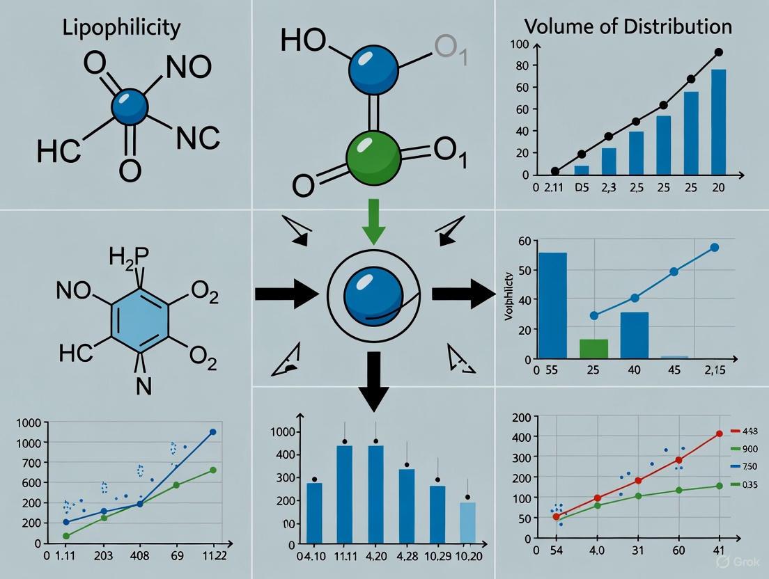
Lipophilicity (LogP and LogD): Core Principles and Measurement
Definitions and Theoretical Foundation
Lipophilicity is a fundamental physicochemical parameter representing a compound's affinity for a lipophilic environment versus an aqueous environment. It is most commonly quantified using the partition coefficient (LogP) and the distribution coefficient (LogD) [1].
- LogP is the logarithm of the partition coefficient, defined as the ratio of the concentration of the unionized compound in an organic phase (typically n-octanol) to its concentration in an aqueous phase (water) at equilibrium [2]. LogP is a constant for a given molecule in its neutral form and is independent of pH [3] [4].
- LogD is the logarithm of the distribution coefficient, which accounts for all forms of the compound present at a specific pH—unionized, ionized, and partially ionized species [3]. Unlike LogP, LogD is pH-dependent, making it a more relevant descriptor for ionizable compounds under physiological conditions [3] [1]. LogD at pH 7.4 (LogD7.4) is of particular interest in drug discovery as it reflects lipophilicity at blood pH [5].
The following conceptual diagram illustrates the partitioning process that defines these coefficients:
Diagram 1: The equilibrium process of a compound partitioning between aqueous and organic phases.
Experimental Methodologies for Determination
Accurate determination of lipophilicity is crucial for reliable structure-activity relationships. The following table summarizes the key experimental protocols.
Table 1: Core Experimental Methods for LogP and LogD Determination
| Method | Principle | Typical Workflow | Key Advantages | Key Limitations |
|---|---|---|---|---|
| Shake-Flask [4] [5] | Direct measurement of equilibrium concentrations between octanol and buffer phases. | 1. Combine 1 mL octanol and 1 mL buffer in vial.2. Add compound stock solution (10 mM in DMSO).3. Shake for 1 hour at room temperature.4. Allow phases to separate.5. Analyze concentrations in each phase via LC-MS/MS. | Considered a "gold standard"; direct measurement. | Labor-intensive; requires compound synthesis; low throughput [5]. |
| Chromatographic Techniques (e.g., HPLC) [5] | Indirect measurement via correlation of retention time with lipophilicity of standards. | 1. Inject compound onto reverse-phase column (e.g., C18).2. Elute with gradient of aqueous and organic mobile phases.3. Measure retention time.4. Compare to calibration curve from compounds with known LogD/LogP. | Simplicity, stability against impurities, higher throughput. | Indirect measure; less accurate than shake-flask [5]. |
| Potentiometric Titration [5] | Measures pKa and logP via titration in a two-phase system (octanol/water). | 1. Dissolve sample in n-octanol/water mixture.2. Titrate with KOH or HCl.3. Monitor pH changes.4. Calculate logP from the titration curve. | Can be automated; provides pKa data simultaneously. | Limited to ionizable compounds; requires high sample purity [5]. |
The standard shake-flask method workflow can be visualized as follows:
Diagram 2: Detailed workflow for the shake-flask determination of LogD.
The Scientist's Toolkit: Essential Reagents for Lipophilicity Assays
Table 2: Key Research Reagents for LogP/LogD Experiments
| Reagent / Material | Function in Assay |
|---|---|
| 1-Octanol | Organic phase simulating lipid membranes in partition experiments [4] [5]. |
| Buffer Solutions (e.g., Phosphate Buffer, pH 7.4) | Aqueous phase maintaining physiological pH during LogD7.4 determination [4]. |
| Internal Standards (e.g., Testosterone) | Quality controls to verify assay performance and consistency across runs [4]. |
| Reverse-Phase HPLC Columns (e.g., C18) | Stationary phase for chromatographic separation in HPLC-based methods [4] [5]. |
| LC-MS/MS System | High-sensitivity instrument for accurate quantification of compound concentrations in each phase [4]. |
Volume of Distribution (VDss): A Key Pharmacokinetic Parameter
Definition and Clinical Significance
The Volume of Distribution at Steady State (VDss) is a pharmacokinetic parameter that represents a drug's propensity to distribute from the plasma into the tissues [6]. It is defined as the theoretical volume required to uniformly distribute the total amount of drug in the body at the same concentration observed in the plasma [6]. The fundamental equation is:
VDss (L) = Total Amount of Drug in the Body (mg) / Plasma Drug Concentration (mg/L) [6]
- A low VDss (e.g., < 5 L) indicates that the drug is primarily confined to the plasma compartment, often due to high plasma protein binding or high molecular weight [6].
- A high VDss (e.g., >> 20 L) signifies extensive tissue distribution beyond the plasma volume, implying strong tissue binding or sequestration [6].
The primary clinical utility of VDss lies in calculating the loading dose required to rapidly achieve a target therapeutic plasma concentration [6]: Loading Dose (mg) = [Target Plasma Concentration (mg/L) x VDss (L)] / Bioavailability (F) [6]
Physicochemical and Physiological Drivers of VDss
A drug's VDss is not a physiological volume but a reflection of its relative binding to tissue components versus plasma proteins. Key factors influencing distribution include:
- Acid-Base Characteristics: Basic molecules tend to have higher VDss because they interact strongly with negatively charged phospholipid membranes in tissues. Acidic molecules often bind more extensively to albumin in plasma, leading to a lower VDss [6].
- Lipophilicity: Hydrophobic interactions drive distribution. Lipophilic drugs more readily pass through lipid bilayers and distribute into lipid-rich tissues like adipose, generally resulting in a higher VDss [6].
- Plasma and Tissue Binding: The critical balance is defined by the fraction unbound in plasma (fup) and tissues (fut). A drug with low fup (high plasma protein binding) and high fut (low tissue binding) will have a lower VDss, and vice versa [7] [8].
The interplay of these factors in determining distribution is a dynamic process:
Diagram 3: Key physicochemical factors influencing a drug's volume of distribution.
Interrelationship: How Lipophilicity Influences Volume of Distribution
Lipophilicity is a primary driver of a compound's distribution characteristics. The connection is not linear but follows a general trend: increasing lipophilicity typically enhances a drug's ability to leave the plasma, cross cell membranes, and bind to tissue components, thereby leading to a higher VDss [6] [7]. However, this relationship plateaus or becomes complex for highly lipophilic drugs (LogP > 4-5), as they may exhibit extremely high plasma protein binding or encounter solubility limitations, which can counterintuitively restrict distribution [7].
This complex relationship is critical for predicting VDss using mechanistic models. A 2024 study highlighted that the accuracy of different VDss prediction methods is highly sensitive to the input LogP value, especially for lipophilic drugs [7]. Methods like Rodgers-Rowland are highly sensitive to LogP and can overpredict VDss for compounds with high LogP, while methods like Oie-Tozer and TCM-New are more robust [7]. The TCM-New method, which incorporates vegetable oil/water partitioning as a surrogate for tissue lipids, was identified as the most accurate for highly lipophilic drugs [7].
Predictive Computational Models and Current Research
The integration of machine learning (ML) and artificial intelligence (AI) has significantly advanced the in silico prediction of both lipophilicity and VDss.
- LogD Prediction: Traditional quantitative structure-property relationship (QSPR) models are being superseded by sophisticated graph neural networks (GNNs). To overcome data scarcity, novel approaches like RTlogD use transfer learning from large chromatographic retention time datasets and multitask learning with LogP and microscopic pKa predictions to enhance model generalization and accuracy [5]. Pharmaceutical companies leverage their massive proprietary datasets (e.g., AstraZeneca's AZlogD74 model trained on >160,000 molecules) to achieve superior predictive performance [5].
- VDss Prediction: ML models now integrate chemical structure, physicochemical properties, and in silico predicted animal PK data to forecast human PK parameters. For instance, PKSmart uses a two-stage pipeline, first predicting rat, dog, and monkey PK parameters from chemical structure, then using these as features in a Random Forest model for human VDss and clearance, achieving performance comparable to industry-standard models [9]. PBPK modeling, particularly when informed by prior animal PBPK data, provides a mechanistic and highly accurate approach for predicting not just VDss but also distribution volumes in different elimination phases (V1, Vβ), outperforming conventional allometric scaling [8].
Table 3: Comparison of Modern VDss Prediction Methods
| Prediction Method | Underlying Principle | Key Inputs | Reported Performance (External R²/Accuracy) | Notable Strengths |
|---|---|---|---|---|
| PKSmart (ML Model) [9] | Two-stage machine learning using predicted animal PK. | Molecular fingerprints, physicochemical properties, predicted animal VDss/CL. | VDss R² = 0.39 | Fully in silico; no animal experiment needed; open-access. |
| PBPK Modeling (with animal model inference) [8] | Physiologically-based mechanistic modeling of drug disposition. | LogP, pKa, fup, BPR, tissue composition data. | Predicts V1, Vss, Vβ within 3-fold error for most compounds. | Mechanistic; predicts full concentration-time profile. |
| TCM-New [7] | Empirical method using vegetable oil/water partitioning. | LogP (octanol/water & vegetable oil/water), pKa. | Most accurate for high LogP drugs; robust to LogP variability. | Addresses limitations of octanol for highly lipophilic drugs. |
| Oie-Tozer Method [7] | Mechanistic model based on plasma and tissue binding. | LogP, pKa, fup, Blood-to-Plasma Ratio (BPR). | Accurate for many lipophilic drugs; modest sensitivity to LogP. | Well-established mechanistic framework. |
The workflow for modern integrated PK prediction tools is complex and multi-staged:
Diagram 4: Generalized workflow of a machine learning pipeline for predicting human pharmacokinetic parameters.
Lipophilicity (LogP/LogD) and volume of distribution (VDss) are inextricably linked parameters that form the cornerstone of pharmacokinetic optimization in drug discovery. A deep and nuanced understanding of their definitions, measurement techniques, and interrelationships is essential for researchers aiming to design effective and safe therapeutics. The field is moving beyond simplistic rules like the Rule of Five into the chemical space "beyond the Rule of 5" (bRo5), where understanding ionization and pH-dependent distribution via LogD becomes even more critical [3]. Concurrently, the advent of sophisticated AI-driven prediction tools and mechanistic PBPK models provides an unprecedented ability to forecast human PK outcomes from molecular structure and in vitro data, thereby de-risking the drug development process. Mastery of these key parameters and the modern tools used to study them empowers scientists to make data-driven decisions, accelerating the delivery of novel medicines.
Volume of distribution at steady state (VDss) is a fundamental pharmacokinetic parameter that quantifies a drug's propensity to distribute throughout the body beyond the plasma compartment. By definition, VDss represents the apparent volume into which a drug dose would need to be diluted to achieve the observed plasma concentration [10] [6]. This parameter provides critical insights into a drug's distribution characteristics, informing dosing regimens and predicting drug behavior in vivo. The physiological basis of VDss encompasses complex interactions between a drug's physicochemical properties and the biological environment, with lipophilicity emerging as a primary determinant of tissue distribution and binding [11] [6]. Understanding these principles is essential for researchers and drug development professionals seeking to optimize therapeutic candidates and predict human pharmacokinetics.
Fundamental Principles of Volume of Distribution
Definition and Quantitative Basis
Volume of distribution (Vd) is mathematically defined as the proportionality constant relating the total amount of drug in the body to its plasma concentration at a given time [10] [6]. The fundamental equation is:
Vd = A(t) / C(t)
Where A(t) is the amount of drug in the body at time t, and C(t) is the drug concentration in plasma at the same time point [10]. For intravenous bolus administration at time zero, where the amount of drug in the body (A₀) equals the dose (D), the volume can be calculated as:
Vd = D / C₀
Here, C₀ represents the initial plasma concentration estimated through back-extrapolation of the concentration-time curve [10]. It is crucial to recognize that VDss is an apparent volume that rarely corresponds to any real physiological volume but serves as a valuable indicator of a drug's distribution extent [10] [12].
Clinical and Pharmacokinetic Significance
VDss has profound implications for drug behavior in the body:
- Dosing Requirements: Drugs with high VDss require higher doses to achieve target plasma concentrations, while those with low VDss require lower doses [6].
- Half-Life Determination: VDss directly influences elimination half-life, which is calculated as t½ = 0.693 × (Vd/CL), where CL is clearance [12] [6]. Drugs with high VDss typically exhibit longer half-lives because only the fraction in plasma is susceptible to elimination [6].
- Loading Dose Calculation: The loading dose needed to achieve target plasma concentration rapidly is derived from LD = Cp × Vd, where Cp is the desired plasma concentration [10] [6].
Table 1: Representative Fluid Volumes in a 70kg Human for Contextualizing VDss [12]
| Compartment | Volume (L) | Percentage of Body Weight |
|---|---|---|
| Plasma | 3 | 4% |
| Blood | 5.5 | 8% |
| Extracellular Fluid | 14 | 20% |
| Intracellular Fluid | 28 | 40% |
| Total Body Water | 42 | 60% |
Physiological Determinants of VDss
Plasma Protein Binding
Drug binding to plasma proteins represents a primary factor restricting distribution from the vascular compartment. Extensive plasma protein binding results in lower VDss values, as the drug remains largely confined to the plasma space [12] [6]. The major plasma proteins involved in drug binding include:
- Albumin (concentration: 3.5-5.0 g/dL): Primarily binds acidic and neutral drugs [12]
- Alpha-1-acid glycoprotein (concentration: 0.04-0.1 g/dL): Preferentially binds basic drugs [12]
- Lipoproteins: Bind highly lipophilic basic drugs [12]
The fraction unbound in plasma (fup) critically influences VDss, with highly bound drugs (fup < 0.01) demonstrating restricted distribution [11]. Experimental methods for assessing plasma protein binding include equilibrium dialysis (considered the gold standard) and ultrafiltration (suited for rapid screening) [12].
Tissue Binding and Composition
Tissue binding represents the counterforce to plasma protein binding, driving drug distribution out of the vascular compartment. Drugs with high affinity for tissue components exhibit elevated VDss values, often far exceeding total body water [10] [6]. Tissue binding occurs through interactions with:
- Phospholipid membranes: Particularly relevant for basic drugs that interact with negatively charged phospholipid head groups [6]
- Intracellular proteins and nucleic acids
- Neutral lipids in adipose tissue: Especially significant for highly lipophilic compounds [11]
The extent of tissue binding is quantified through the fraction unbound in tissues (fut), which influences the steady-state volume of distribution according to the relationship: Vd = Vp + (fup/fut) × Vt, where Vp is plasma volume and Vt is tissue water volume [10].
Physicochemical Properties
A drug's physicochemical characteristics profoundly influence its distribution behavior:
- Lipophilicity: Increased lipophilicity generally enhances membrane permeability and tissue distribution, particularly to lipid-rich tissues like adipose [6]. However, this relationship may plateau for extremely lipophilic compounds (logP > 4) [11].
- Acid-Base Characteristics: Basic molecules tend to exhibit higher VDss than acidic molecules at similar lipophilicity, reflecting their differential binding to plasma versus tissue components [6].
- Molecular Size and Hydrogen Bonding Capacity: These influence permeability across membrane barriers and interaction with binding proteins [11].
Table 2: Volume of Distribution Examples Illustrating Distribution Principles [12] [6]
| Drug | VDss (L) | Physicochemical Properties | Distribution Pattern |
|---|---|---|---|
| Warfarin | 8 | Acidic, PPB = 99% | High plasma protein binding |
| Theophylline | 30 | PPB = 40% | Distribution in total body water |
| Chloroquine | 15,000 | Basic, PPB = 55%, Lipophilic | Extensive tissue distribution |
| Digoxin | 440-700 L | Moderate lipophilicity | Binds to skeletal muscle |
Figure 1: Dynamic Equilibrium Governing VDss. The diagram illustrates the continuous movement of drug molecules between plasma and tissue compartments, driven by binding interactions and physicochemical properties.
Lipophilicity as a Primary Determinant of VDss
Mechanisms of Lipophilicity-Mediated Distribution
Lipophilicity, commonly quantified as logP (partition coefficient between octanol and water), serves as a master variable controlling drug distribution through multiple mechanisms:
- Membrane Permeability: Lipophilic drugs readily traverse lipid bilayers, accessing intracellular spaces and tissue compartments beyond the vascular system [6].
- Tissue Binding Affinity: Lipophilicity enhances interactions with hydrophobic domains of tissue proteins and partitioning into neutral lipids within tissues, particularly adipose [11].
- Plasma Protein Binding: While lipophilicity generally increases plasma protein binding, the differential affinity between plasma and tissue binding sites ultimately determines distribution extent [12].
Recent research indicates that the relationship between logP and VDss may not be linear across extreme values. For highly lipophilic drugs (logP > 3-4), VDss predictions using standard methods tend to overestimate actual distribution, suggesting saturation of partitioning mechanisms or limitations in experimental logP determination for these compounds [11].
Acid-Base Properties and Lipophilicity Interplay
The influence of lipophilicity on VDss is modulated by a drug's ionization state at physiological pH [6]:
- Basic Drugs: Exhibit enhanced tissue binding due to electrostatic interactions with negatively charged phospholipid membranes, amplifying the distribution-enhancing effects of lipophilicity [6].
- Acidic Drugs: Demonstrate high affinity for albumin at relatively low lipophilicity, resulting in restricted distribution despite increasing logP [12] [6].
- Neutral Drugs: Distribution correlates more directly with lipophilicity, though very high logP may promote binding to lipoproteins, somewhat limiting distribution [12].
This interplay explains why basic drugs typically display larger VDss values than acidic drugs at equivalent lipophilicity [6].
Predictive Models and Methodologies
Established VDss Prediction Methods
Several mechanistic approaches have been developed to predict human VDss from drug properties:
- Oie-Tozer Method: Incorporates fup, fut, and physiological volumes to estimate VDss, showing modest sensitivity to logP variations [11].
- Rodgers-Rowland Method: Uses fup, pKa, and logP to predict tissue-specific partition coefficients (Kp); highly sensitive to logP and may overpredict for lipophilic drugs [11].
- TCM-New Method: Utilizes blood-to-plasma ratio (BPR) as a surrogate for tissue partitioning, avoiding direct fup use; demonstrates improved accuracy for lipophilic compounds [11].
Recent comparative analyses indicate that the TCM-New method provides the most accurate VDss predictions across drugs with varying lipophilicity, particularly for compounds with high logP values [11].
Advanced Computational Approaches
Modern VDss prediction incorporates sophisticated computational techniques:
- Quantitative Structure-Property Relationship (QSPR) Models: Employ molecular descriptors and machine learning to predict VDss from chemical structure [13] [14].
- Generative AI Frameworks: Integrate VDss prediction with other pharmacokinetic parameters in multi-objective optimization for drug design [14].
- Quantitative Systems Pharmacology (QSP): Incorporates VDss into mechanistic models simulating drug effects in physiological systems [14].
Table 3: Performance Comparison of VDss Prediction Methods for Lipophilic Drugs [11]
| Prediction Method | Sensitivity to logP | Accuracy for High logP (>4) | Key Input Parameters |
|---|---|---|---|
| Oie-Tozer | Modest | Good | fup, pKa, logP |
| Rodgers-Rowland | High | Poor (Overpredicts) | fup, pKa, logP |
| GastroPlus | High | Variable | fup, pKa, logP |
| Korzekwa-Nagar | High | Variable (Drug-dependent) | logP, pKa, structural features |
| TCM-New | Modest | Excellent | Blood-to-plasma ratio |
Figure 2: VDss Prediction Method Workflow. The diagram compares different methodological approaches to predicting VDss, highlighting their varying dependencies on lipophilicity (logP).
Experimental Protocols and Research Tools
Key Methodologies for Distribution Studies
Plasma Protein Binding Assays:
- Equilibrium Dialysis: The gold standard method where plasma containing the drug is separated from buffer by a semi-permeable membrane; after 4-24 hour incubation at 37°C, concentrations in both chambers are measured to determine free fraction [12].
- Ultrafiltration: A rapid screening approach where drug-plasma mixtures are centrifuged through molecular weight cutoff filters; the filtrate contains unbound drug for quantification [12].
Tissue Binding Assessment:
- Tissue Homogenate Binding: Similar to plasma protein binding methods but using tissue homogenates instead of plasma.
- In Vivo Tissue Distribution Studies: Measuring drug concentrations in various tissues at multiple time points after administration to determine tissue-to-plasma partition coefficients [15].
In Silico Prediction Protocols:
Recent methodologies integrate machine learning with physicochemical descriptors. The typical workflow includes: data collection and curation, descriptor calculation, model training with techniques like random forests or support vector machines, and validation using external test sets [13] [14].
Essential Research Reagent Solutions
Table 4: Key Research Reagents for VDss and Distribution Studies
| Reagent/Resource | Function in VDss Research | Application Notes |
|---|---|---|
| Human Plasma | Plasma protein binding studies | Source of albumin, alpha-1-acid glycoprotein |
| Equilibrium Dialysis Devices | Separation of protein-bound and free drug | Preferred over ultrafiltration for accuracy |
| Tissue Homogenates | Assessment of tissue binding affinity | Liver, muscle, adipose most relevant |
| Radiolabeled Drug Compounds | Quantitative tissue distribution studies | Enables precise tracking and quantification |
| Protein Solutions (HSA, AGP) | Mechanistic binding studies | Isolated protein systems for binding specificity |
| In Silico Prediction Software (GastroPlus, ADMET Predictor) | VDss prediction from structure | Incorporates QSPR and mechanistic models |
The physiological basis of VDss encompasses dynamic equilibrium processes between plasma and tissue compartments governed by fundamental physicochemical principles. Lipophilicity emerges as a central determinant of distribution, operating through complex interactions with plasma protein binding, tissue affinity, and membrane permeability. Contemporary research has refined our understanding of these relationships, revealing nuances such as the saturable nature of extreme lipophilicity effects. The ongoing development of predictive models, particularly those less sensitive to logP variability like the TCM-New method, represents significant advancement in accurately forecasting human VDss. For drug development professionals, integrating these insights with modern computational approaches enables more rational design of compounds with optimized distribution characteristics, ultimately enhancing therapeutic efficacy and safety profiles.
Lipophilicity, quantitatively expressed as the partition coefficient (logP), is a fundamental physicochemical property that profoundly influences the absorption, distribution, metabolism, excretion, and toxicity (ADMET) of drug candidates. Within drug discovery and development, predicting a compound's volume of distribution (VDss) is essential, as it impacts drug half-life and dosing regimen. The distribution of a drug throughout the body is governed by its partitioning into various tissues relative to plasma, defined by the tissue-to-plasma concentration ratio (Kp). Understanding the mechanistic relationship between lipophilicity and tissue partitioning is therefore critical for optimizing pharmacokinetic profiles and de-risking development pipelines. This whitepaper synthesizes current research to provide a mechanistic framework for how lipophilicity dictates tissue partitioning, with a specific focus on the challenges and advanced methodologies associated with predicting the distribution of highly lipophilic compounds.
Mechanistic Foundations of Tissue Partitioning
At its core, tissue partitioning is a process of distribution equilibrium between circulating plasma and the diverse cellular environments of bodily tissues. The tissue-to-plasma partition coefficient (Kp) is the key parameter describing this equilibrium. The primary mechanistic role of lipophilicity is in driving a drug's affinity for biological membranes and tissue components relative to the aqueous plasma environment.
Lipophilic drugs exhibit high affinity for neutral lipids and phospholipids that constitute cellular membranes and adipose tissue. Consequently, lipophilicity is a strong positive driver for a drug's volume of distribution at steady state (VDss) [7] [11]. However, this relationship is not linear across the entire lipophilicity spectrum. For highly lipophilic drugs (logP > ~4-5), traditional prediction models that rely solely on octanol-water partitioning and experimentally measured unbound fraction in plasma (fup) tend to substantially overpredict Kp and VDss [16] [7]. This overprediction occurs for several mechanistic reasons. Firstly, the in vitro measurement of fup for highly lipophilic compounds can be inaccurate, as these compounds may exhibit nonspecific binding to apparatus or membrane proteins, leading to overestimations of the true free fraction available for partitioning in vivo [16]. Secondly, the octanol-water partition coefficient (logP) may not perfectly mimic partitioning into the complex lipid milieu of tissues, which includes triglycerides, diglycerides, and cholesterols [7]. Evidence suggests that for highly lipophilic compounds, tissue distribution becomes limited by the physiological neutral lipid content of tissues rather than by the compound's intrinsic affinity for octanol alone [16]. This plateau effect in adipose tissue Kp is a key consideration that modern prediction methods seek to address.
Quantitative Analysis of Prediction Methods and Lipophilicity Sensitivity
Various mechanistic methods have been developed to predict human VDss, a parameter directly derived from tissue Kp values. A recent comparative analysis evaluated six prominent methods, highlighting their sensitivity to logP and their performance for lipophilic drugs [7] [11].
Table 1: Comparison of VDss Prediction Methods and Their Sensitivity to Lipophilicity
| Prediction Method | Core Mechanistic Basis | Sensitivity to logP | Performance for High logP Drugs |
|---|---|---|---|
| Rodgers-Rowland | Drug dissolution in intra/extracellular water & partitioning into tissue lipids (neutral lipids, phospholipids); binding to albumin/lipoprotein [7]. | High | Inaccurate; substantial overprediction of VDss due to limitations of logP and fup inputs [7] [11]. |
| Oie-Tozer | Empirical equation based on drug binding in plasma and extracellular fluid, with a correction factor for tissue binding [11]. | Modest | Accurate for griseofulvin, posaconazole, isavuconazole [7]. |
| GastroPlus | Perfusion-limited model implementing the Rodgers-Rowland equations for Kp prediction [7]. | High | Accurate for itraconazole and isavuconazole; performance variable [7]. |
| Korzekwa-Nagar | Uses microsomal partitioning (fum) as a surrogate for general cell membrane partitioning [11]. | High | Accurate for posaconazole; performance variable [7]. |
| TCM-New | Uses blood-to-plasma ratio (BPR) as a surrogate for drug partitioning into tissues, avoiding the use of fup [7] [11]. | Modest | Most accurate method across multiple drugs and logP sources; best for highly lipophilic drugs [7] [11]. |
The sensitivity analysis reveals that as logP increases, the Rodgers-Rowland methods (and those based on it) show the greatest fluctuation in predicted VDss, while Oie-Tozer and TCM-New are more robust [7]. The TCM-New model, which uniquely incorporates vegetable oil-water partition data to better represent partitioning into physiological triglycerides, emerged as the most accurate for highly lipophilic drugs, suggesting BPR is a favorable and more reliable surrogate for tissue partitioning [7] [11].
Table 2: Impact of logP Variation on Predicted VDss for Sample Drugs
| Drug | logP Range | Clinically Observed VDss (L/kg) | TCM-New Prediction (L/kg) | Rodgers-Rowland Prediction (L/kg) |
|---|---|---|---|---|
| Griseofulvin | 2.41 - 3.53 | ~1.1 - 1.4 | Accurate | Overpredicted |
| Itraconazole | 4.89 - 6.89 | ~10 - 11 | Accurate (across logP sources) | Overpredicted (~100-fold in some cases) |
| Posaconazole | 4.41 - 6.72 | ~5 - 8 | Accurate | Overpredicted |
| Isavuconazole | 3.56 - 4.93 | ~5 - 6 | Accurate | Overpredicted |
Experimental Protocols for Key Studies
Protocol: Predicting Kp Using Volume of Distribution and Lipophilicity
This empirical method uses preclinical data to predict tissue Kp values for PBPK modeling [17].
- Step 1: Data Collection. Collect in vivo pharmacokinetic data from laboratory animals, including a measured volume of distribution and a descriptor of drug lipophilicity (e.g., logP).
- Step 2: Correlation Development (Training Set). Using a training set of compounds (e.g., 49 drugs), establish correlations between the Kp values for muscle tissue and other tissues.
- Step 3: Kp Prediction (Test Set). For a test set of compounds (e.g., 22 drugs), predict Kp values for various tissues based on the compound's volume of distribution, lipophilicity, and the pre-established muscle-to-other-tissue correlations.
- Step 4: Validation. Compare predicted Kp values against experimentally determined in vivo Kp values (e.g., n=118). The reported accuracy for non-eliminating tissues was 72% within a factor of ±2 [17].
Protocol: Advancing Prediction for Highly Lipophilic Compounds
This protocol addresses the overprediction of VDss for highly lipophilic drugs (logP ≥ 5.8) by proposing a simplified tissue-composition-based model [16].
- Step 1: Problem Identification. Identify a set of highly lipophilic compounds for which existing models (e.g., original tissue-composition model) substantially overpredict Vss.
- Step 2: Model Adjustment. Recognize the potential inaccuracy of experimentally determined fup for these compounds. Alternatively, use the tissue-plasma ratio of neutral lipids (nl) equivalent as the primary factor governing Kp, in combination with logP.
- Step 3: Model Evaluation. Calculate the average fold error between predicted and observed human Vss for both the published model and the proposed simplified model. The simplified model reduced the average fold error from 124 to 1.5 [16].
- Step 4: Sensitivity Analysis. Perform sensitivity analysis to confirm the importance of neutral lipid content and drug lipophilicity as the dominant parameters in the adjusted model [16].
Protocol: Veterinary Drug Residue Study Linking Lipophilicity and Tissue Half-Life
This in vivo study investigates the relationship between lipophilicity, volume of distribution, and tissue residue persistence in food-producing animals [18].
- Step 1: Formulation Administration. Administer two different sulphonamide formulations (A and B) intramuscularly to piglets for six days according to prescribed dosing schedules.
- Step 2: Tissue Sampling. Sacrifice animals on predetermined days post-treatment (e.g., days 10, 12, 14, 16, 18) and collect tissue samples (muscle, liver, kidney, fat, and skin).
- Step 3: Tissue Preparation and HPLC Analysis.
- Homogenize 1g of tissue with 3 mL of 0.1 M ammonium acetate solution and 2 mL of hexane.
- Centrifuge the suspension and collect the organic layer.
- Perform solid-phase extraction (SPE) using Oasis HLB cartridges (pre-conditioned with methanol and water).
- Elute analytes with ethyl acetate and acetonitrile, concentrate to dryness under a nitrogen stream, and reconstitute in mobile phase for HPLC injection.
- Step 4: Data Analysis. Validate the HPLC method according to European Commission Decision 2002/657/EC. Estimate tissue half-lives and relate the clearance period and residue persistence to the drugs' volume of distribution and physicochemical characteristics [18].
Visualizing Mechanistic Relationships and Workflows
Diagram: Mechanistic Pathways of Lipophilicity-Driven Tissue Partitioning
Diagram: Workflow for Evaluating VDss Prediction Methods
The Scientist's Toolkit: Essential Research Reagents and Materials
Table 3: Key Reagents and Materials for Tissue Partitioning Research
| Item | Function/Application |
|---|---|
| Oasis HLB Solid-Phase Extraction (SPE) Cartridges | Used for cleaning up and extracting drugs from complex tissue homogenates prior to analytical quantification [18]. |
| Octanol and Water | Solvent system for experimentally determining the partition coefficient (logP), a fundamental descriptor of lipophilicity [7]. |
| Vegetable Oil | Alternative to octanol for measuring partition coefficients, potentially providing a better surrogate for partitioning into physiological triglycerides [7]. |
| Human/Animal Liver Microsomes | Used in the Korzekwa-Nagar method to determine fraction unbound in microsomes (fum) as a surrogate for cellular membrane partitioning [11]. |
| In Vivo Tissues (Muscle, Liver, Fat, etc.) | Essential for measuring experimental tissue-to-plasma concentration ratios (Kp) for model validation [17] [18]. |
| HPLC System with UV/VIS Detector | Standard analytical equipment for quantifying drug concentrations in plasma and tissue samples [18]. |
| ADMET Predictor Software | In silico tool for predicting key parameters like logP, pKa, and blood-to-plasma ratio (BPR) when experimental data is unavailable [7]. |
The volume of distribution (Vd) is a fundamental pharmacokinetic parameter that quantifies the extent of a drug's distribution throughout the body relative to its plasma concentration [6]. It is a proportionality constant relating the total amount of drug in the body to its plasma concentration at a given time. Understanding the factors that govern Vd is crucial in drug discovery and development, as it directly influences the dosing regimen and the elimination half-life of a drug [6] [19]. The physicochemical properties of a drug, primarily its acid-base characteristics and lipophilicity, are key determinants of its distribution profile [6] [20]. These properties dictate how a drug partitions between plasma and various tissues, thereby defining its apparent Vd. This review examines the mechanistic interplay between ionization, lipophilicity, and physiological factors in determining drug distribution, providing a technical guide for researchers and drug development professionals.
Core Principles: Vd, Acid-Base Properties, and Lipophilicity
Volume of Distribution (Vd): Definition and Clinical Significance
The volume of distribution (Vd) is defined by the equation: Vd (L) = Amount of drug in the body (mg) / Plasma concentration of drug (mg/L) [6]. It is a theoretical volume that a drug would occupy to produce the observed plasma concentration. A high Vd indicates a drug has a high propensity to leave the plasma and distribute into extravascular tissues, necessitating a higher loading dose to achieve a target plasma concentration. Conversely, a low Vd suggests the drug is largely confined to the plasma, requiring a lower loading dose [6].
The most clinically relevant value is the steady-state volume of distribution (Vss), which represents the dynamic equilibrium between drug in the plasma and tissues. Vss is used to calculate the loading dose of a drug: Loading dose (mg) = [Cp (mg/L) x Vd (L)] / F (where Cp is the desired plasma concentration and F is bioavailability) [6]. A drug's half-life is also directly dependent on Vd and clearance (CL): Half-life (hours) = 0.693 x (Vd (L) / CL (L/hr)) [6].
Fundamental Physicochemical Properties
Acid-Base Properties and pKa
The acid dissociation constant (pKa) measures the strength of an acid or base in solution. It is the pH at which 50% of the molecule is ionized [20] [21]. The fraction of a drug that is ionized or unionized at a specific pH is calculated using the Henderson-Hasselbalch equation [20] [21]:
- For acids: pH = pKa + log([A⁻]/[HA])
- For bases: pH = pKa + log([B]/[BH⁺])
This relationship is critical because the unionized form of a drug typically diffuses more readily across biological membranes, which are primarily lipophilic in nature [20].
Lipophilicity: LogP and LogD
Lipophilicity is a measure of how a substance distributes itself between a hydrophobic (nonpolar) phase and a hydrophilic (polar) phase.
- LogP is the logarithm of the partition coefficient (P) for the unionized form of a compound in an octanol-water system: LogP = log([Drug]octanol / [Drug]water) [22] [21]. It is a constant for a given compound.
- LogD is the logarithm of the distribution coefficient, which accounts for the ionization of the compound at a specific pH (typically 7.4): LogD = log( [Drug]octanol / ([Drug]water + [Ion]_water) ) [22]. LogD provides a more physiologically relevant measure of lipophilicity. A theoretical relationship exists: LogD ≈ LogP - log(1 + 10^(pH - pKa)) for acids and LogD ≈ LogP - log(1 + 10^(pKa - pH)) for bases [22].
Table 1: Key Physicochemical Properties Influencing Drug Distribution
| Property | Definition | Physiological Implication |
|---|---|---|
| pKa | pH at which 50% of the molecule is ionized [20] | Determines the fraction of unionized (membrane-permeable) drug at physiological pH. |
| LogP | Partition coefficient of the unionized drug in octanol/water [22] [21] | Intrinsic measure of lipophilicity, influencing membrane permeability and tissue binding. |
| LogD | Distribution coefficient at a specific pH (e.g., 7.4) [22] | Combined measure of lipophilicity and ionization; better predictor of passive diffusion and tissue partitioning. |
| fup | Fraction of drug unbound in plasma [19] | Determines the fraction of drug available to leave the plasma and distribute into tissues. |
The Mechanistic Interplay Governing Distribution
Drug distribution is a competitive process between binding in the plasma and binding in the tissues, both of which are heavily influenced by acid-base properties and lipophilicity.
Plasma Protein Binding
Drugs in plasma can bind to proteins such as albumin, alpha-acid glycoprotein (AAG), and lipoproteins [19]. The fraction unbound in plasma (fup) is a critical parameter. In general:
- Acidic drugs tend to bind primarily to albumin [6] [19].
- Basic drugs have a higher affinity for AAG and phospholipid membranes [6] [19]. High plasma protein binding tends to restrict a drug to the vascular space, leading to a lower Vd, as only the unbound fraction is available to distribute into tissues [19].
Tissue Partitioning
Tissue partitioning is characterized by the tissue-to-plasma partition coefficient (Kp). Drugs with high tissue partitioning will generally have a large Vd [19]. This partitioning is driven by:
- Binding to tissue components: Drugs can bind to tissue membranes (phospholipids, neutral lipids), intracellular proteins, and other cellular components [19].
- Ionic interactions: Basic molecules have strong electrostatic interactions with negatively charged phospholipid head groups on cell membranes, promoting their exit from the systemic circulation and leading to a higher Vd [6].
- Lipophilicity: Lipophilic drugs more readily pass through lipid bilayers and distribute into lipid-rich tissues like adipose, resulting in a higher Vd [6].
The following diagram illustrates the core mechanistic relationship between a drug's physicochemical properties and its resulting volume of distribution.
Integrated Effect on Volume of Distribution
The apparent Vd is the net result of the competition between plasma and tissue binding [19]. Key general principles are:
- Basic, lipophilic drugs: Tend to have high Vd due to extensive tissue binding (e.g., interaction with phospholipids) [6].
- Acidic, hydrophilic drugs: Tend to have low Vd due to high plasma protein binding (e.g., to albumin) and lower tissue permeability [6].
- Notable exception: Extensive plasma protein binding does not necessarily result in a low Vd if tissue partitioning is also extensive. For example, tamoxifen is >98% plasma protein bound but has a very high Vd of ~4000 L due to extreme tissue binding [19].
Table 2: Impact of Drug Properties on Volume of Distribution (Vd)
| Drug Type | Plasma Protein Binding | Tissue Partitioning | Typical Vd | Underlying Mechanism |
|---|---|---|---|---|
| Acidic | High (to albumin) [6] [19] | Low | Low (close to plasma volume) | Ionized at pH 7.4; high affinity for plasma proteins; low membrane permeability. |
| Basic | Variable (to AAG) [6] [19] | High | High (often >> total body water) | Unionized at pH 7.4; high affinity for negatively charged tissue phospholipids [6]. |
| Neutral, Lipophilic | Variable | High | High | Passive diffusion through membranes; partitioning into lipid-rich tissues [6]. |
| Hydrophilic | Low | Low | Low | Inability to cross lipid membranes; confined to plasma and extracellular fluid. |
Experimental Protocols for Key Determinations
Determining Lipophilicity (LogP/LogD) and pKa
Gradient High-Performance Liquid Chromatography (HPLC) is a key technique for the rapid determination of lipophilicity and pKa estimates [23].
Protocol for Lipophilicity and pKa Estimation via HPLC:
- Instrumentation: HPLC system with a reversed-phase C18 column, UV or mass spectrometric detector, and solvents: buffer and organic modifier (e.g., acetonitrile) [23].
- Lipophilicity Estimate (CHI or log kw):
- A single run using a wide-range linear gradient of organic modifier (e.g., 0-100% acetonitrile) at a pH where the analyte is non-ionized.
- The retention time is used to calculate a chromatographic hydrophobicity index (CHI) or the log kw, which correlates with log P [23].
- pKa Estimate:
- A subsequent run is performed at a fixed, optimized organic modifier content (%B) determined from step 2, using a pH gradient of the aqueous buffer component.
- The initial pH is set to ensure the analyte is non-ionized. The retention time shift allows for the estimation of the pKa value in the solvent of the selected %B [23].
- Data Analysis: Correlation of the chromatographic parameters (CHI, log k_w) with reference log P values, and the retention time from the pH gradient with known pKa values of standards [23].
Determining Plasma Protein Binding (fup)
Equilibrium Dialysis is a standard method for determining the fraction of drug unbound in plasma (fup) [19].
- Apparatus: A dialysis chamber divided into two compartments by a semi-permeable membrane that allows passage of only the unbound drug.
- Procedure: Plasma containing the drug is placed on one side (donor), and buffer is placed on the other side (receiver). The system is incubated at 37°C with controlled pH (in the presence of 5–10% CO₂) until equilibrium is reached [19].
- Measurement: The concentration of the drug in both the buffer and plasma compartments is measured after equilibrium.
- Calculation: fup = Concentration in buffer / Concentration in plasma. This method is considered more consistent than ultracentrifugation [19].
Predictive Modeling of Volume of Distribution
Accurate prediction of human Vss prior to clinical studies is a major goal in drug discovery. Several mechanistic and in silico methods have been developed, with varying sensitivity to lipophilicity (logP) [19] [7] [11].
- TCM-New: This method uses the blood-to-plasma ratio (BPR) as a surrogate for drug partitioning into tissues, avoiding the direct use of fup. It is the most accurate method for highly lipophilic drugs and shows low sensitivity to variability in logP values [7] [11].
- Oie-Tozer Method: A physiological-based method that uses fup, logP, and pKa to predict Vss. It is only modestly sensitive to logP and provides accurate predictions for many lipophilic drugs [7] [11].
- Rodgers-Rowland Method: A tissue composition-based approach that predicts tissue-to-plasma partition coefficients (Kp) using fup, pKa, and logP. It is highly sensitive to logP and tends to overpredict Vss for compounds with high logP (e.g., > 3.5) [7] [11].
- Quantitative Structure-Pharmacokinetic Relationships (QSPkR): These are computational models that correlate structural descriptors of drugs (e.g., presence of specific cycles, atom types, polar groups) with Vss. They are useful for lead optimization without requiring in vitro experiments [24].
Comparative Performance of Vss Prediction Methods
A 2024 sensitivity analysis compared the performance of six prediction methods for lipophilic drugs, highlighting the critical impact of logP accuracy and choice of method [7] [11].
Table 3: Relative Performance of Vss Prediction Methods for Lipophilic Drugs
| Prediction Method | Sensitivity to logP | Key Input Parameters | Reported Performance for High logP Drugs |
|---|---|---|---|
| TCM-New | Low | Blood-to-Plasma Ratio (BPR) [7] [11] | Most accurate method across different logP sources; avoids fup challenges [7] [11]. |
| Oie-Tozer | Moderate | fup, pKa, logP [7] [11] | Accurate for griseofulvin, posaconazole, isavuconazole [7] [11]. |
| GastroPlus (PBPK) | High | fup, pKa, logP [7] | Accurate for itraconazole and isavuconazole; performance varies [7]. |
| Korzekwa-Nagar | High | logP, pKa, structural descriptors [7] | Accurate for posaconazole; sensitive to logP input [7]. |
| Rodgers-Rowland | Very High | fup, pKa, logP [7] [11] | Overpredicts Vss for drugs with logP > 3.5; can be 100-fold over [7] [11]. |
The workflow for such a comparative analysis is outlined below.
The Scientist's Toolkit: Essential Reagents and Materials
Table 4: Key Research Reagent Solutions for Distribution Studies
| Reagent / Material | Function in Experimental Protocol |
|---|---|
| n-Octanol | Organic solvent in the standard system for measuring LogP/LogD, mimicking biological membranes [22] [21]. |
| Phosphate Buffers (at varying pH) | Used in HPLC mobile phases and equilibrium dialysis to control pH, crucial for determining pKa and LogD [23]. |
| Human Plasma (or Serum) | Biological matrix for determining fraction unbound (fup) via equilibrium dialysis and for measuring blood-to-plasma ratio (BPR) [19]. |
| Immobilized Artificial Membrane (IAM) HPLC Columns | Chromatographic stationary phase that mimics phospholipid cell membranes; used to study membrane partitioning [25]. |
| Equilibrium Dialysis Devices | Apparatus with a semi-permeable membrane to separate protein-bound and unbound drug for fup determination [19]. |
| Acetonitrile & Methanol (HPLC Grade) | Organic modifiers in reversed-phase HPLC for lipophilicity and pKa measurements [23]. |
| Human Serum Albumin (HSA) & Alpha-1-Acid Glycoprotein (AAG) | Specific plasma proteins for studying binding mechanisms of acidic and basic drugs, respectively [19]. |
The distribution of a drug within the body is a complex process governed by a well-defined interplay between its acid-base properties (pKa) and lipophilicity (LogP/LogD). These fundamental physicochemical properties determine the critical balance between plasma protein binding and tissue partitioning, which in turn defines the volume of distribution (Vd). A deep understanding of these relationships, combined with robust experimental protocols for determining fup, pKa, and LogD, is essential for rational drug design. Furthermore, selecting the appropriate predictive model for Vss—with careful consideration of a drug's lipophilicity and the model's inherent limitations—is crucial for making accurate first-in-human dose predictions and optimizing the pharmacokinetic profile of drug candidates. The continued refinement of these models, particularly for highly lipophilic compounds, remains a vital area of research in pharmaceutical sciences.
Volume of distribution at steady state (VDss) is a fundamental pharmacokinetic parameter that quantifies the extent of a drug's distribution throughout the body beyond the plasma compartment. This technical guide explores the critical relationship between VDss, drug half-life, and dosing regimens, framed within contemporary research on lipophilicity and tissue distribution. VDss serves as a key determinant of a drug's elimination half-life and directly influences loading dose calculations, dosing frequency, and the time to reach therapeutic steady-state concentrations. With advances in predictive modeling, including mechanism-based and in silico approaches, researchers can now optimize drug candidates for desirable distribution characteristics early in development. This whitepaper examines the theoretical principles, experimental methodologies, and practical applications of VDss for research scientists and drug development professionals seeking to design compounds with optimized pharmacokinetic profiles.
Volume of distribution at steady state (VDss) represents a key pharmacokinetic parameter that defines the hypothetical volume into which a drug must be distributed to achieve the observed plasma concentration [13]. It provides a critical proportionality constant relating the total amount of drug in the body to its plasma concentration under steady-state conditions [26]. Unlike the physiologically constrained central compartment (blood/plasma volume), VDss values can range dramatically—from 0.04 L/kg (indicating confinement to plasma) to hundreds of L/kg (indicating extensive tissue distribution) [27].
VDss is not a physical volume but rather a mathematical concept that reflects a drug's relative partitioning between plasma and tissues [13]. A low VDss indicates that a drug remains primarily within the plasma compartment, often due to high plasma protein binding or high water solubility [13]. Conversely, a high VDss suggests significant extra-vascular distribution, typically resulting from tissue binding or high lipid solubility [13]. Understanding these distribution patterns is essential for predicting a drug's pharmacokinetic behavior, particularly its elimination half-life and dosing requirements.
In the broader context of lipophilicity research, VDss serves as a crucial link between a compound's physicochemical properties and its in vivo behavior. As noted in recent distribution studies, "Lipophilicity and ionization are important drug physicochemical properties that affect VDss" because they "impact drug permeability and binding to cell membranes, intracellular and extracellular protein binding, and affinity for enzymes and cell transporters" [11]. This establishes VDss as an integrative parameter that encapsulates complex drug-tissue interactions influenced fundamentally by lipophilic character.
The Fundamental Relationship Between VDss, Clearance, and Half-Life
Mathematical Dependencies
The elimination half-life (t½) of a drug represents the time required for its plasma concentration to decrease by 50% and is governed by both clearance (CL) and volume of distribution (VDss) according to the following fundamental equation [28]:
t½ = 0.693 × VDss / CL
This relationship demonstrates that half-life depends on two independent physiological processes: clearance (representing drug elimination) and distribution volume (representing drug distribution) [29] [28]. Consequently, a drug can exhibit a prolonged half-life either due to slow clearance (CL↓) or extensive tissue distribution (VDss↑), with each scenario having distinct implications for dosing regimen design.
The clinical relevance of this relationship manifests in several critical ways:
- Dosing Frequency Determination: Drugs with large VDss values typically have longer half-lives and may require less frequent dosing [29] [28].
- Time to Steady State: The time to reach steady-state concentrations is determined primarily by half-life, requiring approximately 4-5 half-lives to achieve equilibrium between administration and elimination [30] [28].
- Drug Accumulation Potential: Compounds with high VDss and consequent long half-lives may accumulate in tissues with repeated dosing, potentially leading to toxicity or prolonged effects even after discontinuation [29].
Impact on Dosing Regimen Design
VDss directly influences both loading and maintenance dose strategies. A loading dose is often necessary for drugs with extensive tissue distribution to rapidly achieve therapeutic concentrations, calculated as:
Loading Dose = Target Concentration × VDss
This relationship explains why drugs with high VDss values require substantially larger loading doses to fill the peripheral compartments quickly. For instance, the anti-epileptic drug valproic acid, when formulated as an extended-release preparation (divalproex-ER), demonstrates a functional half-life of approximately 40 hours in uninduced subjects, largely influenced by its distribution characteristics [31].
Table 1: Impact of VDss on Drug Pharmacokinetic Parameters
| VDss Category | Typical Range (L/kg) | Half-Life Implications | Dosing Considerations |
|---|---|---|---|
| Low VDss | 0.04-0.07 | Short to moderate | Frequent dosing often required |
| Moderate VDss | 0.07-2.8 | Variable | Standard dosing intervals |
| High VDss | >2.8-3.5 | Often prolonged | Larger loading doses; less frequent maintenance dosing |
Lipophilicity as a Key Determinant of VDss
The Role of Lipophilicity and Ionization
Lipophilicity, commonly quantified as the partition coefficient (logP), represents one of the principal physicochemical properties governing drug distribution behavior [32]. This parameter significantly influences VDss through multiple mechanisms:
- Membrane Permeation: Lipophilic compounds more readily cross biological membranes, accessing intracellular spaces and lipid-rich tissues [11] [32].
- Tissue Binding Affinity: Lipophilicity enhances binding to cellular components, including lipids and proteins, promoting tissue accumulation [11].
- Plasma Protein Binding: While increasing lipophilicity generally enhances plasma protein binding, the effect on tissue binding often predominates, resulting in net increased distribution [26].
The impact of lipophilicity on VDss demonstrates a complex, non-linear relationship. For highly lipophilic compounds (logP > 3-4), VDss predictions become challenging due to potential permeation limitations and difficulties in accurately measuring plasma protein binding [11] [26]. Recent research indicates that "lipophilicity and ionization are important drug physicochemical properties that affect VDss" and that "logP was the most influential parameter in determining drug tissue-to-plasma partition coefficient (Kp) for neutral and weakly basic drugs" [11].
Ionization and Specialized Distribution Mechanisms
Ionization state (pKa) interacts with lipophilicity to determine distribution patterns through specialized mechanisms:
- Lysosomal Trapping: Basic compounds can accumulate in acidic intracellular organelles (lysosomes) through ion trapping, significantly increasing their VDss [27].
- pH Partitioning: Ionization gradients across biological membranes create differential distribution between compartments with varying pH [26].
- Protein Binding Specificity: Acidic drugs tend to bind albumin, while basic compounds often prefer α1-acid glycoprotein, creating distinct distribution patterns [11].
These mechanisms explain why basic compounds frequently exhibit larger VDss values compared to acidic or neutral compounds of similar lipophilicity, a trend observed in comprehensive analyses of clinical pharmacokinetic data [27].
Table 2: Impact of Drug Properties on Volume of Distribution
| Drug Property | Effect on VDss | Mechanism | Clinical Example |
|---|---|---|---|
| High Lipophilicity (logP > 3) | Increased | Enhanced tissue partitioning and binding | Posaconazole (antifungal) |
| Basic pKa | Markedly increased | Lysosomal trapping in tissues | Distribution patterns for basic drugs often exceed neutral compounds |
| High Plasma Protein Binding | Decreased | Restricted exit from vascular compartment | Warfarin (VDss ~0.1 L/kg) |
| High Tissue Binding | Increased | Sequestration in peripheral tissues | Digoxin (VDss ~5 L/kg) |
Predictive Models for VDss
Mechanism-Based Prediction Approaches
Several mechanism-based models have been developed to predict human VDss, each with distinct strengths and limitations:
Tissue Composition-Based Models (TCM)
- TCM-Rodgers-Rowland (TCM-RR): Incorporces drug physiochemistry and physiological data to predict tissue-plasma partition coefficients [11] [26].
- TCM-Simulations Plus (TCM-SP): Modifies the Rodgers-Rowland approach by replacing experimentally determined plasma binding with calculated values [26].
- TCM-New: A recently developed model that accentuates the impact of blood-to-plasma ratio (BPR) and uses alternative approaches for estimating tissue binding, showing improved accuracy for neutral drugs [26].
The TCM-New model has demonstrated significant improvements in prediction accuracy, with 83% of predictions within twofold error compared to only 50% for the TCM-RR method [26]. This enhanced performance is attributed to its treatment of BPR as "a favorable surrogate for drug partitioning into tissues" which "avoids the use of fup" (fraction unbound in plasma) [11].
Oie-Tozer Method This method uses an equation that incorporates physiological volumes, drug binding to plasma proteins, and binding to tissue proteins to estimate VDss [11]. It demonstrates only modest sensitivity to logP variations compared to other methods [11].
In Silico and Machine Learning Approaches
Advances in computational methods have enabled the development of sophisticated VDss prediction models:
Descriptor-Parsimonious Models Recent research describes "a novel, descriptor-parsimonious in silico model to predict human VDss" that performs equivalently to benchmark models while utilizing fewer molecular descriptors [27]. These models leverage statistical approaches including:
- Random Forests (RF): Ensemble learning method that builds multiple decision trees [13] [27].
- Radial Basis Functions (RBF): Neural network-inspired approach demonstrating high prediction accuracy [13].
- Gaussian Processes (GP): Probabilistic models that provide uncertainty estimates with predictions [13].
A comparative study of six prediction methods revealed that "TCM-New was the most accurate method for VDss prediction of highly lipophilic drugs," while "the Rodgers-Rowland methods provided inaccurate predictions due to the overprediction of VDss" for high logP compounds [11].
Table 3: Comparison of VDss Prediction Methods
| Prediction Method | Key Input Parameters | Strengths | Limitations |
|---|---|---|---|
| TCM-New | logP, pKa, BPR | High accuracy for neutral drugs; 83% within 2-fold error | Limited validation for zwitterions |
| Oie-Tozer | pKa, logP, fup | Modest sensitivity to logP variations | Accuracy affected by fup measurement errors |
| Rodgers-Rowland | fup, pKa, logP | Comprehensive mechanistic basis | Overpredicts VDss for lipophilic drugs (logP > 3.5) |
| In Silico (Random Forest) | Molecular descriptors only | Animal-sparing; rapid predictions | Limited interpretability of structural influences |
| GastroPlus | fup, pKa, logP | Incorporces perfusion-limited model | Similar limitations as Rodgers-Rowland for lipophilic drugs |
Experimental Protocols for VDss Assessment
Preclinical Protocol for VDss Determination
In Vivo VDss Determination in Animal Models
- Animal Preparation: Use healthy adult animals (typically rodent and non-rodent species) with indwelling venous catheters for drug administration and serial blood sampling.
- Dosing Protocol: Administer the test compound intravenously as a bolus or short infusion to ensure complete bioavailability.
- Sample Collection: Collect serial blood samples at predetermined time points (e.g., 2, 5, 15, 30 min and 1, 2, 4, 8, 12, 24 hours post-dose) into anticoagulant-treated containers.
- Sample Processing: Separate plasma by centrifugation and store at -80°C until analysis.
- Bioanalysis: Quantify drug concentrations in plasma using validated analytical methods (typically LC-MS/MS).
- Non-Compartmental Analysis (NCA): Calculate VDss using statistical moment theory:
- VDss = Dose × AUMC / (AUC)²
- Where AUMC is the area under the first moment curve and AUC is the area under the concentration-time curve.
This protocol directly supports the "extrapolation of human pharmacokinetic parameters from rat, dog, and monkey data" [27].
In Vitro Protocols for VDss Prediction
Plasma Protein Binding Measurement
- Equilibrium Dialysis: Place plasma spiked with test compound on one side of a semi-permeable membrane and buffer on the other side.
- Incubation: Incubate the system at 37°C for 4-24 hours to reach equilibrium.
- Quantification: Measure drug concentrations in both compartments using HPLC-UV or LC-MS/MS.
- Calculation: Determine fraction unbound (fup) = Concentrationbuffer / Concentrationplasma.
Blood-to-Plasma Ratio (BPR) Determination
- Incubation: Income whole blood with test compound at 37°C for 30-60 minutes.
- Centrifugation: Separate plasma by centrifugation.
- Quantification: Measure drug concentrations in plasma and calculate BPR based on initial blood concentration.
- Application: Use BPR as a "surrogate for drug partitioning into tissues" in the TCM-New model [26].
Chromatographic Lipophilicity Assessment
RP-TLC Protocol for Lipophilicity Determination
- Stationary Phase: Use reversed-phase C18 TLC plates.
- Mobile Phase: Prepare acetone-TRIS buffer (pH 7.4) mixtures with varying ratios.
- Application: Spot test compounds on TLC plates and develop in chromatographic chambers.
- Detection: Visualize spots under UV light or using appropriate detection methods.
- Calculation: Determine lipophilicity parameter RM0, which correlates with logP values [32].
This method provides a "low-cost tool in the evaluation of examined drug candidates during the early stages of the development process" [32].
Research Toolkit: Essential Reagents and Methods
Table 4: Research Reagent Solutions for VDss Studies
| Reagent/Method | Function in VDss Research | Application Context |
|---|---|---|
| Artificial Membranes | Predict passive diffusion and partitioning | Early screening of tissue distribution potential |
| Immobilized Artificial Membranes (IAM) | Chromatographic measurement of membrane affinity | Prediction of tissue-plasma partition coefficients (Kp) |
| Human Plasma | Experimental determination of plasma protein binding | Measurement of fraction unbound (fup) for mechanistic models |
| Human Hepatocytes | Assessment of metabolic clearance | Integration of clearance and distribution for half-life prediction |
| Tissue Homogenates | Evaluation of tissue binding properties | Estimation of drug partitioning into specific organs |
| In Vivo Animal Models | Comprehensive pharmacokinetic profiling | Empirical determination of VDss for extrapolation to humans |
| Blood-to-Plasma Ratio Assay | Measurement of blood cell partitioning | Key input parameter for TCM-New model |
Visualization of VDss Workflows and Relationships
VDss Prediction and Application Workflow
Diagram 1: VDss Prediction and Application Workflow
Relationship Between Lipophilicity, VDss, and Half-Life
Diagram 2: Relationship Between Lipophilicity, VDss, and Half-Life
VDss represents a critical pharmacokinetic parameter that fundamentally influences drug dosing and half-life through its role as a primary determinant of elimination kinetics. The strong dependence of VDss on lipophilicity underscores the importance of physicochemical optimization in drug design, particularly for compounds requiring specific distribution profiles to achieve therapeutic goals. Contemporary predictive models, especially the recently developed TCM-New and advanced in silico approaches, provide increasingly accurate tools for anticipating human VDss during early development phases. For research scientists and drug development professionals, a comprehensive understanding of VDss principles and their application to dosing regimen design remains essential for developing compounds with optimal pharmacokinetic characteristics and therapeutic outcomes.
Predictive Methods in Action: From Theory to Preclinical Application
The volume of distribution at steady state (VDss) is a fundamental pharmacokinetic parameter that quantifies the extent of drug distribution between the plasma and tissues throughout the body. In conjunction with clearance, VDss determines the drug's half-life and directly influences dosing regimen design, therapeutic index, and safety margin assessment [11] [33]. Accurate prediction of human VDss is therefore crucial in drug discovery and development, particularly for designing first-in-human studies and for drugs with a narrow therapeutic window [11] [8]. Among the various physicochemical properties that govern drug distribution, lipophilicity has been consistently identified as a primary factor. Lipophilicity, most often quantified as the partition coefficient (logP), impacts drug distribution by influencing permeability, cell membrane binding, and intracellular and extracellular protein binding [11]. A recent sensitivity analysis confirmed that logP is the most influential parameter in determining the tissue-to-plasma partition coefficient (Kp) for neutral and weakly basic drugs [11]. This technical guide provides an in-depth overview of major mechanistic VDss prediction models, examining their theoretical foundations, methodological applications, and performance, with a specific focus on their relationship to drug lipophilicity.
Core Mechanistic Models for VDss Prediction
Mechanistic models for predicting VDss aim to simulate the physiological processes governing drug distribution, primarily by estimating tissue-to-plasma partition coefficients (Kp) for various organs. These models incorporate drug-specific physicochemical properties and system-specific physiological parameters to provide a rational basis for distribution prediction.
The Oie-Tozer Model
The Oie-Tozer model is a foundational mechanistic approach that uses a simplified equation to predict VDss based on the drug's binding in plasma and tissues [8] [33].
- Theoretical Basis: The model posits that VDss can be described as a function of the fraction of unbound drug in plasma (fup), the volume of extracellular fluid, and the ratio of drug binding between tissue and plasma proteins [8].
- Methodology: The basic Oie-Tozer equation is:
VDss = Vp + Vt * (fup/fut), where Vp is plasma volume, Vt is the physiological volume of tissue fluid, and fut is the fraction of unbound drug in tissue. The model often uses average fut values from preclinical species for human predictions [33]. - Role of Lipophilicity: While the original equation does not explicitly include logP, lipophilicity is an implicit driver of fut. The model's performance is generally less sensitive to variations in logP compared to other mechanistic methods, making it relatively robust for highly lipophilic compounds [11].
The Rodgers-Rowland Model
The Rodgers-Rowland model is a more detailed, tissue-composition-based approach that has gained widespread use in physiologically based pharmacokinetic (PBPK) modeling [34] [35] [8].
- Theoretical Basis: This mechanistic model predicts tissue-to-plasma water partition coefficients (Kpus) for individual tissues by considering drug dissolution in water and partitioning into specific tissue components, including neutral lipids, phospholipids, and intracellular proteins [34] [35]. A key advancement is its explicit incorporation of the impact of drug ionization (pKa) on tissue partitioning [34] [35].
- Methodology: The method requires input parameters such as fup, pKa, and logP. It accounts for distinct distribution processes for acids, bases, neutrals, and zwitterions. Generic distribution processes identified include lipid partitioning (higher for lipophilic unionized drugs), electrostatic interactions with acidic phospholipids (for ionized bases), and dominant albumin binding for acidic drugs with high plasma protein binding [34] [35].
- Role of Lipophilicity: The Rodgers-Rowland method is highly sensitive to logP values [11]. For highly lipophilic drugs (e.g., logP > 3), the method can substantially overpredict VDss, sometimes by as much as 100-fold, due to challenges in accurately measuring fup for these compounds and potential limitations in how octanol:water partitioning represents partitioning into biological lipids [11].
Other Notable Mechanistic Models
- Poulin and Theil Model: This approach also uses tissue composition to predict Kp, modeling distribution as a function of dissolution in water and binding to neutral lipids and phospholipids [8] [36]. It was later modified by Berezhkovskiy to adjust for assumptions regarding drug binding in tissue water [8] [36].
- TCM-New Model: A newer method that avoids using fup and instead utilizes the blood-to-plasma ratio (BPR) as a surrogate for drug partitioning into tissues and plasma. This model has demonstrated high accuracy for predicting VDss of highly lipophilic drugs and is notably less sensitive to logP variations than the Rodgers-Rowland method [11].
- Korzekwa-Nagar Model: This method incorporates tissue-lipid partitioning represented by the fraction unbound in microsomes (fum) and uses parameters like the number of hydrogen bond donors/acceptors and logP to predict distribution [11].
Table 1: Summary of Key Mechanistic VDss Prediction Models
| Model Name | Core Input Parameters | Mechanistic Basis | Sensitivity to logP |
|---|---|---|---|
| Oie-Tozer | fup, fut (from preclinical species) | Plasma & tissue protein binding ratio | Modest [11] |
| Rodgers-Rowland | fup, pKa, logP | Partitioning into tissue lipids & phospholipids; drug ionization | High [11] |
| Poulin and Theil | fup, logP | Partitioning into tissue lipids & phospholipids | Moderate to High |
| TCM-New | BPR, logP | Blood-to-plasma ratio as a surrogate for tissue partitioning | Modest [11] |
| Korzekwa-Nagar | logP, pKa, structural descriptors | Tissue-lipid partitioning represented by fum | High [11] |
Performance Comparison and the Central Role of Lipophilicity
The relative performance of VDss prediction models is heavily influenced by the physicochemical properties of the drug candidates, with lipophilicity being a dominant factor.
- Comparative Accuracy: A 2024 comparative assessment of six methods (Oie-Tozer, two Rodgers-Rowland approaches, GastroPlus, Korzekwa-Nagar, and TCM-New) for lipophilic drugs found that TCM-New was the most accurate, followed by Oie-Tozer [11]. Both Rodgers-Rowland methods provided inaccurate predictions due to the overprediction of VDss for highly lipophilic compounds [11].
- The logP Sensitivity Challenge: The accuracy of mechanistic models, particularly Rodgers-Rowland, is highly dependent on the source and accuracy of the logP value [11]. For compounds with high lipophilicity (logP > 3), the model's assumptions can break down, leading to significant overpredictions. This is compounded by the fact that highly lipophilic drugs often lack reliable experimentally measured logP values, and computationally estimated values can be unreliable [11].
- Addressing Model Limitations: Research indicates that the lipophilicity plateau in human adipose tissue for highly lipophilic drugs may not be adequately captured by some models [11]. Furthermore, the use of octanol:water partition coefficients may not perfectly represent partitioning into biological lipids like triglycerides, prompting exploration of alternatives such as vegetable oil:water partitions [11].
Table 2: Impact of logP on VDss Prediction Accuracy of Different Models
| Model | Performance for High logP Drugs | Key Limitation | Recommended Use Case |
|---|---|---|---|
| Oie-Tozer | Good accuracy, modest sensitivity to logP [11] | Requires in vivo data from multiple species [33] | When preclinical Vss data is available |
| Rodgers-Rowland | Overpredicts VDss for logP > 3 [11] | High sensitivity to logP; fup measurement challenges [11] | Early screening for drugs with moderate logP |
| TCM-New | High accuracy, best for lipophilic drugs [11] | Does not use fup, relies on BPR [11] | Primary choice for highly lipophilic compounds |
| Poulin and Theil | Variable performance | May not fully capture ionization effects | General screening |
Emerging Trends and Integrated Approaches
The field of VDss prediction continues to evolve with the integration of new computational techniques and data sources.
PBPK Modeling and Prior Animal Model Informing
PBPK modeling integrates mechanistic tissue partition equations into a comprehensive physiological framework. Studies demonstrate that informing human PBPK model development with prior animal PBPK models significantly improves the prediction accuracy of distribution volumes, not only at steady state (Vss) but also in initial (V1) and terminal (Vβ) phases [8]. This approach allows for the optimization of critical input parameters like lipophilicity and aids in selecting the most appropriate mechanistic tissue partition method for a given compound [8].
Machine Learning and Hybrid Methods
Machine learning (ML) is emerging as a powerful tool to overcome the limitations of traditional, labor-intensive methods [37] [38].
- QSAR Models: Quantitative Structure-Activity Relationship (QSAR) models can predict PK parameters like VDss directly from chemical structures, achieving reasonable accuracy for a significant percentage of test compounds [37] [33].
- Hybrid Frameworks: A promising approach involves using ML to predict physicochemical and PK parameters from chemical structures, which are then used as inputs for PBPK or PK simulations to predict full plasma concentration-time profiles [37] [38] [36]. This leverages the pattern recognition power of ML while retaining the physiological relevance of mechanistic models.
- Model Integration Platforms: Computational platforms now integrate ML optimization with mechanistic modeling to simulate plasma profiles and predict tissue-plasma partition coefficients, sometimes by varying the lipophilicity descriptor to overcome identifiability issues in PBPK models [36].
Diagram 1: A hybrid ML-PBPK framework for PK prediction.
Experimental Protocols and Research Toolkit
Detailed Methodology for Key Experiments
Protocol for Evaluating VDss Prediction Accuracy Using a Test Set of Compounds [33]
- Compound Selection: Curate a set of structurally diverse drug compounds with reliably measured human VDss values following intravenous administration. The set should encompass a wide range of lipophilicity (logP) and ionization classes (acids, bases, neutrals, zwitterions).
- Input Parameter Acquisition: For each test compound, gather the necessary input parameters for the mechanistic models to be evaluated. This includes:
- Lipophilicity (logP): Prefer experimentally measured values from consistent assays (e.g., shake-flask). If using in silico predictions, note the specific algorithm used [11] [32].
- Ionization (pKa): Both acidic and basic pKa values.
- Plasma Protein Binding (fup): Experimentally determined fraction unbound in plasma.
- Blood-to-Plasma Ratio (BPR): Measured or predicted.
- Model Predictions: Apply each mechanistic model (e.g., Oie-Tozer, Rodgers-Rowland, Poulin & Theil, TCM-New) to predict the VDss for every compound in the test set using the collected input parameters.
- Accuracy Assessment: Compare the predicted VDss values to the observed in vivo values. Calculate error metrics such as:
- Geometric Mean Fold Error (GMFE): A measure of typical prediction error.
- Percentage within 2-fold or 3-fold error: The proportion of predictions falling within a specified range of the observed value.
- Stratified Analysis: Analyze the prediction errors based on drug characteristics such as logP, ionization class, and fup to identify model strengths and weaknesses for specific compound types [11] [33].
The Scientist's Toolkit: Essential Research Reagents and Materials
Table 3: Key Research Reagents and Materials for VDss Research
| Item / Reagent | Function in VDss Research | Application Context |
|---|---|---|
| Octanol-Water Partition System | Standardized system for measuring lipophilicity (logP) [32] | Core input parameter for most mechanistic models (Rodgers-Rowland, Poulin & Theil) |
| Human Plasma | Experimental determination of fraction unbound in plasma (fup) | Critical for Oie-Tozer, Rodgers-Rowland, and other models; highly influential parameter |
| Liver Microsomes | Determination of fraction unbound in microsomes (fum) | Used as a parameter in the Korzekwa-Nagar model [11] |
| Vegetable Oil-Water System | Alternative lipophilicity measurement system [11] | Used in TCM-New model; may better represent partitioning into biological triglycerides |
| Phosphate Buffers (various pH) | Maintain physiological pH during in vitro assays | Essential for ensuring reliable fup and logP measurements |
| RP-HPLC / RP-TLC Systems | Chromatographic methods for experimental lipophilicity determination (RM0, logk) [32] | Faster, low-consumption alternatives to shake-flask for logP estimation |
Mechanistic models for VDss prediction, including the foundational Oie-Tozer and more granular Rodgers-Rowland approaches, provide critical tools for understanding and forecasting drug distribution. The performance of these models is intrinsically linked to drug lipophilicity, a relationship that must be carefully considered during model selection and application. While traditional models remain widely used, emerging trends point toward a future of integrated strategies. The combination of PBPK modeling informed by animal data, the development of novel models like TCM-New that are less sensitive to logP variability, and the powerful integration of machine learning for parameter and profile prediction are collectively advancing the field. For researchers, the optimal approach involves a strategic integration of these in silico, in vitro, and in vivo methods, selected based on the specific chemical space and available data, to achieve the most accurate VDss predictions in drug development.
The Role of In Vitro and In Silico Data in Model Inputs
In modern drug discovery and development, the integration of in vitro (experimental) and in silico (computational) data has become indispensable for building predictive models of critical pharmacokinetic parameters. This technical guide examines the synergistic roles of these data types, with a specific focus on applications within lipophilicity (logP) and volume of distribution (Vss) research. These two parameters are fundamental determinants of a drug's fate within the body; lipophilicity significantly influences a compound's absorption, distribution, metabolism, and excretion (ADME) characteristics, while the volume of distribution provides a key indicator of its extent of tissue distribution [39] [19]. The accurate prediction of these properties prior to clinical studies is crucial for informing decisions on drug safety, efficacy, and first-in-human dosing, ultimately reducing late-stage attrition rates [19] [40]. This document provides an in-depth analysis of the methodologies, protocols, and integrative strategies employed by researchers to leverage these data types effectively.
Core Concepts: Lipophilicity and Volume of Distribution
Lipophilicity (logP)
Lipophilicity, quantified as the partition coefficient logP, is a fundamental physicochemical property defined as the ratio of a compound's concentration in n-octanol to its concentration in water at equilibrium [39]. It is a primary indicator of a molecule's absorption, distribution, metabolism, and elimination (ADME) characteristics. An optimal logP value (typically between 1 and 3) is often a prerequisite for a successful drug, as compounds with excessively high logP may exhibit poor solubility and absorption, while those with very low logP may demonstrate inadequate membrane permeability [39].
Volume of Distribution (Vss)
The steady-state volume of distribution (Vss) is a pharmacokinetic parameter representing the apparent volume into which a drug dose must distribute to achieve the observed concentration in blood plasma [19] [13]. It is a critical determinant of a drug's elimination half-life and provides insight into the extent of a drug's distribution into tissues versus its retention in the plasma. Vss is governed by a competition between plasma protein binding and tissue partitioning [19]. Drugs with high plasma protein binding and low tissue affinity exhibit a low Vss, while those with high tissue affinity can have a very high Vss, even if plasma protein binding is significant [19].
Table 1: Key Determinants of Volume of Distribution
| Factor | Impact on Vss | Biological/Physicochemical Basis |
|---|---|---|
| Plasma Protein Binding | Inverse relationship | High binding sequesters drug in plasma, reducing distribution to tissues. |
| Tissue Partitioning | Direct relationship | High affinity for tissue components (e.g., lipids, proteins) increases distribution out of plasma. |
| Membrane Permeability | Direct relationship | Governs the rate and extent of drug entry into tissues, particularly in permeability-limited organs like the brain. |
| Active Transport | Variable | Uptake transporters increase tissue concentration (increasing Vss); efflux transporters decrease it. |
In Silico Prediction Methods and Protocols
Predicting Lipophilicity with Machine Learning
Objective: To accurately predict the logP value of a chemical compound from its structural representation using supervised machine learning models.
Protocol:
- Data Collection: Obtain a dataset of compounds with experimentally measured logP values. Publicly available sources, such as the lipophilicity dataset from MoleculeNet/DeepChem, which contains ~4200 molecules and their SMILES strings, are commonly used [39].
- Molecular Featurization: Convert the structural representation of each molecule (e.g., SMILES string) into a numerical vector suitable for machine learning.
- Mol2Vec: This method treats a molecule as a "sentence" and its substructures (derived from Morgan fingerprints) as "words." Using an unsupervised algorithm (Word2Vec), it generates high-dimensional, dense vector embeddings for each substructure. The vector for an entire molecule is obtained by summing the vectors of its constituent substructures [39].
- Other descriptors may include molecular fingerprints (e.g., Morgan fingerprints) or calculated physicochemical properties.
- Model Training and Validation:
- Split the dataset into training, validation, and independent test sets.
- Train various deep learning models on the featurized data. Commonly employed architectures include [39]:
- Multi-Layer Perceptron (MLP): A standard fully connected neural network.
- Convolutional Neural Network (Conv1D): Applied to a sequence of substructure vectors.
- Long Short-Term Memory (LSTM): A recurrent neural network capable of processing sequential data.
- Ensemble Models: Combining multiple architectures (e.g., MLP and Conv1D) to improve predictive performance.
- Validate model performance on the held-out test set using metrics such as Root Mean Square Error (RMSE) and coefficient of determination (R²).
Performance: Recent studies using Mol2Vec with ensemble deep learning models have reported RMSE scores that rank among the best in literature, demonstrating the efficacy of this approach [39]. For more complex molecules like peptides and their derivatives, consensus machine learning models tailored specifically to these chemical classes have shown superior accuracy compared to general-purpose small molecule predictors [41].
Lipophilicity Prediction Workflow
Predicting Volume of Distribution with QSAR and PBPK Approaches
Objective: To predict human Vss using quantitative structure-activity relationship (QSAR) models or physiology-based pharmacokinetic (PBPK) approaches.
Protocol:
- Data Source: Utilize curated datasets of clinical Vss values for model training and validation. For example, a published dataset of 569 compounds with clinical data can be divided into training, validation, and test sets using chemical similarity clustering [13].
- Model Building:
- QSAR Models: Use automated model-building platforms (e.g., StarDrop's Auto-Modeller) to generate a variety of models from the training data.
- Descriptors: A wide range of molecular descriptors is typically calculated and the most relevant are selected automatically.
- Algorithms: Common algorithms include Partial Least Squares (PLS), Random Forests (RF), Radial Basis Functions (RBF), and Gaussian Processes (GP) [13].
- Tissue Composition-Based (PBPK) Models: These methods predict Vss by first estimating tissue-to-plasma partition coefficients (Kp) for individual organs based on drug physicochemical properties and tissue composition (e.g., lipid and water content). The overall Vss is then calculated as the sum of the products of each tissue's Kp and its physiological volume [19].
- QSAR Models: Use automated model-building platforms (e.g., StarDrop's Auto-Modeller) to generate a variety of models from the training data.
- Model Validation and Comparison:
- Validate the performance of the generated models on an independent test set.
- Compare key performance metrics against established models from literature, such as those from Gombar and Hall [13].
- Metrics include RMSE, median fold deviation (Med FD), and the percentage of predictions within 3-fold error (% <3FD).
Performance: As reported in a comparative study, modern QSAR models for Vss prediction, such as RBF and RF models, can achieve a median fold deviation of approximately 1.6-1.8 on an external test set, outperforming older multiple linear regression models [13]. Overall, current state-of-the-art methods generally predict Vss with an absolute average fold error of about 2-fold [19].
Table 2: Comparison of Vss Prediction Model Performance on an External Test Set (Berellini et al.)
| Model Type | RMSE (log units) | Median Fold Deviation | % <3FD |
|---|---|---|---|
| RBF (StarDrop) | 0.29 | 1.7 | 91% |
| RF (StarDrop) | 0.30 | 1.8 | 91% |
| PLS (StarDrop) | 0.36 | 1.7 | 77% |
| SVR (Gombar & Hall) | 0.35 | 1.9 | 86% |
| MLR (Gombar & Hall) | 0.63 | 2.1 | 59% |
Advanced In Vitro Models and Protocols
Complex In Vitro Models (CIVMs) for Better Physiological Relevance
Objective: To bridge the translational gap between traditional 2D cell cultures and in vivo conditions by using advanced 3D models that better recapitulate tissue-specific microenvironments [42].
Protocol for Generating Organoids:
- Cell Source Selection: Choose an appropriate stem cell source.
- Pluripotent Stem Cells (PSCs): Including embryonic stem cells (ESCs) or induced pluripotent stem cells (iPSCs), used for modeling embryonic organ development (e.g., kidney, cerebral organoids) [42].
- Adult Stem Cells (ASCs): Organ-specific resident stem cells (e.g., Lgr5+ intestinal stem cells), used for maintaining mature organ homeostasis [42].
- Matrix Embedding: Embed the cells in a 3D extracellular matrix (ECM) surrogate, such as Matrigel, which provides crucial biochemical and structural support for self-organization [42].
- Specialized Media Formulation: Culture the embedded cells in a defined, serum-free medium supplemented with specific exogenous signals to recapitulate the in vivo stem cell niche. These include:
- Growth Factors: (e.g., EGF, FGF-10, BMP4, FGF9) to drive proliferation and patterning.
- Signaling Agonists and Inhibitors: (e.g., Wnt-3A, SB 431542) to activate or inhibit key developmental pathways like Wnt/β-catenin and TGF-β signaling [42].
- Differentiation and Maintenance: Culture the organoids over time, with media changes and passaging as needed, to allow for self-organization and maturation into functional, organ-specific structures.
Applications: CIVMs like patient-derived organoids (PDOs) and organ-on-a-chip (OoC) technology are increasingly used for disease modeling, drug screening, and toxicity testing, offering a more physiologically relevant platform than traditional models [42] [43]. OoC devices further enhance physiological correlation by incorporating mechanical factors such as fluid shear stress and cyclic stretch to mimic blood flow and organ movement [42].
Organoid Generation Protocol
Experimental Determination of Key Parameters
Lipophilicity (logP) Measurement:
- Shake-Flask Method: The classical experimental method involves dissolving the compound in a mixture of n-octanol and water, agitating the mixture to reach equilibrium, separating the phases, and quantifying the drug concentration in each phase via a validated analytical method (e.g., HPLC-UV) [39].
Volume of Distribution Inputs:
- In Vitro Plasma Protein Binding: Determined using equilibrium dialysis under controlled pH conditions to measure the fraction of drug unbound in plasma (fup) [19]. Accurate fup is critical for predicting both clearance and Vss.
- Blood-to-Plasma Ratio: Measured by comparing drug concentrations in whole blood versus plasma after incubation to determine partitioning into red blood cells [19].
Integration and Future Perspectives
The Synergy of In Silico and In Vitro Data
The most powerful predictive frameworks seamlessly integrate in silico and in vitro data. For instance, a PBPK model predicting Vss relies on in silico-predicted tissue partition coefficients, which can be refined and validated using in vitro data on membrane permeability and plasma protein binding [19]. Furthermore, in vitro results from complex models like organoids can be used to validate and refine in silico predictions of biological activity and toxicity, creating a virtuous cycle of model improvement.
The Role of Data Connectivity and AI
The exponential growth of biomedical data presents both a challenge and an opportunity. Application Programming Interfaces (APIs) are critical for overcoming the "data connectivity challenge," enabling communication between siloed data sources (e.g., electronic lab notebooks, LIMS) and advanced analysis software [44]. This connectivity is foundational for leveraging Artificial Intelligence (AI).
AI and machine learning are transforming drug discovery by [40]:
- Target Identification: Analyzing multi-omics data to uncover novel therapeutic targets.
- Protein Structure Prediction: Using tools like AlphaFold to predict protein structures with high accuracy, aiding druggability assessment and structure-based drug design.
- De Novo Drug Design: Generating novel, optimized molecular structures with desired properties.
- Clinical Trial Optimization: Improving trial design and patient recruitment through predictive modeling.
The Scientist's Toolkit: Essential Research Reagent Solutions
Table 3: Key Reagents and Platforms for Integrated Research
| Item / Platform | Function / Application | Relevance to Model Inputs |
|---|---|---|
| Matrigel | A basement membrane extract used as a 3D scaffold for cell culture. | Essential extracellular matrix for cultivating organoids and other complex 3D models [42]. |
| Specialized Media Kits | Pre-formulated media containing growth factors and signaling molecules (e.g., Wnt, FGF, BMP). | Critical for the differentiation and long-term maintenance of organoids from various tissues [42]. |
| Equilibrium Dialysis Systems | Devices for measuring plasma protein binding (fup). | Provides critical experimental input for PBPK models and Vss prediction [19]. |
| Mol2Vec / DeepChem | Software libraries for molecular featurization and machine learning. | Converts chemical structures into numerical vectors for AI/ML-based logP and ADMET prediction [39]. |
| StarDrop | Software platform for drug discovery with integrated QSAR modeling. | Used for building, validating, and applying predictive models for Vss and other parameters [13]. |
| IGX Platform | Data integration and analysis platform with API connectivity. | Enables synchronization of disparate data sources (ELN, LIMS) for machine learning and analysis [44]. |
| Organ-on-a-Chip | Microfluidic devices that emulate human organ physiology. | Generates high-quality in vitro data on drug effects and toxicity in a physiologically relevant context [42] [43]. |
The strategic integration of in silico predictions and in vitro data from advanced, physiologically relevant models is paramount for enhancing the accuracy of pharmacokinetic and pharmacodynamic forecasts. In the specific research context of lipophilicity and volume of distribution, this synergy allows for more reliable predictions of a drug's disposition early in the discovery process. As Complex In Vitro Models (CIVMs) like organ-on-a-chip technologies continue to evolve and generate more human-relevant data, and as AI and machine learning models become increasingly sophisticated by leveraging these rich datasets, the drug development pipeline is poised to become significantly more efficient, cost-effective, and successful. The future lies in creating tightly coupled, iterative loops where in silico models guide in vitro experimentation, and in vitro results continuously refine and validate the computational predictions.
Volume of distribution at steady-state (VDss) is a fundamental pharmacokinetic parameter that quantifies the extent of a drug's distribution throughout the body relative to its plasma concentration [6]. Together with clearance, it determines the elimination half-life of a drug, directly influencing dosing frequency and regimen [11]. Lipophilicity, commonly measured as the octanol-water partition coefficient (LogP), is a critical physicochemical property that profoundly impacts VDss. It governs a drug's permeability through lipid bilayers and its binding to cellular components, thereby influencing its propensity to remain in the plasma or redistribute into various tissue compartments [11] [45]. Highly lipophilic drugs (high LogP) typically exhibit a high VDss, extensively distributing into tissues, while hydrophilic drugs (low LogP) are often confined to the plasma and extracellular fluid, resulting in a low VDss [6] [45]. Accurately predicting VDss is therefore crucial in drug discovery and development for designing first-in-human studies, predicting half-life, and determining appropriate loading doses [11].
This guide provides a detailed, technical protocol for applying two key methods for predicting human VDss: the established Oie-Tozer model and the newer TCM-New model. A recent comparative study highlighted TCM-New as the most accurate method for predicting VDss for highly lipophilic drugs across multiple LogP value sources, with the Oie-Tozer model also demonstrating robust performance [11].
Theoretical Foundations of the Models
The Oie-Tozer Model
The Oie-Tozer model, developed in 1979, is a physiologically-based equation that describes VDss in terms of physiological volumes and drug-specific binding factors [46] [47]. Its strength lies in providing a mechanistic understanding of the determinants of drug distribution.
The classic Oie-Tozer equation is: VDss = Vp + (1 - α)Ve + (fup / fut) * (Vi - α * Ve) [46]
Where:
- Vp is the plasma volume
- Ve is the extracellular fluid volume
- Vi is the intracellular fluid volume
- α is the extravascular/intravascular albumin ratio (RE/I)
- fup is the fraction unbound in plasma
- fut is the fraction unbound in tissue
The model posits that fut is the same in all tissues and can be estimated from a rearranged form of the equation if VDss and fup are known [46]. However, for about 18% of drugs—particularly very hydrophilic compounds (LogP < 0) or those subject to active transport—the model yields aberrant fut values (fut < 0 or fut > 1), indicating its limitations in these specific chemical spaces [46] [47].
The TCM-New Model
The TCM-New model is a recent refinement of tissue composition-based models (TCM) designed to address the poor prediction accuracy of its predecessors, specifically for neutral drugs [48]. While earlier models like Rodgers-Rowland (TCM-RR) are highly sensitive to LogP and tend to overpredict VDss for lipophilic drugs (LogP > 3), TCM-New introduces two key modifications [11] [48]:
- Incorporation of Blood-to-Plasma Ratio (BPR): It accentuates the role of BPR to account for neutral molecules that permeate red blood cell membranes, a factor lacking in previous models for neutral compounds [48].
- Refined Tissue Binding Estimation: It employs a different, more accurate approach to estimate drug binding in tissues [48].
A significant advantage of TCM-New is that it uses BPR as a surrogate for overall tissue partitioning, which helps it avoid the reliance on experimentally challenging fup measurements for highly lipophilic drugs [11].
Step-by-Step Application and Protocols
Data Requirements and Pre-Processing
Before applying either model, gather the necessary input parameters. The following table summarizes the data requirements for each method.
Table 1: Essential Input Parameters for the Oie-Tozer and TCM-New Models
| Parameter | Description | Oie-Tozer Model | TCM-New Model | Common Experimental/In Silico Methods |
|---|---|---|---|---|
| LogP | Octanol-water partition coefficient (lipophilicity measure) | Required (for fut estimation) | Required | Shake-flask method, HPLC retention time, computational prediction |
| fup | Fraction unbound in plasma | Required | Not Required/Used | Equilibrium dialysis, ultracentrifugation |
| BPR | Blood-to-plasma concentration ratio | Not Used | Required | Incubation of drug in blood vs. plasma, followed by concentration measurement |
| pKa | Acid dissociation constant | Required (for fut estimation) | Required | Potentiometric titration, capillary electrophoresis |
| fut | Fraction unbound in tissue | Estimated (output) | Calculated internally | Not required as direct input |
Handling Lipophilicity Data: Special attention must be paid to LogP values, especially for highly lipophilic compounds (LogP > 4). The accuracy of reported LogP values can be highly variable, and computationally estimated values may be unreliable [11]. It is recommended to use experimentally determined LogP values (e.g., via HPLC) where possible. For highly lipophilic drugs, the TCM-New model's performance is less sensitive to variations in LogP, making it a more robust choice [11].
Applying the Oie-Tozer Method
The following workflow outlines the steps for applying the Oie-Tozer model, from data collection to interpretation.
Step 1: Gather Input Data. Collect the essential drug-specific parameters: the fraction unbound in plasma (fup), the octanol-water partition coefficient (LogP), and the acid dissociation constant (pKa) [46].
Step 2: Incorporate Physiological Constants. Use the standard human physiological volumes and ratios as defined in the original model [46]:
- Plasma Volume (Vp) = 0.043 L/kg
- Extracellular Fluid Volume (Ve) = 0.151 L/kg
- Intracellular Fluid Volume (Vi) = 0.380 L/kg
- Extravascular/Intravascular Albumin Ratio (α or RE/I) = 1.4
Step 3: Calculate the Fraction Unbound in Tissue (fut). The fraction unbound in tissue is not a direct input but is calculated internally by the model based on the provided inputs [46].
Step 4: Predict VDss. The model computes the steady-state volume of distribution (VDss) using the physiological constants and the calculated fut [46].
Step 5: Interpret Results and Identify Outliers. A key advantage of the Oie-Tozer model is its ability to self-diagnose. After calculation, check the derived fut value.
- If fut is between 0 and 1, the prediction is considered physiologically plausible [46].
- If the model returns an aberrant fut value (fut < 0 or fut > 1), this flags that the drug's distribution is not adequately described by the model's assumptions. This is common for drugs with VDss < 0.6 L/kg and fup > 0.1, which are often very hydrophilic (LogP < 0), have a high polar surface area (>150 Ų), or are known substrates for active transport systems (e.g., many anti-infectives) [46] [47].
Applying the TCM-New Method
The TCM-New method offers a streamlined workflow that is particularly advantageous for neutral and lipophilic drugs.
Step 1: Gather Input Data. Collect the essential drug-specific parameters: the blood-to-plasma ratio (BPR), the octanol-water partition coefficient (LogP), and the acid dissociation constant (pKa) [48].
Step 2: Emphasize BPR for Neutral Drugs. The model explicitly uses BPR to account for the distribution of neutral drugs into red blood cells and other tissues, which is a cornerstone of its improved accuracy [48].
Step 3: Estimate Tissue Binding. The model uses its novel approach to estimate drug binding in tissues, which differs from earlier methods like Rodgers-Rowland [48].
Step 4: Predict VDss. The model computes the steady-state volume of distribution (VDss) [11] [48].
Step 5: Interpret Results. The TCM-New model has been validated to provide accurate predictions, particularly for neutral and highly lipophilic drugs, where it outperforms other methods [11].
Performance Analysis and Comparative Data
Quantitative Comparison of Model Performance
A 2024 study directly compared the performance of six VDss prediction methods, including Oie-Tozer and TCM-New, for four lipophilic drugs (griseofulvin, itraconazole, posaconazole, and isavuconazole) using different LogP sources [11]. The results are summarized below.
Table 2: Comparative Accuracy of VDss Prediction Methods for Lipophilic Drugs [11]
| Prediction Method | Sensitivity to LogP | Overall Accuracy | Performance with High LogP (>3) | Key Strengths and Limitations |
|---|---|---|---|---|
| TCM-New | Modestly Sensitive | Most Accurate | Accurate regardless of LogP source | Best for highly lipophilic drugs; uses BPR to avoid fup issues. |
| Oie-Tozer | Modestly Sensitive | Accurate (2nd Best) | Accurate for most drugs, but may fail for outliers. | Provides mechanistic insight; can self-identify outliers via aberrant fut. |
| Rodgers-Rowland | Highly Sensitive | Inaccurate | Overpredicts VDss, unreliable for LogP > 4. | Recognized tendency for overprediction of VDss for lipophilic compounds. |
| GastroPlus | Highly Sensitive | Variable | Inconsistent accuracy. | Similar assumptions to Rodgers-Rowland; performance depends on LogP source. |
| Korzekwa-Nagar | Highly Sensitive | Variable | Inconsistent accuracy. | Performance varies significantly with the source of LogP data. |
Key Experimental Findings
- LogP Sensitivity: The Rodgers-Rowland methods were the most sensitive to changes in LogP values, often leading to significant overpredictions of VDss for lipophilic drugs. In contrast, both TCM-New and Oie-Tozer were only modestly sensitive, making them more robust given the uncertainty in LogP values for highly lipophilic compounds [11].
- Accuracy by Drug: The study found that TCM-New provided accurate predictions for griseofulvin, posaconazole, and isavuconazole. The Oie-Tozer method was also accurate for these three drugs, while GastroPlus was accurate for itraconazole and isavuconazole [11].
- Impact of Obesity: External research confirms the central role of lipophilicity in distribution. The VDss of lipophilic drugs is disproportionately increased in obese individuals compared to hydrophilic drugs, leading to a significantly prolonged elimination half-life. This has critical clinical implications for drug accumulation and washout [45].
The Scientist's Toolkit: Essential Research Reagents and Materials
Table 3: Key Reagents and Materials for VDss Prediction Studies
| Reagent / Material | Function in VDss Research | Technical Notes |
|---|---|---|
| Human Plasma | Experimental determination of fup (for Oie-Tozer) and for plasma concentration analysis. | Use fresh or freshly frozen samples. Equilibrium dialysis is the gold standard for fup measurement. |
| Human Whole Blood | Experimental determination of Blood-to-Plasma Ratio (BPR), a critical input for TCM-New. | Incubate drug in blood, then separate and compare plasma and whole blood concentrations. |
| Octanol & Water | Experimental measurement of the partition coefficient (LogP), a key descriptor of lipophilicity. | The shake-flask method is a standard, though HPLC-derived LogP can be more practical. |
| Physiological Buffers | (e.g., Phosphate Buffered Saline). Used for sample dilution, as a solvent, and in protein binding assays. | Maintain physiological pH (7.4) and ionic strength to mimic in-vivo conditions. |
| In Silico Prediction Software | (e.g., ADMET Predictor). For computationally estimating LogP, pKa, and other physicochemical parameters. | Crucial for high-throughput screening; be aware of potential inaccuracies with high LogP compounds [11]. |
Discussion and Practical Guidance
Model Selection and Strategic Implementation
Choosing the right model depends on the drug's characteristics and the available data.
- For Neutral and Highly Lipophilic Drugs (LogP > 3): The TCM-New model is recommended as the primary tool. Its incorporation of BPR and refined tissue binding approach makes it the most accurate and least sensitive to LogP uncertainties for these challenging compounds [11] [48].
- For a Broad Screening of Drug-like Molecules: The Oie-Tozer model remains a valuable and robust choice. Its ability to flag outliers by calculating aberrant fut values provides a built-in reliability check, offering not just a prediction but also diagnostic insight into the compound's distribution behavior [46].
- When to Be Cautious: Be aware that the Oie-Tozer model may be less applicable for very hydrophilic drugs (LogP < 0), those with high polar surface area, or compounds known to be substrates for active transport, as these often fall into its outlier category [46] [47].
Within the critical context of lipophilicity and volume of distribution research, both the TCM-New and Oie-Tozer models are powerful, mechanism-based tools. TCM-New represents a significant advance for predicting the distribution of neutral and highly lipophilic drugs, addressing a key weakness of previous models. The Oie-Tozer model continues to provide a solid physiological framework with valuable self-diagnostic capabilities. By applying these models step-by-step as outlined, researchers can make more reliable predictions of human VDss, thereby de-risking drug discovery and improving the design of first-in-human studies.
Integrating VDss Predictions into Physiologically Based Pharmacokinetic (PBPK) Models
The Volume of Distribution at steady state (VDss) is a fundamental pharmacokinetic (PK) parameter that quantifies the extent of a drug's distribution between plasma and body tissues [9]. Accurately predicting VDss is essential for predicting drug half-life and designing appropriate dosing regimens, playing a critical role in first-in-human studies, pediatric dose prediction, and therapeutic drug monitoring for narrow therapeutic index drugs [11]. Physiologically Based Pharmacokinetic (PBPK) models provide a powerful framework for predicting drug concentrations in plasma and various tissues by integrating drug-specific physicochemical properties with species-specific physiological parameters [49] [50]. The integration of robust VDss predictions into these models is a critical step in enhancing their predictive accuracy, thereby supporting model-informed drug development and reducing reliance on animal studies [49] [50].
The reliability of PBPK model simulations is highly dependent on the accuracy of its input parameters, with VDss being among the most influential. This parameter is governed by complex interactions between a drug's physicochemical characteristics—particularly its lipophilicity (logP) and ionization (pKa)—and biological components such as tissue composition and plasma protein binding [11] [51]. Lipophilicity is a primary driver of drug distribution, impacting permeability, binding to cell membranes, and intracellular and extracellular protein binding [11]. However, for highly lipophilic drugs (logP > 3), many traditional VDss prediction methods tend to overpredict tissue distribution, leading to inaccurate PBPK simulations [11] [16]. This technical guide details the methodologies for predicting VDss, protocols for their integration into PBPK models, and strategies to address challenges posed by high lipophilicity.
Core Methods for Predicting VDss
Multiple in silico methods exist for predicting VDss, ranging from purely mechanistic to modern machine learning (ML) approaches. The choice of method often depends on the available data, the chemical space of the compound, and the required level of mechanistic insight.
Mechanistic and Semi-Mechanistic Methods
Mechanistic methods predict VDss by estimating tissue-to-plasma partition coefficients (Kp) based on drug-tissue interactions. These predicted Kp values are then used to calculate VDss [9] [11].
- Øie-Tozer Method: This method uses a simplified equation that incorporates fraction unbound in plasma (fup), drug binding in plasma and tissues, and physiological volumes to estimate VDss [9] [11]. It is only modestly sensitive to changes in logP and has shown reasonable accuracy for a range of compounds, including some highly lipophilic drugs like griseofulvin, posaconazole, and isavuconazole [11].
- Rodgers-Rowland Method: A widely used tissue-composition-based model that predicts Kp for individual tissues using fup, pKa, and logP. It assumes drugs partition into intracellular and extracellular water and bind to tissue lipids (neutral lipids and phospholipids) and plasma proteins [11]. A key limitation is its high sensitivity to logP, leading to significant overpredictions of VDss for lipophilic compounds (logP > 3) [11] [16].
- TCM-New Method: A simplified model developed specifically for highly lipophilic drugs. It uses the blood-to-plasma ratio (BPR) as a surrogate for drug partitioning into tissues, thereby avoiding the use of fup, which can be difficult to measure accurately for lipophilic compounds [11]. This method has demonstrated superior accuracy for highly lipophilic drugs and is the least sensitive to variations in logP values from different sources [11].
Table 1: Comparison of Key Mechanistic VDss Prediction Methods
| Method | Key Inputs | Underlying Assumptions | Sensitivity to logP | Best For |
|---|---|---|---|---|
| Øie-Tozer | fup, pKa, logP | fut is the same in all tissues; Re/i is the same for all binding proteins. | Modest | Neutral and basic drugs with moderate lipophilicity [11]. |
| Rodgers-Rowland | fup, pKa, logP | Drug partitions into tissue water and lipids; binding to albumin and lipoproteins. | High | A wide range of drugs, but not recommended for high logP (>3) [11]. |
| TCM-New | BPR, logP | BPR is a representative surrogate for drug partitioning into tissues and plasma. | Low | Highly lipophilic drugs (logP > 3) [11]. |
Machine Learning (ML) and Integrated Approaches
Machine learning models predict VDss directly from chemical structure and properties, often achieving high accuracy by learning complex, non-linear relationships from large datasets.
- PKSmart: An open-source, two-stage ML pipeline that uses Random Forest models. It first predicts rat, dog, and monkey PK parameters (VDss, CL, fu) from chemical structure. These predicted animal PK values are then integrated with molecular descriptors and fingerprints (e.g., Morgan Fingerprints, Mordred Descriptors) to predict human VDss and other PK parameters [9] [52] [53]. This approach demonstrated an external R² of 0.39 for human VDss, performance comparable to proprietary industry models [9] [53].
- SMAG VDss Model: An ensemble learning model that uses 120 optimized 2D Mordred descriptors derived from SMILES strings. It combines multiple base algorithms (ExtraTrees, RandomForest, LightGBM, etc.) with a ElasticNet meta-learner, achieving an R² of 86.46% on its test set [54].
- ML-PBPK Integration: A platform that uses ML models (including Directed-Message Passing Neural Networks/D-MPNN) to predict critical input parameters for PBPK models—such as fup, Caco-2 permeability, and total plasma clearance (CLt)—directly from chemical structures. This "bottom-up" approach uses these ML-predicted inputs to simulate full PK profiles in a PBPK model, achieving 65% accuracy for AUC predictions within a 2-fold error range [50].
Table 2: Performance Metrics of Featured VDss Prediction Models
| Model / Method | Dataset Size (Compounds) | Validation Type | Performance Metric | Result |
|---|---|---|---|---|
| PKSmart (VDss) [9] | 1,283 | External Test Set (n=302) | R² | 0.39 |
| SMAG VDss Model [54] | 3,035 | Test Set | R² | 86.46% |
| ML-PBPK (AUC) [50] | 40 | External Test Set | % within 2-fold error | 65.0% |
| TCM-New (Lipophilic Drugs) [11] | 4 | Specific Drugs | Accuracy | Most accurate across drugs and logP sources |
Experimental and Computational Protocols
Protocol 1: Implementing a QSAR-PBPK Workflow for Novel Analogs
This protocol is ideal for predicting human PK for understudied structural analogs (e.g., fentanyl analogs, new chemical entities) where experimental data is scarce [49].
- Compound Identification and Structure Preparation: Identify target compounds and obtain their canonical SMILES from public databases like PubChem.
- In Silico Parameter Prediction: Input the molecular structures into a QSAR software platform (e.g., ADMET Predictor). Predict critical physicochemical and PK parameters:
- logD and pKa: For modeling ionization and pH-dependent partitioning.
- Fraction Unbound in Plasma (fup): A key determinant of distribution.
- Tissue-to-Plasma Partition Coefficients (Kp): Use a structure-driven QSAR method (e.g., the Lukacova method in GastroPlus) [49].
- Systemic Clearance (CL): If available, use in vivo data from a closely related analog for more accurate parameterization; otherwise, use QSAR-predicted values [49].
- PBPK Model Construction: Import the QSAR-predicted parameters into PBPK software (e.g., GastroPlus). Construct a whole-body PBPK model with compartments for key tissues (e.g., plasma, brain, heart, liver, kidney).
- Model Validation and Simulation:
- Validate the model by comparing simulated concentration-time profiles and PK parameters (AUC, Vss, T1/2) against any available in vivo data for a closely related analog.
- Simulate the human PK and tissue distribution for the target novel analogs. Analyze outputs like brain/plasma ratio to assess distribution potential and abuse risk [49].
QSAR-PBPK Workflow for Novel Analogs
Protocol 2: Selecting and Applying VDss Prediction Methods Based on Lipophilicity
This protocol guides the selection of the most appropriate VDss prediction method based on a compound's lipophilicity to ensure accurate input for PBPK models [11].
- Determine Compound logP: Obtain an experimental or reliable in silico logP value.
- Method Selection Logic:
- If logP < 3: Use standard methods like Øie-Tozer or Rodgers-Rowland. ML models like PKSmart are also suitable.
- If logP ≥ 3 (Lipophilic compounds):
- Prioritize the TCM-New method, as it is designed for high lipophilicity and is less sensitive to logP variation.
- If TCM-New cannot be applied, use the Øie-Tozer method.
- Avoid the standard Rodgers-Rowland method due to its known overprediction bias for lipophilic drugs [11].
- Generate VDss Prediction: Calculate VDss using the selected method.
- Sensitivity Analysis: Conduct a local sensitivity analysis on the PBPK model by varying the input VDss value (e.g., ± 2-fold) to understand its impact on key model outputs like half-life and Cmax.
VDss Method Selection Based on Lipophilicity
The Scientist's Toolkit: Essential Research Reagents and Software
Table 3: Key Software and Reagents for VDss Integration into PBPK
| Category | Item / Software | Specific Function in VDss/PBPK Workflow |
|---|---|---|
| Software & Platforms | GastroPlus | Industry-standard software for whole-body PBPK modeling and simulation; incorporates various Kp prediction methods [49]. |
| ADMET Predictor | QSAR software for predicting physicochemical properties (logP, pKa), fup, and Kp values from molecular structure [11] [49]. | |
| PKSmart Web Application | Open-source tool for predicting human VDss, CL, and other IV PK parameters using a machine learning approach [9] [53]. | |
| Phoenix WinNonlin | Software for non-compartmental analysis (NCA) used to estimate PK parameters from experimental data for model validation [49]. | |
| Computational Libraries | RDKit / Mordred | Open-source cheminformatics libraries for calculating molecular descriptors and fingerprints used in ML models like PKSmart and SMAG [54] [50]. |
| Chemprop (D-MPNN) | A deep learning package implementing Directed-Message Passing Neural Networks for molecular property prediction [50]. | |
| Experimental Reagents | Human/Animal Plasma | Used in in vitro experiments to measure the fraction unbound (fup), a critical input for many mechanistic VDss prediction methods [11]. |
| Liver Microsomes / Hepatocytes | Used in in vitro stability assays to estimate hepatic metabolic clearance for PBPK models [50]. | |
| Caco-2 Cell Line | A model of human intestinal permeability; its predicted permeability is used as a surrogate for tissue cell permeability in some PBPK models [50]. |
The integration of accurate VDss predictions is a cornerstone of reliable PBPK modeling. The choice of prediction method must be strategic, particularly for lipophilic compounds, where traditional methods like Rodgers-Rowland are prone to significant error. For this challenging chemical space, the TCM-New method provides a more robust alternative. The emergence of open-source, machine-learning tools like PKSmart and integrated ML-PBPK platforms offers a powerful, scalable strategy for predicting human pharmacokinetics early in drug discovery, reducing the need for extensive in vitro and in vivo experimentation. By following the structured protocols and method selection criteria outlined in this guide, researchers can more effectively integrate VDss predictions into PBPK models, thereby enhancing the model's power to inform critical decisions in drug development.
Volume of distribution at steady state (VDss) is a critical pharmacokinetic parameter that influences drug dosing regimens and half-life. Predicting VDss for lipophilic drugs presents significant challenges due to their physicochemical properties and tissue binding characteristics. This technical review examines a recent comprehensive analysis comparing six VDss prediction methodologies for lipophilic antifungal drugs. The evaluation demonstrates that the TCM-New method provides the most accurate predictions across multiple lipophilic antifungal agents, offering significant advantages for drug development professionals seeking reliable pharmacokinetic modeling for compounds with high lipophilicity.
Volume of distribution at steady state (VDss) represents a key pharmacokinetic parameter that quantifies a drug's propensity to distribute into tissues relative to plasma concentration [6]. For antifungal agents, particularly those targeting systemic infections, understanding VDss is crucial for determining appropriate dosing regimens to achieve therapeutic concentrations at infection sites. The parameter is defined by the equation:
VDss (L) = Amount of drug in the body (mg) / Plasma concentration of drug (mg/L) [6]
Lipophilicity, commonly expressed as logP (the partition coefficient between octanol and water), significantly influences VDss by affecting drug partitioning into tissues and binding to plasma proteins [11] [6]. Lipophilic molecules demonstrate higher membrane permeability and greater distribution to lipid-rich tissues, generally resulting in higher VDss values [6]. For antifungal drugs, which often require penetration into fungal cells and various body tissues, optimal lipophilicity is essential for therapeutic efficacy.
The relationship between lipophilicity and VDss is not linear, particularly for highly lipophilic compounds (logP > 3), creating challenges in accurate prediction [11]. This review analyzes specific case studies comparing VDss prediction methods for lipophilic antifungal drugs to identify optimal approaches for research and development.
Comparative Analysis of VDss Prediction Methods
A 2024 systematic evaluation assessed six established VDss prediction methods for lipophilic drugs, including several antifungal agents [11] [55]. The study focused on methodology performance with varying logP values while keeping pKa and fraction unbound in plasma (fup) constant. The evaluated methods included:
- Oie-Tozer Method: Uses an equation incorporating fup, fraction unbound in tissue (fut), and physiological volumes [11]
- Rodgers-Rowland Method: Predicts tissue-plasma partition coefficients (Kp) using drug-specific properties including logP [11]
- GastroPlus (PBPK): Physiologically-based pharmacokinetic modeling incorporating Rodgers-Rowland equations [11]
- Korzekwa-Nagar Method: Utilizes tissue-lipid partitioning represented by fraction unbound in microsomes (fum) [11]
- TCM-New Method: Employs blood-to-plasma ratio (BPR) as a surrogate for drug partitioning, avoiding direct fup usage [11]
Methodological Workflows
The experimental protocol for comparing these methods followed a standardized approach:
Step 1: Input Parameter Collection
- Compilation of drug-specific parameters (logP, pKa, fup)
- Acquisition of physiological system parameters (tissue volumes, blood flows)
- Literature-derived input values from multiple sources
Step 2: VDss Calculation
- Application of each prediction method using consistent input parameters
- Calculation of intermediate parameters (fut, Kp values) where applicable
- Derivation of final VDss predictions
Step 3: Validation and Error Analysis
- Comparison of predicted VDss values against clinical literature values
- Calculation of prediction errors
- Sensitivity analysis focusing on logP variation
Figure 1: VDss Prediction Methodology Workflow. The diagram illustrates the systematic approach for predicting volume of distribution using various methods, from parameter collection through validation.
Case Studies: VDss Prediction for Lipophilic Antifungal Drugs
Test Compounds and Lipophilicity Ranges
The evaluation examined four lipophilic antifungal drugs with substantial variation in reported logP values [11]:
Griseofulvin
- Antifungal used for dermatophytoses [56]
- logP range: 2.411 to 3.53 across different measurement methods and sources
Itraconazole
- Broad-spectrum triazole antifungal [56]
- logP range: 4.893 to 6.888
Posaconazole
- Triazole antifungal used for invasive fungal infections
- logP range: 4.405 to 6.716
Isavuconazole
- Triazole antifungal with broad-spectrum activity
- logP range: Approximately 3 (specific range not detailed in available excerpts)
These antifungal agents represent a spectrum of lipophilic characteristics, providing a robust test set for comparing prediction methodologies.
Performance Comparison Across Prediction Methods
The analysis revealed significant differences in method accuracy and logP sensitivity:
Table 1: VDss Prediction Method Performance for Lipophilic Antifungals
| Prediction Method | Sensitivity to logP | Best Performance Cases | Key Limitations |
|---|---|---|---|
| TCM-New | Low to moderate | Griseofulvin, Posaconazole, Isavuconazole | Requires blood-to-plasma ratio (BPR) data |
| Oie-Tozer | Moderate | Griseofulvin, Posaconazole, Isavuconazole | Accuracy depends on reliable fut estimation |
| GastroPlus | High | Itraconazole, Isavuconazole | Overpredicts VDss for high logP compounds |
| Korzekwa-Nagar | High | Posaconazole | Requires fum parameter estimation |
| Rodgers-Rowland | Very high | None (consistently overpredicted) | Severe overprediction for logP > 3 |
The TCM-New method demonstrated superior accuracy across multiple antifungal compounds, successfully predicting VDss for three of the four evaluated drugs [11]. This method was notably the only approach that maintained accuracy regardless of the logP value source, highlighting its robustness for lipophilic compounds where logP measurement variability is common.
The Oie-Tozer method also performed well for griseofulvin, posaconazole, and isavuconazole, showing moderate sensitivity to logP variations [11]. In contrast, both Rodgers-Rowland methods consistently overpredicted VDss, particularly for high logP compounds, due to inadequate adjustment for the plateau effect in adipose tissue partitioning for highly lipophilic drugs [11].
Impact of logP Variability on Prediction Accuracy
The study highlighted how logP uncertainty significantly impacts different prediction methods:
Table 2: logP Sensitivity Analysis Across VDss Prediction Methods
| Prediction Method | Sensitivity to logP Variation | Impact on VDss Prediction | Recommended logP Range |
|---|---|---|---|
| TCM-New | Low | Minimal impact on accuracy | Effective across full range |
| Oie-Tozer | Moderate | Moderate accuracy degradation | logP < 5 |
| GastroPlus | High | Significant overprediction at high logP | logP < 4 |
| Korzekwa-Nagar | High | Substantial variability | logP < 4 |
| Rodgers-Rowland | Very high | Severe overprediction | logP < 3 |
The Rodgers-Rowland methods showed the highest sensitivity to logP values, followed by GastroPlus and Korzekwa-Nagar [11]. As logP values increased beyond 3.5, these methods exhibited progressively greater overpredictions of VDss, with some compounds showing up to 100-fold overprediction [11]. This phenomenon occurs because these methods do not adequately account for the plateau effect observed in human adipose tissue partitioning for highly lipophilic drugs [11].
The TCM-New method's lower sensitivity to logP variation stems from its use of blood-to-plasma ratio (BPR) as a surrogate for drug partitioning into tissues, avoiding direct dependence on fup and logP for tissue distribution estimation [11].
Experimental Protocols for VDss Prediction
TCM-New Method Protocol
The TCM-New method, identified as the most accurate approach for lipophilic antifungals, employs the following methodology:
Principle: Uses blood-to-plasma ratio (BPR) as a surrogate for drug partitioning into tissues, avoiding the use of fup and minimizing logP dependency [11].
Procedure:
- Measure or obtain experimental blood-to-plasma ratio (BPR)
- Apply TCM-New equation incorporating BPR and physiological parameters
- Calculate VDss without intermediate Kp values
Advantages:
- Bypasses challenges in fup measurement for highly lipophilic compounds
- Reduces sensitivity to logP variability
- Accounts for actual drug partitioning behavior in blood components
Oie-Tozer Method Protocol
The Oie-Tozer method, which demonstrated good performance for several antifungal agents:
Principle: Uses a physiological approach incorporating fup, fraction unbound in tissue (fut), and physiological volumes [11].
Procedure:
- Determine fraction unbound in plasma (fup)
- Calculate fraction unbound in tissue (fut) using logP and pKa
- Apply Oie-Tozer equation:
- VDss = Vp + Vex(1+Re/i) + Vlfup/fut + Vr(fup/fut)
- Where Vp, Ve, Vl, Vr are physiological volumes of plasma, extracellular fluid, liver, and rest of body
Advantages:
- Incorporates physiological tissue volumes
- Accounts for drug binding in plasma and tissues
- Moderate sensitivity to logP variations
Essential Research Reagents and Tools
Table 3: Research Reagent Solutions for VDss Studies
| Reagent/Tool | Function in VDss Prediction | Application Notes |
|---|---|---|
| Chromatographic Systems (RP-HPLC, RP-TLC) | Lipophilicity measurement | Determine experimental logP values [57] [58] |
| ADMET Predictor Software | in silico logP prediction | Generate computational logP estimates [11] |
| Human Serum Albumin Columns | Plasma protein binding studies | Determine fup using HPAC methods [58] |
| Equilibrium Dialysis Apparatus | Protein binding measurement | Experimental fup determination |
| Blood-to-Plasma Ratio Assay Kits | BPR measurement | Critical for TCM-New method [11] |
| Physiologically-Based Pharmacokinetic Software | PBPK modeling | Implement GastroPlus and other PBPK methods [11] |
Discussion and Implementation Guidelines
Strategic Method Selection for Antifungal Development
Based on the comparative analysis, researchers should consider the following implementation strategy for VDss prediction of lipophilic antifungal compounds:
For Early-StDiscovery with Limited Experimental Data:
- Utilize TCM-New method with estimated BPR values
- Apply Oie-Tozer method with computational chemistry inputs
- Use results for initial screening and compound prioritization
For Lead Optimization with Partial Experimental Data:
- Implement TCM-New method with experimental BPR measurements
- Apply Oie-Tozer method with experimental fup and logP values
- Compare results from both methods for consensus prediction
For Clinical Candidate Advancement:
- Employ TCM-New as primary prediction method
- Use Oie-Tozer for verification
- Conduct sensitivity analysis with logP variability ranges
Addressing logP Uncertainty in Practice
The challenge of logP variability for lipophilic compounds necessitates specific approaches:
Experimental logP Determination:
- Employ chromatographic methods (RP-HPLC, RP-TLC) for direct measurement [57] [58]
- Standardize measurement conditions to minimize variability
- Conduct multiple measurement replicates
Computational logP Assessment:
- Utilize several in silico prediction algorithms
- Compare results across different computational methods
- Establish correlation with experimental values when available
Sensitivity Analysis:
- Calculate VDss predictions across the range of possible logP values
- Establish prediction confidence intervals based on logP uncertainty
- Identify critical logP thresholds that significantly impact predictions
Accurate prediction of VDss for lipophilic antifungal drugs remains challenging due to the significant influence of logP and uncertainties in its measurement. The recent comparative analysis demonstrates that the TCM-New method provides the most reliable predictions across multiple lipophilic antifungal agents, showing minimal sensitivity to logP variability. The Oie-Tozer method also delivers acceptable accuracy for several compounds with moderate logP sensitivity.
For drug development professionals working with lipophilic antifungal agents, implementing the TCM-New method with experimental BPR measurements offers the most robust approach to VDss prediction. This methodology outperforms traditional approaches, particularly for compounds with logP > 3, where Rodgers-Rowland and related methods exhibit substantial overprediction tendencies. As antifungal drug discovery advances to address resistance and expand spectrum, incorporating these optimized prediction approaches will enhance pharmacokinetic profiling and development efficiency.
Solving the Lipophilicity Challenge: Accuracy and Overprediction in VDss Forecasting
The volume of distribution at steady state (VDss) is a fundamental pharmacokinetic parameter that quantifies the propensity of a drug to distribute into tissues relative to the plasma compartment [6]. In drug discovery and development, accurate prediction of human VDss is crucial for designing first-in-human studies, predicting dosing regimens, and estimating elimination half-life [11] [7]. However, the prediction of VDss for highly lipophilic drugs presents a persistent challenge, as traditional prediction methods tend to systematically overpredict distribution for compounds with high lipophilicity (logP > 3), leading to what is termed "the high lipophilicity trap" [11] [7] [16].
Lipophilicity, most commonly expressed as logP (the partition coefficient between octanol and water), significantly influences a drug's pharmacokinetic behavior [32]. For a given passively distributed lipophilic drug, the extent of in vivo distribution increases with lipophilicity, as demonstrated by strong correlations between VDss and both HPLC retention indices (R² = 0.65) and logP values (R² = 0.78) [45]. While moderate lipophilicity enhances membrane permeability and tissue penetration, excessive lipophilicity leads to disproportionate distribution, particularly into adipose tissue, and presents substantial prediction challenges that can compromise drug development decisions [11] [16].
The Overprediction Problem: Magnitude and Mechanisms
Quantitative Evidence of Overprediction
Multiple studies have documented substantial overprediction of VDss for lipophilic compounds using established prediction methods. Berry et al. (2011) reported that predicted VDss using Rodgers-Rowland methods resulted in more than fourfold overprediction for compounds with logP > 3.5, with some cases showing overpredictions of approximately 100-fold [11] [7]. Chan et al. (2018) further confirmed that Rodgers-Rowland methods become increasingly unreliable when logP > 4, even when using experimentally determined logP values [11] [7].
The magnitude of this overprediction problem is further quantified in Table 1, which summarizes the performance of various VDss prediction methods across different lipophilicity ranges.
Table 1: Performance of VDss Prediction Methods for Lipophilic Drugs
| Prediction Method | Sensitivity to logP | Accuracy for logP > 3 | Key Limitations |
|---|---|---|---|
| Rodgers-Rowland (tissue-specific Kp) | High | Poor (systematic overprediction) | Overestimates adipose tissue partitioning; fails to plateau at high logP |
| Rodgers-Rowland (muscle Kp only) | High | Poor (systematic overprediction) | Same limitations as tissue-specific version |
| GastroPlus | Moderate | Variable | Overreliance on octanol:water partitioning |
| Korzekwa-Nagar | Moderate | Variable for specific drugs (accurate for posaconazole) | Limited validation across diverse chemotypes |
| Oie-Tozer | Modest | Good for griseofulvin, posaconazole, isavuconazole | depends on accurate fup measurements |
| TCM-New | Modest | Best overall accuracy across drugs and logP sources | Uses BPR instead of fup, avoiding measurement issues |
Mechanistic Basis for Overprediction
The overprediction of VDss for highly lipophilic compounds stems from several interrelated factors:
Inadequate Surrogate Partitioning Systems: Traditional methods rely on octanol:water partitioning, which may not adequately represent drug partitioning into biological lipids like triglycerides, diglycerides, monoglycerides, and cholesterol found in plasma and tissues [11] [7]. Vegetable oil:water partitioning has been proposed as a potentially better surrogate for triglyceride-rich adipose tissue, but variability in oil composition presents challenges [11] [7].
Plateauing of Adipose Tissue Partitioning: Human adipose tissue Kp has been observed to plateau for highly lipophilic drugs, a phenomenon not adequately captured by methods like Rodgers-Rowland, which continue to predict increasing partitioning with logP [11] [7].
Measurement Challenges for Highly Lipophilic Compounds: Accurate determination of fraction unbound in plasma (fup) becomes technically challenging for highly lipophilic drugs, potentially leading to underestimated fup values and consequent overprediction of VDss [16]. Additionally, there is a notable lack of reliable experimentally determined logP values for highly lipophilic compounds in the literature, with computational estimates often showing significant variability [11] [7].
Lysosomal Trapping and Phospholipid Binding: Basic lipophilic compounds may undergo lysosomal trapping and bind to acidic phospholipids, mechanisms not fully accounted for in traditional prediction methods [59]. This binding can lead to high total tissue concentrations that may be misinterpreted in distribution models.
Comparative Analysis of VDss Prediction Methods
Methodological Approaches and Their Sensitivities
Six primary methods are commonly employed for VDss prediction, each with distinct approaches and sensitivity to lipophilicity:
Oie-Tozer Method: Uses a physiological approach based on drug binding to plasma and tissues, incorporating fut (fraction unbound in tissue) predictions derived from pKa, logP, and fup. This method demonstrates only modest sensitivity to logP variations [11] [7].
Rodgers-Rowland Methods (both tissue-specific Kp and muscle Kp only): Calculate tissue-plasma partition coefficients (Kp) based on drug dissolution in intra- and extracellular tissue water and partitioning into intracellular lipids (neutral lipids and phospholipids). These methods show high sensitivity to logP values [11] [7].
GastroPlus (perfusion-limited model): Employs similar principles to Rodgers-Rowland for Kp prediction, assuming instant drug equilibrium between extracellular, intracellular, and plasma spaces. Shows moderate sensitivity to logP [11] [7].
Korzekwa-Nagar Method: Uses tissue-lipid partitioning represented by fraction unbound in microsomes (fum), assuming two compartments (tissue and plasma) where only unionized drugs bind to neutral lipids. Demonstrates moderate sensitivity to logP [11] [7].
TCM-New Method: Utilizes blood-to-plasma ratio (BPR) as a surrogate for drug partitioning into tissues and plasma, avoiding the use of fup altogether. Shows only modest sensitivity to logP and has demonstrated superior accuracy for highly lipophilic drugs [11] [7].
Relative Performance Across Lipophilicity Ranges
A comprehensive 2024 evaluation of these six prediction methods across four lipophilic drugs (griseofulvin, itraconazole, posaconazole, and isavuconazole) with varying logP sources revealed distinct performance patterns [11] [7] [55]:
Table 2: Method Performance Across Specific Lipophilic Drugs
| Drug | logP Range | Most Accurate Methods | Least Accurate Methods |
|---|---|---|---|
| Griseofulvin | 2.411 - 3.53 | TCM-New, Oie-Tozer | Rodgers-Rowland (both) |
| Itraconazole | 4.893 - 6.888 | GastroPlus | Rodgers-Rowland (both) |
| Posaconazole | 4.405 - 6.716 | TCM-New, Oie-Tozer, Korzekwa-Nagar | Rodgers-Rowland (both) |
| Isavuconazole | 3.56 - 4.934 | TCM-New, Oie-Tozer, GastroPlus | Rodgers-Rowland (both) |
The superior performance of TCM-New across multiple drugs and logP sources highlights the potential value of BPR as a more reliable surrogate for tissue partitioning than traditional approaches relying on fup and octanol-water partitioning [11] [7].
Experimental Protocols for Accurate VDss Assessment
Sensitivity Analysis Protocol
To assess the impact of logP uncertainty on VDss predictions, researchers can implement the following sensitivity analysis protocol adapted from Coutinho et al. (2024) [11] [7]:
Parameter Ranging: For each drug, keep pKa and fup constant while varying logP at 0.5-unit intervals across the range of reported values (e.g., 2.0, 2.5, 3.0, 3.5, 4.0 for a drug with reported logP between 2-4).
Multi-Method Evaluation: Calculate VDss using all six prediction methods (Oie-Tozer, both Rodgers-Rowland approaches, GastroPlus, Korzekwa-Nagar, and TCM-New) at each logP value.
Intermediate Parameter Tracking: Monitor how intermediate parameters (fut, Kp values) change with logP for each method to identify sources of sensitivity.
Error Quantification: Calculate prediction errors relative to clinical IV VDss data for each method and logP value source (computational, literature, HPLC-based).
This systematic approach allows researchers to identify which prediction methods are most robust to logP uncertainties for their specific chemical series.
logP Determination Methodologies
Accurate logP determination is fundamental to reliable VDss prediction. The following experimental and computational approaches are commonly employed:
Table 3: logP Determination Methods and Their Applications
| Method | Procedure | logP Range | Advantages | Limitations |
|---|---|---|---|---|
| Shake-Flask | Direct measurement of partition between octanol and water | -2 to 4 | OECD standard; direct measurement | Time-consuming; requires pure compounds; limited range |
| RP-HPLC | Measurement of retention time in reversed-phase system | Wide range | Small sample amount; high throughput | Requires correlation to shake-flask; stationary phase effects |
| RP-TLC | Measurement of retention factor in reversed-phase TLC | Wide range | Minimal sample; rapid analysis; parallel processing | Less precise than HPLC |
| Computational (iLOGP, XLOGP, etc.) | Algorithmic prediction from structure | No limit | Rapid; no compound needed | Variable accuracy; method-dependent results |
For highly lipophilic compounds, chromatographic methods (RP-HPLC, RP-TLC) often provide more reliable measurements than traditional shake-flask approaches, which face technical challenges at extreme logP values [32].
Alternative Partitioning Systems
Given the limitations of octanol-water partitioning for predicting tissue distribution of highly lipophilic compounds, researchers should consider:
Vegetable Oil-Water Partitioning: Provides a better surrogate for triglyceride-rich adipose tissue partitioning, though variability in vegetable oil composition requires standardization [11] [7].
Membrane Partitioning Assays: Measurement of drug partitioning into phospholipid vesicles or cell membranes may better predict lysosomal trapping and cellular distribution [59].
Blood-to-Plasma Ratio (BPR): TCM-New method successfully uses BPR as an integrated measure of drug partitioning between blood cells and plasma, circumventing some limitations of traditional approaches [11] [7].
The Scientist's Toolkit: Essential Research Reagents and Methods
Table 4: Essential Research Tools for VDss Prediction of Lipophilic Drugs
| Tool/Reagent | Function | Application Notes |
|---|---|---|
| ADMET Predictor (Simulations Plus) | In silico prediction of logP, pKa, BPR | Provides consistent computational estimates; version 10.4 used in recent studies |
| GastroPlus (Simulations Plus) | PBPK modeling and VDss prediction | Implements perfusion-limited model with Rodgers-Rowland equations |
| SwissADME/pkCSM platforms | Free ADMET parameter prediction | Useful for rapid screening of drug-likeness and pharmacokinetic properties |
| Rapid Equilibrium Dialysis (RED) Device | Measurement of fraction unbound in plasma (fup) | Critical parameter for most VDss methods; technical challenges for lipophilic compounds |
| Pooled Liver Microsomes | Metabolic stability assessment | Used in Korzekwa-Nagar method for fum determination |
| HPLC with MS/MS Detection | Analytical quantification of drugs in biological matrices | Essential for experimental PK studies and parameter determination |
This table summarizes key computational and experimental tools referenced in the literature for VDss prediction research [11] [7] [60].
The systematic overprediction of VDss for highly lipophilic drugs represents a significant challenge in drug development, potentially leading to flawed candidate selection and dosing decisions. Based on current evidence, researchers can employ several strategic approaches to manage this lipophilicity trap:
Method Selection: Prioritize TCM-New and Oie-Tozer methods for VDss prediction of lipophilic compounds, as these demonstrate superior accuracy and reduced sensitivity to logP variations compared to Rodgers-Rowland approaches [11] [7].
Parameter Quality: Invest in experimental logP determination using chromatographic methods (RP-HPLC, RP-TLC) rather than relying solely on computational estimates, particularly for compounds with suspected high lipophilicity [32].
Alternative Surrogates: Explore blood-to-plasma ratio (BPR) as a potentially more reliable parameter than traditional fup for predicting tissue distribution of lipophilic drugs [11] [7].
Systematic Sensitivity Analysis: Implement logP sensitivity analysis as a standard practice during candidate optimization to identify compounds whose predicted VDss is highly dependent on logP uncertainty [11] [7].
By adopting these strategies, researchers can significantly improve VDss predictions for lipophilic compounds, enhancing decision-making in drug discovery and development while avoiding the pitfalls of the high lipophilicity trap.
In the realm of drug development, the lipophilicity of a compound, most often quantified as its octanol-water partition coefficient (logP), and its fraction unbound in plasma (fup) are fundamental physicochemical properties that critically influence a compound's pharmacokinetic and pharmacodynamic profile. Lipophilicity drives a molecule's passive membrane permeability, its distribution into tissues, and its propensity for nonspecific binding. The fup, representing the fraction of drug not bound to plasma proteins like albumin or alpha-1-acid glycoprotein, determines the pharmacologically active concentration available for interaction with cellular targets and for elimination processes. These parameters are not isolated; they are intimately linked in determining a drug's overall distribution and efficacy. Inaccurate measurement or prediction of either logP or fup can lead to catastrophic failures in candidate selection, flawed dose predictions in clinical trials, and an incomplete understanding of a drug's behavior in the human body. This guide details the critical pitfalls associated with determining these values and provides a framework for robust experimental and computational practices.
The Pitfalls of Lipophilicity (logP) Measurement and Prediction
The partition coefficient (logP) describes the equilibrium concentration ratio of the neutral, unionized species of a substance between octanol and water. Despite being a cornerstone property, its determination is fraught with challenges.
Methodological Variability and Limitations
Different experimental techniques often yield conflicting results for the same compound, complicating comparisons and predictions.
Table 1: Comparison of Common logP/logD Determination Methods
| Method | Principle | Applicable Range | Key Advantages | Key Limitations and Pitfalls |
|---|---|---|---|---|
| Shake-Flask (Gold Standard) | Direct partitioning between 1-octanol and water/buffer phases [61] [62] | logP ~ -2 to 4 [62] | Thermodynamically sound; measures the definitive physicochemical property [62]. | Labor-intensive; prone to operational errors and impurities; inaccurate for highly lipophilic/hydrophilic compounds [62]. |
| Reverse-Phase HPLC | Retention time on a non-polar stationary phase (e.g., C8, C18) correlated to logP [63] [64] | logP ~ 0 to 6 [62] | High-throughput, automated, insensitive to impurities [62] [64]. | Not a direct measure; requires calibration; free Si-OH groups can interact with acids/bases [62]. |
| Thin-Layer Chromatography (TLC) | Retention factor (RM) on non-polar plates correlated to logP [63] | Varies with system | Simple, rapid, cost-effective [63]. | Indirect measure; requires calibration and multiple runs with different mobile phases [63]. |
A comprehensive study comparing shake-flask, HPLC, and calculated logP values for 121 molecules found a significant discrepancy between the different methods, concluding that data from one method are often not comparable to another [62]. The shake-flask method, while considered the gold standard, is operationally sensitive. The OECD guidelines themselves recommend a limited logP range of -2.0 to 4.0 for this method, as measurements outside this range become inaccurate [62]. Furthermore, the presence of inverse micelles in water-saturated octanol creates a heterogeneous environment that is difficult to model accurately in simulations and can impact partitioning behavior [65].
The Challenge of Computational Predictions
Computational (in silico) methods for predicting logP (ClogP) are straightforward and provide results for vast virtual compound libraries. However, their accuracy is highly variable. They work well for compounds similar to those in their training sets but show significant deviations for novel chemical structures or molecules with specific intramolecular interactions not represented in the underlying database [62]. The SAMPL6 logP blind challenge, which assessed 91 prediction methods, found that while many methods could achieve good accuracy, the performance of physical modeling methods varied significantly, with quantum mechanics-based methods generally outperforming molecular mechanics-based approaches [65]. This highlights that no single computational method is universally superior.
Figure 1: The Cascade of Errors from Inaccurate logP and fup Measurements. Inaccurate input parameters trigger a cascade of flawed predictions in pharmacokinetics and pharmacodynamics, ultimately leading to negative clinical outcomes.
The Complexities of Determining Fraction Unbound in Plasma (fup)
The fraction unbound in plasma (fup) is a critical parameter that directly influences a drug's volume of distribution and clearance. Its accurate determination is paramount, yet it is subject to several sources of error.
Experimental and Biological Variance
The two primary experimental methods for determining fup are equilibrium dialysis and ultrafiltration. While robust, these techniques are not infallible. Non-specific binding to the experimental apparatus (e.g., dialysis membrane or ultrafiltration device) can lead to an overestimation of binding (lower measured fup). Furthermore, the common practice of using pooled plasma from multiple subjects to determine a single fup value obscures a critical source of variability: inter-individual differences [66].
Significant work has demonstrated that inter-individual differences in plasma protein concentrations (e.g., albumin, alpha-1-acid glycoprotein) due to genetics, disease, or age can lead to substantial variation in fup. A striking example was observed with a novel Syk inhibitor, AZ8399, in beagle dogs, where an approximately five-fold difference in fup was observed between individual animals, directly correlating with significant differences in total plasma clearance and volume of distribution [66]. Applying a population mean fup value in such a context introduces significant errors in scaling in vitro data and interpreting pharmacokinetic and pharmacodynamic relationships [66].
Limitations of Quantitative Structure-Property Relationship (QSPR) Models
When experimental fup data are unavailable, researchers turn to QSPR models. However, these models have notable limitations. A comparative evaluation of QSPR models found that they often result in over-prediction of fup for highly binding compounds and under-prediction for low or moderately binding compounds [67]. The most significant uncertainty lies in predicting fup for highly protein-bound compounds (fup ≤ 0.25) [67]. The chemical descriptors most critical for predicting fup include positive polar surface area, the number of basic functional groups, and lipophilicity [67]. This underscores that these models are approximations and should be used with caution, especially for data-poor compounds outside the chemical space of their training sets.
The Interplay with Volume of Distribution (Vd)
The volume of distribution (Vd) is a pharmacokinetic parameter that relates the total amount of drug in the body to its plasma concentration [6]. It is a theoretical volume, but it is critically important for determining the loading dose required to rapidly achieve a target plasma concentration [6] [68].
Table 2: Key Formulae for Volume of Distribution (Vd) and Dosing
| Parameter | Formula | Clinical Application |
|---|---|---|
| Volume of Distribution | Vd (L) = Amount of drug in the body (mg) / Plasma concentration (mg/L) [6] | Describes the extent of a drug's distribution outside the plasma compartment. |
| Loading Dose | Loading dose (mg) = [Cp (mg/L) x Vd (L)] / F (Bioavailability) [6] | Calculates the initial dose needed to achieve the target plasma concentration Cp rapidly. |
The Vd is heavily influenced by a drug's physicochemical properties and its binding to plasma and tissue proteins. The following relationship is key:
- Drugs with high lipophilicity and basic character tend to readily leave the plasma, distribute into tissues, and exhibit a high Vd [6] [68] [64].
- Acidic drugs often have a higher affinity for plasma albumin and, consequently, a lower Vd [6] [64].
Crucially, it is the unbound drug that is free to diffuse out of the plasma and distribute into tissues. Therefore, the fup is a direct determinant of Vd. A lower fup (high plasma protein binding) generally restricts a drug to the plasma compartment, resulting in a lower Vd. Conversely, a higher fup, combined with favorable tissue binding, promotes distribution and leads to a higher Vd. Inaccurate fup measurements will, therefore, directly translate into inaccurate predictions of Vd and the required loading dose.
Best Practices and Robust Methodologies
To mitigate the pitfalls described, adherence to robust and well-understood methodologies is essential.
Experimental Protocols for logP and fup
Standardized Shake-Flask Method for logP [61] [62]:
- Preparation: Saturate 1-octanol with the aqueous buffer (e.g., phosphate buffer, pH 7.4) and vice-versa before use.
- Partitioning: Add the compound to the pre-saturated solvent system in a vial or tube. Seal and shake mechanically for a defined period (e.g., 1 hour) at a constant temperature to reach equilibrium.
- Separation: Allow the phases to separate completely. Centrifugation may be used to aid separation.
- Analysis: Carefully sample from each phase and quantify the compound concentration in each using a sensitive and specific method like HPLC-MS/MS [61].
- Calculation: logP = log10([solute]octanol / [solute]water).
Equilibrium Dialysis for fup [67]:
- Setup: Use a device with two chambers separated by a semi-permeable membrane with a specific molecular weight cutoff.
- Loading: Add blank buffer (e.g., phosphate-buffered saline, pH 7.4) to one chamber and drug-spiked plasma to the other.
- Incubation: Dialyze at 37°C with gentle agitation for a sufficient time (e.g., 4-6 hours) to reach equilibrium. The duration must be determined to ensure equilibrium is achieved without compound degradation.
- Sampling and Analysis: After dialysis, collect samples from both the buffer and plasma chambers. Measure the drug concentration in both samples (Cbuffer and Cplasma).
- Calculation: fup = Cbuffer / Cplasma. Correct for volume shifts if necessary.
The Scientist's Toolkit: Essential Research Reagents and Materials
Table 3: Essential Research Reagents and Materials for logP and fup Studies
| Item | Function/Benefit |
|---|---|
| HPLC-MS/MS System | Provides high sensitivity and specificity for quantifying analyte concentrations in complex matrices like octanol and plasma [61]. |
| 1-Octanol (HPLC Grade) | The standard organic solvent for partition coefficient measurements, ensuring consistency and low UV-absorbing impurities. |
| Phosphate Buffered Saline (PBS), pH 7.4 | Mimics physiological pH for logD and fup determinations, providing biologically relevant data. |
| Human Serum Albumin (HSA) & Alpha-1-Acid Glycoprotein (AGP) | For immobilization on HPLC columns to create biomimetic surfaces that predict plasma protein binding behavior [64]. |
| Equilibrium Dialysis Devices | The standard apparatus for experimental determination of fup, allowing free diffusion of unbound drug to equilibrium [67]. |
| Immobilized Artificial Membrane (IAM) HPLC Columns | Stationary phases that mimic phospholipid cell membranes, useful for predicting membrane permeability and volume of distribution [64]. |
Figure 2: Determinants of Volume of Distribution. The Volume of Distribution (Vd) of a drug is not a physical volume but an apparent one that is determined by the interplay of its physicochemical properties, particularly its ionization state, lipophilicity, and the balance between plasma and tissue binding.
Accurate determination of logP and fup is non-negotiable for efficient drug design and correct prediction of human pharmacokinetics. The pitfalls are significant and varied, stemming from methodological inconsistencies, biological variability, and the inherent limitations of predictive models. Researchers must be vigilant, selecting experimental methods with a clear understanding of their constraints and validating in silico predictions with experimental data whenever possible. A robust, multi-method approach that acknowledges and accounts for these critical pitfalls is essential for de-risking the drug development process and increasing the likelihood of clinical success.
Limitations of Octanol-Water Systems and the Role of Alternative Partitioning Models
Lipophilicity is a fundamental physicochemical property that profoundly influences the absorption, distribution, metabolism, excretion, and toxicity (ADMET) of pharmaceutical compounds. For decades, the octanol-water partition coefficient (logP) has served as the gold-standard metric for quantifying lipophilicity in drug discovery and development. Its widespread adoption is rooted in historical precedent, experimental convenience, and the vast accumulated data sets that enable robust quantitative structure-activity relationships (QSAR). Within pharmacokinetics, lipophilicity is a key driver of volume of distribution (VDss), a critical parameter that determines the dosing regimen and elimination half-life of a drug [11].
However, the octanol-water system presents a simplified representation of the complex biological environment. While it captures hydrophobic interactions, its ability to model specific intermolecular interactions with distinct biological phases—such as phospholipid bilayers, storage lipids, and plasma proteins—remains limited. This discrepancy becomes particularly problematic for highly lipophilic drugs (logP > 3), where octanol-water-based prediction methods often lead to significant overestimation of tissue partitioning and, consequently, the volume of distribution [11]. Recognizing these limitations has spurred research into alternative partitioning models and experimental systems that more accurately mimic the heterogeneous nature of biological environments.
This whitepaper examines the inherent limitations of the octanol-water system, explores emerging alternative partitioning models with a focus on their application in predicting human VDss, and provides a detailed guide to the experimental and computational tools available to modern drug development scientists.
Critical Limitations of the Octanol-Water System
The octanol-water partition coefficient, while a useful initial descriptor, suffers from several fundamental limitations that can compromise its predictive power in pharmacokinetics.
Inadequate Biomimicry of Biological Membranes and Tissues
The octanol-water system primarily captures nonspecific hydrophobic interactions (London dispersion forces) but is a poor surrogate for the complex chemistry of biological membranes. Membranes are composed of phospholipids with polar head groups and lipid tails, creating an environment with distinct hydrogen-bonding acidity and basicity compared to octanol [69]. Storage lipids (e.g., triglycerides in adipose tissue) and membrane phospholipids exhibit different partitioning mechanisms due to their distinct chemical structures and intermolecular interactions with contaminants [69]. Consequently, a drug's partitioning into real tissues often deviates significantly from its partitioning into octanol.
Systematic Overprediction for Lipophilic Compounds
A significant practical shortcoming is the systematic overprediction of tissue distribution for highly lipophilic drugs. A 2024 study assessing VDss prediction methods for lipophilic drugs found that models heavily reliant on octanol-water logP, particularly the Rodgers-Rowland method, demonstrated high sensitivity to logP values and resulted in substantial overpredictions of VDss [11]. This overprediction occurs because the partitioning into octanol does not plateau for high-logP compounds, whereas partitioning into human adipose tissue has been observed to reach a plateau [11]. This fundamental difference leads to increasingly erroneous predictions as compound lipophilicity increases.
Challenges with Ionizable Compounds and Experimental Variability
For ionizable compounds, the measured distribution coefficient (logD) is pH-dependent, adding a layer of complexity. The shake-flask method (OECD TG 107), the default experimental method for logP determination, is only reliable for a logP range of -2 to 4 [70]. Highly lipophilic compounds often have poor water solubility, making direct measurement challenging. Furthermore, the variability between different experimental methods or computational approaches for determining logP can exceed 1 log unit, and sometimes even 2 log units, across the entire hydrophobicity range [70]. This uncertainty directly propagates into unreliable VDss predictions.
Alternative Partitioning Models and Their Applications
To overcome the limitations of the octanol-water system, several advanced models have been developed, offering more biologically relevant partitioning insights.
The TCM-New Model: Leveraging Blood-to-Plasma Ratio
The TCM-New model represents a significant advancement in VDss prediction for lipophilic drugs. It is unique among the methods assessed in a recent 2024 study because it does not directly rely on fraction unbound in plasma (fup) or octanol-water logP alone. Instead, it uses the blood-to-plasma ratio (BPR) as a surrogate for drug partitioning into tissues [11]. The model's performance was notably superior for highly lipophilic drugs, showing only modest sensitivity to the source of logP values. The study concluded that TCM-New was the most accurate method for predicting human VDss across four lipophilic drugs and three different logP sources, suggesting that "BPR is a favorable surrogate for drug partitioning in the tissues" [11]. This approach effectively bypasses the challenges associated with measuring fup for highly lipophilic compounds.
Lipid-Water Partition Coefficients
Recognizing that octanol is an imperfect surrogate for biological lipids, researchers have turned to direct measurement of lipid-water partition coefficients.
- Storage Lipid-Water Partition Coefficient (logKlw): This parameter uses triglycerides or natural oils (e.g., fish oil, olive oil) to model partitioning into adipose tissue and other storage lipids.
- Phospholipid-Water Partition Coefficient (logKpw): This parameter uses liposomes or other phospholipid membranes to model partitioning into cellular membranes.
Recent research has developed robust two-parameter Linear Free Energy Relationship (tp-LFER) models that predict logKlw and logKpw using the linear combination of logKow and the dimensionless Henry's Law Constant (logKaw) [69]. These models successfully describe the variations in experimental data (for logKlw: n=305, R²=0.971, RMSE=0.375; for logKpw: n=131, R²=0.953, RMSE=0.413) and perform similarly to or better than more complex poly-parameter LFER models [69]. The inclusion of logKaw helps account for specific intermolecular interactions like dipole-dipole and hydrogen bonding, which are not fully captured by logKow alone.
In Silico and Chromatographic Methods
Computational and high-throughput methods are increasingly important for characterizing partitioning.
- Consolidated logKOW Estimates: A 2025 study proposes a "consolidated logKOW" approach to reduce uncertainties. This involves combining multiple estimates from different experimental and computational methods in a weight-of-evidence (WoE) or averaging approach. The study found that the mean of at least five valid data points from different independent methods yields a robust measure of hydrophobicity, with variability mostly within 0.2 log units [70].
- Quantitative Structure-Retention Relationship (QSRR) Models: For ionizable basic compounds, multi-parameter QSRR models based on Ion-Suppression Reversed-Phase Liquid Chromatography (IS-RPLC) retention behavior can accurately predict logD across a wide pH range (7.0-10.0). By incorporating molecular structure parameters like electrostatic charge and hydrogen bonding parameters, these models overcome the poor linearity observed in single-parameter models, especially for ionized species [71].
Table 1: Performance Comparison of VDss Prediction Methods for Lipophilic Drugs [11]
| Prediction Method | Sensitivity to logP | Relative Performance for High logP | Key Assumptions and Features |
|---|---|---|---|
| TCM-New | Modest | Most accurate; reliable across logP sources | Uses Blood-to-Plasma Ratio (BPR) as a surrogate for tissue partitioning; avoids using fup. |
| Oie-Tozer | Modest | Accurate for several lipophilic drugs | Assumes fraction unbound in tissue (fut) is the same in all tissues. |
| GastroPlus | High | Accurate for specific drugs (e.g., itraconazole) | Uses tissue-specific Kp predictions based on the Rodgers-Rowland method. |
| Korzekwa-Nagar | High | Accurate for some drugs (e.g., posaconazole) | Represents tissue-lipid partitioning via fraction unbound in microsomes (fum). |
| Rodgers-Rowland | Very High | Inaccurate; overpredicts VDss | Assumes drug dissolves in intra/extracellular water and partitions into neutral lipids/phospholipids. |
Experimental Protocols for Advanced Partitioning Analysis
Determining Consolidated Octanol/Water Partition Coefficients
Objective: To obtain a scientifically valid and reproducible logKOW estimate with known variability by combining multiple independent determinations [70].
Workflow:
Methodology:
- Data Generation/Collection: Assemble a minimum of five logKOW values for the target compound. These should be derived from a variety of independent methods:
- Experimental: Shake-flask (OECD TG 107), slow-stirring (OECD TG 123), generator column, or reversed-phase HPLC (OECD TG 117).
- Computational: Various QSAR models based on fragment constants (e.g., KOWWIN), linear solvation energy relationships (LSER), or quantum-chemical calculations.
- Data Validation: Apply exclusion criteria to remove erroneous estimates. This includes checking for methodological failures, compound-specific issues (e.g., impurity, surface adsorption), and values that are extreme outliers.
- Consensus Modeling: Calculate the mean (or median) of the validated logKOW values. The standard deviation of these values represents the uncertainty of the estimate.
- Reporting: Report the final consolidated logKOW as the mean value, along with its standard deviation or range. This consolidated value is a more robust and reliable measure for hazard and risk assessment [70].
Predicting Lipid-Water Partition Coefficients via tp-LFER
Objective: To estimate the storage lipid-water (logKlw) and phospholipid-water (logKpw) partition coefficients using a two-parameter Linear Free Energy Relationship (tp-LFER) [69].
Methodology:
- Parameter Acquisition: Obtain the required input parameters for the target chemical.
- logKow: The octanol-water partition coefficient. Using a consolidated value is recommended.
- logKaw: The dimensionless Henry's Law Constant (air-water partition coefficient). This can be experimentally determined or estimated using software like the U.S. EPA's EPI Suite.
- Model Application: Input the parameters into the following calibrated tp-LFER equations [69]:
- For logKlw:
logKlw = 0.92 × logKow + 0.33 × logKaw - 0.66 - For logKpw:
logKpw = 0.92 × logKow + 0.55 × logKaw - 0.35
- For logKlw:
- Interpretation: The calculated logKlw and logKpw provide estimates that are often more mechanistically relevant for predicting bioconcentration and tissue distribution than logKow alone.
The Scientist's Toolkit: Essential Research Reagents and Materials
Table 2: Key Reagents and Materials for Partitioning Studies
| Item | Function/Application | Specific Examples |
|---|---|---|
| 1-Octanol | Standard organic solvent for the classical partition coefficient measurement. | Used in shake-flask (OECD TG 107) and slow-stirring (OECD TG 123) methods [70]. |
| Biological Lipids | To measure biomimetic partition coefficients. | Olive oil, fish oil, or goose fat for logKlw; phosphatidylcholine liposomes for logKpw [69]. |
| Chromatography Columns | For high-throughput determination of relative hydrophobicity via HPLC. | Silica-based C18 columns, often used with octanol-saturated mobile phases [71]. |
| In Vitro Test Systems | For studying chemical distribution in bioassays. | Multi-well plates used in mass balance models (e.g., Fischer, Armitage models) to account for binding to media, cells, and plastic [72]. |
| Reference Compounds | For calibration and validation of experimental and computational methods. | Structurally similar compounds with known logKOW values, essential for HPLC methods (OECD TG 117) [70]. |
The reliance on the octanol-water partition coefficient as a sole descriptor for lipophilicity presents significant limitations in accurately predicting the volume of distribution, especially for modern drug candidates that are often highly lipophilic. The systematic overprediction of tissue distribution by logP-dependent models underscores the critical need for more biologically relevant partitioning models.
Emerging approaches, such as the TCM-New model that utilizes blood-to-plasma ratio, direct measurement of lipid-water partition coefficients, and the use of consolidated logKOW estimates, offer powerful alternatives that can significantly improve prediction accuracy. Integrating these advanced models into the drug discovery workflow provides a more nuanced and reliable understanding of a compound's distribution behavior. This, in turn, enables better-informed decisions in compound selection, lead optimization, and first-in-human dose prediction, ultimately increasing the efficiency and success rate of drug development programs. The future of lipophilicity assessment lies in moving beyond octanol towards a multi-faceted, mechanistic, and biologically-informed paradigm.
In the field of drug development, the ability to make reliable predictions about a compound's behavior in vivo is a critical determinant of success. This challenge is particularly acute in pharmacokinetics, where parameters like the volume of distribution at steady state (VDss) dictate dosing regimens and therapeutic efficacy [11]. For highly lipophilic drugs, this prediction becomes exceptionally complex, as traditional models often struggle to accurately capture the intricate relationships between physicochemical properties and distribution characteristics. The core thesis of this whitepaper is that reliable prediction in this domain is not a function of any single element, but rather emerges from the systematic integration of high-quality data, appropriate model selection, and a deep understanding of compound-specific properties like lipophilicity. The strategies outlined herein provide a framework for researchers and scientists to navigate this complexity, with a specific focus on the context of lipophilicity and volume of distribution research.
The Foundation: Predictive Data Quality
The accuracy of any predictive model is inextricably linked to the quality of the data upon which it is built. For models predicting volume of distribution, where lipophilicity (often quantified as logP) is a highly influential parameter, ensuring data quality is paramount [11] [73]. Predictive data quality is the set of practices that ensures the data used for predictive modeling is as accurate, complete, and timely as possible [73]. The following pillars are essential for creating a reliable foundation for predictive modeling in drug development.
- Data Accuracy: This foundational pillar ensures that the values in a dataset genuinely represent what they are supposed to. Incorrect or inaccurate data, such as erroneous logP measurements, can be highly misleading, causing models to make wrong assumptions and predictions [73]. For lipophilic drugs, the challenge of data accuracy is pronounced, as there is often a lack of reliable, experimentally determined logP values in the literature, and computationally estimated values can be particularly unreliable for high logP compounds [11].
- Data Completeness: A dataset should not have missing or incomplete information, as this can create gaps in analysis and lead to incorrect predictive models [73]. In the context of VDss prediction, this means ensuring that all necessary input parameters—such as logP, pKa, fraction unbound in plasma (fup), and blood-to-plasma ratio (BPR)—are available for the compounds of interest.
- Data Consistency: Consistency is crucial when pulling information from multiple sources or when data is stored in different formats. Inconsistent data can give mixed signals to a predictive model, reducing its effectiveness and accuracy [73]. This is especially relevant when aggregating data from various literature sources or experimental batches.
- Data Relevance: Not all data is relevant for every predictive model. Irrelevant features can introduce noise and lead to overfitting, where the model performs well on training data but poorly on new, unseen data [73]. Feature selection techniques are vital for identifying the most relevant variables, such as logP for lipophilic drugs, for a specific VDss prediction model.
- Data Timeliness and Integrity: Timeliness refers to how current the data is, which is important for ensuring models are built on relevant information [73]. Data integrity involves the overall reliability, security, and governance of the data, ensuring it is not only accurate but also trustworthy and protected from unauthorized access or manipulation.
A Taxonomy of Predictive Models and Algorithms
Selecting the correct type of predictive model is a critical step in the research workflow. Different models are designed to answer different types of questions and are suited to different kinds of data. The table below summarizes the key predictive model types and their common applications, which can be leveraged in various stages of drug discovery and development.
Table 1: Key Types of Predictive Models and Algorithms
| Model Type | Primary Function | Common Algorithms | Example Applications in Drug Development |
|---|---|---|---|
| Classification | Categorizes data into distinct groups [74]. | Decision Trees, Random Forest, Naive Bayes, Support Vector Machines (SVM) [75]. | Classifying compounds as high/low VDss; identifying potential toxicants [74]. |
| Regression | Predicts a continuous numerical value [75]. | Linear Regression, Polynomial Regression, Generalized Linear Model (GLM) [74] [75]. | Predicting exact VDss values; forecasting compound solubility. |
| Clustering | Groups data points based on similar attributes [74]. | K-means Clustering, Hierarchical Clustering [75]. | Patient stratification for clinical trials; segmenting compounds with similar PK properties. |
| Time Series | Analyzes and forecasts time-dependent data [74]. | ARIMA, Exponential Smoothing [75]. | Modeling drug concentration decay over time; predicting long-term stability. |
| Ensemble | Combines multiple models to improve accuracy and stability [75]. | Random Forest, Bagging, Boosting [74] [75]. | Improving the robustness of VDss or clearance predictions. |
The choice of algorithm within a model type is also crucial. For instance, Random Forest, an ensemble method, is popular for its high accuracy, resistance to overfitting, and ability to handle thousands of input variables without deletion, making it suitable for complex biological datasets [74]. Conversely, Generalized Linear Models (GLMs), while training quickly and being straightforward to interpret, require relatively large datasets and are more susceptible to outliers [74].
Experimental Protocols for Volume of Distribution Prediction
For researchers focusing on lipophilicity and VDss, specific methodological approaches have been benchmarked. A critical study evaluated the impact of logP on human VDss predictions for lipophilic drugs using six different methods, providing a protocol for sensitivity analysis and error prediction [11].
Core Methodology for Comparative Analysis
The following workflow was employed to assess the performance and logP-sensitivity of different prediction methods [11]:
- Drug Selection: Four lipophilic drugs (griseofulvin, itraconazole, posaconazole, and isavuconazole) were selected based on high experimental logP (>3) and the availability of a wide range of reported logP values.
- Sensitivity Analysis: For each drug, pKa and fraction unbound in plasma (fup) were held constant while logP was systematically varied. This isolated the impact of logP on the predicted VDss across the six methods.
- Error Prediction Analysis: VDss was also calculated for each drug using specific literature logP values. Prediction errors were then analyzed by the source of the logP value, by drug, and overall.
Detailed Description of VDss Prediction Methods
The study compared the following six methods, each with distinct assumptions and intermediate parameters as summarized in the table below [11].
Table 2: Key Methods for Predicting Volume of Distribution at Steady State (VDss)
| Method Name | Key Intermediate Parameters | Model Assumptions & Notes |
|---|---|---|
| Oie-Tozer | Fraction unbound in tissue (fut) [11]. | fut is the same in all tissues. Only modestly sensitive to logP [11]. |
| Rodgers-Rowland | Tissue-to-plasma partition coefficient (Kp) [11]. | May overpredict VDss for compounds with high logP (e.g., >3) [11]. Highly sensitive to logP values. |
| GastroPlus | Tissue-to-plasma partition coefficient (Kp) [11]. | Uses a perfusion-limited model. Follows Rodgers-Rowland equations for Kp. Highly sensitive to logP [11]. |
| Korzekwa-Nagar | L*KL (a parameter related to tissue-lipid partitioning) [11]. | Tissue-lipid partitioning is represented by fraction unbound in microsomes (fum). Highly sensitive to logP [11]. |
| TCM-New | None (direct prediction) [11]. | Uses blood-to-plasma ratio (BPR) as a surrogate for drug partitioning, avoiding the use of fup. Most accurate across drugs and logP sources; modestly sensitive to logP [11]. |
The Scientist's Toolkit: Essential Research Reagents and Materials
The following table details key reagents, materials, and data inputs essential for conducting research and experiments in VDss prediction, particularly for lipophilic compounds.
Table 3: Research Reagent Solutions for Lipophilicity and VDss Studies
| Item / Solution | Function / Application | Technical Notes |
|---|---|---|
| Lipophilic Drug Compounds | Serve as test subjects for method validation and sensitivity analysis. | Compounds like griseofulvin, itraconazole, posaconazole, and isavuconazole are prototypical [11]. |
| logP Determination Systems | Quantifies the partition coefficient (lipophilicity), a critical input parameter. | Can be experimental (e.g., HPLC-based) or in silico (e.g., ADMET Predictor). Source and measurement method significantly impact VDss prediction accuracy [11]. |
| Plasma Protein Binding Assays | Determines the fraction unbound in plasma (fup), a key input for most VDss methods. | Accurate measurement is challenging for highly lipophilic drugs and can substantially affect predictions [11]. |
| Blood-to-Plasma Ratio (BPR) Assays | Measures partitioning into blood cells, a key parameter for the TCM-New method. | Serves as a favorable surrogate for overall tissue partitioning, avoiding the use of fup [11]. |
| In Silico Prediction Software | Platforms for implementing Oie-Tozer, Rodgers-Rowland, GastroPlus, and other models. | Allows for sensitivity analysis and high-throughput prediction by varying input parameters like logP. |
Workflow Visualization: From Data to Prediction
The logical sequence from data preparation to model selection and final prediction can be visualized as a workflow. This diagram encapsulates the core strategies discussed in this guide, emphasizing the cyclical nature of model refinement.
Diagram 1: VDss Prediction Workflow
Model Selection and Interpretation of Results
The ultimate goal of this process is to arrive at a reliable VDss prediction that can inform critical decisions in drug development. Interpretation of results must be conducted with an understanding of the relative performance and limitations of the models used.
Key Findings from Comparative Analysis
The sensitivity analysis revealed that the relative performance of VDss prediction methods is highly dependent on the source of the logP value and the specific drug [11]. Key conclusions include:
- Model Sensitivity: The Rodgers-Rowland methods (both tissue-specific and muscle Kp) were highly sensitive to logP values, followed by GastroPlus and Korzekwa-Nagar. The Oie-Tozer and TCM-New methods were only modestly sensitive to logP [11].
- Prediction Accuracy: For highly lipophilic drugs, TCM-New was the most accurate method across the four drugs and three logP sources tested, followed by Oie-Tozer [11]. TCM-New was the only method that was accurate regardless of the logP value source.
- Handling High logP: As logP values increased, TCM-New and Oie-Tozer were the most accurate methods. Both Rodgers-Rowland methods provided inaccurate predictions due to the overprediction of VDss, a recognized issue for compounds with logP > 3 [11].
Strategic Recommendations for Practice
Based on these findings, the following strategic recommendations are proposed for researchers:
- Prioritize Robust Models: For the VDss prediction of highly lipophilic drugs, the TCM-New method should be considered a primary tool due to its accuracy and low sensitivity to variations in logP input.
- Conduct Sensitivity Analysis: Researchers should perform their own sensitivity analyses on critical input parameters like logP, especially when working with compounds at the extremes of physicochemical property space.
- Quality Over Quantity: Prioritize the acquisition of accurate, experimentally determined input data, particularly for logP and fup, over the volume of data. The adage "garbage in, garbage out" holds particularly true in this domain.
- Utilize Model Consensus: For critical decisions, employing a consensus approach from multiple high-performing models (e.g., TCM-New and Oie-Tozer) can provide a more robust and reliable prediction than relying on a single method.
Advanced Formulations and Delivery Systems for Highly Lipophilic Drugs
Highly lipophilic drugs represent a significant portion of modern therapeutic pipelines, with nearly 90% of newly discovered active pharmaceutical ingredients (APIs) classified as poorly soluble and belonging to Class II of the Biopharmaceutics Classification System (BCS) [76]. The inherent chemical nature of these compounds, characterized by their hydrophobic (water-repelling) properties and high affinity for lipids rather than aqueous environments, creates substantial challenges for pharmaceutical scientists [77]. These challenges are particularly pronounced in the context of oral drug delivery, where the aqueous environment of the gastrointestinal tract severely limits dissolution and absorption, ultimately restricting systemic bioavailability [78].
The significance of lipophilicity extends beyond solubility concerns to profoundly influence pharmacokinetic behavior, particularly the volume of distribution (VDss), which is a critical parameter governing drug disposition and dosing regimens [11]. Lipophilic drugs tend to exhibit extensive tissue distribution and binding, leading to a high volume of distribution that impacts both half-life and therapeutic efficacy [79]. For instance, many basic drugs (e.g., amphetamine, meperidine) are extensively taken up by tissues and thus have an apparent volume of distribution larger than the volume of the entire body [79]. This extensive distribution is modulated by the ratio of drug present in tissues, blood, and plasma, with lipophilicity and ionization being important physicochemical properties that affect VDss by influencing drug permeability, binding to cell membranes, intracellular and extracellular protein binding, and affinity for enzymes and cell transporters [11].
The development of advanced formulation strategies has become imperative to overcome the bioavailability challenges posed by high lipophilicity while simultaneously leveraging the distribution characteristics of these compounds. Modern drug delivery systems are specifically engineered to address the paradoxical nature of lipophilic drugs – their poor solubility yet favorable membrane permeability – by employing sophisticated nanocarrier systems and lipid-based approaches that enhance dissolution, protect against degradation, and potentially modulate distribution patterns [76] [78]. These advanced systems represent a convergence of formulation science and pharmacokinetic principles, offering solutions that can transform problematic lipophilic compounds into viable therapeutics.
Lipophilicity and Volume of Distribution: Fundamental Relationships
The relationship between lipophilicity and volume of distribution forms a critical foundation for understanding the pharmacokinetic behavior of highly lipophilic drugs. Volume of distribution at steady-state (VDss) is a key pharmacokinetic parameter that, in conjunction with clearance, impacts half-life and dosing regimen [11]. This parameter represents the theoretical volume of fluid into which the total drug administered would have to be diluted to produce the concentration in plasma, though it is essential to recognize that VDss has nothing to do with the actual volume of the body or its fluid compartments but rather involves the distribution of the drug within the body [79].
Lipophilicity, typically quantified as logP (the partition coefficient between octanol and water), serves as a primary determinant of tissue distribution behavior. For a drug that is highly tissue-bound, very little drug remains in the circulation; thus, plasma concentration is low and volume of distribution is high [79]. This relationship emerges from the fundamental tendency of lipophilic compounds to partition into cellular membranes and lipid-rich tissues throughout the body, leading to extensive tissue accumulation relative to plasma concentrations. The extent of drug distribution into tissues depends on the degree of plasma protein and tissue binding, with only unbound drug available for passive diffusion to extravascular or tissue sites where the pharmacologic effects occur [79].
Recent research has illuminated the complexities of predicting VDss for highly lipophilic compounds, with traditional prediction methods demonstrating varying degrees of accuracy depending on the specific computational approach employed. A comprehensive evaluation of six prediction methods revealed that the Rodgers-Rowland methods were highly sensitive to logP values, followed by GastroPlus and Korzekwa-Nagar, while the Oie-Tozer and TCM-New methods were only modestly sensitive to logP [11]. This sensitivity becomes particularly important for highly lipophilic drugs, as methods like Rodgers-Rowland may overpredict VDss for compounds with high logP (e.g., logP > 3), with some compounds showing overprediction of about 100-fold [11]. The TCM-New method, which incorporates vegetable oil:water partition in addition to octanol:water logP, has emerged as the most accurate prediction method for human VDss across multiple drugs and logP sources, suggesting blood-to-plasma ratio (BPR) as a favorable surrogate for drug partitioning in tissues [11].
The accurate prediction of VDss for lipophilic drugs is complicated by several factors, including challenges in accurately measuring fraction unbound in plasma (fup) for highly lipophilic drugs and the lack of reliable experimentally determined logP values in the literature, particularly for high logP compounds [11]. Furthermore, the fundamental assumption that partitioning into octanol adequately represents drug partition into the complex classes of neutral lipids present in plasma and tissues (e.g., triglycerides, diglycerides, monoglycerides, cholesterol) may not hold true for all compounds, necessitating more sophisticated modeling approaches [11].
Figure 1: Relationship Between Lipophilicity and Volume of Distribution
Advanced Delivery Systems for Lipophilic Drugs
Lipid-Based Nanocarriers
Liposomes represent a cornerstone technology in lipophilic drug delivery due to their versatile, biomimetic architecture, structural similarity to cellular membranes, and high biocompatibility [76]. These spherical nanocarriers are composed of one or more concentric lipid bilayers enclosing an aqueous core, with their amphiphilic nature allowing them to encapsulate a wide variety of therapeutic agents [76]. The structural configuration of liposomes enables efficient loading of lipophilic compounds within the lipid bilayers, protecting them from degradation and improving their solubility characteristics. A key clinical feature of liposomal systems is their ability to accumulate in malignant tissues via the enhanced permeability and retention (EPR) effect, offering improved pharmacokinetics and reduced systemic toxicity [76]. This passive targeting mechanism is particularly valuable in oncology applications, where the leaky vasculature of tumors facilitates the extravasation and retention of nanocarriers within the tumor microenvironment.
Stealth liposomes, achieved through PEGylation (the integration of polyethylene glycol into the liposome structure), represent a major advancement in liposomal technology [76]. The presence of PEG significantly extends circulation time in the bloodstream while simultaneously reducing uptake by the mononuclear phagocyte system, thereby improving both target specificity and therapeutic efficacy [76]. PEG is biocompatible, water- and organic solvent-soluble, and exhibits low toxicity, minimal immunogenicity, and favorable excretion kinetics [76]. Additionally, modifications of PEG terminal groups allow for the attachment of monoclonal antibodies or ligands, enabling active targeting of specific cells [76]. Despite these advantages, recent studies have identified significant limitations of PEGylation, including the "Accelerated Blood Clearance" (ABC) phenomenon, whereby repeated administration induces anti-PEG IgM production and subsequent rapid clearance [76]. Research efforts are increasingly investigating PEG alternatives, including poly(zwitterions), poly(2-oxazoline)s (POx), polyglycerols, and natural polysaccharides such as hyaluronic acid and dextran [76].
Lyotropic Liquid Crystal (LLC) Nanoparticles have gained significant attention as drug delivery systems owing to their unique self-assembly properties, biocompatibility, and ability to encapsulate both hydrophilic and hydrophobic drugs [80]. These systems form highly ordered internal structures that self-assemble into lamellar, hexagonal, and cubic phases, with the specific LLC phase influencing the properties of the encapsulated drug and allowing for customized release profiles [80]. The amphiphilic nature of polar lipids plays a crucial role in their ability to self-assemble into unique structures that exhibit polymorphism due to their interactions with water molecules [80]. Different phase types offer unique advantages: lamellar phases are simple to produce but lack durability, cubic phases provide excellent encapsulation but require complex characterization, and hexagonal phases have high viscosity but are challenging to scale up [80].
Table 1: Comparison of Lyotropic Liquid Crystalline Phases for Drug Delivery
| Phase Type | Structural Characteristics | Drug Delivery Advantages | Limitations |
|---|---|---|---|
| Lamellar | One-dimensional lipid bilayer sheets with interspersed water layers [80] | Simple production; ideal for transdermal delivery due to structural resemblance to stratum corneum lipids [80] | Lower stability; faster drug release compared to other phases [80] |
| Cubic | Complex three-dimensional bicontinuous network [80] | Superior structural stability for sustained release; tortuous diffusion pathway enables prolonged release [80] | High viscosity complicates administration; complex characterization [80] |
| Hexagonal | Cylindrical structures arranged in hexagonal lattices [80] | High viscosity suitable for depot formulations; ideal for lipophilic drugs where prolonged retention is desired [80] | Challenging to scale up; complex preparation [80] |
Lipid-Based Formulations (LBFs)
Self-Emulsifying Drug Delivery Systems (SEDDS) represent a prominent formulation strategy for lipophilic drugs, consisting of isotropic mixtures of oils, surfactants, and co-solvents that spontaneously form fine oil-in-water emulsions upon mild agitation in the aqueous environment of the gastrointestinal tract [77]. These systems significantly enhance the solubility and absorption of lipophilic compounds by maintaining the drug in a solubilized state throughout the gastrointestinal transit, thereby overcoming the dissolution rate-limited absorption that plagues conventional formulations [78]. The efficiency of SEDDS can be further enhanced through the development of Self-Microemulsifying Drug Delivery Systems (SMEDDS), which form even finer emulsions with droplet sizes less than 100 nm, providing a larger surface area for absorption and potentially more consistent performance [77].
The mechanism of action for lipid-based formulations involves multiple complementary pathways that collectively enhance the oral bioavailability of lipophilic drugs. First, these systems increase solubility by maintaining the drug in a solubilized state within the lipid matrix, preventing precipitation in the gastrointestinal fluids [77]. Second, lipids stimulate endogenous secretion of bile salts and pancreatic enzymes, leading to the formation of intestinal mixed micelles that can further solubilize lipophilic compounds [78]. Third, certain lipid formulations promote lymphatic transport of lipophilic drugs, bypassing first-pass hepatic metabolism and thereby increasing the amount of drug that enters systemic circulation [77]. This lymphatic transport pathway is particularly relevant for highly lipophilic compounds with logP > 5 and significant triglyceride solubility, as they can associate with chylomicrons and directly enter the systemic circulation via the thoracic duct [78].
Solid Lipid Nanoparticles (SLNs) and Nanostructured Lipid Carriers (NLCs) represent advanced generations of lipid-based nanocarriers that combine the advantages of various lipid systems while minimizing their limitations. In SLNs, the drug is encapsulated in a solid lipid matrix, which improves stability and controls the release of the drug over time [77]. NLCs were developed to overcome the limited drug loading capacity and potential expulsion during storage associated with SLNs by incorporating a mixture of solid and liquid lipids that creates a less ordered crystalline structure with more space for drug accommodation [78]. These systems offer enhanced physical stability, protection of sensitive drugs from degradation, and the potential for controlled release kinetics, making them particularly valuable for challenging therapeutic applications.
Experimental Protocols and Methodologies
Formulation Development and Characterization
Liposome Preparation and PEGylation Protocol: The most common method for preparing stealth liposomes involves anchoring the polyethylene glycol (PEG) polymer within the lipid bilayer using a cross-linked lipid during liposome synthesis [76]. The standard protocol begins with dissolving lipid components (typically phospholipids such as phosphatidylcholine, cholesterol, and PEG-lipid conjugates) in an organic solvent, followed by solvent removal under reduced pressure to form a thin lipid film [76]. The film is then hydrated with an aqueous buffer solution containing the drug candidate, resulting in the formation of multilamellar vesicles. Subsequent size reduction through sonication or extrusion through polycarbonate membranes yields unilamellar vesicles with controlled particle sizes typically ranging from 60 to 150 nm [76]. Critical quality attributes that must be characterized include particle size distribution (via dynamic light scattering), zeta potential (indicating surface charge and colloidal stability), encapsulation efficiency (determined through separation of unencapsulated drug followed by quantitative analysis), and in vitro drug release profile using dialysis methods [76].
Lyotropic Liquid Crystal Nanoparticle Fabrication: The preparation of LLC nanoparticles involves a precise protocol that begins with the selection of appropriate amphiphilic lipids (such as glyceryl monooleate or phytantriol) and the lipophilic drug candidate [80]. The lipid and drug are melted together and mixed under controlled temperature conditions, followed by the addition of aqueous phase with constant stirring to facilitate self-assembly into the desired liquid crystalline phase [80]. Phase characterization is critical and is typically performed using small-angle X-ray scattering (SAXS) to identify the specific mesophase (lamellar, cubic, or hexagonal) based on characteristic scattering patterns [80]. Additional characterization includes polarized light microscopy to assess birefringence patterns, differential scanning calorimetry to determine thermal behavior, and rheological measurements to evaluate viscoelastic properties [80]. The encapsulation efficiency is determined by separating the unencapsulated drug through centrifugation or dialysis and quantifying the drug content in the purified nanoparticles using appropriate analytical methods such as HPLC or UV-Vis spectroscopy [80].
Volume of Distribution Prediction Methodologies
TCM-New Method Protocol: The TCM-New method has emerged as the most accurate approach for predicting human VDss of highly lipophilic drugs, utilizing blood-to-plasma ratio (BPR) as a representative of drug partitioning into tissues and plasma [11]. The experimental protocol begins with the determination of BPR through in vitro incubation of the drug candidate with fresh blood samples at physiological temperature (37°C), followed by separation of plasma and measurement of drug concentrations in both whole blood and plasma using LC-MS/MS [11]. The key advantage of this method is its avoidance of fraction unbound in plasma (fup) measurements, which are particularly challenging for highly lipophilic compounds due to nonspecific binding issues [11]. The VDss prediction is calculated using the established TCM-New equation with experimentally determined BPR values. Validation studies should include comparison with in vivo VDss data from preclinical species and correlation with human VDss values from clinical studies when available [11].
Oie-Tozer Method Implementation: The Oie-Tozer method represents another valuable approach for VDss prediction, though it requires additional input parameters compared to the TCM-New method [11]. The experimental protocol involves determination of fraction unbound in plasma (fup) using equilibrium dialysis or ultrafiltration methods, with special considerations for highly lipophilic drugs to prevent artifacts from nonspecific binding to apparatus materials [11]. Additionally, the method requires measurement of fraction unbound in tissues (fut), which is typically estimated using a combination of experimental data and computational approaches based on the drug's logP and pKa values [11]. The Oie-Tozer equation incorporates these parameters along with physiological constants representing extracellular fluid volume, plasma volume, and tissue-specific binding components to generate VDss predictions [11]. This method assumes that fut is the same in all tissues and that human fut can be estimated as the average animal fut across species [11].
Figure 2: Integrated Workflow for Formulation Development and VDss Prediction
In Vitro Release and Permeability Assessment
Dissolution Testing under Biorelevant Conditions: Standard dissolution methods are often inadequate for evaluating lipid-based formulations due to their unique release mechanisms and dependence on digestive processes [78]. A more appropriate approach involves the use of biorelevant media that simulate the gastrointestinal environment, including the addition of bile salts (BS), phosphatidylcholine (PL), and cholesterol (CL) to represent fasting or fed state conditions [78]. The experimental protocol typically utilizes the USP apparatus with suitable modifications, maintaining physiological temperature (37°C) and pH gradients that simulate transit from stomach to small intestine [78]. For formulations containing digestible lipids, the inclusion of digestive enzymes such as pancreatic lipase is essential to account for the impact of lipid digestion on drug release [78]. Samples are collected at predetermined time points and analyzed for drug concentration using validated analytical methods, with sink conditions maintained throughout the experiment to ensure physiological relevance.
Permeability Assessment Using Cell-Based Models: The Caco-2 cell model represents the gold standard for in vitro permeability assessment and can provide valuable insights into the absorption enhancement potential of novel formulations [78]. The experimental protocol involves growing Caco-2 cells on permeable supports until they form differentiated monolayers with tight junctions, typically requiring 21-28 days in culture [78]. Test formulations are applied to the apical compartment (representing intestinal lumen), and samples are collected from the basolateral compartment (representing systemic circulation) at designated time points [78]. Measurement of transepithelial electrical resistance (TEER) before and after experiments confirms monolayer integrity, while apparent permeability coefficients (Papp) are calculated from the transport rates [78]. For lipophilic compounds, additional studies examining potential inhibition of efflux transporters (such as P-glycoprotein) and cytochrome P450 enzymes can provide mechanistic insights into the formulation's performance [78].
Table 2: Key Reagents and Materials for Lipophilic Drug Formulation Research
| Research Reagent/Material | Function and Application | Technical Considerations |
|---|---|---|
| Polyethylene Glycol (PEG)-Lipid Conjugates | Steric stabilization of liposomes to prolong circulation half-life; reduces recognition by mononuclear phagocyte system [76] | Molecular weight (typically 2000-5000 Da) and surface density of PEG chains affect pharmacokinetics; potential for ABC phenomenon with repeated administration [76] |
| Glyceryl Monooleate | Amphiphilic lipid for constructing lyotropic liquid crystalline nanoparticles; forms cubic and hexagonal phases upon hydration [80] | Phase behavior depends on temperature and water content; characterization using SAXS essential to confirm mesophase structure [80] |
| Labrasol | Non-ionic surfactant for self-emulsifying drug delivery systems; enhances solubility and permeation of lipophilic drugs [78] | Typically used in combination with other lipids and surfactants; concentration optimization required to balance efficacy and potential cytotoxicity [78] |
| Pancreatic Lipase | Digestive enzyme for in vitro lipid digestion models; simulates physiological processing of lipid-based formulations [78] | Activity must be standardized and validated; calcium concentration and pH must be controlled to maintain enzymatic activity [78] |
| Equilibrium Dialysis Apparatus | Determination of fraction unbound in plasma (fup) for volume of distribution predictions [11] | Nonspecific binding to apparatus can be significant for highly lipophilic drugs; use of validated methods with appropriate controls essential [11] |
Emerging Trends and Future Perspectives
The field of advanced drug delivery for lipophilic compounds is rapidly evolving, with several emerging trends poised to shape future research and development. Stimuli-responsive systems represent a particularly promising direction, with lyotropic liquid crystals and other nanocarriers being engineered to respond to specific physiological triggers such as pH, temperature, or enzyme activity [80] [81]. These systems ensure on-demand drug release based on the pathological environment, enabling precision targeting that enhances therapeutic efficacy while minimizing off-target effects [81]. For instance, hydrogels that release anti-inflammatory drugs in response to heat or inflammation markers, pH-sensitive systems for acne-prone or infected skin, and enzyme-activated formulations for wound healing applications represent the next generation of intelligent delivery systems [81].
Hybrid nanocarriers and combination approaches are gaining traction as researchers seek to overcome the limitations of individual delivery technologies. These systems integrate multiple functional components to create synergistic effects, such as liposomes incorporating liquid crystalline structures or polymeric nanoparticles with lipid coatings [76]. The development of multifunctional nanocarriers is anticipated to broaden further, offering combination therapy, on-demand drug release capacities, and real-time diagnostics [82]. Similarly, the integration of digital technologies with drug delivery systems, including connected injectors and wearable systems, is enhancing patient adherence and providing valuable data for customized treatments [82]. These smart delivery devices represent a convergence of pharmaceutical sciences and digital health, potentially revolutionizing the management of chronic conditions requiring long-term therapy with lipophilic drugs.
The personalization of formulation approaches represents another significant trend, driven by advances in pharmacogenomics and diagnostic tools that enable topical therapies and other delivery systems to be tailored to individual patient needs [81]. This approach considers factors such as skin type and barrier function for transdermal delivery, genetic variations that affect drug metabolism and response, and disease-specific needs that require targeted treatments [81]. The movement toward personalized formulations aligns with the broader shift toward precision medicine and acknowledges the substantial interindividual variability in response to lipid-based formulations and other advanced delivery systems.
From a regulatory perspective, agencies are increasingly recognizing the potential of innovative delivery systems, leading to streamlined approval processes for novel formulations [81]. In parallel, sustainability considerations are driving the adoption of green chemistry principles in formulation development, with eco-friendly solvents and biodegradable materials gaining traction in response to environmental concerns [81]. These developments, combined with ongoing advances in formulation science and pharmacokinetic understanding, promise to expand the therapeutic potential of highly lipophilic drugs and address the longstanding challenges associated with this important class of pharmaceutical compounds.
Benchmarking Performance: A Comparative Analysis of Modern Prediction Methods
Volume of distribution at steady state (VDss) is a fundamental pharmacokinetic parameter that significantly influences drug half-life and dosing regimens. For lipophilic drugs, accurate VDss prediction presents a substantial challenge due to the profound influence of lipophilicity (logP) and uncertainties in its measurement. This whitepaper provides a systematic evaluation of four prominent VDss prediction methodologies—Oie-Tozer, Rodgers-Rowland, GastroPlus, and TCM-New—within the context of contemporary lipophilicity research. Based on recent comparative analyses examining highly lipophilic compounds, we assess the relative performance, logP sensitivity, and underlying mechanistic basis of each approach. The findings demonstrate that method performance varies significantly with lipophilicity, with TCM-New emerging as the most robust method for highly lipophilic drugs, while Oie-Tozer provides reliable predictions across a moderate lipophilicity range. This analysis aims to guide researchers in selecting appropriate prediction methods based on drug physicochemical properties and underscores the critical importance of accurate logP determination in pharmacokinetic modeling.
Volume of distribution at steady state (VDss) is a key pharmacokinetic parameter that, in conjunction with clearance, determines the half-life and dosing regimen of a drug [11]. VDss represents the apparent volume into which a drug distributes at equilibrium and is influenced by factors such as plasma protein binding, tissue binding, and drug physicochemical properties [6]. Accurate prediction of VDss is particularly crucial during drug discovery and development for designing first-in-human studies, predicting pediatric doses, and establishing therapeutic regimens for narrow therapeutic index drugs [11].
Lipophilicity, quantified as logP (the partition coefficient between octanol and water), is among the most significant physicochemical properties affecting drug distribution [11]. Lipophilicity impacts drug permeability, binding to cell membranes, intracellular and extracellular protein binding, and affinity for enzymes and transporters. Prior research has established logP as the most influential parameter in determining tissue-to-plasma partition coefficients (Kp) for neutral and weakly basic drugs [11]. However, highly lipophilic drugs (logP > 3) present particular challenges for accurate VDss prediction due to potential plateauing effects in adipose tissue distribution and difficulties in experimental logP determination [11].
This technical review provides a comprehensive comparison of four established VDss prediction methodologies, with specific emphasis on their performance for lipophilic drugs and sensitivity to logP variations. The analysis is situated within the broader research thesis that understanding the mechanistic basis and limitations of these models is essential for advancing predictive pharmacokinetics in drug development.
Theoretical Foundations of VDss Prediction Methods
Core Principles of Drug Distribution
Drug distribution throughout the body involves complex processes whereby drugs move between intravascular (blood/plasma) and extravascular (intracellular and extracellular) compartments [6]. The extent of distribution depends on a drug's ability to cross biological membranes and its binding to proteins and tissues. VDss is mathematically defined as the proportionality constant relating the total amount of drug in the body to its plasma concentration at steady state [6]. Drugs with high VDss values tend to extensively distribute to tissues outside the bloodstream, while those with low VDss remain primarily in the plasma compartment.
The acid-base characteristics and lipophilicity of a drug significantly influence its distribution pattern [6]. Basic molecules typically display higher VDss values due to interactions with negatively charged phospholipid membranes, while acidic molecules often exhibit lower VDss because of preferential binding to plasma albumin. Lipophilic compounds generally have higher membrane permeability and greater distribution to lipid-rich tissues, resulting in higher VDss values compared to hydrophilic compounds [6].
Oie-Tozer Method
The Oie-Tozer method employs a physiological approach that incorporates terms for plasma protein binding (fup), tissue binding (fut), and the extravascular/intravascular albumin ratio, along with physiological constants for plasma, extracellular fluid, and tissue volumes [46]. A key assumption of this model is that fut is consistent across all tissues, and human fut represents the average animal fut across species [11]. The method also assumes that the ratio of extracellular binding proteins (Re/i) remains constant for all binding proteins in both animals and humans [11]. The model can be expressed through the following equation:
VDss = Vp + Vt × (fup/fut)
Where Vp represents plasma volume and Vt represents tissue volume [83].
Rodgers-Rowland Method
The Rodgers-Rowland approach represents a mechanistic, tissue composition-based model that predicts tissue-to-plasma partition coefficients (Kpu) using drug-specific parameters including fup, pKa, and logP [34] [84]. The method assumes that drugs dissolve in intra- and extracellular tissue water, with unbound unionized drug partitioning into intracellular lipids (neutral lipids and phospholipids) [11]. It further posits that in the extracellular space, neutral and weakly acidic drugs bind primarily to albumin, while neutral drugs may also bind to lipoproteins [11]. The model initially developed for rats is assumed to be applicable to humans through allometric scaling [11]. Recent extensions have incorporated lysosomal trapping, particularly relevant for basic lipophilic compounds [84].
GastroPlus Method
The GastroPlus software implements a perfusion-limited physiological based pharmacokinetic (PBPK) model that largely builds upon the Rodgers-Rowland equations for predicting tissue-plasma partition coefficients [11]. It shares the same fundamental assumptions regarding drug distribution processes but operates within a comprehensive PBPK framework that enables simultaneous modeling of absorption, distribution, metabolism, and excretion [11]. The model assumes instantaneous drug equilibrium between extracellular, intracellular, and plasma spaces [11].
TCM-New Method
The TCM-New method represents a novel approach that utilizes the blood-to-plasma ratio (BPR) as a surrogate for drug partitioning into tissues [11] [55]. This method is distinctive in that it avoids using fup measurements entirely, instead relying on BPR and vegetable oil:water partition coefficients in addition to octanol:water logP [11]. The fundamental premise is that blood components (cells and plasma) are comparable to tissues, and the red blood cell membrane regulates drug distribution similarly to tissue cell membranes [11]. The incorporation of vegetable oil partitioning addresses limitations of octanol-based systems in representing partitioning into physiological triglycerides [11].
Comparative Performance Analysis
Sensitivity to Lipophilicity (logP)
A critical differentiator among VDss prediction methods is their sensitivity to variations in logP values, particularly important given the uncertainties in experimental logP determination for highly lipophilic compounds.
Table 1: Sensitivity of VDss Prediction Methods to logP Variations
| Prediction Method | Sensitivity to logP | Key Factors in logP Relationship |
|---|---|---|
| Rodgers-Rowland | High | Direct use in Kpu predictions for lipid partitioning |
| GastroPlus | High | Inherits sensitivity from Rodgers-Rowland foundation |
| Korzekwa-Nagar | Moderate-High | Utilizes logP for tissue-lipid partitioning via fum |
| Oie-Tozer | Moderate | logP incorporated in fut calculations |
| TCM-New | Low | Uses BPR and vegetable oil partitioning alongside logP |
Recent sensitivity analyses demonstrate that Rodgers-Rowland methods exhibit the highest sensitivity to logP values, followed by GastroPlus and Korzekwa-Nagar [11] [55]. This pronounced sensitivity manifests as substantial overpredictions of VDss for compounds with logP > 3, with some instances of nearly 100-fold overprediction [11]. The Oie-Tozer and TCM-New methods show only modest sensitivity to logP variations, making them more robust when logP values are uncertain [11] [55].
The relationship between logP and prediction accuracy follows a distinct pattern across methods. As logP increases, TCM-New and Oie-Tozer maintain the most accurate predictions, while Rodgers-Rowland methods demonstrate systematic overpredictions [11]. This trend is particularly evident for highly lipophilic drugs like itraconazole (logP 4.9-6.9) and posaconazole (logP 4.4-6.7), where Rodgers-Rowland methods significantly overestimate VDss due to unrealistic accumulation predictions in adipose and other lipid-rich tissues [11].
Prediction Accuracy Across Drug Classes
Table 2: Performance of VDss Prediction Methods for Lipophilic Drugs
| Prediction Method | Overall Accuracy | Best Performing Drugs | Notable Limitations |
|---|---|---|---|
| TCM-New | Highest across multiple logP sources | Griseofulvin, Posaconazole, Isavuconazole | Limited validation for very hydrophilic drugs |
| Oie-Tozer | Moderate-High | Griseofulvin, Posaconazole, Isavuconazole | Aberrant fut values for certain chemotypes |
| GastroPlus | Moderate | Itraconazole, Isavuconazole | Overprediction for high logP compounds |
| Rodgers-Rowland | Low for high logP | Limited accuracy across dataset | Systematic overprediction for logP > 3 |
Comprehensive evaluations using structurally diverse lipophilic drugs reveal that TCM-New provides the most accurate predictions across multiple logP sources and drug compounds [11] [55]. Specifically, TCM-New generated accurate VDss predictions for griseofulvin, posaconazole, and isavuconazole, demonstrating robust performance regardless of the logP value source [55].
The Oie-Tozer method also performed well for griseofulvin, posaconazole, and isavuconazole, but may yield aberrant fut values (fut < 0 or fut > 1) for certain compounds, particularly those with VDss < 0.6 L/kg and fup > 0.1 [46]. These aberrant values are often associated with specific physicochemical properties, including very low lipophilicity (logP < 0), high polar surface area (>150 Ų), and significant differences between logP and logD (>2.5) [46].
GastroPlus showed moderate accuracy, performing adequately for itraconazole and isavuconazole but suffering from overpredictions characteristic of its Rodgers-Rowland foundation [11]. Both standard Rodgers-Rowland approaches (tissue-specific and muscle-only Kp) provided consistently inaccurate predictions for lipophilic drugs due to substantial overprediction of VDss [11] [55].
Distribution Mechanisms Captured by Different Prediction Methods
Experimental Protocols and Methodologies
Sensitivity Analysis Protocol
Recent comparative analyses employed systematic sensitivity analyses to evaluate logP effects on VDss predictions [11] [55]. The following protocol details the key methodological aspects:
Input Parameters and Constants:
- Constant parameters: pKa and fraction unbound in plasma (fup) were maintained at literature-derived values for each drug
- Varied parameter: logP was systematically varied across a physiologically relevant range
- Drug set: Four lipophilic drugs with substantial logP variation in literature: griseofulvin (logP 2.4-3.5), itraconazole (logP 4.9-6.9), posaconazole (logP 4.4-6.7), and isavuconazole (logP ~3)
- logP sources: Experimental literature values, HPLC-derived measurements, and in silico predictions from ADMET Predictor
Calculation Procedure:
- Baseline VDss values were computed using literature-specific logP values for each method
- Sensitivity analyses were conducted by incrementally varying logP while holding other parameters constant
- Intermediate parameters (fut, Kp values) were tracked for mechanistic interpretation
- Prediction errors were calculated against clinically observed VDss values from intravenous administration studies
Error Analysis:
- Prediction errors were quantified as absolute-fold errors comparing predicted versus observed VDss
- Errors were analyzed by logP source, drug compound, and overall method performance
- Statistical comparisons identified significant differences in method accuracy
Method-Specific Implementation Details
Oie-Tozer Implementation
The Oie-Tozer method requires calculation of the fraction unbound in tissue (fut) using drug pKa and logP values [11]. Human fut is typically estimated as the average animal fut across species [11]. The method assumes consistent Re/i values across all binding proteins in extracellular fluid and plasma [11]. Implementation involves solving the Oie-Tozer equation that incorporates plasma volume, extracellular fluid volume, tissue volume, and the fup/fut ratio [83].
Rodgers-Rowland Implementation
The standard Rodgers-Rowland implementation predicts Kpu values for 13 individual tissues using drug-specific parameters (fup, pKa, logP) [34]. The simplified approach uses only muscle Kpu values to estimate VDss, reducing computational complexity while maintaining reasonable accuracy [34]. Recent extensions incorporate lysosomal trapping by estimating lysosomal volume fractions across tissues (0.03% in adipose to 5.3% in spleen) and calculating pH-driven lysosomal sequestration, particularly impactful for basic compounds with moderate lipophilicity [84].
GastroPlus Implementation
As a commercial PBPK platform, GastroPlus implements the perfusion-limited model using Rodgers-Rowland equations for Kp prediction [11]. The software incorporates additional physiological details including organ blood flows, tissue compositions, and pH gradients that influence drug distribution [11].
TCM-New Implementation
The TCM-New method requires blood-to-plasma ratio (BPR) measurements and vegetable oil:water partition coefficients in addition to octanol:water logP [11] [55]. The method uniquely avoids using fup measurements entirely, relying instead on BPR as a surrogate for tissue partitioning behavior [11]. Implementation involves correlating BPR with empirical distribution patterns without explicit fut calculations.
VDss Prediction Workflow Across Methods
Research Reagent Solutions and Essential Materials
Table 3: Key Research Reagents and Materials for VDss Prediction Studies
| Reagent/Material | Function in VDss Research | Method Application |
|---|---|---|
| Human Serum Albumin | Plasma protein binding studies | Oie-Tozer, Rodgers-Rowland, GastroPlus |
| Liver Microsomes | Metabolic stability assessment | Korzekwa-Nagar (fum determination) |
| Artificial Biomimetic Membranes | Permeability and partitioning studies | Rodgers-Rowland (phospholipid binding) |
| Octanol-Water System | Standard lipophilicity measurement | All methods except TCM-New as primary |
| Vegetable Oil-Water System | Alternative partitioning system | TCM-New (triglyceride representation) |
| Blood Cells | Blood-to-plasma ratio determination | TCM-New (primary parameter) |
| Tissue Homogenates | Tissue binding assessments | Rodgers-Rowland validation |
| Acidic Phospholipids | Basic drug binding studies | Rodgers-Rowland (electrostatic interactions) |
Discussion and Research Implications
Method Selection Guidelines
Based on comprehensive performance comparisons, method selection should be guided by drug lipophilicity and the reliability of available input parameters:
For Highly Lipophilic Drugs (logP > 4): TCM-New demonstrates superior accuracy and minimal sensitivity to logP variations, making it the preferred method when BPR data are available [11] [55]. The method's use of vegetable oil partitioning better represents physiological triglyceride interactions than octanol-based systems [11].
For Moderately Lipophilic Drugs (logP 2-4): Both Oie-Tozer and TCM-New provide accurate predictions, with Oie-Tozer offering advantages when tissue binding data are available from preclinical species [11]. The Rodgers-Rowland method may be applicable but requires careful logP verification [11].
For Drugs with Uncertain logP Values: TCM-New and Oie-Tozer are recommended due to their lower sensitivity to logP variations [11] [55]. When experimental logP values show significant variability, these methods provide more robust predictions than Rodgers-Rowland approaches [11].
Limitations and Research Gaps
Current VDss prediction methods exhibit several limitations that warrant further investigation. The systematic overprediction of VDss for highly lipophilic compounds by Rodgers-Rowland methods suggests fundamental limitations in representing adipose tissue distribution, possibly due to plateauing effects not captured by current models [11]. Additionally, accurate measurement of fup for highly lipophilic drugs remains technically challenging, potentially compromising methods reliant on this parameter [11].
Emerging research directions include the incorporation of lysosomal trapping mechanisms into mechanistic models, particularly relevant for basic lipophilic compounds [84]. Additionally, improved representation of adipose tissue distribution through better surrogates for triglyceride partitioning may address current limitations with high-logP compounds [11]. The development of standardized experimental protocols for critical input parameters (especially logP and fup) would significantly enhance prediction reliability across all methods.
This systematic comparison of VDss prediction methods reveals significant differences in performance, particularly for lipophilic drugs where logP sensitivity substantially impacts accuracy. TCM-New emerges as the most robust method for highly lipophilic compounds, demonstrating consistent accuracy across diverse logP sources and minimal sensitivity to input variability. Oie-Tozer provides reliable predictions for moderately lipophilic drugs and benefits from established physiological foundations. Rodgers-Rowland methods and GastroPlus, while valuable for their mechanistic insights, exhibit substantial sensitivity to logP variations that limit their utility for highly lipophilic compounds without careful parameter verification.
These findings underscore the importance of method selection based on drug-specific properties, particularly lipophilicity, and highlight the critical need for accurate logP determination in pharmacokinetic prediction. Future advances in VDss prediction will likely involve hybrid approaches that combine the mechanistic insights of tissue-composition methods with the empirical robustness of blood-based distribution metrics, ultimately enhancing our ability to predict human pharmacokinetics during drug development.
Volume of distribution at steady-state (VDss) is a critical pharmacokinetic parameter influencing drug half-life and dosing regimens. Predicting VDss is particularly challenging for highly lipophilic drugs, where the octanol-water partition coefficient (logP) significantly influences drug distribution. This technical guide presents a comprehensive sensitivity analysis examining how six established VDss prediction methods respond to variations in logP values. The analysis reveals substantial differences in model sensitivity, with methods like Rodgers-Rowland showing high logP-dependence while Oie-Tozer and TCM-New demonstrate only modest sensitivity. For drugs with high logP values, TCM-New emerged as the most accurate and robust method across multiple drugs and logP sources, followed by Oie-Tozer. These findings provide crucial guidance for researchers selecting appropriate VDss prediction methods based on compound lipophilicity and available logP data sources.
Lipophilicity, quantified as logP, represents one of the most fundamental physicochemical properties in drug disposition, significantly impacting permeability, tissue binding, and overall distribution [55]. For highly lipophilic drugs, accurate prediction of volume of distribution at steady-state (VDss) presents particular challenges due to complex partitioning behavior and limitations in experimental measurements [16]. The reliability of logP values themselves is concerning, especially for highly lipophilic compounds, where experimentally determined values may be inaccurate or entirely unavailable [7].
Six established methods are commonly employed for VDss prediction: Oie-Tozer, Rodgers-Rowland (in two variations: tissue-specific Kp and muscle Kp only), GastroPlus, Korzekwa-Nagar, and TCM-New [55] [7]. Each method incorporates logP differently in its calculations, suggesting potential variations in sensitivity to this parameter. Understanding these methodological sensitivities is crucial for selecting appropriate prediction approaches, especially when working with lipophilic compounds or when logP values are uncertain.
Table 1: Overview of VDss Prediction Methods and Their Key Characteristics
| Method | Key Input Parameters | Theoretical Basis | Reported Limitations |
|---|---|---|---|
| Oie-Tozer | logP, pKa, fup | Physiological based on drug binding in plasma and tissues | Moderate sensitivity to logP [55] |
| Rodgers-Rowland (tissue-specific) | logP, pKa, fup | Tissue composition-based | High sensitivity to logP; overprediction for lipophilic compounds [55] [7] |
| Rodgers-Rowland (muscle Kp only) | logP, pKa, fup | Simplified tissue composition | High sensitivity to logP; overprediction issues [55] |
| GastroPlus | logP, pKa, fup | Mechanistic PBPK modeling | Moderate to high sensitivity to logP [55] |
| Korzekwa-Nagar | logP, pKa, fup, fum | Incorporates microsomal partitioning | Moderate to high sensitivity to logP [55] [7] |
| TCM-New | logP, vegetable oil:water partition | Uses BPR as surrogate; avoids fup | Minimal sensitivity to logP; handles high lipophilicity well [55] [7] |
Methodology for Sensitivity Analysis
Experimental Design and Drug Selection
The sensitivity analysis followed a structured workflow to ensure systematic evaluation of logP impact across prediction methods [7]. Four lipophilic drugs with substantially different logP ranges were selected: griseofulvin (logP range: 2.411-3.53), itraconazole (logP range: 4.893-6.888), posaconazole (logP range: 4.405-6.716), and isavuconazole (logP range: 3.56-4.934) [7]. These compounds were chosen specifically because they represent highly lipophilic substances with wide variations in reported logP values (at least one logP unit difference between sources), enabling robust sensitivity testing.
The experimental approach maintained constant pKa and fraction unbound in plasma (fup) values for each drug while systematically varying logP values. This controlled manipulation allowed isolation of logP effects from other confounding factors [7]. LogP values were sourced from multiple origins to capture real-world variability: computational predictions from ADMET Predictor software, literature-reported values, and high-performance liquid chromatography (HPLC)-based measurements [7].
Analytical Approach
The sensitivity analysis employed a two-objective framework [7]:
Objective 1: Sensitivity Analysis of Intermediate and Final Parameters
- Analysis conducted at 0.5 logP unit increments across the reported range for each drug
- Examination of how fraction unbound in tissue (fut), tissue-to-plasma partition coefficients (Kp), and final VDss predictions changed with logP variations
- Evaluation of sensitivity magnitude and direction for each method
Objective 2: VDss Prediction Error Analysis
- Calculation of VDss using specific literature logP values
- Comparison of predictions to clinical VDss values from intravenous administration studies
- Error assessment grouped by: (1) logP source, (2) specific drug, and (3) overall combined analysis
All VDss predictions for Oie-Tozer, both Rodgers-Rowland methods, Korzekwa-Nagar, and TCM-New were calculated using Microsoft Excel (Version 2308), while GastroPlus predictions were generated using GastroPlus software (Simulations Plus, Version 10.4) [7].
Sensitivity Analysis Workflow: This diagram illustrates the systematic approach for evaluating logP sensitivity across VDss prediction methods, showing the controlled parameter manipulation and dual analytical objectives.
Key Findings: Model Sensitivities to logP Variations
Comparative Sensitivity Across Methods
The sensitivity analysis revealed substantial differences in how each VDss prediction method responds to variations in logP values [55]. The Rodgers-Rowland methods (both tissue-specific and muscle Kp only) demonstrated the highest sensitivity to logP values, followed by GastroPlus and Korzekwa-Nagar methods [55]. In contrast, the Oie-Tozer and TCM-New methods showed only modest sensitivity to logP variations [55]. This differential sensitivity directly impacted the relative performance of these methods, which varied considerably depending on the source of logP values used for calculations [55].
As logP values increased, TCM-New and Oie-Tozer emerged as the most accurate methods for VDss prediction [55]. Notably, TCM-New was the only method that maintained accuracy regardless of the logP value source, demonstrating particular robustness for highly lipophilic compounds [55]. This robustness likely stems from TCM-New's unique approach of using blood-to-plasma ratio (BPR) as a favorable surrogate for drug partitioning in tissues while avoiding the use of fup, which can be problematic to measure accurately for lipophilic compounds [55] [7].
Table 2: Model Sensitivity and Performance with Increasing Lipophilicity
| Method | Sensitivity to logP | Performance with High logP | Key Advantages | Key Limitations |
|---|---|---|---|---|
| Rodgers-Rowland (both versions) | High | Overpredicts VDss; inaccurate | Comprehensive tissue composition | Overpredicts for logP > 3; fup measurement issues [7] |
| GastroPlus | Moderate to High | Accurate for some azoles | Mechanistic PBPK framework | Variable performance across drug classes [55] |
| Korzekwa-Nagar | Moderate to High | Accurate for posaconazole only | Incorporates microsomal partitioning | Limited accuracy for highly lipophilic drugs [55] |
| Oie-Tozer | Modest | Accurate for 3 of 4 drugs | Established physiological method | Limited by fup accuracy [55] |
| TCM-New | Modest | Accurate across all drugs | Robust to logP source; avoids fup issues | Requires vegetable oil partition data [55] [7] |
Method-Specific Performance and Error Patterns
The investigation revealed distinct performance patterns across methods when applied to specific lipophilic drugs [55]. The Oie-Tozer method provided accurate VDss predictions for griseofulvin, posaconazole, and isavuconazole, while GastroPlus showed accuracy for itraconazole and isavuconazole [55]. The Korzekwa-Nagar method was accurate only for posaconazole among the four drugs studied [55]. Most notably, TCM-New demonstrated accuracy for griseofulvin, posaconazole, and isavuconazole, representing the most consistently reliable method across the compound set [55].
Both Rodgers-Rowland methods provided consistently inaccurate predictions due to substantial overprediction of VDss, particularly concerning for highly lipophilic drugs [55]. This overprediction aligns with previous reports indicating that Rodgers-Rowland methods can overpredict VDss by more than fourfold for compounds with logP greater than 3.5, with some extreme cases showing approximately 100-fold overprediction [7]. The underlying mechanism for this overprediction appears related to the method's handling of adipose tissue partitioning, which fails to plateau appropriately for highly lipophilic compounds, unlike observed physiological behavior [7].
Model Sensitivity to logP Variations: This diagram illustrates the relationship between logP input and model performance, showing the spectrum of sensitivity across different VDss prediction methods and their characteristic outcomes.
Experimental Protocols for VDss Prediction Methods
Method Implementation Framework
The accurate implementation of VDss prediction methods requires careful attention to input parameters and calculation procedures. For the Oie-Tozer method, essential inputs include logP, pKa, fraction unbound in plasma (fup), and drug blood-to-plasma ratio (BPR) [7]. The method calculates VDss based on physiological volumes and drug-specific binding terms, incorporating both plasma and tissue binding components [16]. The Rodgers-Rowland methods (both tissue-specific and muscle Kp only) similarly require logP, pKa, and fup, but utilize a more comprehensive tissue composition approach, calculating individual tissue-plasma partition coefficients (Kp) for multiple tissues before determining overall VDss [7].
The Korzekwa-Nagar method distinctively incorporates microsomal partitioning as a surrogate for general cell membrane partitioning, requiring additional parameters including fraction unbound in microsomes (fum) [7]. The TCM-New method represents the most unique approach, utilizing both octanol-water and vegetable oil-water partition coefficients while employing BPR as a surrogate for tissue partitioning and avoiding the use of fup entirely [55] [7]. This avoidance of fup may explain its superior performance with highly lipophilic compounds, as fup measurements can be particularly unreliable for these substances [7] [16].
Data Quality and Measurement Considerations
Accurate VDss prediction depends heavily on the quality of input parameters, with logP and fup representing particularly challenging measurements for lipophilic compounds [7]. For highly lipophilic drugs (logP > 5), experimentally determined fup values may be unexpectedly high compared to predicted values, creating a fundamental limitation for methods relying on this parameter [16]. This measurement bias can lead to substantial overpredictions of VDss, particularly for methods like Rodgers-Rowland that are highly sensitive to fup values [16].
The source of logP values significantly impacts prediction accuracy, with substantial variations existing between computational predictions, literature values, and HPLC-based measurements [7]. Researchers should document the source and measurement method for logP values and consider conducting sensitivity analyses using a range of plausible logP values when working with lipophilic compounds. For the highest prediction accuracy with highly lipophilic drugs, the TCM-New method is recommended as it demonstrated the lowest sensitivity to logP variations and maintained accuracy across different logP sources [55].
Research Reagent Solutions and Computational Tools
Table 3: Essential Research Materials and Computational Tools for VDss Studies
| Category | Specific Tools/Reagents | Function/Application | Considerations |
|---|---|---|---|
| Software Platforms | ADMET Predictor (Simulations Plus) | logP, pKa, and BPR prediction | Computational estimates may differ from experimental values [7] |
| GastroPlus (Simulations Plus) | PBPK modeling and VDss prediction | Implements proprietary algorithms for distribution [7] | |
| Microsoft Excel | Custom implementation of Oie-Tozer, Rodgers-Rowland, Korzekwa-Nagar, TCM-New methods | Flexible but requires validation [7] | |
| PKPlus (Simulations Plus) | Non-compartmental and compartmental analysis | For comparison with clinical VDss values [7] | |
| Experimental Assays | HPLC-based logP measurement | Experimental logP determination | More reliable for lipophilic compounds than computational methods [7] |
| Vegetable oil:water partition | Alternative partition coefficient for TCM-New method | Better represents triglyceride partitioning [7] | |
| Plasma protein binding assays | Experimental fup measurement | Potentially biased for highly lipophilic compounds [16] | |
| Microsomal binding assays | Experimental fum measurement | Required for Korzekwa-Nagar method [7] |
This sensitivity analysis demonstrates that the choice of VDss prediction method significantly impacts results, particularly for lipophilic drugs where logP variations exert method-dependent effects. The Rodgers-Rowland methods show concerning sensitivity to logP and tendency for overprediction, while Oie-Tozer offers moderate sensitivity with generally good accuracy. The TCM-New method emerges as the most robust approach, demonstrating minimal sensitivity to logP variations and maintaining accuracy across diverse compounds and logP sources. These findings strongly support using TCM-New for VDss prediction of highly lipophilic drugs, particularly when logP values are uncertain or derived from different sources. Researchers should prioritize method selection based on compound lipophilicity and quality of available input parameters, with TCM-New representing the preferred approach for challenging lipophilic compounds where distribution prediction is most problematic.
The accurate prediction of a drug's volume of distribution at steady state (VDss) is a critical determinant in the drug development process, directly influencing dosing regimens, half-life, and therapeutic efficacy [11]. For lipophilic drugs, this prediction is particularly challenging. Lipophilicity, most commonly quantified as logP (the partition coefficient between octanol and water), is a fundamental physicochemical property that profoundly influences how a drug distributes into various tissues versus remaining in the plasma [55] [11].
This technical guide is framed within the broader research context that recognizes lipophilicity and VDss as deeply interconnected. A prior global sensitivity analysis established that among parameters like pKa, fraction unbound in plasma (fup), and drug blood-to-plasma ratio (BPR), logP was the most influential parameter in determining the drug tissue-to-plasma partition coefficient (Kp) for neutral and weakly basic drugs [11]. However, the accuracy of VDss predictions for highly lipophilic compounds has been hampered by two major factors: the sensitivity of prediction methods to logP variability and challenges in obtaining accurate, experimentally determined logP values for these compounds [55] [11]. This guide provides a comprehensive evaluation of the performance metrics for various VDss prediction methods when applied to lipophilic drugs, detailing methodologies and providing a quantitative assessment of their accuracy.
Core VDss Prediction Methods for Lipophilic Drugs
Several mechanistic methods have been developed to predict human VDss. These methods primarily differ in their underlying assumptions regarding drug partitioning into tissues and their reliance on lipophilicity as an input parameter [11].
- Oie-Tozer Method: This method uses a physiological-based equation that considers drug binding in plasma and tissues. It calculates the fraction unbound in tissue (fut) as an intermediate parameter using pKa, logP, and fup, assuming that fut is the same in all tissues [11].
- Rodgers-Rowland Method: This tissue-composition-based approach predicts tissue-specific partition coefficients (Kp). It models that drugs dissolve in intra- and extracellular tissue water and that unbound unionized drug partitions into cellular lipids (neutral lipids and phospholipids). It requires fup, pKa, and logP as inputs [11]. A variation uses only muscle Kp for prediction.
- GastroPlus (Perfusion Limited Model): This method utilizes the same Rodgers-Rowland equation for Kp prediction within a physiologically based pharmacokinetic (PBPK) software framework, adhering to the same core assumptions [11].
- Korzekwa-Nagar Method: This model uses a different surrogate for tissue partitioning, represented by the fraction unbound in microsomes (fum). It incorporates the number of hydrogen bond donors and acceptors, specific chemical groups, logP, and pKa values to predict VDss [11].
- TCM-New Method: This approach is distinct in that it uses the blood-to-plasma ratio (BPR) as a favorable surrogate for drug partitioning into tissues, thereby avoiding the direct use of fup in its central calculation. It is also the only method among those compared that incorporates vegetable oil:water partition data in addition to octanol:water logP [11].
The table below summarizes the core characteristics of these methods.
Table 1: Overview of Key VDss Prediction Methods for Lipophilic Drugs
| Method | Key Intermediate Parameter | Critical Inputs | Core Model Assumptions |
|---|---|---|---|
| Oie-Tozer [11] | Fraction unbound in tissue (fut) | pKa, logP, fup | fut is consistent across all tissues; binding characteristics are similar across species. |
| Rodgers-Rowland [11] | Tissue:plasma partition coefficient (Kp) | fup, pKa, logP | Drug distributes into tissue water and partitions into intracellular lipids; binding to extracellular proteins is considered. |
| GastroPlus [11] | Tissue:plasma partition coefficient (Kp) | fup, pKa, logP | Same as Rodgers-Rowland; integrated within a PBPK software structure. |
| Korzekwa-Nagar [11] | Tissue-lipid partitioning (L*KL) | # of H-bond donors/acceptors, logP, pKa | Tissue partitioning is represented by fum; unbound drug in plasma is in equilibrium with tissue. |
| TCM-New [11] | None (uses BPR directly) | BPR, logP (octanol & vegetable oil) | BPR is a representative surrogate for overall drug partitioning into tissues and plasma. |
Methodological Workflow
The following diagram illustrates the general workflow for predicting VDss and the points where different methods incorporate the critical logP parameter, highlighting the shared and unique pathways.
Experimental Protocols for Key Inputs
Measuring Lipophilicity (logP)
Accurate logP values are foundational for reliable VDss predictions. The following are established experimental protocols for its determination.
Shake-Flask Method: This is the traditional and most direct method for measuring distribution coefficients [61] [85].
- Procedure: The compound is dissolved in a mixture of pre-saturated 1-octanol and aqueous buffer (typically at pH 7.4). The mixture is shaken vigorously to allow partitioning and then centrifuged to separate the two phases.
- Analysis: The concentration of the compound in each phase is quantified using a suitable analytical technique, such as high-performance liquid chromatography (HPLC) with UV detection or liquid chromatography-tandem mass spectrometry (LC-MS/MS) [61]. The logP is calculated from the ratio of concentrations in the octanol and aqueous phases.
- Advantages/Limitations: This method is considered a gold standard and provides accurate results. However, it is relatively slow, requires high compound purity, and has a limited practical measurement range (approximately -2 < logP < 4) [85]. For higher throughput, a modified shake-flask technique can be used where mixtures of up to 10 compounds are simultaneously measured using LC-MS/MS, though potential ion-pair partitioning interactions between compounds must be considered [61].
Reversed-Phase High-Performance Liquid Chromatography (RP-HPLC): This method is widely used for its speed and broad applicability [85].
- Procedure and Calibration: A set of reference compounds with known logP values is analyzed on a qualified RP-HPLC system with a non-polar stationary phase (e.g., C18) and a polar mobile phase. The retention time of each standard is used to calculate its capacity factor (k). A calibration curve is constructed by plotting the logk values against their known logP values to establish a standard equation.
- Sample Analysis: The test compound is run under the same chromatographic conditions. Its retention time is measured, the capacity factor is calculated, and its logP is determined by interpolating from the standard equation.
- Advantages/Limitations: RP-HPLC offers higher throughput, requires smaller sample volumes, has lower purity requirements, and can effectively measure logP for highly lipophilic compounds (logP > 6) [85]. Its accuracy depends on the quality of the calibration curve and the similarity of the test compound to the reference compounds.
Protocol for VDss Prediction Sensitivity Analysis
A systematic methodology to evaluate the impact of logP on different VDss prediction methods involves a sensitivity analysis [55] [11].
- Drug Selection: Select a set of lipophilic drugs with a wide range of reported logP values and available clinical human VDss data from intravenous administration. Example drugs include griseofulvin, itraconazole, posaconazole, and isavuconazole [11].
- Input Parameter Control: For each drug, keep the pKa and fraction unbound in plasma (fup) constant while varying the logP input across a physiologically relevant range. This isolates the effect of logP on the prediction.
- VDss Calculation: Calculate the VDss using each of the prediction methods (Oie-Tozer, both Rodgers-Rowland methods, GastroPlus, Korzekwa-Nagar, and TCM-New) for each logP value.
- Error Analysis: Perform a prediction error analysis by comparing the predicted VDss values against the observed clinical VDss values. Analyze errors by the source of the logP value (e.g., computational, HPLC-derived, shake-flask), by drug, and overall.
Quantitative Performance Metrics and Comparison
A 2024 study conducted precisely this type of sensitivity analysis on four highly lipophilic drugs, providing critical performance metrics for the six prediction methods [55] [11].
Sensitivity to logP Variation
The methods exhibited significantly different levels of sensitivity to changes in the logP input, which is a crucial metric for robustness.
Table 2: Sensitivity of VDss Prediction Methods to logP Input Variation
| Method | Sensitivity to logP | Key Findings |
|---|---|---|
| Rodgers-Rowland | High | Showed high sensitivity, leading to significant over-prediction of VDss as logP values increased [55] [11]. |
| GastroPlus | High | Performance closely followed the Rodgers-Rowland method upon which it is based [11]. |
| Korzekwa-Nagar | High | Demonstrated considerable sensitivity to variations in logP values [11]. |
| Oie-Tozer | Modest | Showed only modest sensitivity to logP, making it more robust for drugs where logP is uncertain [55] [11]. |
| TCM-New | Modest | Exhibited only modest sensitivity to logP, contributing to its consistent accuracy across different logP sources [55] [11]. |
Prediction Accuracy Across Methods
The relative accuracy of each method was assessed across multiple drugs and logP sources, with the results summarized in the table below.
Table 3: Overall Accuracy of VDss Prediction Methods for Lipophilic Drugs
| Method | Overall Accuracy | Drug-Specific Performance (Examples) |
|---|---|---|
| TCM-New | Most Accurate | Accurate across all four drugs (griseofulvin, posaconazole, isavuconazole) and three logP sources. The only method that was accurate regardless of logP value source [55] [11]. |
| Oie-Tozer | Second Most Accurate | Provided accurate predictions for griseofulvin, posaconazole, and isavuconazole [11]. |
| GastroPlus | Variable | Provided accurate predictions for itraconazole and isavuconazole [11]. |
| Korzekwa-Nagar | Variable | Provided accurate predictions for posaconazole [11]. |
| Rodgers-Rowland | Least Accurate (Over-prediction) | Both tissue-specific and muscle Kp methods provided inaccurate predictions due to systematic over-prediction of VDss, especially for high logP [55] [11]. |
The relationship between increasing lipophilicity and the performance of the different prediction methods can be visualized as follows, illustrating the divergence in accuracy.
The Scientist's Toolkit: Essential Research Reagents and Materials
The following table details key reagents, software, and materials essential for conducting the experiments and analyses described in this guide.
Table 4: Essential Research Reagents and Solutions for VDss and Lipophilicity Studies
| Item Name | Function/Application |
|---|---|
| 1-Octanol & Aqueous Buffers | The two immiscible phases used in the shake-flask method to measure the partition coefficient (logP) of a compound [61] [85]. |
| RP-HPLC System with C18 Column | A core analytical system using a non-polar stationary phase for the high-throughput determination of lipophilicity (logP) via retention time analysis [85]. |
| LC-MS/MS System | Used for highly sensitive and specific quantification of drug concentrations in complex matrices, such as from shake-flask experiments or biological samples [86] [61]. |
| Reference Compounds with Known logP | A calibrated set of compounds used to establish the standard equation for determining the logP of unknown compounds via the RP-HPLC method [85]. |
| PBPK Software (e.g., Simcyp, GastroPlus) | Commercial simulation platforms that incorporate mechanistic tissue partition equations (e.g., Rodgers-Rowland) to predict human VDss and pharmacokinetics using drug-specific biochemical parameters [11] [8]. |
| Human Plasma/Serum | Used for experimental determination of critical input parameters like the fraction unbound in plasma (fup) and blood-to-plasma ratio (BPR) [11]. |
The evaluation of performance metrics across a range of lipophilic drugs leads to a clear conclusion: the TCM-New method is the most accurate and robust approach for predicting human VDss, followed by the Oie-Tozer method. The superior performance of the TCM-New method, which uses the blood-to-plasma ratio (BPR) and avoids direct use of fup, suggests that BPR is a highly favorable surrogate for drug partitioning into tissues [55] [11]. Furthermore, the high sensitivity and systematic over-prediction observed with the Rodgers-Rowland and related methods for highly lipophilic drugs (logP > 3) underscore a critical limitation that researchers must account for in their models [55] [11]. Therefore, for researchers working with lipophilic drug candidates, prioritizing the TCM-New method or the more robust Oie-Tozer method, while investing in accurate experimental logP measurement techniques like RP-HPLC or modern shake-flask protocols, is essential for achieving reliable human VDss predictions in drug development.
Accurate prediction of the volume of distribution at steady-state (VDss) is a critical challenge in pharmacokinetics, particularly for highly lipophilic drugs. Lipophilicity, quantified as logP, is a key physicochemical property that profoundly influences drug distribution, yet it also introduces significant prediction uncertainties. This whitepaper evaluates emerging mechanistic models for VDss prediction, with a focused analysis of the Tissue Composition-Based Model-New (TCM-New). Evidence demonstrates that TCM-New provides superior predictive accuracy for high logP compounds compared to established methods like Rodgers-Rowland, Oie-Tozer, and GastroPlus, largely due to its innovative use of the blood-to-plasma ratio (BPR) as a surrogate for tissue partitioning. This review synthesizes comparative performance data, delineates detailed experimental protocols, and provides a scientific toolkit for researchers to implement this robust prediction method in drug discovery and development.
Volume of distribution at steady-state (VDss) is a fundamental pharmacokinetic parameter that determines the dosing regimen and half-life of a drug candidate. For lipophilic compounds, predicting VDss is particularly challenging. Lipophilicity, characterized by the partition coefficient (logP), directly impacts a drug's ability to permeate membranes and bind to cellular components, leading to extensive tissue distribution [11].
The core challenge lies in the fact that traditional prediction models exhibit high sensitivity to logP values, which are often uncertain for highly lipophilic drugs. As logP increases, methods such as Rodgers-Rowland have been shown to overpredict VDss by more than fourfold for compounds with logP > 3.5, with some cases showing overpredictions of up to 100-fold [11]. This inaccuracy stems from an oversimplified representation of drug partitioning into complex biological tissues using octanol-water systems alone. Furthermore, highly lipophilic drugs present practical challenges in accurate measurement of fraction unbound in plasma (fup), which further complicates prediction efforts [11] [26].
Within this context, the TCM-New model has emerged as a robust alternative that specifically addresses these limitations through its mechanistic incorporation of BPR, avoiding dependence on problematic fup measurements and providing more reliable predictions for the lipophilic compounds that dominate modern drug pipelines [11] [26].
Mechanistic Basis of VDss Prediction Methods
Several mechanistic models predict VDss by estimating tissue-to-plasma partition coefficients (Kp) using drug physicochemical properties and physiological data.
- Oie-Tozer Method: This method assumes the fraction unbound in tissues (fut) is consistent across all tissues and uses pKa, logP, and fup for its predictions [11].
- Rodgers-Rowland Method: This approach calculates Kp values by considering drug dissolution in intra- and extracellular tissue water, with unbound unionized drug partitioning into cellular lipids (neutral lipids and phospholipids) [11]. It is recognized that this method may overpredict for compounds with logP > 3 [11].
- GastroPlus (Perfusion Limited Model): This method operates on similar assumptions to Rodgers-Rowland, with the added principle of instant drug equilibrium between intracellular, extracellular, and plasma spaces [11].
- Korzekwa-Nagar Method: This model represents tissue-lipid partitioning using fraction unbound in microsomes (fum) and considers binding to proteins and lipids [11].
- TCM-New: This method introduces a paradigm shift by using BPR as a surrogate for drug partitioning into tissues. It posits that the membrane of red blood cells regulates drug distribution similarly to tissue cell membranes, and that blood components (cells and plasma) are comparable to tissues (cells and interstitial fluid) [11] [26].
The TCM-New Model: A Paradigm Shift
The TCM-New model incorporates two key modifications over its predecessors (TCM-RR and TCM-SP):
- It accentuates the effect of BPR to account for permeation limitations across biomembranes for neutral compounds, a factor lacking in previous models.
- It employs a different approach to estimate binding in tissues [26].
The mechanistic rationale for using BPR is sound: red blood cells contain similar binding components (neutral lipids, phospholipids, proteins) as tissue cells, and the three-dimensional architecture of their membranes better represents in vivo conditions than simple partitioning systems [26]. Consequently, BPR serves as an integrated, readily measurable parameter that reflects a drug's overall partitioning behavior between cellular and fluid compartments, making it a powerful predictor for tissue distribution [11] [26].
Comparative Performance Analysis
Sensitivity to logP Variation
A critical assessment of VDss prediction methods involves their sensitivity to variations in logP input values. A recent sensitivity analysis kept pKa and fup constant for four lipophilic drugs while varying logP, revealing stark differences between methods [11].
Table 1: Sensitivity of VDss Prediction Methods to logP Variation
| Prediction Method | Sensitivity to logP | Key Findings |
|---|---|---|
| Rodgers-Rowland | High | Highly sensitive; overpredicts VDss for high logP compounds (logP > 3) [11] |
| GastroPlus | High | Shows significant sensitivity to logP changes [11] |
| Korzekwa-Nagar | High | Sensitivity to logP is notably high [11] |
| Oie-Tozer | Modest | Only modestly sensitive to logP variations [11] |
| TCM-New | Modest | Demonstrates only modest sensitivity; robust across logP sources [11] |
The high sensitivity of methods like Rodgers-Rowland means that the uncertainty in logP values for lipophilic drugs translates directly into large, often unrealistic, uncertainties in VDss predictions. In contrast, the modest sensitivity of TCM-New and Oie-Tozer makes them more reliable when precise logP values are not available [11].
Prediction Accuracy for Specific Drugs
The relative performance of these methods was tested on four lipophilic drugs—griseofulvin, itraconazole, posaconazole, and isavuconazole—which have a wide range of reported logP values [11].
Table 2: Accuracy of VDss Prediction Methods for Specific Lipophilic Drugs
| Prediction Method | Griseofulvin | Itraconazole | Posaconazole | Isavuconazole |
|---|---|---|---|---|
| Oie-Tozer | Accurate | Inaccurate | Accurate | Accurate |
| Rodgers-Rowland | Inaccurate | Inaccurate | Inaccurate | Inaccurate |
| GastroPlus | Inaccurate | Accurate | Inaccurate | Accurate |
| Korzekwa-Nagar | Inaccurate | Inaccurate | Accurate | Inaccurate |
| TCM-New | Accurate | Inaccurate | Accurate | Accurate |
Note: Accuracy is determined based on the method's ability to predict VDss close to clinically observed values with acceptable error [11].
Overall, TCM-New was the most accurate method across the four drugs and three different logP sources, providing accurate predictions for three of the four drugs (griseofulvin, posaconazole, and isavuconazole). Oie-Tozer was the second most accurate. Both Rodgers-Rowland methods provided consistently inaccurate predictions due to substantial overprediction of VDss [11].
Large-Scale Validation of TCM-New
In a comprehensive validation study involving 202 commercial and proprietary neutral compounds, TCM-New demonstrated marked superiority over established models [26].
Table 3: Large-Scale Validation of TCM-New for Neutral Drugs
| Prediction Model | % Predictions within 2-fold Error | % Predictions within 3-fold Error | Key Improvement |
|---|---|---|---|
| TCM-RR (Rodgers-Rowland) | ~50% | N/R | Baseline |
| TCM-New | 83% | >95% | Accentuates BPR impact and uses a different tissue binding estimation approach [26] |
This validation confirms that the structural modifications in TCM-New significantly enhance prediction accuracy for neutral drugs, a class for which previous models were notably less accurate [26].
Experimental Protocols and Methodologies
Protocol for Sensitivity Analysis of logP
The following protocol was used to assess the impact of logP on various VDss prediction methods [11].
Objective: To determine the sensitivity of VDss predictions from different methods (Oie-Tozer, Rodgers-Rowland, GastroPlus, Korzekwa-Nagar, TCM-New) to variations in drug logP.
Materials:
- Selected lipophilic drugs with known clinical IV VDss data (e.g., griseofulvin, itraconazole, posaconazole, isavuconazole).
- Literature or software-derived pKa and fup values for each drug.
- A range of logP values from different sources (e.g., ADMET Predictor, literature values, HPLC-based values) for each drug.
Procedure:
- For each drug, fix the pKa and fup values at a constant literature-derived value.
- Systematically vary the logP input across the range of values collected for that drug.
- For each logP value, calculate the predicted VDss using all six prediction methods.
- Record all predicted VDss values and the intermediate parameters (e.g., fut, Kp) for each method.
- Analyze the variation in predicted VDss as a function of logP for each method to determine sensitivity.
Analysis:
- Methods with large changes in predicted VDss for small changes in logP are classified as "highly sensitive."
- Methods with minimal variation in predicted VDss are classified as "modestly sensitive."
Protocol for Validation of TCM-New Model
This protocol outlines the steps for validating the TCM-New model against a large dataset of compounds [26].
Objective: To validate the predictive performance of the TCM-New model for VDss of neutral drugs against observed in vivo data and compare it to existing models (TCM-RR, TCM-SP).
Materials:
- A large dataset of drugs (e.g., 202 commercial and proprietary neutral compounds) with known observed VDss values from preclinical and clinical studies.
- Required input parameters for each drug: logP, pKa, BPR, and other physicochemical properties as needed.
- Software or computational scripts to implement the TCM-New, TCM-RR, and TCM-SP algorithms.
Procedure:
- Compile the dataset of drugs and generate all necessary input parameters.
- For each compound, predict the VDss using the TCM-New, TCM-RR, and TCM-SP models.
- Record the predicted VDss values from all three models for each compound.
Analysis:
- Calculate statistical parameters to assess accuracy and precision: average fold error (AFE), absolute average fold error (AAFE), root mean square error (RMSE), and correlation coefficient (R²).
- Determine the percentage of predictions that fall within 2-fold and 3-fold errors of the observed values for each model.
- Perform comparative assessments across different prediction scenarios (e.g., by species, by drug class).
The Scientist's Toolkit: Essential Research Reagents and Materials
Implementing and validating VDss prediction models requires specific data inputs and computational tools. The following table details key resources for this research.
Table 4: Essential Research Reagents and Computational Tools for VDss Prediction
| Item / Resource | Function / Description | Relevance to Experiment |
|---|---|---|
| Lipophilic Drug Compounds | Test substrates with high logP (e.g., > 3) for model validation. | Essential for evaluating model performance on challenging compounds; e.g., griseofulvin, itraconazole [11]. |
| Clinical IV VDss Data | Observed human VDss values from intravenous clinical studies. | Gold standard for validating the accuracy of prediction methods [11]. |
| logP Values | Experimental (e.g., HPLC-based) and in silico partition coefficients. | Primary input variable for sensitivity analysis; source variability is a key factor [11]. |
| pKa Values | Acid dissociation constant, measuring compound ionization. | Critical input parameter for all mechanistic models that accounts for ionization effects [11] [26]. |
| Fraction Unbound in Plasma (fup) | Measured or calculated fraction of drug not bound to plasma proteins. | Key input for most models (except TCM-New); challenging to measure accurately for lipophilic drugs [11] [26]. |
| Blood-to-Plasma Ratio (BPR) | Ratio of drug concentration in blood to plasma. | Core surrogate parameter in TCM-New model for predicting tissue partitioning; routinely measurable [11] [26]. |
| ADMET Predictor (Simulations Plus) | Commercial software for predicting physicochemical and ADMET properties. | Source for in silico logP and other input parameters; used in comparative studies [11]. |
| GastroPlus (Simulations Plus) | Commercial PBPK modeling and simulation software. | Implements one of the evaluated prediction methods (GastroPlus) [11]. |
The evidence consolidated in this whitepaper firmly establishes TCM-New as a robust and often superior method for predicting human VDss of highly lipophilic drugs. Its mechanistic innovation—using BPR as a surrogate for tissue partitioning—confers two major advantages: reduced sensitivity to uncertain logP values and independence from problematic fup measurements.
For researchers and drug development professionals, this translates to more reliable predictions for challenging compounds early in the discovery process, enabling better candidate selection and more accurate first-in-human dose predictions. While TCM-New does not accurately predict all compounds (e.g., it was inaccurate for itraconazole in one study), its overall performance, especially for neutral and highly lipophilic drugs, makes it an essential tool in the modern pharmacokineticist's arsenal [11] [26].
Future development should focus on expanding the model's applicability to a wider chemical space, including zwitterions and acids, and further integrating in vitro BPR data with in silico predictions to create a fully streamlined workflow. As drug candidates continue to explore lipophilic chemical space, the adoption of robust, mechanism-based models like TCM-New will be critical for improving the efficiency and success rate of drug development.
Guidelines for Selecting the Right Prediction Method for Your Compound
In drug discovery and development, predicting the volume of distribution at steady state (VDss) is essential, as this parameter—in conjunction with clearance—directly impacts drug half-life and dosing regimen design [7]. For lipophilic compounds, this prediction becomes particularly challenging. Lipophilicity, most commonly quantified as the logarithm of the partition coefficient (logP), serves as a key determinant of how a drug distributes between tissues and plasma [55] [7]. It significantly influences drug permeability, binding to cell membranes, intracellular and extracellular protein binding, and affinity for enzymes and transporters [7]. However, accurately predicting VDss for highly lipophilic drugs has been a persistent hurdle, as many traditional prediction methods tend to overpredict distribution for these compounds, sometimes by orders of magnitude [55] [16]. This guide synthesizes current research to provide a structured framework for selecting the most appropriate VDss prediction method based on your compound's properties, with special emphasis on navigating the complexities introduced by high lipophilicity.
Core Principles: Understanding VDss and the Impact of Lipophilicity
Volume of Distribution at Steady State (VDss)
VDss is a pharmacokinetic parameter that quantifies the theoretical volume required to distribute the total amount of drug in the body at the same concentration observed in plasma. It is a reflection of a drug's relative affinity for tissue binding sites compared to plasma proteins and blood cells. A high VDss suggests significant tissue distribution beyond the vascular compartment, while a low VDss indicates confinement primarily to the bloodstream.
The Lipophilicity Paradox
While increased lipophilicity generally promotes tissue penetration and can lead to a larger VDss, this relationship is not linear and eventually plateaus for highly lipophilic drugs [7] [16]. The overprediction of VDss for lipophilic compounds by many methods stems from several factors:
- Inaccurate Fraction Unbound in Plasma (fup) Measurements: The experimentally determined fup is often unreliable for highly lipophilic compounds, as these molecules may exhibit nonspecific binding to apparatus components during in vitro assays, leading to artificially high fup values that subsequently cause overprediction of VDss [16].
- Limitations of Octanol-Water Partitioning: The octanol-water system (from which logP is derived) may not adequately represent drug partitioning into the complex lipid environment of human tissues, particularly neutral lipids like triglycerides that dominate adipose tissue [7].
- Physiological Saturation: Tissue distribution becomes limited by the actual physiological capacity of tissues and their lipid content, a factor not fully accounted for in some mechanistic models [16].
Comparative Analysis of Popular VDss Prediction Methods
Multiple in silico methods have been developed to predict human VDss. These models generally aim to estimate drug distribution by considering mechanisms behind drug partition into tissues, often incorporating physicochemical properties and in vitro data.
Table 1: Key VDss Prediction Methods and Their Characteristics
| Method Name | Underlying Principle | Key Input Parameters | Reported Strengths | Reported Limitations |
|---|---|---|---|---|
| TCM-New | Tissue-composition-based model incorporating both octanol-water and vegetable oil-water partitions [55] [7]. | logP, pKa, BPR, vegetable oil-water partition [7]. | Most accurate for highly lipophilic drugs; modest sensitivity to logP variability; avoids use of fup [55] [7]. | Requires vegetable oil-water partition data. |
| Oie-Tozer | Physiological method based on drug binding in plasma and tissues [55] [7]. | logP, pKa, fup [55] [7]. | Moderately accurate for lipophilic drugs; modest sensitivity to logP [55]. | Accuracy compromised if fup measurement is inaccurate [16]. |
| GastroPlus | PBPK software platform simulating ADME processes [55] [7]. | logP, pKa, fup, BPR [55]. | Accurate for some lipophilic drugs (e.g., itraconazole) [55]. | Highly sensitive to logP values [55]. |
| Rodgers-Rowland | Tissue composition-based model predicting tissue-specific Kp values [55] [7]. | logP, pKa, fup [55] [7]. | Well-established methodology. | High sensitivity to logP; significant overprediction of VDss for lipophilic drugs (logP > 3) [55] [7]. |
| Korzekwa-Nagar | Uses microsomal partitioning as a surrogate for general cell membrane partitioning [55] [7]. | logP, pKa, fup, fum (fraction unbound in microsomes) [55] [7]. | Accurate for some lipophilic drugs (e.g., posaconazole) [55]. | Highly sensitive to logP values [55]. |
Sensitivity to LogP and Performance
The choice of prediction method is critically dependent on the lipophilicity of the compound under investigation. A recent 2024 study conducted a systematic sensitivity analysis to determine how variation in logP impacts VDss predictions across six different methods [55] [7].
Table 2: Method Sensitivity to LogP and Performance with Lipophilic Drugs
| Method | Sensitivity to LogP Variability | Performance with High Lipophilicity (logP > 4) | Reported Accuracy for Case Study Drugs* |
|---|---|---|---|
| TCM-New | Modestly Sensitive [55] | Most accurate method; predictions plateau physiologically [55] [7] | Accurate for Griseofulvin, Posaconazole, Isavuconazole [55] |
| Oie-Tozer | Modestly Sensitive [55] | Accurate predictions if fup is reliable [55] | Accurate for Griseofulvin, Posaconazole, Isavuconazole [55] |
| GastroPlus | Highly Sensitive [55] | Variable performance [55] | Accurate for Itraconazole, Isavuconazole [55] |
| Korzekwa-Nagar | Highly Sensitive [55] | Variable performance [55] | Accurate for Posaconazole [55] |
| Rodgers-Rowland | Highly Sensitive [55] | Consistent overprediction due to Kp overestimation [55] [7] | Inaccurate for all case study drugs [55] |
| *Case study drugs: Griseofulvin (logP ~2.4-3.5), Itraconazole (logP ~4.9-6.9), Posaconazole (logP ~4.4-6.7), Isavuconazole (logP ~3.6-4.9) [7]. |
The study concluded that the relative performance of VDss prediction methods depends heavily on the source and accuracy of the logP value, with TCM-New being the most accurate across a range of drugs and logP sources, followed by Oie-Tozer [55].
A Structured Workflow for Method Selection
Navigating the various prediction methods requires a systematic approach. The following workflow provides a logical pathway for selecting the most appropriate VDss prediction method based on compound-specific characteristics.
VDss Prediction Method Selection Workflow
Essential Experimental Protocols and Reagents
The Scientist's Toolkit: Key Research Reagent Solutions
Successful prediction of VDss relies on accurate input parameters. The following table details essential materials and their functions in obtaining these critical inputs.
Table 3: Essential Research Reagents and Materials for VDss Prediction Inputs
| Reagent/Material | Function in VDss Prediction Context | Key Considerations |
|---|---|---|
| Octanol-Water System | Experimental determination of the partition coefficient (LogP) [7]. | Standardized shake-flask or HPLC methods; may not fully represent tissue partitioning [7]. |
| Vegetable Oil | Determination of vegetable oil:water partition coefficient for TCM-New method [7]. | More closely related structurally to tissue triglycerides than octanol; variability in composition exists [7]. |
| Human Plasma/Serum | Measurement of fraction unbound in plasma (fup) via equilibrium dialysis or ultrafiltration [16]. | High nonspecific binding of lipophilic compounds can lead to inaccurate fup; use low-binding materials [16]. |
| Liver Microsomes | Determination of fraction unbound in microsomes (fum) for Korzekwa-Nagar method [55] [7]. | Serves as a surrogate for general cell membrane partitioning [7]. |
| In Silico Prediction Software | Computation of molecular descriptors (e.g., logP, pKa) and execution of PBPK models [87]. | Examples: ADMET Predictor, GastroPlus; validate predictions when possible [7]. |
Protocol for Addressing fup Measurement Challenges in Lipophilic Compounds
A major source of error in VDss prediction for lipophilic compounds is inaccurate fup measurement. The following protocol, adapted from research by Poulin et al., outlines an approach to mitigate this issue [16]:
- Problem Recognition: Acknowledge that for highly lipophilic compounds (logP > 5.8), experimentally determined fup is often inaccurate and can lead to significant Vss overprediction (average fold error of 124 reported in one study) [16].
- Alternative Calculation: For highly lipophilic compounds, consider using an adjusted fup value. This can be derived by relating the tissue-plasma ratio of neutral lipids equivalent as the main factor governing Kp, in addition to logP [16].
- Model Application: Use the adjusted fup value as an input to the tissue-composition-based model instead of the experimentally determined value. This approach has been shown to reduce the average fold error of deviation between predicted and observed human Vss from 124 to 1.5 [16].
- Sensitivity Analysis: Perform a sensitivity analysis to confirm the relative importance of neutral lipid content and drug lipophilicity compared to the fup parameter in the final model output [16].
Validation and Acceptance Criteria for Predictions
Defining Prediction Success
Evaluating the success of a VDss prediction requires predefined criteria. A common approach in IVIVE (In Vitro to In Vivo Extrapolation) is using fold-error metrics, where a prediction is deemed successful if it falls within a certain fold (e.g., 2-fold, 3-fold) of the observed clinical value [88]. However, it is important to note:
- There is no universal standard: Published studies disagree on which fold error represents an accurate prediction, with some using 1.5-fold and others up to 5-fold [88].
- Consider clinical variability: "Observed" clinical PK parameters themselves are subject to inter- and intra-study variability due to factors like genetics, health status, and study design. A PK parameter from a single clinical study may not always be a perfect gold standard [88].
- A more refined approach: A more reasonable method links success criteria to the variation in the observed clinical value, considering factors like sample size and variance of the parameter of interest, rather than applying a rigid n-fold criterion universally [88].
The Role of QSAR and Machine Learning
Quantitative Structure-Activity Relationship (QSAR) models are mathematical models that relate a set of "predictor" variables (chemical descriptors) to the potency of a response variable (like VDss) [89]. These models are continuously evolving:
- Traditional QSAR: Utilizes physicochemical properties or theoretical molecular descriptors to build regression or classification models [89] [90]. Validation through techniques like internal cross-validation and external validation on test sets is crucial for robustness [89] [90].
- Advanced Integrations: Newer approaches like quantitative Read-Across Structure-Activity Relationship (q-RASAR) combine the merits of traditional QSAR and similarity-based read-across techniques. This hybrid method uses machine learning-derived similarity functions to enhance the external predictivity of models, demonstrating improved performance in predicting endpoints like hERG cardiotoxicity [91].
- AI and Deep Learning: Artificial intelligence, particularly deep learning, has shown significant predictivity advantages over traditional machine learning approaches for complex ADMET (Absorption, Distribution, Metabolism, Excretion, Toxicity) endpoints, though they often require large, high-quality datasets [87].
Predicting the volume of distribution for lipophilic compounds remains a complex but essential task in drug development. The selection of an appropriate prediction method must be guided by a compound's lipophilicity and the availability of reliable input parameters, particularly fup. Current evidence suggests that for highly lipophilic drugs, the TCM-New method demonstrates superior accuracy, largely because it avoids the pitfalls of experimental fup measurements and incorporates a more physiologically relevant partitioning system [55] [7]. The Oie-Tozer method provides a robust alternative for a wider range of lipophilicity, provided fup data is trustworthy [55].
Future advancements will likely come from more sophisticated hybrid models that integrate chemical, biological, and phenotypic features using machine learning and AI [87] [92]. Furthermore, a growing understanding of the physiochemical nuances of tissue partitioning and continued refinement of in vitro experimental protocols to reduce measurement errors for lipophilic compounds will steadily enhance our predictive capabilities. By applying these structured guidelines, researchers can make more informed decisions, ultimately accelerating the development of safer and more effective therapeutics.
Conclusion
The intricate relationship between lipophilicity and volume of distribution remains a cornerstone of pharmacokinetic science. A nuanced understanding confirms that while logP is a dominant factor influencing VDss, its impact is not linear, and highly lipophilic drugs present unique prediction challenges. Contemporary research validates that methods like TCM-New and Oie-Tozer offer superior accuracy for these compounds, largely by addressing the limitations of traditional models and problematic experimental inputs like fup. The future of VDss prediction lies in the continued refinement of mechanistic models, the adoption of more physiologically relevant partitioning systems, and the improved accuracy of fundamental physicochemical data. For drug developers, integrating these insights is crucial for de-risking candidate selection, optimizing pharmacokinetic profiles, and designing more efficient and predictive preclinical studies.
References
- 1. Lipophilicity - an overview [https://www.sciencedirect.com/topics/biochemistry-genetics-and-molecular-biology/lipophilicity]
- 2. LogP and logD calculations [https://docs.chemaxon.com/display/docs/logp-and-logd-calculations.md]
- 3. LogP vs LogD - What is the Difference? [https://www.acdlabs.com/blog/partitioning-logp-or-logd-are-you-using-measuring-the-right-descriptor/]
- 4. ADME LogP LogD Assay [https://www.bioduro.com/adme-logp-logd-assay.html]
- 5. LogD7.4 prediction enhanced by transferring knowledge from ... [https://jcheminf.biomedcentral.com/articles/10.1186/s13321-023-00754-4]
- 6. Volume of Distribution - StatPearls - NCBI Bookshelf - NIH [https://www.ncbi.nlm.nih.gov/books/NBK545280/]
- 7. Relative Performance of Volume of Distribution Prediction ... [https://link.springer.com/article/10.1007/s11095-024-03703-4]
- 8. Prediction of Human Distribution Volumes of Compounds ... [https://www.sciencedirect.com/science/article/abs/pii/S0090955624078826]
- 9. PKSmart: an open-source computational model to predict ... [https://pmc.ncbi.nlm.nih.gov/articles/PMC12466039/]
- 10. Volume of Distribution - an overview | ScienceDirect Topics [https://www.sciencedirect.com/topics/pharmacology-toxicology-and-pharmaceutical-science/volume-of-distribution]
- 11. Relative Performance of Volume of Distribution Prediction ... [https://pmc.ncbi.nlm.nih.gov/articles/PMC11196289/]
- 12. Distribution and plasma protein binding | Cambridge MedChem Consulting [https://www.cambridgemedchemconsulting.com/resources/ADME/distribution.html]
- 13. Human volume of distribution models [https://optibrium.com/resources/human-volume-of-distribution-models/]
- 14. Integrating Pharmacokinetics and Quantitative Systems ... [https://pmc.ncbi.nlm.nih.gov/articles/PMC12117666/]
- 15. Distribution, lipophilicity and tissue half-life as key factors in ... [https://pubmed.ncbi.nlm.nih.gov/40151414/]
- 16. Advancing Prediction of Tissue Distribution and Volume ... [https://www.sciencedirect.com/science/article/abs/pii/S0022354915315616]
- 17. Prediction of drug tissue to plasma concentration ratios using a measured volume of distribution in combination with lipophilicity - PubMed [https://pubmed.ncbi.nlm.nih.gov/17724666/]
- 18. Distribution, lipophilicity and tissue half-life as key factors in ... [https://pmc.ncbi.nlm.nih.gov/articles/PMC11949273/]
- 19. Methods to Predict Volume of Distribution - PMC [https://pmc.ncbi.nlm.nih.gov/articles/PMC8221585/]
- 20. The Significance of Acid/Base Properties in Drug Discovery [https://pmc.ncbi.nlm.nih.gov/articles/PMC3641858/]
- 21. pKa and Log P [https://www.sevenstarpharm.com/pka-and-logp]
- 22. LogP, LogD, pKa and LogS: A Physicists guide to basic ... [https://doktormike.gitlab.io/posts/navigating-logp-logd-pka-and-logs-a-physicists-guide/]
- 23. Lipophilicity and pKa estimates from gradient high ... [https://www.sciencedirect.com/science/article/abs/pii/S0021967302001140]
- 24. Prediction of Steady-State Volume of Distribution of Acidic ... [https://www.sciencedirect.com/science/article/abs/pii/S0022354915316737]
- 25. Drug development Prediction of human clearance (CL) and ... [https://www.sciencedirect.com/science/article/abs/pii/S1740674904000423]
- 26. A New Version of the Tissue Composition-Based Model for ... [https://www.sciencedirect.com/science/article/abs/pii/S0022354923003374]
- 27. In Silico Models of Human PK Parameters. Prediction ... [https://www.sciencedirect.com/science/article/abs/pii/S0022354920304937]
- 28. - StatPearls - NCBI Bookshelf Half Life [https://web.archive.org/web/20230324172621/https://www.ncbi.nlm.nih.gov/books/NBK554498/]
- 29. (PDF) clinical of significance of drugs half life [https://www.academia.edu/24763979/CLINICAL_SIGNIFICANCE_OF_HALF_LIFE_OF_DRUGS]
- 30. Elimination-Rate Constant, Half - Life ... - Free Sketchy Medical Lesson [https://www.sketchy.com/medical-lessons/elimination-rate-constant-half-life-steady-state]
- 31. Functional Half - Life is Meaningful in Steady-State Pharmacokinetics... [https://www.medscape.com/viewarticle/549089_4]
- 32. Study of the Lipophilicity and ADMET Parameters of New ... [https://www.mdpi.com/1424-8247/17/6/725]
- 33. Evaluation of Prediction Accuracy for Volume ... [https://www.sciencedirect.com/science/article/abs/pii/S002235492030784X]
- 34. Mechanistic approaches to volume of distribution predictions [https://pubmed.ncbi.nlm.nih.gov/17372687/]
- 35. Mechanistic Approaches to Volume of Distribution ... [https://www.semanticscholar.org/paper/Mechanistic-Approaches-to-Volume-of-Distribution-Rodgers-Rowland/08d33be36d0a9442f1272023c047bd781371eb44]
- 36. A hybrid modeling approach for assessing mechanistic ... [https://www.nature.com/articles/s41598-021-90637-1]
- 37. Application of Machine Learning and Mechanistic ... [https://pmc.ncbi.nlm.nih.gov/articles/PMC11998014/]
- 38. Application of machine learning in combination with ... [https://www.frontiersin.org/journals/systems-biology/articles/10.3389/fsysb.2023.1180948/full]
- 39. Efficient lipophilicity prediction of molecules employing ... [https://www.sciencedirect.com/science/article/pii/S0169743921000770]
- 40. Integrating artificial intelligence in drug discovery and early ... [https://pmc.ncbi.nlm.nih.gov/articles/PMC11909971/]
- 41. Lipophilicity prediction of peptides and peptide derivatives ... [https://pubmed.ncbi.nlm.nih.gov/30288227/]
- 42. Complex in vitro model: A transformative model in drug ... [https://pmc.ncbi.nlm.nih.gov/articles/PMC10828975/]
- 43. Innovations in Drug Development: The Increased Utility of ... [https://labtesting.wuxiapptec.com/2024/03/07/innovations-in-drug-development-the-increased-utility-of-in-vitro-testing/]
- 44. Solving the data connectivity challenge in drug discovery ... [https://enpicom.com/news/solving-the-data-connectivity-challenge-to-accelerate-drug-discovery/]
- 45. Effect of lipophilicity on drug distribution and elimination [https://pubmed.ncbi.nlm.nih.gov/33450083/]
- 46. Use of the Øie-Tozer model in understanding mechanisms ... [https://pubmed.ncbi.nlm.nih.gov/20375179/]
- 47. Use of the Øie-Tozer Model in Understanding Mechanisms ... [https://www.sciencedirect.com/science/article/abs/pii/S0090955624024097]
- 48. A New Version of the Tissue Composition-Based Model for ... [https://pubmed.ncbi.nlm.nih.gov/37634869/]
- 49. QSAR-based physiologically based pharmacokinetic (PBPK ... [https://pmc.ncbi.nlm.nih.gov/articles/PMC12575352/]
- 50. A Combination of Machine Learning and PBPK Modeling ... [https://link.springer.com/article/10.1007/s11095-024-03725-y]
- 51. Distribution, lipophilicity and tissue half-life as key... [https://reference-global.com/article/10.2478/jvetres-2025-0002]
- 52. PKSmart: an open-source computational model to predict ... [https://pharmb.io/publication/2025-pksmart-intravenous-pharmacokinetics-small-molecules/]
- 53. PKSmart: an open-source computational model to predict ... [https://jcheminf.biomedcentral.com/articles/10.1186/s13321-025-01066-5]
- 54. VDss Prediction Model to Advance Pharmacokinetics [https://smag-ai.com/vdss-prediction-model.html]
- 55. Relative Performance of Volume of Distribution Prediction ... [https://pubmed.ncbi.nlm.nih.gov/38720034/]
- 56. Clinical study on antifungal drug resistance among cases of ... [https://pmc.ncbi.nlm.nih.gov/articles/PMC10746813/]
- 57. Lipophilic Studies and In Silico ADME Profiling of ... [https://www.mdpi.com/1422-0067/24/15/12230]
- 58. Experimental and computational analysis of lipophilicity ... [https://www.sciencedirect.com/science/article/abs/pii/S1570023225000339]
- 59. Total drug's last stand on phospholipidosis: Unbound ... [https://www.sciencedirect.com/science/article/abs/pii/S0090955625094917]
- 60. Prediction of Compound Plasma Concentration–Time ... [https://pmc.ncbi.nlm.nih.gov/articles/PMC10245373/]
- 61. A Method for Measuring the Lipophilicity of Compounds in ... [https://www.sciencedirect.com/science/article/pii/S2472555222077619]
- 62. LogP, a yesterday's value? [https://www.sciencedirect.com/science/article/abs/pii/S0969805117300628]
- 63. Use of TLC and Computational Methods to Determine ... [https://pmc.ncbi.nlm.nih.gov/articles/PMC12472637/]
- 64. HPLC-Based Measurements of Various Lipophilicity ... [https://www.chromatographyonline.com/view/hplc-based-measurements-various-lipophilicity-parameters-aid-drug-design]
- 65. Assessing the accuracy of octanol-water partition ... [https://pmc.ncbi.nlm.nih.gov/articles/PMC7138020/]
- 66. Impact of Interindividual Differences in Plasma Fraction ... [https://pubmed.ncbi.nlm.nih.gov/34135088/]
- 67. Evaluation of Quantitative Structure Property Relationship ... [https://pmc.ncbi.nlm.nih.gov/articles/PMC8128700/]
- 68. Volume of distribution [https://derangedphysiology.com/main/cicm-primary-exam/pharmacokinetics/Chapter-202/volume-distribution]
- 69. Exploring the role of octanol-water partition coefficient and ... [https://www.nature.com/articles/s41598-022-19452-6]
- 70. Consolidated octanol/water partition coefficients ... [https://link.springer.com/article/10.1186/s12302-025-01072-2]
- 71. Prediction of the n-Octanol/Water Partition Coefficients ... [https://www.mdpi.com/1420-3049/28/5/2270]
- 72. Comparative Analysis of Chemical Distribution Models for ... [https://www.mdpi.com/2305-6304/13/6/439]
- 73. Predictive Data Quality: What is It & How to Go About It [https://atlan.com/predictive-data-quality/]
- 74. Top Predictive Analytics Models and Algorithms to Know [https://insightsoftware.com/blog/top-5-predictive-analytics-models-and-algorithms/]
- 75. What is Predictive Modeling? Types & Techniques [https://www.qlik.com/us/predictive-analytics/predictive-modeling]
- 76. Advances in Liposomal Drug Delivery - PubMed Central - NIH [https://pmc.ncbi.nlm.nih.gov/articles/PMC12298344/]
- 77. Optimizing the Oral Delivery of Lipophilic Drugs - Symmetric [https://www.symmetric.events/blog-lipid-based-formulations-optimizing-the-oral-delivery-of-lipophilic-drugs/?srsltid=AfmBOooTyU2S4ZW5FKIGDbbiY5yZ5jTscEjQeXKYZjKkYL-biiUGZNm8]
- 78. Exploring LIPIDs for their potential to improves ... [https://pmc.ncbi.nlm.nih.gov/articles/PMC10694332/]
- 79. to Tissues - Clinical Pharmacology - Merck Manual... Drug Distribution [https://www.merckmanuals.com/professional/clinical-pharmacology/pharmacokinetics/drug-distribution-to-tissues]
- 80. Recent advances in lyotropic liquid crystal nanoparticle ... [https://www.frontiersin.org/journals/soft-matter/articles/10.3389/frsfm.2025.1658466/full]
- 81. Topical Drug Delivery Trends 2025 [https://tiogaresearch.com/topical-drug-delivery-trends-2025-advanced-formulation-science-and-smart-delivery-systems/]
- 82. Drug Delivery Technologies Impacting 2025 and Beyond [https://www.pharmaadvancement.com/pharma-trends/drug-delivery-technologies-impacting-2025-and-beyond/]
- 83. Use of unbound volumes of drug distribution in ... [https://www.sciencedirect.com/science/article/abs/pii/S0928098710003568]
- 84. Extension of the Mechanistic Tissue Distribution Model ... [https://www.sciencedirect.com/science/article/abs/pii/S0090955624078991]
- 85. How to Evaluate Lipophilicity Rapidly? The Significance of ... [https://dmpkservice.wuxiapptec.com/blogs/91-how-to-evaluate-lipophilicity-rapidly-the-significance-of-reversed-phase-liquid-chromatography-rp-hplc/]
- 86. Performance Metrics for Liquid Chromatography-Tandem ... [https://pmc.ncbi.nlm.nih.gov/articles/PMC2830836/]
- 87. Artificial intelligence in drug discovery and development - PMC [https://pmc.ncbi.nlm.nih.gov/articles/PMC7577280/]
- 88. Articles Deciding on Success Criteria for Predictability of ... [https://www.sciencedirect.com/science/article/abs/pii/S0090955624100013]
- 89. Quantitative structure–activity relationship [https://en.wikipedia.org/wiki/Quantitative_structure%E2%80%93activity_relationship]
- 90. Quantitative Structure-Activity Relationship - an overview [https://www.sciencedirect.com/topics/pharmacology-toxicology-and-pharmaceutical-science/quantitative-structure-activity-relationship]
- 91. Machine-learning-based similarity meets traditional QSAR [https://www.sciencedirect.com/science/article/abs/pii/S0169743923000795]
- 92. Machine Learning Techniques for Predicting Drug-Related ... [https://pmc.ncbi.nlm.nih.gov/articles/PMC11206653/]