Lipophilicity and Plasma Protein Binding: The Critical Partnership in Drug Discovery and Development
This article provides a comprehensive analysis of the intricate relationship between lipophilicity and plasma protein binding (PPB), a cornerstone of pharmacokinetic optimization.
Lipophilicity and Plasma Protein Binding: The Critical Partnership in Drug Discovery and Development
Abstract
This article provides a comprehensive analysis of the intricate relationship between lipophilicity and plasma protein binding (PPB), a cornerstone of pharmacokinetic optimization. Tailored for researchers and drug development professionals, we explore the foundational principles governing this interaction, detail cutting-edge methodological approaches for its assessment, and address key challenges in troubleshooting and optimization. Through validation and comparative analysis of case studies across diverse therapeutic modalities—from small molecules to oligonucleotides—we illuminate the critical impact of this relationship on drug distribution, efficacy, and safety. The content synthesizes contemporary research and regulatory perspectives to offer a practical guide for leveraging lipophilicity and PPB in designing candidates with superior pharmacological profiles.
The Fundamental Interplay: How Lipophilicity Drives Plasma Protein Binding
In modern drug discovery and development, predicting the pharmacokinetic behavior of a new chemical entity is paramount for ensuring its efficacy and safety. Among the numerous factors influencing a drug's fate in the body, three parameters stand out for their foundational role in describing distribution: lipophilicity (LogP), the fraction unbound in plasma (fu), and the volume of distribution (Vd). These parameters are deeply interconnected, governing how a drug partitions between vascular and tissue compartments, its access to pharmacological targets, and its eventual elimination. Framed within the critical context of lipophilicity and plasma protein binding relationship research, this whitepaper provides an in-depth technical guide to these core parameters. It details their theoretical basis, summarizes their quantitative relationships, and outlines established and emerging experimental and computational methods for their determination, serving as a comprehensive resource for researchers and scientists aiming to optimize the pharmacokinetic profiles of novel therapeutic agents.
Theoretical Foundations and Key Relationships
Defining the Core Parameters
Lipophilicity (LogP/LogD): Lipophilicity is a measure of a molecule's affinity for a lipophilic environment versus an aqueous environment. It is most frequently quantified as LogP, the logarithm of the partition coefficient of the neutral species of a compound between n-octanol and water. For ionizable compounds, the distribution coefficient LogD (typically at pH 7.4) is more physiologically relevant, as it accounts for the distribution of all ionized and neutral species at a given pH. LogP/LogD is a fundamental descriptor in medicinal chemistry, as it profoundly influences passive membrane permeability, solubility, and the tendency to bind to proteins and cellular membranes [1].
Fraction Unbound (fu): The fraction unbound in plasma (fu) is the proportion of the total drug concentration in plasma that is not bound to plasma proteins (e.g., albumin, α1-acid glycoprotein, γ-globulins) and is thus freely dissolved. Only this unbound fraction is considered capable of diffusing through capillary walls, interacting with therapeutic targets, and undergoing metabolism and excretion—a concept often referred to as the "free drug hypothesis" [2]. The value of fu is drug-specific and is a critical parameter for understanding a drug's pharmacokinetic and pharmacodynamic properties.
Volume of Distribution (Vd): The volume of distribution at steady state (Vss) is an apparent volume that relates the total amount of drug in the body to its concentration in plasma. It is a theoretical parameter that quantifies the extent of a drug's distribution into tissues. A high Vss indicates significant tissue binding and sequestration outside the plasma compartment, while a low Vss suggests the drug is largely confined to the vascular system. Vss is a key determinant of a drug's half-life and loading dose.
Mechanistic Interrelationships
The relationship between these three parameters is mechanistic and can be described by models that account for drug partitioning between plasma and tissues. The well-established Øie-Tozer equation is one such model, describing Vss as a function of fu, the unbound fraction in tissues (fut), and physiological volumes [3] [4].
A simplified but powerful approach is the tissue-composition-based model, which predicts tissue-plasma ratios (Kp) and Vss based on a drug's lipophilicity and its binding to plasma and tissue components like neutral lipids and phospholipids [5]. The core relationship can be summarized as:
Vss ≈ f (fu / fut)
Where the unbound fraction in tissue (fut) is itself strongly influenced by the drug's lipophilicity (LogP/LogD). As lipophilicity increases, fut typically decreases (tissue binding increases), which, depending on the relative change in fu and fut, can lead to a larger Vss [6].
Table 1: Summary of Core Parameter Interrelationships
| Parameter Relationship | Mechanistic Basis | Impact on Vd |
|---|---|---|
| Lipophilicity (↑LogP/LogD) & fu | Increased lipophilicity generally decreases fu (higher plasma protein binding) and decreases fut (higher tissue binding). | Vd increases if the decrease in fut is proportionally greater than the decrease in fu. This is often the case for highly lipophilic compounds [5]. |
| Lipophilicity (↑LogP/LogD) & Vd,u | The unbound volume of distribution (Vd,u = Vd/fu) shows a good correlation with LogD. As lipophilicity increases, both fu and fut decrease, but not necessarily at the same rate, leading to an increase in Vd,u [6]. | Vd,u increases with lipophilicity, providing medicinal chemists a rational means to modulate tissue distribution. |
| fu & Vss | According to the Øie-Tozer equation, Vss is directly proportional to fu when other factors are held constant. A lower fu (higher plasma protein binding) typically results in a lower Vss, confining the drug to the plasma compartment. | A decrease in fu generally leads to a decrease in Vss, assuming no compensatory change in tissue binding. |
The following diagram illustrates the logical relationships and the combined influence of LogP/LogD and fu on the volume of distribution, integrating the concepts of plasma and tissue binding.
Diagram 1: Logical relationship between LogP, fu, and Vd.
Experimental Protocols and Methodologies
Determining Lipophilicity (LogP/LogD)
1. Reversed-Phase Chromatographic Methods: These are the most widely used indirect methods for determining lipophilicity due to their need for only small amounts of sample and relatively short analysis times.
- RP-TLC (Reversed-Phase Thin-Layer Chromatography): The lipophilicity parameter RM0 is determined experimentally using RP18 plates and a mobile phase such as acetone-TRIS buffer (pH 7.4). RM0 can be correlated to or converted into a calculated logP value (logPTLC) [1].
- RP-HPLC (Reversed-Phase High-Performance Liquid Chromatography): The retention factor (logk0) serves as a descriptor of lipophilicity. Furthermore, Immobilized Artificial Membrane (IAM) Chromatography uses stationary phases that mimic cell membranes (log kIAM), providing a lipophilicity measure that can be a better predictor for biological partitioning than traditional LogP [3].
2. Shake-Flask Method: This is the classic procedure recommended by the OECD, involving direct measurement of the partition coefficient between n-octanol and water buffers. While considered a reference, it is time-consuming, requires relatively large amounts of pure compound, and is generally limited to a logP range of -2 to 4 [1].
Measuring Plasma Protein Binding (fu)
Accurate determination of fu is critical, especially for highly bound drugs, as errors can significantly impact Vd predictions and Drug-Drug Interaction (DDI) assessments [2].
Equilibrium Dialysis (ED): This is the most commonly used and recommended method. It employs a semi-permeable membrane separating plasma (donor) from buffer (receiver). After incubation at 37°C until equilibrium, the drug concentration in both chambers is measured. fu is calculated as the ratio of the free concentration in the buffer to the total concentration in the plasma. The use of Rapid Equilibrium Dialysis (RED) devices has improved the throughput and convenience of this method [7]. For compounds with very high binding (fu < 0.01), methods like pre-saturation, dilution, or flux dialysis may be necessary to achieve accurate results [2].
Ultrafiltration: This method involves centrifuging a plasma sample containing the drug through a molecular weight cut-off (MWCO) filter. The free drug passes through the filter, and its concentration is measured. While faster than ED, it can be prone to non-specific binding to the device and concentration effects. Assay conditions, such as membrane pretreatment with surfactants (e.g., Tween-20), are critical to mitigate non-specific binding and ensure recovery >70% [8].
Ultracentrifugation: This technique separates free drug via high-speed centrifugation without a membrane, thereby avoiding non-specific binding issues. However, it is costly and lower throughput compared to other methods [8].
The general workflow for determining fu using ultrafiltration is detailed below.
Diagram 2: Experimental workflow for determining fu via ultrafiltration.
Predicting Volume of Distribution (Vss)
- In Vivo Methods: The clinical gold standard for determining Vss involves administering the drug intravenously to human subjects, measuring plasma concentration-time profiles, and using non-compartmental analysis to calculate the parameter.
- In Vitro-to-In Vivo Extrapolation (IVIVE) using Mechanistic Models: A prominent animal-free method involves predicting the fraction unbound in tissue (fut) from in vitro data, which is then used in the Øie-Tozer equation to calculate Vss. For example, one model uses log kIAM (from IAM chromatography), the fraction of compound ionized at pH 7.4, and log fu in a multiple linear regression to predict fut and subsequently Vss with an average fold error of less than 2 [3].
- Tissue-Composition-Based Model (PBPK): For highly lipophilic compounds (LogP > 5.8), a simplified mechanistic model has been developed that uses the tissue-plasma ratio of neutral lipids and the drug's LogP as the main factors governing Kp and Vss, significantly improving prediction accuracy over models relying solely on experimentally determined fu [5].
The Scientist's Toolkit: Essential Reagents and Materials
Table 2: Key Research Reagents and Materials for Parameter Determination
| Item / Reagent | Function / Application | Example from Literature |
|---|---|---|
| Immobilized Artificial Membrane (IAM) Columns | Chromatographic stationary phase to determine log kIAM, a biomimetic measure of lipophilicity that often correlates better with Vd than traditional LogP [3]. | Used in a model to predict Vss for 121 structurally diverse acids, bases, neutrals, and ampholytes [3]. |
| Rapid Equilibrium Dialysis (RED) Device | High-throughput system for measuring plasma protein binding (fu); consists of a Teflon base with plasma and buffer chambers separated by a semi-permeable membrane (e.g., 8K MWCO) [7]. | Used to determine the PPB of neonicotinoids and metabolites in human plasma [7]. |
| Ultrafiltration Devices (e.g., Nanosep Centrifugal Filters) | Devices with a defined MWCO membrane (e.g., 30K) used to separate unbound drug from protein-bound drug in plasma for fu measurement [8]. | Employed with pretreatment (Tween-80) to measure fu of antisense oligonucleotides (ASOs) [8]. |
| Human Plasma Proteins (HSA, α1-AGP, HG, HDL, LDL) | Isolated proteins used to characterize specific binding interactions and identify the major binding partners for a drug in plasma. | Human γ-globulins (HG) were identified as a predominant binding protein for both MOE/PS and PMO antisense oligonucleotides [8]. |
| Surfactants (e.g., Tween-20, Tween-80) | Used to pre-treat filters and containers to block non-specific binding (NSB) sites, critical for achieving high recovery of analytes, especially in ultrafiltration [8]. | Essential for recovering >70% of target ASOs during ultrafiltration method development [8]. |
Computational Prediction of Parameters
Computational (in silico) approaches are indispensable in early drug discovery for rapid property screening before compounds are synthesized or tested experimentally.
- Lipophilicity Prediction: Numerous software tools and online platforms can predict logP values from molecular structure. Commonly used algorithms include iLOGP, XLOGP3, WLOGP, and MLOGP. Studies suggest that while these are useful for rapid prediction, their results should be complemented with experimental data, as different algorithms may perform better for specific chemical classes [1].
- Parallel Prediction of Vss and fu: Multivariate statistical approaches, such as Partial Least Squares (PLS) regression, can be applied to predict Vss and fu simultaneously from molecular descriptors. The hypothesis is that modeling these interrelated parameters together improves prediction accuracy. These models have shown performance comparable or superior to commercial software like Volsurf+ [4]. Non-linear methods like Recursive Partitioning (RP) classification can also identify compounds with high or low Vss with good sensitivity [4].
- Integration into PBPK Software: Comprehensive PBPK modeling platforms (e.g., PK-Sim) integrate these fundamental parameters. The software allows users to input measured or calculated values for lipophilicity, fu, pKa, and solubility, which are then used to mechanistically simulate the drug's absorption, distribution, metabolism, and excretion (ADME) in silico [9].
Lipophilicity stands as one of the most critical physicochemical parameters in pharmaceutical research, profoundly influencing the pharmacological profile of drug-like compounds. This property encapsulates a molecule's affinity for lipid versus aqueous environments, directly governing its ability to passively penetrate biological membranes—a fundamental process underlying the absorption, distribution, metabolism, excretion, and toxicity (ADMET) of potential therapeutics [10]. The hydrophobic effect, a thermodynamic phenomenon driven by the entropy gain when water molecules are released from structured hydration shells around non-polar surfaces, provides the fundamental driving force for numerous biological processes, including protein-ligand recognition, membrane association, and drug binding to plasma proteins [11] [12].
Achieving a balanced lipophilicity represents a cornerstone of rational drug design. Excessively low lipophilicity often leads to high aqueous solubility but may compromise a compound's ability to traverse biological barriers, particularly the blood-brain barrier for central nervous system targets. Conversely, highly lipophilic drugs tend to bind more strongly to plasma proteins, potentially reducing the free (pharmacologically active) fraction available to reach target tissues [10]. Within this complex balancing act, understanding the molecular mechanisms of the hydrophobic effect in protein-ligand interactions becomes paramount for optimizing drug distribution and bioavailability, particularly within the context of plasma protein binding relationships [10].
Molecular Mechanisms of Hydrophobic Interactions
Thermodynamic Foundations
Protein-ligand complex formation represents a delicate balance between various non-covalent interactions and their associated thermodynamic parameters. The overall binding process is governed by the Gibbs free energy equation (ΔGbind = ΔH - TΔS), where a negative ΔGbind indicates a spontaneous reaction [11]. Hydrophobic interactions differ significantly from other molecular forces—while hydrogen bonds and ionic interactions are primarily enthalpy-driven through electrostatic attractions, the hydrophobic effect is largely entropy-driven. When non-polar ligand surfaces approach protein hydrophobic pockets, structured water molecules surrounding these apolar regions are released into the bulk solvent, increasing system disorder and driving complex formation through this entropy gain [11].
The hydrophobic effect operates through multifaceted mechanisms depending on the size and nature of the interacting species. For small, molecular solutes, the phenomenon can be rationalized through considerations of atomic partial charges and hydrogen-bonding capabilities. However, in concentrated biological environments or with macromolecular species, non-trivial conformational fluctuations and intermolecular interactions lead to more complex behavior, including the formation of solute-rich and water-rich regions [13].
Molecular Recognition Models
Three primary models describe the mechanistic basis of molecular recognition in protein-ligand interactions:
Lock-and-Key Model: This early theory proposes rigid complementarity between protein binding sites and ligand surfaces, representing an entropy-dominated process with minimal conformational adaptation [11].
Induced-Fit Model: This model introduces flexibility, suggesting that conformational changes occur in the protein during binding to optimally accommodate the ligand—akin to a "hand in glove" mechanism rather than rigid complementarity [11].
Conformational Selection Model: Ligands selectively bind to pre-existing conformational states from an ensemble of protein substates, with the population distribution shifting toward ligand-compatible conformations without necessarily undergoing further rearrangement [11].
Hydrophobic interactions contribute significantly to each of these recognition mechanisms, particularly through the burial of non-polar surface areas during complex formation. The displacement of ordered water molecules from both the protein binding pocket and ligand surface creates a substantial entropic advantage that often dominates the binding free energy, even when individual hydrophobic interactions are weak compared to hydrogen bonds or ionic pairs [11].
Lipophilicity-Driven Binding Mechanisms
Recent research has revealed surprising nuances in how ligand lipophilicity dictates molecular mechanisms of biological interactions. Studies of ligand-functionalized nanoparticles demonstrate a non-monotonic dependence of adsorption free energy barriers on ligand end group lipophilicity [12]. Intermediate lipophilicity promotes favorable nanoparticle-lipid contacts through ligand intercalation within the bilayer, while highly lipophilic end groups may remain sequestered within the ligand monolayer rather than engaging with the membrane, resulting in larger free energy barriers despite their lipophilic character [12].
This phenomenon underscores how subtle variations in ligand lipophilicity dictate adsorption mechanisms and associated kinetics by influencing the interplay of lipid-ligand interactions. In protein-ligand contexts, similar principles apply, where optimal lipophilicity enables productive interactions with hydrophobic binding pockets without incurring excessive desolvation penalties or promoting non-productive sequestration [12].
Experimental Characterization Methods
Lipophilicity Assessment via Chromatography
Chromatographic techniques provide powerful tools for evaluating compound lipophilicity, offering advantages over traditional shake-flask methods through reduced sample requirements, ability to handle impurities, and superior reproducibility [10].
Table 1: Chromatographic Methods for Lipophilicity Assessment
| Method | Principle | Key Parameters | Applications |
|---|---|---|---|
| Reversed-Phase Thin-Layer Chromatography (RP-TLC) | Separation on non-polar stationary phase with aqueous-organic mobile phases | RM0 (lipophilicity parameter), C0 (organic modifier concentration) | High-throughput lipophilicity screening of compound series [10] |
| Reversed-Phase High-Performance Liquid Chromatography (RP-HPLC) | Partitioning between polar mobile phase and non-polar stationary phase | Retention factor (k), LogP/LogD | Accurate lipophilicity measurement for drug candidates [10] |
| High Performance Affinity Chromatography (HPAC) | Retention on protein-immobilized stationary phase | Retention time, binding affinity | Direct assessment of plasma protein binding [10] |
Experimental Protocol: RP-TLC Lipophilicity Determination
- Stationary Phase Preparation: Utilize RP-18W F254s aluminum plates as stationary phase.
- Sample Application: Spot 1.0 μL of compound solutions (0.5 mg/mL in methanol) onto the plate.
- Mobile Phase Composition: Prepare isocratic mobile phases containing methanol or acetonitrile in concentrations ranging from 0.5 to 0.9 volume fraction, with water acidified with formic acid.
- Chromatographic Development: Develop plates in vertical chambers until the mobile front reaches a predetermined distance.
- Detection and Analysis: Visualize under UV light (254 nm), calculate retardation factors (RF), and derive lipophilicity parameters (RM0) via linear regression [10].
Plasma Protein Binding Analysis
Plasma protein binding (PPB) significantly influences drug distribution, with human serum albumin (HSA) serving as the primary binding protein for most exogenous compounds due to its high plasma concentration (5-7.5 × 10⁻⁴ mol/L) [10].
Experimental Protocol: High Performance Affinity Chromatography
- Column Selection: Employ HSA-immobilized stationary phase (e.g., ChromTech HSA column).
- Mobile Phase Preparation: Prepare phosphate buffer (pH 7.0) with 2-propanol as organic modifier.
- Chromatographic Conditions: Apply isocratic elution at controlled flow rate (typically 1.0 mL/min) and temperature (25°C).
- Detection: Monitor elution at appropriate wavelengths (e.g., 254 nm).
- Data Analysis: Calculate percentage plasma protein binding (%PPB) from retention behavior using reference compounds with known literature values [10].
Table 2: Experimental Plasma Protein Binding Data for Tacrine Derivatives
| Compound Series | % PPB Range | Lipophilicity (RM0) | Key Structural Features |
|---|---|---|---|
| Phenyl derivatives | 82.38 - 94.54% | Varies by substituent | Hydrophobic aromatic rings |
| Nicotinoyl derivatives | 84.29 - 98.16% | Varies by substituent | Hydrogen bonding capability |
| Chlorophenyl derivatives | High binding | Increased lipophilicity | Electron-withdrawing groups |
| Fluorophenyl derivatives | Moderate-high binding | Balanced lipophilicity | Moderate hydrophobicity |
| Methoxy derivatives | Moderate binding | Reduced lipophilicity | Electron-donating groups |
Computational Approaches
Molecular docking serves as a pivotal computational tool for predicting protein-ligand interactions, employing algorithms to identify optimal binding modes between macromolecular targets and small molecules [11].
Experimental Protocol: Molecular Docking Analysis
- Protein Preparation: Obtain three-dimensional protein structure from Protein Data Bank; remove water molecules, add hydrogen atoms, and assign partial charges.
- Ligand Preparation: Generate ligand 3D structures; optimize geometry and assign appropriate atom types and charges.
- Grid Generation: Define binding site using grid boxes centered on known binding regions or predicted active sites.
- Docking Execution: Perform multiple docking runs using genetic algorithms or Monte Carlo methods to sample conformational space.
- Pose Analysis: Cluster results, rank by scoring functions, and analyze interaction geometries (hydrogen bonds, hydrophobic contacts, ionic interactions) [11].
Advanced deep learning approaches like LABind further enhance binding site prediction by utilizing graph transformers to capture binding patterns within local spatial contexts of proteins and incorporating cross-attention mechanisms to learn distinct binding characteristics between proteins and ligands [14].
The Scientist's Toolkit: Essential Research Reagents and Materials
Table 3: Key Research Reagent Solutions for Hydrophobic Interaction Studies
| Reagent/Material | Function | Application Context |
|---|---|---|
| RP-TLC Plates (RP-18W F254s) | Stationary phase for lipophilicity screening | Chromatographic lipophilicity assessment [10] |
| HSA-Immobilized Chromatography Columns | Affinity stationary phase | Plasma protein binding studies [10] |
| Lipophilic Dyes (e.g., Solvent Dyes) | Staining and labeling lipids | Cell membrane studies, lipid droplet analysis [15] |
| Methanol & Acetonitrile (HPLC Grade) | Organic modifiers for mobile phases | Chromatographic separation [10] |
| Phosphate Buffer (pH 7.4) | Physiological simulation medium | Biomimetic binding conditions [10] |
| Gold Nanoparticles with Functionalized Ligands | Model drug delivery systems | Studying lipophilicity-dependent membrane interactions [12] |
| DOPC Lipids | Model membrane formation | Bilayer adsorption experiments [12] |
Biological Implications and Research Applications
Plasma Protein Binding Relationships
The relationship between lipophilicity and plasma protein binding represents a critical determinant of drug pharmacokinetics. Research on tacrine-based cholinesterase inhibitors demonstrates that derivatives with higher lipophilicity exhibit increased binding to human serum albumin, primarily at Sudlow site I—the main binding site for heterocyclic aromatic compounds [10]. Docking analyses reveal that these interactions are stabilized through a combination of hydrogen bonding and aromatic interactions, with the hydrophobic effect providing the fundamental driving force for complex formation [10].
Principal component analysis of experimentally determined lipophilicity parameters and distribution data confirms the significant influence of lipophilicity on adsorption and distribution processes [10]. This relationship follows a threshold phenomenon—moderate lipophilicity enhances tissue distribution and target engagement, while excessive lipophilicity leads to high plasma protein binding that can limit the free drug fraction available for pharmacological activity.
Drug Delivery System Optimization
Lipophilicity considerations directly inform the design of advanced drug delivery systems. Studies of nanoparticle interactions with lipid bilayers demonstrate how ligand lipophilicity determines cellular uptake pathways and distribution patterns [12]. Nanoparticles with intermediate ligand lipophilicity exhibit the smallest free energy barriers for membrane adsorption, facilitating tissue penetration, while both highly hydrophilic and extremely lipophilic ligands encounter larger barriers [12].
Lipophilic dyes serve as valuable tools in optimizing these delivery systems, enabling researchers to track distribution patterns and monitor drug release kinetics [15]. The growing investment in drug delivery system research underscores the importance of understanding hydrophobic interactions for next-generation therapeutic development.
Visualization of Methodologies and Interactions
Experimental Workflow for Lipophilicity and PPB Assessment
Molecular Mechanisms of Hydrophobic Interactions
The hydrophobic effect represents a fundamental physical force with profound implications for protein-ligand interactions and drug development. Through entropy-driven mechanisms involving the release of structured water molecules, hydrophobic interactions provide substantial contributions to binding free energies that often determine the success or failure of therapeutic compounds. Contemporary research approaches combining chromatographic lipophilicity assessment, plasma protein binding studies, and computational docking analyses provide multidimensional insights into these molecular mechanisms. As drug discovery advances, integrating this mechanistic understanding of hydrophobicity with emerging computational methods like deep learning-based binding site prediction will continue to enhance our ability to design compounds with optimized binding characteristics and pharmacological profiles. The delicate balance between lipophilicity, plasma protein binding, and target engagement remains central to overcoming development challenges and achieving therapeutic efficacy.
Lipophilicity stands as a pivotal physicochemical parameter in drug design, exerting a profound and dual influence on a compound's pharmacokinetic profile. On one hand, adequate lipophilicity is essential for passive diffusion across cellular membranes, including the critical blood-brain barrier (BBB). On the other, excessive lipophilicity can lead to extensive plasma protein binding (PPB), effectively trapping the drug in the systemic circulation and reducing its free, pharmacologically active concentration. This whitepaper delves into the intricate balance between these competing outcomes, framing the discussion within ongoing research on the lipophilicity-PPB relationship. We summarize key quantitative data, detail experimental protocols for characterizing these properties, and visualize the core concepts. Furthermore, we provide a toolkit of resources to aid researchers and drug development professionals in navigating this critical challenge to optimize the pharmacokinetic and therapeutic profiles of new chemical entities.
The worldwide market for therapies for CNS disorders is worth more than $50 billion, yet central nervous system research and development is associated with significant challenges, including a higher attrition rate for CNS drug candidates than for non-CNS drug candidates [16]. A key factor contributing to this high failure rate is the requirement for CNS drugs to successfully cross the blood-brain barrier, a feat heavily influenced by a molecule's lipophilicity [16]. Lipophilicity, commonly measured as the partition coefficient between n-octanol and water (log P), is a fundamental descriptor that correlates with a drug's ability to permeate lipid bilayers via passive diffusion [17].
However, this beneficial property is a double-edged sword. The same hydrophobic forces that favor membrane permeation also drive the association of drugs with plasma proteins, primarily human serum albumin (HSA) and alpha-1-acid glycoprotein (AAG) [18] [19]. This binding is a reversible process that creates a reservoir of bound drug in the plasma [18]. Since only the unbound fraction (f_u) of a drug is available for diffusion across membranes and interaction with pharmacological targets, excessive plasma protein binding can diminish a drug's therapeutic efficacy, particularly for compounds with a narrow therapeutic index [18] [19]. Consequently, understanding and managing the delicate equilibrium between permeation and trapping is a central endeavor in modern drug discovery, especially for targets behind biological barriers like the BBB.
Theoretical Foundations and Quantitative Relationships
The Mechanistic Basis of Lipophilicity's Effects
The influence of lipophilicity on membrane permeation and protein binding stems from the physicochemical interactions of a drug molecule with its environment.
- Membrane Permeation: The cell membrane is a semipermeable phospholipid bilayer. Passive diffusion, the most common route for drug absorption, favors molecules that are low-molecular-weight, unionized, and possess sufficient lipophilicity to dissolve in the lipid core of the membrane [18]. Lipophilic drugs can more readily traverse the tightly-packed endothelial cells of the BBB, which are characterized by tight junctions that eliminate paracellular transport [16] [18].
- Plasma Protein Trapping: Plasma proteins like HSA possess hydrophobic pockets or domains that interact with lipophilic moieties on drug molecules [19]. The binding is stabilized by non-covalent forces, including hydrophobic interactions, hydrogen bonds, and electrostatic forces [19]. As drug lipophilicity increases, the driving force for these interactions strengthens, leading to a higher fraction of the drug being bound and pharmacologically inert.
Key Parameters and Their Interplay
The relationship between lipophilicity, permeation, and binding is quantified through several key parameters, summarized in the table below.
Table 1: Key Quantitative Parameters in Lipophilicity-PPB Relationships
| Parameter | Description | Influence of High Lipophilicity | Experimental/Computational Methods |
|---|---|---|---|
| log P / log D | Partition coefficient (P) or distribution coefficient (D) at a specified pH. | Directly increases | Shake-flask, Chromatography (RP-TLC, RP-HPLC), SwissADME prediction [17] [1] |
| Unbound Fraction (f_u) | Ratio of unbound drug concentration to total drug concentration in plasma. | Decreases f_u | Equilibrium dialysis, Ultrafiltration [8] [20] [19] |
| Volume of Distribution (V_d) | Apparent volume in which a drug is distributed. | Can be increased (if tissue binding dominates) or decreased (if plasma binding dominates) [18] | Pharmacokinetic modeling from plasma concentration data [16] |
| BBB Permeability (log PS) | Permeability-surface area product across the blood-brain barrier. | Increases, up to a point | In vivo models, in vitro BBB models [18] |
| P-gp Efflux | Susceptibility to P-glycoprotein-mediated efflux. | Often increases for moderate-permeability compounds [21] | MDR1-MDCKII cell assays [21] |
The interplay between passive permeability and active efflux is particularly critical. Research has shown that the functional role of P-glycoprotein (P-gp), a key efflux transporter at the BBB, is highly dependent on a compound's passive permeability. The transport of P-gp substrates with moderate passive permeability is highly attenuated by P-gp, while passive permeability overrules the P-gp-mediated efflux for high-permeability molecules [21]. This underscores that merely increasing lipophilicity is not a sufficient strategy for optimizing brain exposure.
The following diagram illustrates the core conceptual relationship between lipophilicity and its two major pharmacokinetic outcomes.
Experimental Protocols for Characterization
Accurate experimental characterization is fundamental to understanding a compound's position within the lipophilicity-permeation-binding landscape. Below are detailed methodologies for key assays.
Determining Lipophilicity by Reversed-Phase TLC
The RP-TLC method is a robust and low-cost technique for determining the experimental lipophilicity of new drug candidates [1].
- Stationary Phase: Use commercially available RP18 TLC plates.
- Mobile Phase: Prepare a series of mobile phases with varying ratios of a water-miscible organic modifier (e.g., acetone) and a TRIS buffer (pH 7.4) to mimic physiological conditions.
- Application: Spot small volumes of the compound solution onto the TLC plate.
- Chromatography: Develop the chromatogram in a saturated chamber.
- Detection & Calculation: Detect the spots under UV light and calculate the RM value. The lipophilicity parameter (RM0) is derived from the intercept of the RM versus organic modifier concentration plot [1]. This value can be compared to computationally calculated logP values from tools like iLOGP, XLOGP3, and MLOGP [17] [1].
Measuring Plasma Protein Binding via Ultrafiltration
Ultrafiltration is a widely used method for determining the unbound fraction (fu) of drugs in plasma, including challenging molecules like antisense oligonucleotides (ASOs) [8].
- Sample Preparation: Incubate the drug at the desired concentration in plasma (e.g., mouse or human) for a sufficient time to reach binding equilibrium.
- Device Preparation: Pre-treat Nanosep centrifugal filters (e.g., 30K MWCO) with solutions like 0.5% Tween-80 to minimize non-specific binding (NSB) of the drug to the filter device. Validation of recovery (>70%) is critical [8].
- Ultrafiltration: Load the pre-incubated drug-plasma mixture into the pre-treated filter device and centrifuge (e.g., 5,000 × g for 10 min at 20°C). The free drug passes through the membrane into the filtrate.
- Quantification: Analyze the total drug concentration in the initial plasma and the free drug concentration in the filtrate using a sensitive analytical technique (e.g., HPLC-MS/MS or hybridization electrochemiluminescence for ASOs [8]).
- Calculation: Calculate the unbound fraction (fu) using the formula: fu = [Free Drug] / [Total Drug]. The percentage of plasma protein binding (PPB%) is PPB% = (1 - fu) × 100% [18].
Assessing Permeability and P-gp Efflux with MDCKII Monolayers
The MDRI-MDCKII cell monolayer model is a standard in vitro system for evaluating a compound's passive permeability and its susceptibility to P-gp efflux [21].
- Cell Culture: Grow MDRI-MDCKII cells (canine kidney cells transfected with the human MDR1 gene) on permeable filters until they form a confluent, tight monolayer.
- Assay Buffer: Use a suitable physiological buffer (e.g., Hanks' Balanced Salt Solution, HBSS).
- Transport Study: Add the drug to the donor compartment (either apical, A, or basolateral, B). At designated time points, sample from the receiver compartment.
- Bidirectional Assay:
- To measure apparent permeability (Papp), perform A-to-B and B-to-A transport.
- Calculate the efflux ratio (ER): ER = Papp(B-to-A) / Papp(A-to-B). An ER >> 2 suggests the drug is a P-gp substrate.
- Data Interpretation: As established by Varma et al., P-gp significantly limits the absorption of compounds with moderate passive permeability, while for highly permeable compounds, passive diffusion dominates over efflux [21].
Table 2: The Scientist's Toolkit: Essential Research Reagents and Materials
| Tool / Reagent | Function in Research | Specific Example / Note |
|---|---|---|
| RP-TLC Plates | Stationary phase for experimental lipophilicity determination. | RP18 plates used with acetone-TRIS buffer mobile phase [1]. |
| Ultrafiltration Devices | Physically separate protein-bound and free drug fractions. | Nanosep 0.5-mL centrifugal filters (30K MWCO); pre-treatment with Tween-80 is often essential [8]. |
| MDCKII Cell Lines | In vitro model for predicting intestinal absorption and BBB penetration. | MDRI-MDCKII cells overexpress P-gp, allowing for efflux transport studies [21]. |
| Human Plasma | Matrix for plasma protein binding studies. | Frozen pooled, mixed-gender donors from biological suppliers; use of heparin as an anticoagulant is common [8]. |
| In Silico Platforms (SwissADME) | Free web tool for predicting physicochemical properties, pharmacokinetics, and drug-likeness. | Provides multiple logP predictors (iLOGP, XLOGP3), bioavailability radars, and BOILED-Egg model for BBB penetration prediction [17]. |
Predictive Computational Modeling
The expansion of computational resources has enabled the development of various in silico models to predict pharmacokinetic parameters, offering a high-throughput alternative to guide early drug discovery [20].
- SwissADME: This free web tool provides a pool of robust predictive models for physicochemical properties and pharmacokinetics. It is particularly valuable for its consensus logP prediction, which averages the results of five different methods (iLOGP, XLOGP3, WLOGP, MLOGP, SILICOS-IT), and its BOILED-Egg model, which provides an intuitive visual prediction of gastrointestinal absorption and BBB penetration [17].
- Machine Learning (ML) Models: Recent research has leveraged large datasets to build ML-based quantitative structure-activity relationship (QSAR) models for predicting human plasma fraction unbound (fu). Studies have shown that support vector machine (SVM) and random forest (RF) models can achieve superior results in predicting fu and hepatocyte intrinsic clearance, respectively [20]. These models help researchers anticipate and mitigate the risks of high plasma protein trapping based on chemical structure alone.
The interplay between lipophilicity, membrane permeation, and plasma protein trapping represents a fundamental challenge in drug design. While a certain degree of lipophilicity is indispensable for achieving adequate membrane permeability, particularly for CNS targets, an excess leads to pronounced plasma protein binding, reducing the free fraction available for therapeutic activity. Navigating this duality requires a multidisciplinary approach, integrating experimental data from well-established protocols for measuring PPB and permeability with powerful in silico predictive tools. The ultimate goal is to identify an optimal lipophilicity range that maximizes tissue penetration while minimizing unproductive plasma trapping. Successfully balancing these factors is key to reducing attrition in late-stage drug development and delivering effective medicines to patients, especially for complex disorders involving protected physiological compartments.
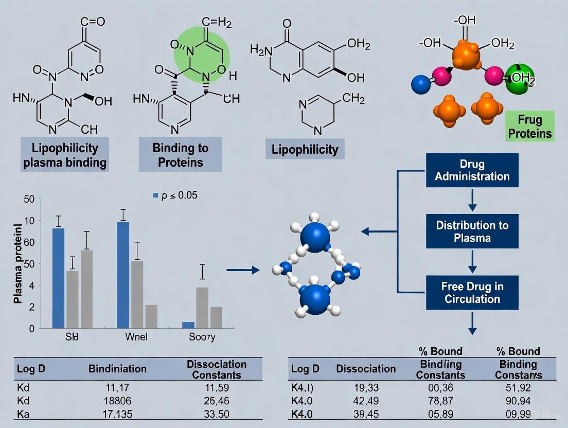
The development of tacrine-based cholinesterase inhibitors represents a significant area of investigation in Alzheimer's disease therapeutics. Although tacrine itself was withdrawn from clinical use due to hepatotoxicity, its high potency and lipophilicity make it a valuable structural motif for designing new inhibitors with improved pharmacological profiles [10]. Within modern drug discovery, the interplay between lipophilicity and plasma protein binding (PPB) serves as a critical determinant of a compound's pharmacokinetic behavior, influencing absorption, distribution, metabolism, excretion, and toxicity (ADMET) [10]. This case study analyzes a series of thirteen tacrine/piperidine-4-carboxamide derivatives, examining their lipophilicity, plasma protein binding properties, and the interrelationship between these parameters within the broader context of rational drug design for central nervous system targets.
Theoretical Background: Lipophilicity and Plasma Protein Binding
The Fundamental Role of Lipophilicity
Lipophilicity is a crucial physicochemical parameter that reflects a substance's ability to passively penetrate cell membranes. For drugs targeting the central nervous system (CNS), such as cholinesterase inhibitors, a well-balanced lipophilicity is essential [10]. If lipophilicity is too low, the drug may fail to cross the blood-brain barrier (BBB), while excessively high lipophilicity can lead to undesirable pharmacokinetic profiles, including high nonspecific binding and increased metabolic clearance [10] [22].
Plasma Protein Binding and its Pharmacokinetic Consequences
Plasma protein binding involves the reversible association of drugs with plasma proteins, primarily human serum albumin (HSA) and α-1-acid glycoprotein (AGP) [23]. Only the unbound drug fraction is considered pharmacologically active, as it can pass through biological membranes and reach its target site [23]. While moderate PPB can prolong a drug's presence in the bloodstream, excessive binding (>95%) can significantly reduce the free fraction available for therapeutic action, potentially limiting efficacy [10]. For tacrine derivatives intended for chronic administration in Alzheimer's disease, optimizing PPB is therefore essential for achieving and maintaining therapeutic concentrations at the target site.
Materials and Experimental Protocols
Investigated Compounds
This study analyzed thirteen tacrine derivatives featuring variously functionalized piperidine-4-carboxamide moieties, including phenyl (1), nicotinoyl (2–4), chlorophenyl (5–7), fluorophenyl (8–10), and methoxy derivatives (11–13) [10]. These compounds had previously demonstrated potent inhibition of both acetylcholinesterase (AChE) and butyrylcholinesterase (BuChE), along with neuroprotective capacity and minimal cytotoxicity toward SH-SY5Y cell lines [10].
Lipophilicity Assessment by Reversed-Phase Thin-Layer Chromatography
Protocol: Lipophilicity was determined using reversed-phase thin-layer chromatography (RP-TLC) on aluminum plates coated with RP-18W F254s stationary phase [10].
- Sample Preparation: Compounds were dissolved in MeOH to approximately 0.5 mg/mL concentration, with 1.0 μL spots applied to plates [10].
- Mobile Phases: Isocratic mobile phases containing organic modifiers (MeOH, ACN, dioxane, or acetone) in concentrations ranging from 0.5 to 0.9 volume fraction, along with formic acid and water [10].
- Lipophilicity Parameters: The RM0 value (extrapolated RM value for zero organic modifier concentration) and C0 value (theoretical modifier concentration for which RM = 0) were calculated from the equation RM = RM0 + bC, where RM = log(1/RF - 1) [10].
Plasma Protein Binding Determination by High Performance Affinity Chromatography
Protocol: PPB properties were analyzed using an HPLC method with an HSA stationary phase [10].
- Chromatographic System: Column: HSA-coated silica stationary phase; Mobile phase: phosphate buffer (pH = 7.0) with 2-propanol as organic modifier [10].
- Binding Calculation: The percentage of plasma protein binding (%PPB) was calculated from two independent experiments based on chromatographic retention behavior [10].
Molecular Docking Studies
Protocol: Docking analyses were performed to investigate binding interactions between the tacrine derivatives and human serum albumin [10].
- Binding Site Focus: Studies targeted Sudlow site I, the primary binding site for heterocyclic aromatic compounds within HSA [10].
- Key Interactions Analyzed: Hydrogen bonding and aromatic interactions were specifically examined to understand the structural basis of PPB [10].
Data Analysis Methods
Principal component analysis (PCA) was conducted on both experimentally determined and predicted lipophilicity values, as well as on predicted adsorption and experimentally determined distribution data, to identify key patterns and relationships [10].
Results and Analysis
Lipophilicity Assessment
The lipophilicity parameters for the thirteen tacrine derivatives, obtained through RP-TLC with different organic modifiers, are summarized in Table 1. Among the evaluated parameters, the RM0 and C0 values obtained using MeOH were identified as the most reliable for characterizing the lipophilicity of the investigated compounds [10]. The observed differences in lipophilicity among the derivatives resulted from a complex interplay of substituent effects (hydrophobicity, polarity, steric hindrance, and electronic effects), positional influence, and characteristics of the organic modifier [10].
Table 1: Lipophilicity Parameters of Tacrine/Piperidine-4-Carboxamide Derivatives
| Compound | Substituent Type | RM0 (MeOH) | C0 (MeOH) | RM0 (ACN) | C0 (ACN) |
|---|---|---|---|---|---|
| 1 | Phenyl | - | - | - | - |
| 2 | Nicotinoyl | - | - | - | - |
| 3 | Nicotinoyl | - | - | - | - |
| 4 | Nicotinoyl | - | - | - | - |
| 5 | Chlorophenyl | - | - | - | - |
| 6 | Chlorophenyl | - | - | - | - |
| 7 | Chlorophenyl | - | - | - | - |
| 8 | Fluorophenyl | - | - | - | - |
| 9 | Fluorophenyl | - | - | - | - |
| 10 | Fluorophenyl | - | - | - | - |
| 11 | Methoxy | - | - | - | - |
| 12 | Methoxy | - | - | - | - |
| 13 | Methoxy | - | - | - | - |
Note: Specific numerical values were not provided in the source material, but the methodology for obtaining these parameters was thoroughly described [10].
Plasma Protein Binding Properties
The plasma protein binding results revealed that all investigated tacrine derivatives efficiently bound to human serum albumin, with calculated %PPB values ranging from 82.38% to 94.54% in the first experiment and 84.29% to 98.16% in the second experiment [10]. These findings suggest that most compounds bind efficiently but not excessively to plasma proteins, maintaining a potentially therapeutic unbound fraction while still benefiting from extended circulation time provided by protein binding.
Molecular Docking Insights
Docking analysis revealed that all investigated ligands bind to Sudlow site I within HSA, which is the main binding site for heterocyclic aromatic compounds such as warfarin, azoprazone, and tacrine itself [10]. The key binding interactions were primarily hydrogen bonding and aromatic interactions [10]. These specific interactions help explain the structural basis for the observed PPB values and provide insights for rational modification of future derivatives to optimize binding characteristics.
Interrelationship Between Lipophilicity and PPB
Principal component analysis highlighted the significant influence of lipophilicity on both adsorption and distribution processes [10]. The positive correlation between lipophilicity parameters and PPB values aligns with established physicochemical principles in pharmacokinetics, confirming that lipophilicity serves as a key driver for plasma protein binding in this series of tacrine derivatives.
The Scientist's Toolkit: Essential Research Reagents and Materials
Table 2: Key Research Reagents and Materials for Lipophilicity and PPB Studies
| Reagent/Material | Specification | Experimental Function |
|---|---|---|
| RP-TLC Plates | Aluminum plates coated with RP-18W F254s stationary phase (Merck, Art. 5559) | Stationary phase for lipophilicity determination by reversed-phase chromatography [10] |
| Organic Modifiers | HPLC-grade MeOH, ACN, dioxane, acetone | Mobile phase components for creating binary solvent systems in RP-TLC [10] |
| HSA Stationary Phase | Silica particles chemically bonded with Human Serum Albumin | Affinity chromatography stationary phase for PPB determination [10] |
| Phosphate Buffer | pH = 7.0 aqueous solution | Aqueous component of mobile phase in HPAC to simulate physiological conditions [10] |
| Formic Acid | High purity acid additive | Mobile phase component to control ionization and improve chromatographic performance [10] |
| 2-Propanol | HPLC-grade organic solvent | Organic modifier in HPAC mobile phase for elution of protein-bound compounds [10] |
Discussion and Broader Implications
Strategic Implications for Tacrine Derivative Design
The findings from this case study carry significant implications for the rational design of tacrine-based therapeutics. The demonstrated relationship between lipophilicity and PPB provides medicinal chemists with a predictive framework for optimizing the pharmacokinetic properties of new derivatives. Recent investigations into novel tacrine-based multi-target directed ligands have similarly emphasized the importance of balanced lipophilicity for achieving favorable CNS penetration, with some advanced compounds demonstrating brain-to-plasma ratios exceeding 2.36 in murine models [24].
Methodological Considerations in Lipophilicity and PPB Assessment
This study highlights several methodological advantages of chromatographic approaches for lipophilicity and PPB determination. RP-TLC offers simplicity, cost-efficiency, and reduced consumption of organic solvents compared to traditional shake-flask methods [10]. Similarly, the HPAC approach with immobilized HSA provides a robust, high-throughput alternative to equilibrium dialysis (considered the gold standard), ultrafiltration, and ultracentrifugation, which often suffer from limitations such as low throughput and poor reproducibility [10] [23].
Computational Approaches and QSAR Modeling
Beyond experimental methods, quantitative structure-activity relationship (QSAR) and quantitative structure-property relationship (QSPR) approaches have demonstrated value in predicting PPB based on molecular descriptors [23] [25]. Studies have identified that hydrophobicity, van der Waals surface area parameters, and aromaticity serve as governing molecular factors for high PPB [23]. However, current models show particular uncertainty in predicting binding for highly protein-bound compounds (fup ≤ 0.25), suggesting that QSPR-predicted values should be used cautiously in physiologically based pharmacokinetic modeling [25].
This comprehensive case study demonstrates the critical interrelationship between lipophilicity and plasma protein binding for a series of tacrine-based cholinesterase inhibitors. Through the integrated application of chromatographic techniques (RP-TLC and HPAC), computational docking, and multivariate analysis, we have established a robust framework for understanding and optimizing the pharmacokinetic properties of this therapeutically relevant chemotype. The findings underscore that well-balanced lipophilicity not only ensures adequate solubility and membrane permeability but also optimizes PPB, which is essential for effective drug distribution and bioavailability. For CNS-targeted agents such as tacrine derivatives, maintaining PPB in the observed range of 82-98% provides an optimal balance between sufficient free fraction for pharmacological activity and adequate protein binding for favorable pharmacokinetic profiles. These insights contribute significantly to the broader thesis that rational optimization of fundamental physicochemical parameters represents a crucial strategy in the development of effective therapeutics for complex neurodegenerative disorders.
In drug discovery and development, the phenomenon of plasma protein binding (PPB) is a critical determinant of a compound's pharmacokinetic (PK) and pharmacodynamic (PD) profile. Historically, research has predominantly focused on human serum albumin (HSA), the most abundant plasma protein, as the primary binding partner for drugs. However, a narrow focus on HSA provides an incomplete picture of the complex binding interactions within plasma. This whitepaper shifts the perspective beyond albumin to elucidate the critical and often underappreciated roles of other major plasma proteins—α1-acid glycoprotein (AGP), lipoproteins, and γ-globulins. Framed within broader research on lipophilicity and PPB relationships, this guide provides a technical deep dive into the binding characteristics, structural determinants, and methodological approaches for studying these key proteins, equipping researchers with the knowledge to optimize drug design and better predict in vivo behavior.
Binding Characteristics of Key Plasma Proteins
The efficacy, distribution, and clearance of a drug are profoundly influenced by its binding to plasma proteins. While HSA binds a wide range of acidic and neutral drugs, other proteins specialize in binding specific drug classes. Understanding the distinct role of each protein is essential for predicting drug disposition.
Human Serum Albumin (HSA) serves as a universal carrier but has specific limitations. It is a 66 kDa globular protein and is the most abundant plasma protein at a concentration of 500–750 µM (35–50 mg/mL), constituting approximately 60% of total plasma protein [26] [27]. It possesses multiple hydrophobic binding sites and primarily binds organic anions, carboxylic acids, and phenols, though it also interacts with some basic and neutral drugs [26]. Its primary physiological functions are to maintain blood pH and osmotic pressure [27].
α1-Acid Glycoprotein (AGP) is the principal carrier for basic drugs. It is a 44 kDa protein with a high carbohydrate content (45%) and an acidic isoelectric point of approximately 3. Its concentration in plasma is significantly lower than HSA, at about 15 µM (0.5–1.0 mg/mL) [26]. AGP has one binding site per molecule and primarily binds basic drugs (e.g., amines) and hydrophobic compounds (e.g., steroids) through nonspecific hydrophobic interactions [26] [27]. A key characteristic is that its concentration is more sensitive to certain disease states (e.g., inflammation, cancer) than HSA, which can significantly alter the free fraction of drugs it carries [26].
Lipoproteins are key binders of lipophilic molecules. This category includes very-low-density lipoprotein (VLDL), low-density lipoprotein (LDL), high-density lipoprotein (HDL), and very-high-density lipoprotein (VHDL). They are particularly important for binding lipophilic basic and neutral drugs, such as probucol and etretinate [26].
γ-Globulins, a class of proteins that includes antibodies, have a historically overlooked role in drug binding. Recent research highlights that human γ-globulins can exhibit a predominant binding affinity for certain therapeutics, even surpassing HSA at physiological concentrations [28] [29]. A 2025 study on antisense oligonucleotides (ASOs) found that γ-globulins had the highest binding affinity for both 2'-O-methoxyethyl/phosphorothioate (MOE/PS)-modified ASOs and phosphorodiamidate morpholino oligomers (PMOs) among the major plasma proteins tested [28] [29].
Table 1: Key Characteristics of Major Drug-Binding Plasma Proteins
| Protein | Molecular Weight | Plasma Concentration | Primary Drug Binding Specificity | Binding Site Capacity |
|---|---|---|---|---|
| Human Serum Albumin | 66 kDa | 500–750 µM (35–50 mg/mL) | Acidic & Neutral Drugs | Multiple sites [26] [27] |
| α1-Acid Glycoprotein | 44 kDa | ~15 µM (0.5–1.0 mg/mL) | Basic Drugs | Single primary site [26] |
| γ-Globulins | Variable (~150 kDa for IgG) | Variable | Diverse (e.g., ASOs) | Variable [28] |
| Lipoproteins | Variable | Variable | Lipophilic Basic/Neutral Drugs | Variable [26] |
Table 2: Comparative Plasma Protein Binding Profiles of Antisense Oligonucleotides (ASOs) [28] [29]
| ASO Chemistry | Unbound Fraction (fu) in Plasma | Saturation Observed | Primary Binding Protein(s) |
|---|---|---|---|
| MOE/PS-modified | Low (high binding) | Yes, above 1 µM | Human γ-Globulins |
| PMO | Higher (low binding) | No, up to 10 µM | Human γ-Globulins |
Methodologies for Characterizing Plasma Protein Binding
Accurately determining the unbound fraction (fu) of a drug is critical, as this is the fraction responsible for pharmacologic activity. The choice of experimental method can significantly influence the results and their interpretation.
Equilibrium Dialysis is widely considered the gold standard for PPB studies [26]. This technique determines the partitioning of a drug across a semi-permeable membrane between a buffer and a plasma compartment. At equilibrium, the concentration of the free drug is identical on both sides of the membrane. The unbound fraction (fu) is calculated as the ratio of the drug concentration in the buffer chamber to that in the plasma chamber. Its main advantage is that it causes minimal disturbance to the equilibrium, but it can be time-consuming and requires membranes with a suitable molecular weight cut-off, which can be a challenge for large molecules like oligonucleotides [29].
Ultrafiltration is a higher-throughput alternative. It involves loading a plasma sample into a device with an ultrafiltration membrane and using centrifugation to separate the unbound drug. The fu is calculated from the drug concentration in the filtrate. A key challenge is nonspecific binding (NSB) of the drug to the device and membrane [26] [29]. Mitigation strategies include pre-treating filters with surfactants like Tween-80 or Tween-20, or using sacrificial oligonucleotides to block binding sites [28] [29]. Recovery experiments are essential to validate that NSB is controlled, with a common acceptance criterion being recovery higher than 70% [29].
Ultracentrifugation avoids the issue of membrane binding altogether. This technique involves centrifuging plasma at high speed (e.g., 100,000 × g) for an extended period (e.g., 24 hours) to separate free drug from protein-bound drug based on density. While advantageous for eliminating NSB to membranes, it is a low-throughput and costly method [26] [29].
Additional Techniques include charcoal adsorption, high-performance affinity chromatography (HPAC), and high-performance frontal analysis (HPFA), each with its own specific applications and limitations [26]. For all methods, it is recommended to test at least three concentrations of the investigational drug to identify potential saturation of binding sites [26].
The Scientist's Toolkit: Key Research Reagent Solutions
The following table details essential materials and reagents used in PPB studies, particularly those featuring the ultrafiltration method for novel therapeutics like ASOs.
Table 3: Essential Research Reagents for Plasma Protein Binding Studies (e.g., Ultrafiltration)
| Reagent / Material | Specification / Example | Function in Experimental Protocol |
|---|---|---|
| Centrifugal Filters | Nanosep 0.5-mL, 30K MWCO | Device for physical separation of unbound drug via centrifugation [29]. |
| Surfactants | Tween-20, Tween-80 | Pre-treatment agent to block non-specific binding sites on filters and consumables [28] [29]. |
| Reference Compounds | (S)-Warfarin, Antipyrine | Small molecule standards for method validation and ensuring reliability [29]. |
| Plasma Proteins | HSA, AGP, Human γ-Globulin, LDL, HDL | Isolated proteins for characterizing individual binding contributions and affinities [28]. |
| Plasma | Pooled, heparin-treated human/mouse plasma | Biologically relevant matrix for measuring binding under near-physiological conditions [28] [29]. |
| Detection Probes | Biotin-/Digoxigenin-conjugated probes | For sensitive, sequence-specific detection of oligonucleotides via hybridization-ECL assays [29]. |
Conceptual Framework and Interrelationships
The binding of a drug to plasma proteins is a dynamic equilibrium process. The relationship between lipophilicity and PPB is a cornerstone of understanding, though it is complex. For congeneric series, lipophilicity is often the dominant factor driving binding, particularly to albumin. However, for a diverse set of molecules, the correlation is weaker, suggesting that specific molecular recognition elements and structural motifs are equally critical [26]. This is especially true for interactions with proteins like AGP and γ-globulins, which may exhibit more stereoselective binding.
The following diagram illustrates the competitive and dynamic equilibrium that exists between a free drug and its potential binding partners in plasma, highlighting the roles of the key proteins discussed beyond albumin.
Diagram: Dynamic Equilibrium of Drug Binding to Plasma Proteins. The free drug is in dynamic equilibrium with multiple plasma proteins. Only the free drug fraction can interact with the therapeutic target or be eliminated from the body.
Implications for Drug Disposition and Efficacy
The binding of a drug to plasma proteins creates a reservoir, prolonging its duration in circulation. However, the impact on efficacy and safety is multifaceted.
Influence on Pharmacokinetics: Plasma protein binding directly impacts a drug's volume of distribution, clearance, and half-life. A highly bound drug with slow dissociation can be 'restrictive,' meaning it is retained in plasma, leading to a lower volume of distribution, potentially decreased clearance, and a longer half-life [26]. Conversely, a drug with high binding but fast dissociation (like propranolol) can be 'permissive,' allowing for high liver extraction [26].
Optimizable Parameter for Efficacy: PPB should not be viewed merely as a fixed property but as an optimizable parameter in drug design [30]. Strategic modulation of PPB can be used to achieve suitable effective half-lives and improve the therapeutic index. For instance, increasing binding can prolong half-life and allow for lower maintenance doses, while decreasing binding can increase the free fraction available for tissue penetration [30].
Drug-Drug Interactions (DDIs): The potential for one drug to displace another from plasma proteins is a classic mechanism of DDI. While this can lead to a transient increase in the free fraction of the displaced drug, in open biological systems, this effect is often self-correcting due to increased distribution and elimination of the now-unbound drug [31]. A clinically significant interaction is more likely for drugs that are highly protein-bound (>95%), have a narrow therapeutic index, and whose clearance is restrictive (e.g., warfarin) [31].
A comprehensive understanding of plasma protein binding that extends beyond albumin is indispensable for modern drug development. The roles of AGP, lipoproteins, and γ-globulins are critical in determining the fate of specific drug classes, as evidenced by the recent discovery of γ-globulin's predominant role in ASO binding. Future research will be guided by advanced computational models, such as those using machine learning on platforms like OCHEM, which show high accuracy in predicting PPB and can inform structural optimization of lead compounds [32]. By integrating detailed knowledge of protein-specific binding with robust experimental methodologies and predictive modeling, researchers can more effectively navigate the complex interplay between lipophilicity, protein binding, and in vivo efficacy, ultimately accelerating the development of safer and more effective therapeutics.
Modern Techniques for Measuring Lipophilicity and Protein Binding
Lipophilicity, quantified as the logarithm of the n-octanol/water partition coefficient (log P) or distribution coefficient (log D), constitutes a crucial physicochemical parameter in quantitative structure-activity relationships (QSARs) for bioactive compounds [33]. It plays a pivotal role in governing pharmacokinetic and pharmacodynamic properties, including absorption, distribution, metabolism, excretion, and toxicity (ADMET) [34] [35]. For any compound to exert its pharmacological effect, it must successfully traverse biological membranes and achieve adequate distribution within the body, processes largely governed by lipophilicity [36]. Particularly critical is the relationship between lipophilicity and plasma protein binding, as only the unbound drug fraction remains pharmacologically active [37]. High lipophilicity often correlates with increased plasma protein binding, reduced free drug concentration, and potential toxicity concerns [38].
Within this context, reliable high-throughput methods for lipophilicity assessment are indispensable for modern drug discovery. Traditional methods like the shake-flask approach, while considered a gold standard, present limitations including being time-consuming, requiring high compound purity, and having a restricted measurement range (-2 < log P < 4) [33] [34] [39]. Chromatographic techniques, specifically Reversed-Phase Thin-Layer Chromatography (RP-TLC) and Reversed-Phase High-Performance Liquid Chromatography (RP-HPLC), have emerged as powerful alternatives that overcome these limitations while offering speed, reproducibility, insensitivity to impurities, and a broad dynamic range [33] [40]. This technical guide elaborates on the application of these chromatographic methods for high-throughput lipophilicity assessment within the framework of drug discovery, with particular emphasis on their relevance to understanding plasma protein binding.
Theoretical Foundations of Lipophilicity Measurement
Defining Lipophilicity Parameters: Log P and Log D
Lipophilicity is fundamentally characterized by two parameters: the partition coefficient (log P) and the distribution coefficient (log D). Log P refers to the partition coefficient logarithm of a compound between an organic phase and an aqueous phase when the compound exists entirely as non-ionized molecules. Its value depends solely on the compound's intrinsic properties, such as molecular volume, dipole moment, and hydrogen bond acidity/basicity [34] [35]. In contrast, Log D describes the distribution coefficient logarithm when the compound exists as both ionized and non-ionized forms at a specific pH, making it a pH-dependent value [34]. For ionizable compounds, including many pharmaceuticals, log D provides a more physiologically relevant measure of lipophilicity.
The Chromatographic Basis for Lipophilicity Assessment
Chromatographic techniques model the partitioning of a compound between a stationary phase, which mimics the lipophilic environment, and a mobile aqueous phase. The retention behavior of a compound in these systems correlates with its lipophilicity [33] [40]. The primary retention parameter in RP-HPLC is the capacity factor (k), calculated as log k = log((tR - t0)/t0), where tR is the solute's retention time and t0 is the retention time of an unretained compound [33]. In RP-TLC, the retardation factor (Rf) is measured, from which the RM value is derived as RM = log(1/Rf - 1) [40]. To obtain a chromatographic index independent of organic modifier effects, the retention parameter (log k or RM) is determined at multiple concentrations of organic modifier (e.g., methanol, acetonitrile) and extrapolated to zero organic modifier concentration, yielding log kw in HPLC or RMW in TLC [33] [40]. These values serve as chromatographic descriptors of lipophilicity that can be correlated to reference log P values through the Collander equation: log P = a × log kw + b [33] [34].
Reversed-Phase High-Performance Liquid Chromatography (RP-HPLC)
RP-HPLC has become a standard procedure for lipophilicity measurement recommended by the Organisation for Economic Co-operation and Development (OECD) [33]. It offers significant practical advantages for high-throughput screening, including operational speed, excellent reproducibility, insensitivity to impurities or degradation products, broad dynamic range, on-line detection, and minimal sample requirements [33] [34] [35]. Particularly notable is its extended measurement range, which can be expanded to compounds with log P > 6 under certain conditions, effectively overcoming the limitations of the shake-flask method for highly lipophilic compounds [34].
Experimental Protocols for RP-HPLC
Basic Protocol for Log P Determination (Method 1)
This method prioritizes speed and efficiency for early drug screening [34]:
- System Calibration: A series of reference compounds with known log P values covering a wide lipophilicity range are selected. An example set is shown in Table 1.
- Chromatographic Analysis: The reference compounds and test compounds are injected into the qualified RP-HPLC system using optimized chromatographic conditions (typically a C18 column with a methanol/water or acetonitrile/water mobile phase gradient).
- Capacity Factor Calculation: The retention time (tR) for each compound is used to calculate the capacity factor (k), and subsequently log k.
- Standard Curve Construction: A standard equation, log P = a × log k + b, is established by plotting the log k values of the reference compounds against their known log P values. The correlation coefficient (R²) should be ≥ 0.97 [34].
- Log P Determination for Unknowns: The log k value of a test compound is substituted into the standard equation to determine its log P value.
Advanced Protocol for Enhanced Accuracy (Method 2)
For late-stage drug development requiring higher accuracy, a modified approach eliminates the interference from organic modifiers [34]:
- Multiple Gradient Measurements: The retention times for reference and test compounds are measured under at least three different mobile phase gradients with varying organic modifier content (φ).
- log kw Determination: For each compound, an equation is established: log k = Sφ + log kw. The y-intercept (log kw) represents the capacity factor in the absence of organic modifier.
- Standard Curve with log kw: A more accurate standard equation is constructed: log P = a × log kw + b, which typically shows excellent correlation (R² > 0.99) with reference log P values [34].
Table 1: Example Reference Compounds for RP-HPLC Method Development [34]
| Compound Name | Reported Log P |
|---|---|
| 4-Acetylpyridine | 0.5 |
| Acetophenone | 1.7 |
| Chlorobenzene | 2.8 |
| Ethylbenzene | 3.2 |
| Phenanthrene | 4.5 |
| Triphenylamine | 5.7 |
Application to Plasma Protein Binding Studies
RP-HPLC can be extended to predict plasma protein binding by using stationary phases that mimic biological components. Immobilized Human Serum Albumin (HSA) columns are particularly valuable, as HSA is the most abundant protein in human plasma and responsible for binding many drugs [37] [38]. The retention factor (log k) obtained from HSA-HPLC demonstrates a significant correlation with experimental plasma protein binding data [37] [38]. For a group of 34 basic drugs, the correlation coefficient (R) between log k and protein binding was 0.63, explaining approximately 40% of the variance in binding [37]. This chromatographic approach is especially useful for reliably ranking molecules in the high-binding region (above 95% bound), facilitating the construction of structure-binding relationships to guide molecular modifications that optimize binding properties [38].
Reversed-Phase Thin-Layer Chromatography (RP-TLC)
RP-TLC serves as a straightforward, cost-effective, and high-throughput alternative for lipophilicity assessment [40]. Its advantages include low solvent consumption, the ability to analyze several samples simultaneously on a single plate, no requirement for sophisticated instrumentation, and high reproducibility of results [40]. The technique is particularly valuable in the initial evaluation of drug candidates and in constructing QSAR models during early discovery stages.
Experimental Protocols for RP-TLC
- Stationary Phase Preparation: Commercially available TLC plates pre-coated with hydrophobic layers (e.g., RP-18, RP-8, or CNF254) are used. The stationary phase can be modified with proteins like Bovine Serum Albumin (BSA) to mimic protein binding environments [37].
- Mobile Phase Selection: Binary mixtures of a water-miscible organic modifier (e.g., methanol, acetone, or acetonitrile) and aqueous buffer (often at physiological pH 7.4) are used as the mobile phase [37] [40].
- Chromatographic Development: Samples are spotted on the plate, which is then developed in a chromatographic chamber saturated with the mobile phase vapor.
- Retention Measurement: The retardation factor (Rf) is calculated for each compound as the ratio of the distance traveled by the solute to the distance traveled by the solvent front.
- Lipophilicity Index Calculation: The RM value is calculated from Rf using RM = log(1/Rf - 1). This measurement is repeated using mobile phases with different volume fractions of the organic modifier (φ).
- Extrapolation to RMW: The relationship between RM and φ is described by the equation RM = RMW + Sφ. The lipophilicity parameter RMW (the value at φ = 0) is determined by linear or polynomial regression [40]. Alternative calculation methods include Soczewiński-Wachtmeister's and Ościk's equations [40].
Correlating RP-TLC Data with Protein Binding
RP-TLC data obtained from BSA-impregnated plates show significant prognostic value for plasma protein binding. In chemometric analyses, multiple linear regression (MLR) models using retention data from normal-phase TLC on BSA-impregnated plates demonstrated high correlation with experimental protein binding values, with coefficients of determination (R²) ranging from 0.73 to 0.91 for different classes of drugs (acids, bases, and neutrals) [37]. This suggests that TLC-based binding indices can serve as convenient quantitative parameters for predicting protein binding affinity.
Comparative Analysis of Chromatographic Methods
Table 2: Comparison of Lipophilicity Measurement Methods [33] [34] [39]
| Method | Measurement Range (log P) | Throughput | Key Advantages | Limitations | Suitability for Protein Binding Studies |
|---|---|---|---|---|---|
| Shake-Flask | -2 to 4 | Low | Considered gold standard, accurate results | Time-consuming, requires high purity, limited range | Requires separate experiments |
| RP-HPLC | 0 to 6+ | High | Broad range, high accuracy, automatable, insensitive to impurities | Requires reference compounds, method development | Excellent with HSA columns |
| RP-TLC | Wide range | Very High | Low cost, parallel analysis, minimal sample prep | Lower precision than HPLC | Good with BSA-impregnated plates |
| Computer Simulation | Broad | Very High | Instantaneous, no compounds needed | Accuracy depends on algorithm and training data | Limited predictive power |
The Scientist's Toolkit: Essential Research Reagents and Materials
Table 3: Key Research Reagent Solutions for Chromatographic Lipophilicity Assessment
| Item | Function/Description | Application Notes |
|---|---|---|
| C18 Chromatographic Columns | The most common reversed-phase stationary phase for RP-HPLC, consisting of silica bonded with octadecyl carbon chains. | Standard for log P determination; choose particle size (e.g., 3-5 μm) and dimensions suitable for throughput needs [33] [34]. |
| HSA-Immobilized Columns | HPLC columns with Human Serum Albumin chemically bonded to the stationary phase. | Mimics drug-protein binding in plasma; directly predicts plasma protein binding affinity [37] [38]. |
| RP-18 TLC Plates | Glass or plastic plates pre-coated with a layer of C18-modified silica gel. | Standard stationary phase for RP-TLC; enables parallel analysis of multiple compounds [40]. |
| BSA (Bovine Serum Albumin) | A protein often used as an effective replacement for HSA in binding studies. | Used to impregnate TLC plates to create a biomimetic surface for protein binding assessment [37]. |
| Reference Compound Sets | A series of compounds with precisely known log P values covering a broad lipophilicity range. | Essential for constructing calibration curves in both RP-HPLC and RP-TLC [34]. |
| Buffers (pH 7.4) | Aqueous mobile phase components, typically phosphate buffers. | Mimics physiological pH, crucial for measuring log D and for biomimetic binding studies [37] [36]. |
Workflow and Data Relationship Diagrams
High-Throughput Lipophilicity and Protein Binding Assessment Workflow
The following diagram illustrates the integrated experimental workflow for assessing lipophilicity and plasma protein binding using RP-HPLC and RP-TLC, and how the resulting data interrelates.
RP-TLC and RP-HPLC provide robust, high-throughput platforms for the reliable assessment of lipophilicity within drug discovery programs. The ability of these chromatographic techniques to be adapted for biomimetic purposes—specifically through the use of HSA columns and BSA-impregnated plates—makes them uniquely powerful for investigating the critical relationship between lipophilicity and plasma protein binding. By implementing the detailed methodologies and standardized protocols outlined in this guide, researchers can efficiently generate high-quality lipophilicity and binding data essential for rational drug design. This data directly informs the optimization of lead compounds, guiding structural modifications to achieve favorable ADMET profiles and increasing the likelihood of clinical success.
Plasma protein binding (PPB) is a fundamental parameter in pharmacokinetics, describing the reversible formation of complexes between drugs and plasma proteins, primarily human serum albumin (HSA) and alpha-1-acid glycoprotein (AAG) [41]. According to the free drug hypothesis, only the unbound drug fraction is biologically active as it can diffuse across cell membranes to interact with target sites [42]. This binding directly influences key pharmacokinetic properties including a drug's volume of distribution, clearance rate, and half-life [42]. For highly bound drugs (>99%), even slight variations in binding can lead to significant changes in free drug concentration, potentially altering therapeutic efficacy and safety profiles [41] [43]. This is particularly important in polytherapy scenarios where displacement interactions may occur, creating a risk of adverse drug reactions [43].
The relationship between PPB and lipophilicity is well-established, with compounds possessing substantial lipophilicity typically exhibiting strong affinity for plasma proteins [41] [44] [10]. This connection forms the basis for various predictive models and experimental approaches in drug discovery. Accurate PPB determination is therefore crucial for understanding both the pharmacokinetics and pharmacodynamics of drug candidates, enabling the design of safer and more effective therapeutics with optimal dosing strategies [41]. This review examines the gold standard methodologies for PPB assessment and their modern variants, contextualized within the critical framework of lipophilicity-PPB relationships.
Core Methodologies: Principles, Protocols, and Applications
Equilibrium Dialysis (ED) – The Recognized Gold Standard
Equilibrium dialysis is widely regarded as the gold standard method for PPB determination due to its fundamental principle of measuring binding at equilibrium, which minimizes disturbance to the natural binding equilibrium [42] [45]. The method operates on a simple principle: a semi-permeable membrane separates a protein-containing compartment (plasma) from a protein-free buffer compartment. The membrane allows small molecules to pass freely while retaining large plasma proteins. During incubation, the unbound drug diffuses across the membrane until equilibrium is established, whereafter the free drug concentration is measured in the buffer compartment [41] [43].
A critical consideration in ED is maintaining physiological pH throughout the experiment. Research has demonstrated that plasma loses carbon dioxide during storage and incubation, causing pH to rise significantly—potentially to levels as high as 9—which dramatically alters protein binding properties [41]. One proven solution involves adjusting the initial aqueous buffer pH to 7.8 and exposing the dialysis device to CO2 using gas-permeable seals, which maintains the incubation mixture at physiological pH 7.4 during typical 18-hour equilibrium experiments [41]. For highly bound compounds, this extended incubation time is essential, as shorter periods (4-6 hours) are often insufficient to reach equilibrium [41].
The basic protocol for ED involves placing drug-spiked plasma in one compartment and buffer in the other, followed by incubation at 37°C with appropriate pH control. Post-dialysis, drug concentrations in both compartments are quantified using techniques such as HPLC-MS [45]. The fraction unbound (fu) is calculated as fu = Cbuffer / Cplasma, where Cbuffer and Cplasma represent drug concentrations in the buffer and plasma compartments, respectively, after equilibrium is established.
Recent innovations have enhanced ED throughput, including the rapid equilibrium dialysis (RED) device and novel approaches for challenging compounds. For highly bound lipophilic compounds, researchers have developed a modified RED method coupled with extraction of the post-dialysis buffer into organic phase (typically octanol). This approach leverages the lipophilicity of strongly bound compounds, enabling their concentration and accurate quantification despite very low free fractions [41]. High-throughput adaptations also include sample pooling approaches, where multiple compounds are dialyzed simultaneously and quantified using LC-MS, significantly increasing screening capacity without sacrificing accuracy [45].
Ultrafiltration (UF) – A Rapid Alternative
Ultrafiltration offers a faster alternative to ED, particularly valuable in high-throughput screening environments. The method employs centrifugal force to separate unbound drug through a semi-permeable membrane with specific molecular weight cut-offs [43]. After centrifugation of drug-spiked plasma in specialized UF devices, the protein-free ultrafiltrate in the lower compartment is analyzed for free drug concentration [43].
The primary advantage of UF is its speed and simplicity, with typical processing times significantly shorter than ED. However, UF faces substantial limitations, most notably non-specific binding (NSB) of compounds to the filter membrane and device components [43]. This issue is particularly problematic for lipophilic compounds, which may adsorb extensively to filtration materials, leading to underestimation of free drug concentrations [43]. Additional considerations include the Donnan effect (ion distribution imbalance across the membrane), protein leakage, and requirement for careful control of experimental conditions including pH and temperature [43].
Strategies to mitigate NSB include membrane pre-treatment with agents such as Tween 80 (effective for acidic and neutral compounds) or benzalkonium chloride (preferable for basic compounds) [43]. However, these treatments require careful optimization as they may potentially interact with plasma proteins or the drug itself [43]. Despite its limitations, UF remains a valuable technique for rapid PPB screening, especially for compounds with low to moderate NSB potential.
Ultracentrifugation
Ultracentrifugation represents a third principal method for PPB determination, though it appears less frequently in recent literature compared to ED and UF. This technique employs high-speed centrifugation to separate protein-bound drug complexes from free drug molecules based on density differences [43]. The method requires specialized equipment capable of generating sufficient gravitational force to sediment plasma proteins while leaving the unbound drug in solution.
A significant advantage of ultracentrifugation is the absence of membrane-related issues such as NSB or the Donnan effect, making it potentially useful for problematic compounds that strongly interact with filtration or dialysis membranes. However, the method faces challenges including potential drug sedimentation at high centrifugal forces, lengthy processing times, and requirements for substantial sample volumes [43]. Additionally, the need for specialized, expensive equipment limits its accessibility and practicality for high-throughput applications.
Methodological Comparison and Technical Considerations
Table 1: Comparative Analysis of Major PPB Determination Methods
| Parameter | Equilibrium Dialysis | Ultrafiltration | Ultracentrifugation |
|---|---|---|---|
| Fundamental Principle | Passive diffusion through semi-permeable membrane until equilibrium | Pressure-driven separation through semi-permeable membrane | Sedimentation based on density differences |
| Throughput | Moderate (improved with 96-well format and pooling) [45] | High | Low |
| Incubation Time | Longer (up to 18h for highly bound compounds) [41] | Short (minutes to hours) | Long (several hours) |
| Key Advantages | Minimal disturbance of equilibrium; considered gold standard [42] | Speed and simplicity; small sample volumes | No membrane interactions |
| Major Limitations | Time-consuming; potential pH shifts [41] | Non-specific binding; Donnan effect [43] | Equipment cost; lengthy process; potential drug sedimentation [43] |
| NSB Concerns | Low | High, especially for lipophilic compounds [43] | None |
| pH Control | Critical; requires careful buffering or CO2 control [41] | Important but easier to maintain | Important but easier to maintain |
| Suitable for Highly Bound Compounds | Yes, especially with organic phase extraction [41] | Problematic due to NSB | Possible, but with limitations |
Table 2: Advanced ED Modifications for Challenging Compounds
| Method Variant | Principle | Applications | Considerations |
|---|---|---|---|
| RED with Organic Phase Extraction [41] | Leverages lipophilicity; extracts post-dialysis buffer into organic solvent | Highly bound compounds with low fu (10⁻¹ to 10⁻⁶); e.g., venetoclax, amiodarone | Requires compatible organic solvent (e.g., octanol); extends quantification range |
| Sample Pooling [45] | Multiple compounds dialyzed simultaneously; analyzed by LC-MS | High-throughput screening; limited plasma availability | Theoretical model suggests ≤10 compounds; validation required |
| Flux Dialysis | Measures drug movement across membrane over time | Compounds with very slow equilibrium | Complex data interpretation |
| Dilution Method | Dilutes plasma to reduce binding | High-affinity binders | Potential changes in protein conformation |
| Presaturation | Saturates NSB sites before experiment | Compounds with significant NSB | Risk of incomplete presaturation or displacement |
The Lipophilicity-PPB Relationship and Predictive Approaches
Fundamental Relationships Between Lipophilicity and PPB
The connection between molecular lipophilicity and plasma protein binding represents a cornerstone of physicochemical property assessment in drug discovery. Compounds with substantial lipophilicity typically exhibit strong affinity for plasma proteins, as they possess hydrophobic functional groups or regions that interact favorably with hydrophobic pockets in proteins like HSA and AAG [41]. This relationship was quantitatively described as early as 1987 using the model fu = 1/(1 + a·D^b), where fu is the unbound fraction, D is the octanol/water partition coefficient, and a and b are fitting parameters [44].
Chromatographic techniques have proven particularly valuable for exploring this relationship. In studies of tacrine-based cholinesterase inhibitors, researchers demonstrated that lipophilicity parameters measured by reversed-phase thin-layer chromatography (RP-TLC) significantly influenced PPB characteristics [10]. Similarly, biomimetic chromatography using stationary phases containing immobilized HSA or AGP has emerged as a high-throughput screening method that effectively predicts PPB based on retention factors that correlate with binding affinity [42].
For acidic drugs specifically, quantitative structure-plasma protein binding relationship (QSPPBR) studies have identified key structural features influencing PPB: lipophilicity, presence of aromatic rings, cyano groups, and H-bond donor-acceptor pairs increase PPB, while tertiary carbon atoms, four-membered rings, and iodine atoms decrease PPB [46]. These findings enable the creation of guidance checklists similar to Lipinski's Rule of Five for preliminary PPB assessment during drug design.
Computational Prediction of PPB
Advances in computational methods have enabled increasingly accurate prediction of PPB from molecular structure alone. Quantitative Structure-Activity Relationship (QSAR) models, particularly those incorporating neural networks, have demonstrated remarkable predictive capability for PPB [47]. One recent model developed using a dataset of 277 drugs achieved external validation with a predictive squared correlation coefficient (Q²) of 0.966 and root mean squared error (RMSE) of 0.063, outperforming previously published models [47] [48].
The development of robust QSAR models involves a multi-step process: (1) curating a high-quality experimental dataset; (2) generating comprehensive molecular descriptors from chemical structures; (3) selecting relevant descriptors using filter methods or other feature selection techniques; (4) model training using machine learning algorithms; and (5) rigorous validation including assessment of the applicability domain [47]. These in silico approaches significantly reduce the need for resource-intensive chemical synthesis and laboratory testing during early drug discovery stages [47].
Machine learning algorithms now routinely combine biomimetic chromatography data with in silico molecular descriptors and/or molecular fingerprints to predict in vivo PPB parameters [42]. This integrated approach leverages the strengths of both experimental and computational methods, providing increasingly accurate predictions of complex biological interactions from structural information alone.
Essential Research Reagents and Experimental Solutions
Table 3: Key Research Reagent Solutions for PPB Studies
| Reagent/Equipment | Function/Role | Application Notes |
|---|---|---|
| Human Serum Albumin (HSA) | Primary binding protein for acidic and neutral drugs | Major plasma protein (5-7.5×10⁻⁴ M); contains Sudlow sites I and II [10] |
| Alpha-1-Acid Glycoprotein (AGP) | Primary binding protein for basic drugs | Acute-phase protein; concentration increases during inflammation [43] |
| RED Device | 96-well format equilibrium dialysis | Enables higher throughput; commercial systems available |
| Ultrafiltration Units | Centrifugal devices with semi-permeable membranes | Various molecular weight cut-offs available; material affects NSB |
| Octanol | Organic solvent for extraction | Used in modified RED for highly lipophilic compounds [41] |
| Biomimetic Columns (HSA/AGP) | HPLC stationary phases with immobilized proteins | High-throughput screening; correlates with in vivo PPB [42] [10] |
| pH-Controlled Buffers | Maintain physiological pH during incubation | Critical for accurate results; CO2-controlled systems recommended [41] |
The determination of plasma protein binding remains an essential component of comprehensive drug characterization, with equilibrium dialysis maintaining its status as the gold standard methodology due to its minimal disturbance of the natural binding equilibrium. Recent innovations, particularly the combination of RED with organic phase extraction, have extended viable measurement ranges to include even highly challenging compounds with extremely low free fractions. While ultrafiltration and ultracentrifugation offer valuable alternatives for specific applications, each method presents distinct advantages and limitations that must be carefully considered in experimental design.
The well-established relationship between lipophilicity and PPB continues to inform both experimental approaches and computational predictions. Modern QSAR models incorporating neural networks demonstrate remarkable predictive accuracy, reducing reliance on resource-intensive laboratory testing. As drug discovery increasingly focuses on highly lipophilic compounds with strong protein binding characteristics, the continued refinement of these methodologies—both experimental and computational—will remain crucial for the development of safer, more effective therapeutics with optimized pharmacokinetic profiles.
Experimental Workflow Visualization
Diagram 1: Methodological workflow for PPB determination, highlighting the role of lipophilicity across experimental and computational approaches.
High-Performance Affinity Chromatography (HPAC) is a robust analytical technique that combines the specificity of biological interactions with the efficiency of high-performance liquid chromatography. In this method, a biologically-related binding agent, or affinity ligand, is immobilized onto a rigid support to create a stationary phase capable of selective interactions [49] [50]. When the affinity ligand is Human Serum Albumin (HSA)—the most abundant plasma protein in human blood—the resulting HSA-immobilized stationary phase becomes a powerful tool for studying drug-protein interactions, which is fundamental to understanding drug disposition, efficacy, and toxicity [51] [52]. The core principle relies on the specific and reversible binding that occurs between HSA and a wide array of pharmaceutical compounds, endogenous substances, and toxins [52]. HPAC using HSA columns provides significant advantages over traditional solution-based methods like equilibrium dialysis or ultrafiltration, including high precision, ease of automation, reusability of the protein stationary phase, and the ability to perform analyses in minutes rather than hours [51] [52].
The relationship between lipophilicity and plasma protein binding is a critical focus in pharmaceutical research, as a drug's lipophilicity often correlates with its binding affinity to HSA [53]. This binding directly influences the free drug fraction, which is the portion available for pharmacological activity, metabolism, and excretion [2]. HPAC serves as an ideal experimental platform for quantifying this relationship, enabling researchers to efficiently screen compound libraries and predict pharmacokinetic behavior early in drug development [53] [54].
Key Methodological Approaches
The two primary chromatographic methods used in HPAC for interaction studies are zonal elution and frontal analysis. Each provides unique insights into the binding parameters between solutes and the immobilized HSA.
Zonal Elution
Zonal elution is performed by injecting a small, discrete volume of analyte onto the HSA column. The resulting retention factor (k) is directly related to the strength of binding [51] [52]. This method is particularly effective for competitive interaction studies, where a mobile phase modifier or competing agent is introduced to investigate site-specific binding and displacement phenomena [53] [52]. For instance, zonal elution has been successfully used to identify the specific HSA binding sites of synthetic cannabinoids by observing the retention shifts in the presence of site-specific probes like warfarin (Site I) and L-tryptophan (Site II) [53].
Frontal Analysis
Frontal analysis involves the continuous application of a known concentration of the analyte onto the HSA column. As the immobilized binding sites become saturated, a breakthrough curve is generated [51] [52]. The mean position of this curve is used to calculate the binding affinity (association constant, K) and the number of active binding sites on the protein [51]. This method is ideal for accurately determining the binding constants for a single solute-protein interaction.
A powerful hybrid approach involves generating a standard plot by combining data from both frontal and zonal methods. The retention factors (k) of reference compounds obtained via zonal elution are plotted against their association constants (K) determined by frontal analysis. This calibrated plot can then be used to rapidly determine the association constants for new compounds using only quick zonal elution measurements, significantly increasing throughput [51].
Table 1: Comparison of Primary HPAC Methodologies for HSA-Based Studies
| Method | Primary Application | Key Measured Parameters | Advantages |
|---|---|---|---|
| Zonal Elution | Competition studies, binding site mapping, rapid screening | Retention factor (k), bound fraction (%b) | High speed, distinguishes binding sites, uses minimal analyte [51] [53] |
| Frontal Analysis | Determination of binding affinity and capacity | Association constant (K), number of binding sites | High accuracy for single-solute binding parameters [51] [52] |
| Hybrid Approach | High-throughput determination of association constants | Association constant (K) derived from retention factor (k) | Combines speed of zonal elution with accuracy of frontal analysis [51] |
Experimental Protocols and Workflows
Preparation of HSA-Immobilized Stationary Phases
The creation of a robust and active HSA stationary phase is critical. A common protocol involves immobilizing HSA to a silica-based support using the Schiff base method (reductive amination) [51] [55].
- Support Activation: Diol-bonded silica is first oxidized with sodium periodate to create an aldehyde-activated support [51].
- Immobilization: The aldehyde silica is reacted with HSA in the presence of a reducing agent, sodium cyanoborohydride (NaCNBH₃), in a potassium phosphate buffer (pH 7.4). The reaction is typically allowed to proceed for several days at low temperature to maximize ligand density [51].
- Capping: Excess aldehyde groups on the support are subsequently converted to inert alcohols by treatment with sodium borohydride (NaBH₄) [51].
An alternative method, entrapment, has also been developed and optimized. This technique involves encapsulating HSA within the pores of a hydrazide-activated silica support using mildly oxidized glycogen as a capping agent. This physical method has been shown to effectively retain the protein's binding activity [56]. More recent advancements have focused on using organic polymer-based monoliths (e.g., copolymers of glycidyl methacrylate and ethylene glycol dimethacrylate) as supports, which offer low back-pressures and excellent mass transfer properties [55]. The on-column entrapment method in these monolithic supports has demonstrated a three-fold higher retention for model solutes like warfarin compared to slurry-based methods, leading to higher capacity stationary phases [56].
Standard HPAC Binding Assay Workflow
The following diagram illustrates a generalized workflow for conducting a binding study using an HSA-HPAC column, incorporating both zonal and frontal analysis principles.
Displacement Studies for Binding Site Identification
To identify the specific site on HSA where a drug binds, a displacement chromatography protocol is used:
- A known site-specific probe (e.g., warfarin for Site I, L-tryptophan for Site II) is added to the mobile phase at a fixed concentration [53].
- The analyte of interest is injected onto the column via zonal elution.
- A significant reduction in the analyte's retention time in the presence of the probe indicates competition for the same binding site.
- This method was key in identifying that synthetic cannabinoids like 5F-AMB and AB-PINACA bind primarily to HSA's warfarin site (Site I) [53].
Key Research Findings and Data
HPAC studies with HSA columns have yielded critical quantitative data on the binding profiles of diverse compounds, directly linking lipophilicity to the extent of plasma protein binding.
Table 2: Binding Affinity of Various Drugs to HSA as Measured by HPAC
| Compound/Drug Class | Example Compound | Bound Fraction (%b) / Association Constant (K) | Primary HSA Binding Site |
|---|---|---|---|
| Synthetic Cannabinoids | 5F-AMB | 98.9% [53] | Site I (Warfarin site) [53] |
| AB-PINACA | 98.7% [53] | Site I (Warfarin site) [53] | |
| AMB-FUBINACA | 99.7% [53] | Site I (Warfarin site) [53] | |
| FUBIMINA | 99.9% [53] | Information Not Specified | |
| Non-Steroidal Anti-Inflammatory Drugs (NSAIDs) | Ibuprofen | K ≈ 2 × 10⁵ to 3.5 × 10⁶ M⁻¹ [51] | Site II (Indole-Benzodiazepine site) |
| Phenylbutazone | K ≈ 1.4 × 10⁵ to 1.5 × 10⁶ M⁻¹ [51] | Information Not Specified | |
| Other Drugs | Tolbutamide | Reference compound for standard plots [51] | Information Not Specified |
| Diazepam | Reference compound for standard plots [51] | Site II (Indole-Benzodiazepine site) | |
| Control Compounds | Chlorpromazine | >90% (Experimental: 82.12%) [53] | Information Not Specified |
| Indomethacin | 97.8% (Experimental: 96.89%) [53] | Information Not Specified |
The data unequivocally shows that highly lipophilic compounds, such as synthetic cannabinoids, exhibit extremely high HSA binding (>98%), which aligns with their high calculated log P values [53]. This strong binding directly impacts their volume of distribution and can lead to a prolonged half-life. Furthermore, HPAC displacement studies confirm that most drugs bind to one of two major sites on HSA (Site I or Site II), highlighting the potential for drug-drug interactions due to competition for these sites [53] [52]. For instance, the binding affinity of synthetic cannabinoids was found to increase in the presence of (S)-ibuprofen, a Site II binder, suggesting an allosteric interaction between the sites [53].
The Scientist's Toolkit: Essential Research Reagents and Materials
Successful implementation of HPAC with HSA stationary phases requires specific, high-quality materials and reagents. The following table details the essential components of the "researcher's toolkit" for this field.
Table 3: Key Research Reagent Solutions for HPAC with HSA Stationary Phases
| Category | Specific Examples | Function/Purpose |
|---|---|---|
| Chromatographic Support | Diol-bonded Silica (e.g., Nucleosil Si-300) [51]; GMA/EDMA or GMA/TRIM Monoliths [55] | Serves as the solid scaffold for immobilizing HSA; determines pressure stability and efficiency. |
| Immobilization Reagents | Sodium Periodate [51]; Sodium Cyanoborohydride [51] [55] | Activate the support (periodate) and stabilize the HSA immobilization via reductive amination (cyanoborohydride). |
| Affinity Ligand | Human Serum Albumin (HSA), Fatty Acid Free [51] [56] [55] | The key biological binding agent immobilized to create the selective stationary phase. |
| Buffer Systems | Potassium Phosphate Buffer (e.g., 67 mM, pH 7.0-7.4) [51] [53] | Mimics physiological conditions for binding studies and maintains protein stability. |
| Site-Specific Probes | R/S-Warfarin [51] [53]; L-Tryptophan [51] [53]; (S)-Ibuprofen [53] | Used in displacement studies to map the binding location of an analyte on HSA. |
| Organic Modifiers | Acetonitrile (ACN) [53] | Added in low percentages to the mobile phase to control retention and elute strongly bound compounds. |
Applications in Drug Discovery and Development
The primary application of HSA-based HPAC in pharmaceutical research is the rapid screening of drug-protein binding [51] [54]. This is crucial for predicting a drug's pharmacokinetic profile, as only the unbound fraction is pharmacologically active [2]. The technique is extensively used to:
- Predict Drug-Drug Interactions (DDIs): By identifying which drugs share common binding sites on HSA, HPAC can forecast potential displacement interactions that may increase the free fraction of a co-administered drug, potentially leading to toxicity [53] [2]. Regulatory guidelines (e.g., ICH M12) now emphasize using experimentally measured free fractions for accurate DDI prediction, moving away from overly conservative defaults [2].
- Study the Impact of Protein Modification: HPAC has been used to investigate how conditions like glycation (which occurs in diabetes) alter HSA's drug-binding properties, providing insights into altered drug efficacy and safety in specific patient populations [56].
- Perform Chiral Separations: HSA exhibits different binding affinities for the enantiomers of a chiral drug. This property allows HSA-HPAC columns to resolve enantiomers, which is vital since different enantiomers often have distinct pharmacological activities [55].
The relationship between lipophilicity, as measured by log P, and the extent of HSA binding is a cornerstone of these applications. HPAC provides the experimental data to validate and refine computational models that predict this relationship, thereby accelerating the design of new chemical entities with optimal binding characteristics [53].
High-Performance Affinity Chromatography utilizing HSA-immobilized stationary phases stands as a versatile and powerful bioanalytical platform. It directly addresses the critical need in drug discovery to understand and quantify the interplay between lipophilicity, plasma protein binding, and pharmacokinetics. The methodology provides robust, reproducible, and rapid data on binding affinity, binding site location, and potential for drug-drug interactions. As exemplified by studies on diverse compounds from synthetic cannabinoids to cardiovascular drugs, HSA-HPAC is an indispensable tool for researchers aiming to streamline the drug development process and enhance the prediction of clinical outcomes based on fundamental physicochemical properties.
The development of modern therapeutics increasingly involves compounds that present significant analytical and pharmacokinetic challenges, primarily driven by their extreme physicochemical properties. Among these, highly lipophilic acids and oligonucleotides represent two distinct classes of challenging compounds where traditional methodological approaches often fall short. For highly lipophilic acids, their tendency for extensive plasma protein binding (>99%) directly stems from their physicochemical characteristics, creating substantial hurdles in accurately determining pharmacologically active (unbound) concentrations [57] [41]. Simultaneously, oligonucleotide therapeutics exhibit unique properties that place them outside traditional drug development paradigms, requiring completely different methodological considerations for assessing their absorption, distribution, metabolism, and excretion (ADME) profiles [58] [59].
The relationship between lipophilicity and plasma protein binding (PPB) is well-established in pharmaceutical research. Compounds with substantial lipophilicity typically exhibit strong affinity for plasma proteins, particularly human serum albumin (HSA) and alpha-1-acid glycoprotein (AAG) [41]. This interaction is crucial because only the unbound drug fraction can cross cell membranes, interact with therapeutic targets, and undergo metabolism or excretion [2]. Consequently, accurate determination of PPB is essential for understanding pharmacokinetics and pharmacodynamics, especially for highly bound compounds where small measurement errors can lead to significant mispredictions of clinical efficacy and drug-drug interaction (DDI) potential [57] [41] [2].
Methodological Challenges for Highly Lipophilic Acids
The Plasma Protein Binding Determination Problem
Accurate determination of plasma protein binding (PPB) for highly lipophilic acids remains technically challenging due to multiple factors. These compounds often demonstrate fraction unbound (fu) values ranging from 10-1 to 10-6, pushing conventional analytical methods to their detection limits [57] [41]. The standard rapid equilibrium dialysis (RED) method, considered the gold standard for PPB determination, frequently fails to provide accurate measurements for these challenging compounds due to several limitations.
The primary challenges include:
- Non-specific binding to laboratory equipment or membrane surfaces
- Low free concentrations in post-dialysis aqueous buffer, challenging analytical detection limits
- Insufficient equilibrium time (4-6 hours is often inadequate, requiring extension to 18 hours)
- Critical pH sensitivity where plasma pH can rise significantly during incubation, dramatically altering protein binding properties [41]
Traditional methodological adaptations for strongly bound compounds include dilution, presaturation, competition, and flux-dialysis methods. While these approaches can extend the measurable range, they still present limitations including solubility issues, potential saturation of plasma proteins, and occasional failure to reach equilibrium [41]. For compounds like venetoclax, amiodarone, montelukast, and fulvestrant, standard RED methods have historically failed to determine PPB values until recent methodological advances [57] [41].
Innovative Solutions: RED Coupled with Organic Phase Extraction
A novel approach that leverages the inherent lipophilicity of highly bound compounds has demonstrated significant improvements in PPB determination. This method modifies the standard RED protocol by coupling equilibrium dialysis with extraction to the organic phase, specifically utilizing octanol due to its favorable distribution characteristics for lipophilic compounds [57] [41].
The experimental workflow involves:
- Extended Equilibrium Dialysis: Performing RED for 18 hours at physiological pH (7.4) using CO2 exposure to maintain proper pH balance
- Organic Phase Extraction: Transferring the post-dialysis aqueous buffer to a smaller volume of organic solvent (octanol)
- Concentration Enhancement: Leveraging the high lipophilicity of the compounds to partition into the organic phase, effectively concentrating the analyte
- Quantitative Analysis: Measuring compound concentrations using HPLC with appropriate detection methods [57] [41]
This innovative methodology takes advantage of the correlation between high lipophilicity and protein binding, recognizing that strongly bound drugs frequently contain hydrophobic functional groups that enable favorable interactions with hydrophobic pockets of plasma proteins [41]. By incorporating an extraction step that aligns with the compounds' physicochemical properties, this approach significantly improves the ability to quantify extremely low unbound fractions that were previously undetectable.
Table 1: Comparison of Methodological Approaches for Determining PPB of Highly Bound Compounds
| Method | Principle | Advantages | Limitations | Optimal Use Cases |
|---|---|---|---|---|
| Standard RED | Equilibrium dialysis across semi-permeable membrane | Gold standard; well-characterized | Falls short for fu < 0.01; non-specific binding issues | Compounds with moderate PPB (fu > 0.01) |
| Dilution Method | Dilutes plasma to reduce binding protein concentration | Reduces protein saturation; extends dynamic range | May alter binding equilibrium; dilution artifacts | Moderately high PPB compounds |
| Flux Dialysis | Measures unbound drug flux over time | Avoids equilibrium limitations; suitable for very high PPB | Complex data interpretation; longer experiment time | Extremely high PPB (fu < 0.001) |
| RED with Organic Extraction | Combines dialysis with solvent extraction | Enhances detection sensitivity; leverages compound lipophilicity | Requires compatibility with organic solvent | Highly lipophilic compounds with very high PPB |
Oligonucleotide Therapeutics: A Paradigm Shift in Analytical Methodology
Unique Properties and ADME Challenges
Oligonucleotide therapeutics, including antisense oligonucleotides (ASOs) and small interfering RNAs (siRNAs), represent a fundamentally distinct class of compounds with unique ADME properties that necessitate specialized methodological approaches. Unlike traditional small molecules, oligonucleotides exhibit:
- Nuclease-mediated metabolism rather than cytochrome P450 metabolism
- Charged backbone and hydrophilic nature preventing passive membrane diffusion
- Subject to endocytosis or receptor-mediated uptake rather than typical transporter pathways
- Tissue distribution that often doesn't correlate with plasma pharmacokinetics [58] [59]
These intrinsic properties create significant challenges for conventional bioanalytical methods. Oligonucleotides present issues with high molecular weight, negative and multiple charged nature, presence of metabolites or impurities, and high nonspecific binding to laboratory surfaces [59]. Furthermore, their systemic PK parameters frequently fail to reflect target tissue distribution and often don't correlate with pharmacodynamic outcomes, highlighting the need for additional pharmacodynamic endpoints in multiple-dose studies [58].
Specialized Bioanalytical Approaches for Oligonucleotides
The distinct nature of oligonucleotide therapeutics demands specialized bioanalytical methods that differ significantly from traditional small molecule approaches. Three major assay platforms have emerged as essential for oligonucleotide bioanalysis, each with specific advantages and limitations.
Table 2: Bioanalytical Method Comparison for Oligonucleotide Therapeutics
| Method Platform | Detection Principle | Sensitivity | Specificity | Key Applications | Example Therapeutics |
|---|---|---|---|---|---|
| Liquid Chromatography-Mass Spectrometry (LC-MS) | Mass-based structural identification | Moderate (approaching sub-ng/mL) | High (differentiates parent from metabolites) | Quantitative bioanalysis; metabolite identification | Givosiran, Inclisiran, Vutrisiran [59] |
| Ligand-Binding Assay (LBA) | Antibody-antigen interaction | High | Moderate | High-throughput screening; clinical monitoring | Nusinersen, Inotersen, Tofersen [59] |
| Polymerase Chain Reaction (PCR)-Based | Nucleic acid amplification | Very high | Lower (may detect related fragments) | Sensitive quantification when specificity is manageable | Research applications [59] |
Chromatography-based assays, particularly LC-MS, have become the primary platform for quantitation of oligonucleotide therapeutics. While historically limited by sensitivity compared to other methods, advancements in sample extraction techniques and instrumentation have improved LC-MS sensitivity to approach sub-ng/mL ranges [59]. This platform offers the significant advantage of differentiating between parent compounds and metabolites without requiring analyte-specific reagents.
Delivery Strategies and Conjugation Approaches
Overcoming the delivery challenges for oligonucleotides has required innovative chemical modification and conjugation strategies. These approaches address fundamental limitations including rapid degradation by nucleases, inefficient cellular uptake, and inadequate tissue targeting.
Key strategies include:
- GalNAc Conjugation: Covalent attachment of N-acetylgalactosamine to target hepatocytes via the asialoglycoprotein receptor (ASGPR), dramatically improving delivery to liver tissues [58] [59]
- Lipid Conjugation: Enhancement of cell membrane interaction, promotion of cellular uptake through endocytosis, facilitation of endosomal escape, and protection from nuclease-mediated degradation [59]
- Antibody-Oligonucleotide Conjugates: Leveraging antibody specificity to deliver oligonucleotides to particular cell types or tissues [59]
- PEGylation: Conjugation with polyethylene glycol to increase solubility and prevent rapid elimination [59]
These conjugation strategies have been instrumental in the clinical success of oligonucleotide therapeutics, with thirteen ASOs and seven siRNAs currently approved by the United States FDA [58]. The continuous refinement of these approaches addresses the unique delivery challenges posed by oligonucleotides' charged backbones, hydrophilic nature, and large molecular size.
Experimental Protocols for Challenging Compounds
Detailed Protocol: RED with Organic Phase Extraction for Lipophilic Acids
Principle: This method determines PPB of highly bound compounds by leveraging their lipophilicity through extraction from post-dialysis aqueous buffer into a smaller volume of organic phase, enhancing detection sensitivity [41].
Materials and Equipment:
- Rapid Equilibrium Dialysis (RED) device with semi-permeable membrane
- Species-specific plasma (human, rat, mouse, canine, or monkey)
- Compound of interest (24 highly bound compounds with fu from 10-1 to 10-6 validated)
- Octanol (organic solvent for extraction)
- HPLC system with appropriate detection
- CO2-regulated incubator for pH maintenance [41]
Procedure:
- Sample Preparation: Spike compound into plasma at the predetermined maximal non-saturating concentration (MNSC)
- pH Control: Adjust initial aqueous buffer pH to 7.8 and expose device to CO2 with gas-permeable seals to achieve physiological pH 7.4 during incubation
- Equilibrium Dialysis: Perform RED for 18 hours at 37°C to ensure equilibrium
- Organic Extraction: Transfer post-dialysis aqueous buffer to octanol (typically 1:0.2-0.5 volume ratio)
- Mixing and Phase Separation: Vortex mix for 30 minutes, then centrifuge for 10 minutes at 10,000 × g
- Analysis: Quantify compound concentration in organic phase using HPLC
- Calculation: Determine fraction unbound (fu) using standard PPB calculations [41]
Critical Considerations:
- Determine MNSC for each compound through preliminary concentration-ranging studies
- Extend incubation time to 18 hours for highly bound compounds to ensure equilibrium
- Maintain strict pH control throughout the procedure
- Validate extraction efficiency for each compound-solvent combination [41]
Solid-Phase Extraction (SPE) Method Selection Guide
Solid-phase extraction serves as a powerful sample preparation technique for both lipophilic acids and oligonucleotides, with method selection dependent on compound and matrix properties.
Diagram: Solid-Phase Extraction Method Selection Workflow
SPE Mechanism Selection Guide:
Nonpolar SPE:
- Sorbent Types: C18, C8, C6, C4, C2, phenyl, cyclohexyl, cyanopropyl
- Retention Mechanism: Van der Waals (dispersive) forces
- Optimal Application: Extraction of nonpolar analytes from polar matrices
- Elution Solvents: Methanol, acetonitrile, isopropanol, tetrahydrofuran [60]
Polar SPE:
- Sorbent Types: Diol, aminopropyl, cyanopropyl, unbonded silica, alumina, Florisil
- Retention Mechanism: Dipole-dipole or hydrogen bonding interactions
- Optimal Application: Extraction of polar analytes from nonpolar matrices
- Elution Solvents: Tetrahydrofuran, ethyl acetate, isopropanol, acetonitrile, methanol, water [60]
Ion Exchange SPE:
- Cation Exchange: Contains negatively charged groups (sulfonic acid, carboxylic acid)
- Anion Exchange: Contains positively charged groups (quaternary ammonium, primary/secondary/tertiary amines)
- Retention Mechanism: Electrostatic (ionic) interactions
- Elution Methods:
- High ionic strength buffer
- pH adjustment (2 units below pKa for acids, 2 units above pKa for bases)
- Counterions with high affinity for sorbent surface [60]
Mixed-Mode SPE:
- Composition: Combination of hydrophobic and ion-exchange functional groups
- Advantage: Highly selective extractions from complex matrices
- Elution Requirement: Must disrupt both retentive interactions simultaneously [60]
The Scientist's Toolkit: Essential Research Reagents and Materials
Table 3: Essential Research Reagents for Challenging Compound Analysis
| Reagent/Material | Technical Function | Application Examples | Key Considerations |
|---|---|---|---|
| Rapid Equilibrium Dialysis (RED) Device | Separation of protein-bound and free drug fractions via semi-permeable membrane | PPB determination for small molecules | Extended incubation (18h) needed for highly bound compounds [41] |
| Octanol | Organic solvent for lipophilic compound extraction | RED with organic phase extraction method | High distribution coefficient suitable for lipophilic compounds [41] |
| Solid-Phase Extraction (SPE) Cartridges | Sample cleanup and analyte concentration using various sorbent chemistries | Sample preparation for HPLC, GC, MS analyses | Sorbent selection critical based on analyte and matrix properties [61] [60] |
| N-Acetylgalactosamine (GalNAc) | Ligand for targeted delivery to hepatocytes via ASGPR receptor | Oligonucleotide conjugation for liver targeting | Dramatically improves therapeutic index for hepatic targets [58] [59] |
| LC-MS/MS Systems | High specificity detection based on structural information | Quantitative bioanalysis of oligonucleotides | Advanced systems now approach sub-ng/mL sensitivity [59] |
| Ion-Pairing Reagents | Facilitate separation of charged molecules in reversed-phase chromatography | Oligonucleotide analysis by LC-MS | Essential for resolving highly charged oligonucleotides [59] |
The evolving landscape of drug development continues to introduce compounds with increasingly challenging physicochemical properties, necessitating continuous innovation in analytical methodologies. For highly lipophilic acids, techniques that leverage their intrinsic properties—such as the RED method coupled with organic phase extraction—provide more accurate determination of critical pharmacokinetic parameters like plasma protein binding. Simultaneously, oligonucleotide therapeutics demand a fundamental rethinking of traditional ADME assessment approaches, requiring specialized bioanalytical methods tailored to their unique characteristics.
The relationship between lipophilicity and plasma protein binding remains a crucial consideration in drug development, particularly as the field advances toward more targeted therapies with extreme physicochemical properties. By developing and implementing specialized methodologies for these challenging compounds, researchers can more accurately predict in vivo behavior, optimize therapeutic indices, and ultimately deliver safer, more effective medicines to patients. The continued refinement of these approaches will be essential for unlocking the full potential of both small molecule and oligonucleotide therapeutics in the coming years.
The modern drug discovery landscape is being transformed by in silico and artificial intelligence (AI)-driven approaches, which enable the rapid exploration of chemical space and the prediction of key molecular properties. For researchers investigating the complex relationship between lipophilicity and plasma protein binding, these computational methods provide powerful tools to decipher the structural determinants that govern pharmacokinetics. Molecular docking predicts how small molecules interact with protein targets at an atomic level, while generative AI models can design novel compounds with optimized properties. These approaches allow for the high-throughput prediction of Absorption, Distribution, Metabolism, Excretion, and Toxicity (ADMET) parameters, including plasma protein binding, which is crucial for understanding a drug's free concentration and therapeutic efficacy [62] [63]. The integration of these computational strategies creates a synergistic framework that accelerates the identification of promising drug candidates with desirable pharmacokinetic profiles.
Molecular Docking in Drug Discovery
Fundamental Principles and Methodologies
Molecular docking is a computational technique that predicts the preferred orientation of a small molecule (ligand) when bound to a target protein. This prediction facilitates the estimation of the binding affinity and interaction energy between the ligand and protein, which are critical parameters in early drug discovery [64]. The docking process typically involves two main components: conformational sampling of the ligand in the protein's binding site and scoring of the resulting poses to identify the most likely binding mode.
Deep learning-based docking methods, such as DiffDock, have demonstrated impressive accuracy and significantly faster performance compared to traditional docking approaches [64]. These methods approach molecular docking as a problem of learning a distribution of ligand poses based on the protein structure, utilizing diffusion generative models developed over a defined mathematical space [64]. The accuracy of docking results is traditionally evaluated using metrics such as Root Mean Square Distance (RMSD), with values below 2 Ångstroms considered nearly accurate [64]. However, studies suggest that RMSD alone may not reliably capture the chemical and physical properties of docked molecules, highlighting the need for more comprehensive assessment tools that evaluate strain energy, steric clashes, and binding affinity [64].
Experimental Protocols and Controls
Successful implementation of molecular docking, particularly for large-scale virtual screening, requires careful experimental design and validation controls. The following protocol outlines key steps for conducting reliable docking studies:
Structure Preparation and Validation: Begin with a high-resolution ligand-bound (holo) structure when available, as these typically outperform ligand-free (apo) structures [65]. The protein structure should be prepared by adding hydrogen atoms, correcting residue protonation states, and removing crystallographic water molecules unless they participate in key interactions. For targets like GPCRs and kinases, ensure the binding site is clearly defined and accessible.
Binding Site Identification: When experimental structures are unavailable, use tools such as SphGen, SiteMap, or FTMap to identify potential ligand binding sites [65]. Small, enclosed binding pockets that complement a ligand's shape typically yield better results than large, flat, solvent-exposed binding sites.
Library Preparation and Filtering: For virtual screening, prepare compound libraries by generating 3D structures, assigning proper tautomeric states, and enumerating stereoisomers. Implement drug-like filters (e.g., Lipinski's Rule of Five) to focus on chemically relevant space. Ultra-large libraries of make-on-demand compounds can encompass billions of synthesizable molecules [65].
Control Docking Calculations: Before undertaking large-scale prospective screens, validate the docking protocol by demonstrating the ability to reproduce known ligand poses from the protein's crystal structure (RMSD < 2 Å). Perform benchmark calculations to recover known active compounds from a decoy library, assessing enrichment factors [65].
Docking Execution and Pose Selection: Utilize docking programs such as DOCK3.7, AutoDock Vina, or GLIDE with appropriate sampling parameters. For deep learning-based methods like DiffDock, leverage the integrated confidence model to rank potential poses [64]. Select top-ranked poses based on both scoring functions and visual inspection of key interactions.
Post-Docking Analysis: Evaluate docking results using comprehensive tools like COMPASS, which integrates PoseCheck for analyzing molecular strain energy, protein-ligand interactions, and steric clashes, along with AA-Score for calculating binding affinity energy [64]. This multi-faceted analysis helps identify poses with both favorable binding energy and physico-chemical feasibility.
Table 1: Performance Metrics from Selected Large-Scale Docking Studies
| Screen Target | Library Size/Type | Best Hit | Docking Rank | Hit Rate |
|---|---|---|---|---|
| D4 dopamine receptor | 138 million (make-on-demand) | ZINC621433144 (EC~50~ = 180 pM) | Top 0.07% | 24% (58/238) |
| MT1 Melatonin receptor | 150 million (make-on-demand) | ZINC442850041 (EC~50~ = 470 pM) | Top 0.005% | 39% (15/38) |
| KEAP1 | 1 billion (make-on-demand) | iKeap1 (K~d~ = 114 nM) | Top 0.0001% | 11.7% (69/590) |
| AmpC β-lactamase | 99 million (make-on-demand) | ZINC339204163 (K~i~ = 1.3 μM) | Top 0.00001% | 11% (5/44) |
Generative AI Models in Molecular Design
Architectures and Implementation Strategies
Generative AI models represent a paradigm shift in molecular design, enabling the creation of novel chemical structures with optimized properties rather than merely screening existing libraries. Several architectures have been developed, each with distinct strengths and applications in drug discovery:
Variational Autoencoders (VAEs) map input molecular representations to a lower-dimensional latent space, allowing for smooth interpolation and controlled generation of molecules with specific properties [66]. Their continuous latent space facilitates integration with active learning cycles, making them particularly valuable for directed exploration of chemical space.
Generative Adversarial Networks (GANs) employ a generator and discriminator in a competitive framework to produce realistic molecular structures. While capable of generating high-quality outputs, they can face training instability and mode collapse issues [67].
Reinforcement Learning (RL) approaches enable goal-directed generation by rewarding the model for producing molecules that satisfy specific criteria, such as target affinity or desirable ADMET properties [67]. These methods can incorporate multi-parameter optimization functions to balance competing objectives.
Transformer Models leverage attention mechanisms to capture long-range dependencies in molecular representations, such as SMILES strings or molecular graphs [66]. Their autoregressive nature allows for sequential generation of molecular structures.
Diffusion Models iteratively denoise random noise into valid molecular structures through a forward and reverse process, typically producing diverse and high-quality chemical outputs [66].
Active Learning Integration for Molecular Optimization
A cutting-edge approach in generative modeling involves integrating VAEs with nested active learning (AL) cycles to iteratively refine molecular generation based on computational feedback [66]. This workflow aims to overcome common limitations of generative models, including insufficient target engagement, poor synthetic accessibility, and limited generalization.
The following diagram illustrates this integrated workflow:
Generative AI Active Learning Workflow
This integrated system employs two nested feedback loops [66]:
- Inner AL cycles utilize chemoinformatic oracles (drug-likeness, synthetic accessibility, and similarity filters) to evaluate generated molecules, with promising candidates used to fine-tune the VAE.
- Outer AL cycles employ molecular docking as an affinity oracle to further refine the model toward structures with improved target engagement.
This approach successfully generated novel scaffolds for CDK2 and KRAS targets, with experimental validation showing 8 out of 9 synthesized molecules exhibiting in vitro activity, including one with nanomolar potency [66].
Successful implementation of in silico drug discovery requires a combination of specialized software tools, computational resources, and compound libraries. The following table details key resources mentioned in recent literature.
Table 2: Essential Research Reagents and Computational Tools
| Resource Name | Type | Primary Function | Application Context |
|---|---|---|---|
| SwissADME/PreADMET [62] | Software Tool | Calculation of ADME-Tox descriptors (Log P, Log S, Caco-2 permeability, CYP450 interactions) | Prediction of pharmacokinetic properties and toxicity profiles |
| DOCK3.7 [65] | Docking Software | Structure-based docking screens of large compound libraries | Virtual screening of ultra-large libraries (up to billions of compounds) |
| DiffDock [64] | Deep Learning Docking | Leverages diffusion generative models for rapid molecular docking | Accelerated pose prediction and binding affinity estimation |
| COMPASS [64] | Analysis Tool | Integrates PoseCheck and AA-Score for comprehensive PCB analysis | Evaluation of strain energy, steric clashes, and binding affinity |
| Chemistry42 [68] | Generative AI Platform | Generative AI for small molecule design and optimization | De novo molecular design with synthetic accessibility constraints |
| PandaOmics [68] | AI-Powered Platform | Target and biomarker discovery using LLM scores and multi-omics data | Identification and validation of novel therapeutic targets |
| Enamine REAL Space [65] | Compound Library | Ultra-large make-on-demand virtual compound library | Source of synthetically accessible compounds for virtual screening |
| Pharma.AI [68] | Integrated Platform | End-to-end generative AI-driven drug discovery platform | Target discovery, molecular design, and experimental validation |
Integrating Docking and Generative AI for ADMET Optimization
Multi-Parameter Optimization Framework
The integration of docking studies and generative AI creates a powerful framework for multi-parameter optimization (MPO) in drug design, particularly crucial for balancing target potency with favorable ADMET properties, including the critical relationship between lipophilicity and plasma protein binding [67]. This integration enables the simultaneous optimization of multiple, often competing, objectives including target-specific bioactivity, chemical synthesizability, and ADMET properties.
Generative AI models can be guided by scoring functions that incorporate predictions from docking studies alongside calculated physicochemical properties such as log P (a key measure of lipophilicity), topological polar surface area, and molecular weight [67]. Reinforcement learning with human feedback (RLHF) further enhances this process by incorporating the nuanced judgment of experienced drug hunters, guiding the AI toward "beautiful molecules" that balance numerical metrics with therapeutic potential [67].
Practical Implementation and Workflow Integration
A practical implementation of this integrated approach involves several key stages. First, initial generative AI models produce candidate molecules with desired scaffold features. These candidates then undergo high-throughput molecular docking to predict binding modes and affinities against the primary target. Concurrently, in silico ADMET predictors estimate key properties, including lipophilicity (log P), plasma protein binding, metabolic stability, and hERG inhibition [62] [63].
The resulting data feeds into an MPO function that weights each parameter according to project priorities, with the scores used to fine-tune the generative AI model in subsequent iterations [67]. This creates a closed-loop design-make-test-analyze cycle that progressively improves compound quality. For instance, a generative model can be explicitly constrained to produce molecules with log P values within a specific range optimized for both permeability and solubility, while maintaining strong target binding [66].
This integrated approach has demonstrated experimental success, with one study reporting a Random Forest model that accurately predicted LD~50~ values (r² = 0.8410; RMSE = 0.1112) and identified compound CC-43 as a promising TLK2 inhibitor candidate with both strong binding affinity (-8.2 kcal/mol) and moderate predicted toxicity [62].
Solving Practical Challenges and Strategic Optimization in Drug Design
Addressing Non-Specific Binding and Low Recovery in PPB Assays
In the investigation of the relationship between lipophilicity and plasma protein binding (PPB), accurate determination of the unbound drug fraction (f~u~) is paramount. Non-specific binding (NSB) and low recovery present formidable methodological challenges that can compromise data integrity, particularly for highly lipophilic compounds that inherently exhibit strong binding to both proteins and experimental apparatus. NSB refers to the undesirable adhesion of drug molecules to various surfaces of the laboratory equipment, such as filter membranes, plastic devices, and pipette tips, effectively reducing the amount of drug available for the primary binding interaction with plasma proteins [8] [69]. Recovery, typically expressed as a percentage, quantifies the amount of analyte successfully measured after the experimental process compared to the initial amount introduced; it serves as a crucial indicator of NSB and overall assay performance [70]. When recovery is low, it signals significant compound loss to NSB, which can distort the apparent f~u~ values, leading to inaccurate PPB estimates. These inaccuracies can subsequently misinform critical drug discovery decisions, including the optimization of lead compounds based on their lipophilicity and the prediction of in vivo pharmacokinetics and pharmacodynamics [71] [2]. Addressing these challenges is therefore not merely a technical exercise but a fundamental requirement for generating reliable structure-activity relationships in lipophilicity-PPB research.
Methodological Approaches and Their Optimization
Various techniques are employed to measure PPB, each with distinct advantages and vulnerabilities to NSB. Selecting the appropriate method and implementing robust optimization protocols are essential steps in mitigating these issues.
Comparison of Primary PPB Assay Techniques
The following table summarizes the core principles, common challenges, and key optimization strategies for the most prevalent PPB measurement methods in the context of NSB and recovery.
Table 1: Comparison of PPB Assay Techniques and Their Handling of NSB and Recovery
| Method | Fundamental Principle | Common NSB and Recovery Challenges | Recommended Optimization Strategies | Typical Workflow Time |
|---|---|---|---|---|
| Equilibrium Dialysis (ED) | Separation of free and protein-bound drug across a semi-permeable membrane at equilibrium [71]. | - NSB to the dialysis membrane and device chambers [71].- Slow equilibration for highly bound compounds, risking under-estimation of f~u~ [71].- Potential for volume shift and pH change during long incubation. | - Use of presaturation or dilution methods to accelerate equilibration for highly bound drugs [71] [2].- Ensure sufficient incubation time to reach true equilibrium. | >6 to 24 hours [72] |
| Ultrafiltration (UF) | Centrifugal force separates protein-bound drug from free drug through a size-exclusion membrane [8] [70]. | - Significant NSB to the filter membrane is a primary concern [8] [70].- Protein leakage can lead to overestimation of free fraction [69].- Pressure-induced complex dissociation may occur. | - Membrane pretreatment with surfactants like Tween-20 or Tween-80 to block NSB sites [8].- Use of mass balance calculation to confirm recovery in plasma, not just PBS [70].- Validate minimal protein leakage via BCA assay [69]. | ~20-30 minutes [70] |
| Ultracentrifugation | High-speed centrifugation separates free drug based on density differences without a membrane [8]. | - Negligible membrane-related NSB.- Potential for sedimentation of the drug itself, leading to low recovery and inaccurate f~u~ [69].- Costly and low-throughput. | - Assess compound behavior in buffer-only controls to identify sedimentation issues [69]. | Several hours [8] |
| Solid-Phase Microextraction (BioSPME) | Non-depletive extraction of free drug using coated pins that exclude macromolecules [72]. | - Minimal NSB by design, as proteins are excluded from binding.- Requires careful calibration and matrix matching. | - Automated workflow reduces manual handling errors.- Use of glass-coated plates for hydrophobic compounds to prevent NSB to plastic [72]. | <2 hours [72] |
Ultrafiltration: A Closer Look at Optimization
Ultrafiltration is a widely used, rapid technique that is particularly susceptible to NSB. The following workflow diagram outlines a comprehensive, optimized UF protocol that integrates key mitigation strategies.
Diagram 1: Optimized ultrafiltration workflow with NSB mitigation. The critical pretreatment step, which involves washing filters with surfactant, is highlighted in green. The final mass balance calculation is a essential check for overall recovery.
The workflow's success hinges on two pivotal strategies:
- Membrane Pretreatment: A proven method involves pre-washing the ultrafiltration filters with 300 µL of a 0.5% Tween-80 solution, incubating at room temperature for 15 minutes, and then centrifuging to prepare the device for the sample [8]. The non-ionic surfactant molecules occupy NSB sites on the filter membrane and device surfaces, creating a barrier that prevents the adhesion of drug molecules during the assay.
- The Mass Balance Approach for Recovery Calculation: Traditional NSB assessment, which involves incubating the compound in phosphate-buffered saline (PBS), can be misleading. Research demonstrates that NSB behavior in pure PBS is not representative of NSB in plasma, as plasma proteins themselves passivate NSB sites on the device [70]. Therefore, it is more accurate and relevant to calculate the overall recovery of the compound directly from the plasma experiment using a mass balance approach [70]. The calculation requires measuring the drug concentration in the initial plasma (C~1~), the retentate (C~2~), and the ultrafiltrate (C~3~), along with their respective volumes (V~1~, V~2~, V~3~):
Special Considerations for Challenging Compounds
Highly Bound Drugs: For compounds with f~u~ values much less than 1% (e.g., >99.9% bound), achieving equilibrium in ED can be slow. Methods such as the dilution method or presaturation of plasma proteins with unlabeled drug can be employed to accelerate equilibration and yield accurate f~u~ measurements [71].
Oligonucleotides: ASOs and siRNAs present unique challenges due to their large molecular weight, linear structure, and strong negative charge, which promote NSB [8] [69]. Beyond surfactant pretreatment, the use of siliconized low-binding tips and tubes is crucial to minimize losses throughout the sample handling process [8]. For siRNA, the Electrophoretic Mobility Shift Assay (EMSA) is also a commonly used alternative method [69].
The Scientist's Toolkit: Essential Research Reagent Solutions
Success in mitigating NSB relies on the use of specific reagents and consumables. The following table catalogs key solutions referenced in the literature.
Table 2: Key Research Reagent Solutions for NSB Mitigation
| Reagent / Consumable | Function in Addressing NSB/Recovery | Example from Literature |
|---|---|---|
| Non-ionic Surfactants (Tween-20, Tween-80) | Membrane Pretreatment: Blocks hydrophobic NSB sites on filter membranes and device surfaces by forming a protective layer [8]. | Used to pretreat Nanosep centrifugal filters before ultrafiltration of ASOs, achieving >70% recovery [8]. |
| Siliconized/Low-Bind Tips & Tubes | Reduced Surface Adsorption: Hydrophobic coatings minimize contact and adhesion of susceptible compounds (e.g., lipophilic drugs, oligonucleotides) to consumable surfaces [8]. | Explicitly used with ASOs to mitigate nonspecific binding during sample handling in PPB assays [8]. |
| Blocking Agents (Casein, BSA) | Surface Passivation: Proteins like casein can be used to block NSB sites in certain assay formats, though this is less common in standard PPB protocols. | Blocker Casein in TBS buffer was listed among materials for PPB research, indicating its utility in assay development [8]. |
| Glass-Coated Well Plates | Prevents NSB to Plastic: Provides an inert surface for hydrophobic compounds that strongly bind to polystyrene plates, improving recovery [72]. | Recommended for extraction of hydrophobic compounds like ketoconazole and imipramine in BioSPME workflows [72]. |
Non-specific binding and low recovery are not insurmountable obstacles in PPB assays. By understanding the mechanisms of NSB and critically evaluating the performance of each method through recovery calculations, researchers can generate highly reliable data. The consistent application of optimized protocols—such as membrane pretreatment for ultrafiltration, the use of mass balance principles, and the selection of appropriate materials and methods for challenging compounds—is essential for advancing a robust understanding of the intricate relationship between lipophilicity and plasma protein binding in drug discovery and development.
The plasma protein binding (PPB) of therapeutics is a critical determinant of their pharmacokinetic (PK) and pharmacodynamic (PD) profiles. For Antisense Oligonucleotides (ASOs), this binding is not always linear, often exhibiting a saturation phenomenon at higher concentrations. This concentration-dependent binding directly influences the fraction of unbound drug, which is the pharmacologically active species available for tissue distribution and target engagement. Understanding this saturation phenomenon is therefore paramount for optimizing the therapeutic index of ASOs and other modalities. This whitepaper explores the core principles of this phenomenon, framed within the critical relationship between lipophilicity and PPB, and provides a technical guide for researchers navigating these complexities in drug development.
The lipophilicity of a compound is a primary driver of its interaction with plasma proteins. Compounds with higher lipophilicity generally demonstrate stronger binding to plasma proteins, which can reduce the unbound fraction available to reach target tissues [10]. For ASOs, chemical modifications fundamentally alter their lipophilic character and thus their PPB profiles. Notably, phosphorothioate (PS) backbones with 2′-O-methoxyethyl (MOE) modifications create negatively charged, hydrophilic oligonucleotides with high protein binding capacity, whereas neutral phosphorodiamidate morpholino oligomers (PMOs) are more hydrophilic and exhibit significantly lower plasma protein binding [8]. This establishes a direct link between the chemistrically-driven lipophilicity of an ASO and its PPB characteristics, a relationship that underpins the observed saturation kinetics.
The Saturation Phenomenon in ASO Plasma Protein Binding
The saturation of plasma protein binding sites is a concentration-dependent effect that leads to a non-linear increase in the unbound fraction (fu) of a drug. Recent investigations with sequence-matched ASOs have quantitatively demonstrated this phenomenon.
Quantitative Evidence of Saturation
Ultrafiltration studies combined with hybridization electrochemiluminescence have revealed distinct saturation profiles for different ASO chemistries. The data show that MOE/PS-modified ASOs reach a saturation point for their unbound fraction in plasma at concentrations above 1 μM. In contrast, PMOs of the same sequence and length do not exhibit saturation even at concentrations as high as 10 μM [8]. This indicates that the binding capacity of plasma proteins for the more strongly binding MOE/PS ASOs is finite and can be exceeded.
Table 1: Comparative Plasma Protein Binding Profiles of ASO Chemistries
| ASO Chemistry | Backbone Charge | Saturation Observed | Concentration at Saturation | Key Binding Proteins |
|---|---|---|---|---|
| MOE/PS | Negative | Yes | >1 μM | Human γ-Globulins, HSA |
| PMO | Neutral | No | Not observed up to 10 μM | Human γ-Globulins |
Mechanistic Insights and the Role of γ-Globulins
A pivotal finding in understanding the saturation phenomenon is the primary role of human γ-globulins (HG). Contrary to traditional assumptions that human serum albumin (HSA)—the most abundant plasma protein—dominates binding, research has demonstrated that HG has a predominant binding affinity for both MOE/PS and PMO ASOs at physiological concentrations [8]. The saturation point for MOE/PS ASOs is likely reached when the binding capacity of this specific protein pool is exceeded. This highlights a previously overlooked mechanism and underscores that protein abundance alone does not predict binding affinity for ASOs.
Diagram 1: Mechanism of binding site saturation. At high concentrations, ASOs exceed γ-globulin binding capacity, increasing the free fraction.
Experimental Analysis of PPB and Saturation
Accurately measuring the unbound fraction and identifying saturation thresholds require carefully optimized and validated experimental methods.
Ultrafiltration Method for ASO PPB Determination
Equilibrium dialysis, the standard for small molecules, is often incompatible with ASOs due to their molecular weight and nonspecific binding to dialytic membranes. Ultrafiltration has emerged as the more feasible and reliable methodology [8].
Detailed Protocol:
- Sample Preparation: ASO stock solutions are prepared in PBS, 0.1% Tween-20, and 1M NaCl in TE Buffer, mouse plasma, or human plasma.
- Mitigation of Non-Specific Binding (NSB): NSB to devices and membranes is a critical challenge. To achieve >70% recovery:
- Use polyethylene tubes and siliconized tips.
- Pre-treat Nanosep 0.5-mL centrifugal filters (30K MWCO) with 300 µL of Milli-Q water or 0.5% Tween-80.
- Incubate filters at room temperature for 15 minutes, then centrifuge at 5,000 × g for 10 minutes at 20°C before sample loading.
- Ultrafiltration: Load the pre-treated filters with the plasma sample containing the ASO. Centrifuge under conditions optimized for the specific ASO to separate the unbound fraction.
- Analysis: Quantify the ASO concentration in the initial plasma and the filtrate (unbound fraction) using a sequence-specific technique like hybridization electrochemiluminescence.
Table 2: Key Research Reagents for ASO PPB Studies via Ultrafiltration
| Reagent / Material | Function | Key Considerations |
|---|---|---|
| Nanosep Centrifugal Filters (30K MWCO) | Physical separation of unbound ASO from protein-bound ASO in plasma. | Molecular weight cutoff must be suitable for ASOs; pretreatment is essential. |
| Tween-20 / Tween-80 | Non-ionic surfactant used to block non-specific binding sites on filter surfaces and consumables. | Critical for achieving high analyte recovery; concentration must be optimized. |
| Sequence-Specific Detection Probes (Biotin/Digoxigenin) | Enables highly specific quantification of ASOs in complex matrices like plasma via hybridization-ECL. | Avoids interference from plasma components; essential for accurate fu measurement. |
| Pooled Plasma (Mouse/Human) | Biologically relevant matrix for PPB studies. | Heparin-treated; mixed-gender; should be from multiple donors to represent population variability. |
Assessing Binding to Individual Plasma Proteins
To identify the specific proteins responsible for binding and saturation, studies can be performed with isolated human proteins.
Protocol for Individual Protein Binding:
- Protein Solutions: Prepare solutions of major human plasma proteins: Human Serum Albumin (HSA), α1-Acid Glycoprotein (AGP), Human γ-Globulins (HG), Low-Density Lipoprotein (LDL), and High-Density Lipoprotein (HDL).
- Binding Measurement: Use the ultrafiltration method described above, replacing plasma with a buffer solution containing the individual protein at physiological concentration.
- Data Analysis: Compare the unbound fraction of the ASO across the different protein solutions. This directly identifies which proteins contribute most significantly to overall binding and are therefore most likely to exhibit saturation [8].
Diagram 2: Experimental workflow for comprehensive ASO protein binding analysis.
Impact of Conjugation and Lipophilicity on PPB
Chemical conjugation is a common strategy to alter the distribution profile of ASOs, and it exerts a profound effect on PPB by modulating lipophilicity.
Cholesterol Conjugation
Research on cholesterol-conjugated heteroduplex oligonucleotides (Chol-HDO) has demonstrated that the addition of a cholesterol moiety drastically changes the PPB profile. While the parent HDO showed lower protein binding, the Chol-HDO exhibited much higher binding, with a marked affinity for high-density and low-density lipoproteins (HDL and LDL) [73]. This shift in binding from globulins to lipoproteins is a direct consequence of increased lipophilicity and is a critical factor in the improved blood retention and brain delivery observed with Chol-HDOs.
Lipophilicity as a Design Tool
The lipophilicity of drug-like compounds reflects their ability to passively penetrate membranes and is intricately connected to their ADMET properties. A well-balanced lipophilicity ensures adequate solubility while optimizing PPB for effective distribution [10]. Chromatographic techniques, such as Reversed-Phase Thin-Layer Chromatography (RP-TLC), are favored for determining the lipophilicity of potential therapeutics due to their simplicity, cost-efficiency, and low consumption of materials [10]. Principal Component Analysis (PCA) can then be used to highlight the significant influence of measured lipophilicity on adsorption and distribution processes, providing a quantitative framework for drug design.
The phenomenon of concentration-dependent saturation in ASO plasma protein binding has significant implications for drug discovery and development. The key takeaways for researchers and drug development professionals are:
- Chemistry Dictates Saturation: The chemical modification of the ASO backbone (e.g., MOE/PS vs. PMO) is a primary determinant of its PPB profile and its potential to exhibit saturation. This must be a key consideration in the early design phase.
- Protein Target Matters: The identity of the binding protein is critical. The dominant role of human γ-globulins, rather than HSA, for many ASOs requires a paradigm shift in how PPB data for these therapeutics is interpreted. Conjugation strategies can further shift binding to other protein classes like lipoproteins.
- Concentration-Dependent PK: Saturation of binding at higher doses leads to a non-linear increase in the unbound fraction (fu). This can result in a disproportionate increase in tissue exposure and potential toxicity at higher dose levels. PK/PD models must account for this non-linearity.
- Protocol Optimization is Critical: Accurate measurement of fu for ASOs requires specialized, optimized protocols like ultrafiltration with stringent controls for non-specific binding. Using standard small molecule protocols can lead to significant inaccuracies.
In conclusion, proactively investigating the saturation phenomenon through the lens of lipophilicity and specific protein interactions is not merely an academic exercise. It is an essential strategic activity for de-risking development, predicting human pharmacokinetics, and ultimately designing safer and more effective oligonucleotide therapeutics.
Plasma protein binding (PPB) has traditionally been considered a fixed parameter in drug discovery, often with a perception that high binding is undesirable as it reduces free drug concentration. However, strategic optimization of PPB represents a viable strategy for acidic drugs where high binding can significantly improve pharmacokinetic profiles. This technical guide examines the mechanistic rationale for PPB optimization, demonstrating how deliberately engineering high plasma protein binding for acidic compounds can reduce clearance and achieve favorable effective half-lives compatible with once or twice-daily dosing regimens. Through case studies, methodological frameworks, and strategic considerations, we establish PPB as an optimizable parameter within the broader context of lipophilicity and plasma protein binding relationship research.
The conventional "free drug hypothesis" posits that only unbound drug molecules can engage pharmacological targets or undergo metabolism and excretion. While this principle remains fundamentally valid, a nuanced understanding reveals that strategic optimization of plasma protein binding—particularly for acidic molecules—can yield significant therapeutic advantages rather than representing a liability to be minimized [74] [75].
This paradigm shift recognizes that for acidic drugs with inherently low volume of distribution, elevating PPB above a critical threshold can simultaneously maintain low distribution volume while reducing clearance through restricted hepatic and renal access of unbound drug [74] [76]. The resultant increase in effective half-life enables dosing regimens compatible with clinical practice, transforming PPB from a passive observation to an actively optimizable parameter in drug design [30].
Within the broader investigation of lipophilicity-PPB relationships, this approach represents a sophisticated application of physicochemical property optimization that challenges traditional drug discovery orthodoxy.
Theoretical Foundation: The Pharmacokinetic Rationale for PPB Optimization
Acidic Drug Pharmacokinetic Challenges
Acidic molecules typically exhibit low volume of distribution (Vss) due to their limited penetration into tissues and confinement primarily to plasma compartments. This pharmacokinetic profile presents a significant challenge: to achieve an effective half-life commensurate with once or twice-daily dosing, clearance (CL) must also be exceptionally low [74].
The mathematical relationship driving this challenge is defined by:
t₁/₂ = (0.693 × Vss) / CL
Where a low Vss demands an equally low CL to maintain adequate half-life. Strategic PPB optimization addresses this constraint through dual mechanisms:
Volume of Distribution Limitation: As PPB increases beyond a certain level, distribution volume plateaus at a constant low value approximately equal to the distribution volume of albumin (∼0.2-0.3 L/kg) [74] [76]
Clearance Reduction: High PPB restricts access of unbound drug to eliminating organs (hepatocytes for metabolism, renal tubules for excretion), thereby reducing intrinsic clearance [74]
The Lipophilicity-PPB Relationship Framework
The strategic optimization of PPB occurs within the broader context of lipophilicity-PPB relationships, where acidic compounds demonstrate preferential binding to serum albumin through specific molecular interactions [75]. This relationship enables medicinal chemists to strategically modulate PPB through controlled increases in lipophilicity while maintaining other optimal drug-like properties.
The following diagram illustrates the strategic decision-making process for PPB optimization of acidic drugs:
Case Studies: Experimental Validation of PPB Optimization
CXCR2 Antagonist Optimization at AstraZeneca
AstraZeneca's optimization of acidic CXC chemokine receptor 2 (CXCR2) antagonists for inflammatory diseases provides compelling validation of strategic PPB optimization. The program yielded two clinical candidates with tailored PPB profiles and corresponding pharmacokinetic properties [74] [76]:
Table 1: PPB-Optimized CXCR2 Antagonists from AstraZeneca
| Compound | PPB (%) | Fraction Unbound (fu) | Human Hepatocyte CLint (μl/min/10⁶ cells) | Predicted Human Vss (L/kg) | Effective Human Half-life (hours) |
|---|---|---|---|---|---|
| AZD5069 | >99 | <0.01 | <5 | <0.3 | 4 |
| AZD4721 | >99 | <0.01 | <5 | <0.3 | 17 |
Both compounds achieved high oral bioavailability despite extreme PPB (>99%), demonstrating the viability of this approach when paired with high pharmacologic potency [74]. The significant difference in half-life between the two structurally related compounds (4 vs. 17 hours) highlights how subtle modifications can fine-tune PPB and its effects on clearance mechanisms.
Regulatory and DDI Assessment Considerations
Strategic PPB optimization requires careful assessment of drug-drug interaction (DDI) potential, as traditional conservative defaults (e.g., assuming fu = 0.01 for highly bound drugs) may overestimate clinical DDI risk [2]. Updated ICH M12 guidelines now emphasize using experimentally determined fu values for more accurate DDI predictions [2].
Table 2: DDI Prediction Impact Using Measured fu vs. Conservative Default
| Case Study | Compound | Measured fu | DDI Prediction with fu=0.01 Default | DDI Prediction with Measured fu | Clinical Observation |
|---|---|---|---|---|---|
| Itraconazole | Antifungal | 0.001-0.003 | AUC increase 10-30x | AUC increase 5-10x | ~6x increase |
| OATP1B1 Inhibitor | Discovery compound | 0.002 | AUCR=6.1 | AUCR=2.0 | AUCR=1.8 |
These cases demonstrate that using actual measured fu values rather than conservative defaults provides more accurate DDI predictions, preventing overestimation of clinical risks that might otherwise preclude development of highly bound drugs [2].
Methodological Framework: Experimental Protocols for PPB Determination
Gold Standard Method: Equilibrium Dialysis
Equilibrium dialysis remains the gold standard for PPB determination due to its minimal non-specific binding and well-characterized principles [75]. The standard protocol involves:
- Device Setup: RED (Rapid Equilibrium Dialysis) device with 8 kDa molecular weight cut-off membrane
- Compartments:
- Donor chamber: 200 μL drug-spiked human plasma
- Receiver chamber: 350 μL phosphate-buffered saline (PBS, pH 7.4)
- Incubation: 4-6 hours at 37°C with constant shaking
- Sample Analysis: LC-MS/MS quantification of both compartments after matrix-matching for consistent instrument response [75]
The fraction unbound (f𝑢) is calculated as:
f𝑢 = Cbuffer / Cplasma
Where Cbuffer is the drug concentration in the buffer compartment and Cplasma is the concentration in the plasma compartment after equilibrium [75].
Advanced Methods for Highly Bound Compounds
For extremely high PPB compounds (f𝑢 < 0.01), standard equilibrium dialysis may encounter technical challenges. Modified approaches include:
- Flux Dialysis: Enhances detection sensitivity for low f𝑢 compounds
- Pre-saturation: Addresses nonspecific binding issues
- Dilution Method: Reduces protein concentration to shift equilibrium
- Competition Methods: Uses competitors to displace binding
- Organic Phase Extraction: Novel approach leveraging compound lipophilicity for enhanced detection [57]
A 2025 comparative study demonstrated that a novel RED method coupled with organic phase extraction successfully determined PPB for challenging compounds like venetoclax, amiodarone, montelukast, and fulvestrant, which had previously resisted measurement with standard approaches [57].
The following workflow diagram illustrates the experimental and decision process for determining PPB of highly bound compounds:
The Scientist's Toolkit: Essential Research Reagents and Methodologies
Successful implementation of PPB optimization strategies requires specific methodological expertise and reagents. The following table catalogs essential components of the experimental toolkit:
Table 3: Research Reagent Solutions for PPB Optimization Studies
| Reagent/Method | Function in PPB Assessment | Key Applications | Technical Considerations |
|---|---|---|---|
| Rapid Equilibrium Dialysis (RED) Device | Primary method for f𝑢 determination | Standard PPB screening for discovery compounds | 4-6 hour incubation; 8 kDa MWCO membrane |
| Human Plasma (Pooled) | Physiological binding medium | Prediction of human PPB | Use heparinized; consider individual variability |
| Species-Specific Plasma | Cross-species PPB comparison | Preclinical to human translation | Address interspecies binding differences |
| LC-MS/MS System | Quantification of free and total drug | Sensitive detection at low f𝑢 | Matrix-matched calibration critical for accuracy |
| Albumin & AGP Purified Proteins | Mechanism of binding studies | Identify primary binding proteins | Acidic drugs preferentially bind albumin |
| Pre-saturation Solutions | Reduce nonspecific binding | Challenging lipophilic compounds | Pre-incubate system with cold compound |
| Organic Solvent Extraction | Enhance detection sensitivity | Extremely high PPB compounds | Coupled with RED for improved quantification |
Strategic Implementation Framework
Property-Based Decision Guidelines
Successful PPB optimization requires integrated consideration of multiple drug properties:
High Potency Prerequisite: The approach necessitates high target affinity (typically sub-nanomolar IC50 values) as the free drug concentration will be substantially reduced [74] [76]
Clearance Mechanism Dependence: PPB optimization is most effective for drugs with restrictive clearance (capacity-limited elimination) [74] [30]
Therapeutic Index Considerations: Extensive PPB provides a reservoir effect that may buffer concentration fluctuations, potentially improving safety margins
DDI Risk Management: Use experimentally measured f𝑢 values rather than conservative defaults for accurate DDI prediction [2]
Lipophilicity Design Space
Strategic PPB optimization operates within a defined lipophilicity design space where controlled increases in log P can enhance binding affinity for albumin while avoiding detrimental increases in tissue distribution or metabolic clearance. The optimal region typically exists in a balanced lipophilicity range (log P ∼ 2-4) that maximizes albumin binding while maintaining acceptable solubility and absorption properties.
Strategic optimization of plasma protein binding represents a sophisticated approach in modern drug design, particularly for acidic molecules with challenging pharmacokinetic profiles. By deliberately engineering high PPB, drug discovery teams can achieve favorable effective half-lives through dual effects on volume of distribution and clearance. This approach demands rigorous experimental characterization of fraction unbound, careful DDI assessment using measured rather than default values, and integration within a holistic understanding of lipophilicity-PPB relationships. When applied to appropriate chemical series with high intrinsic potency, PPB optimization transforms a traditionally passive parameter into a powerful tool for achieving desirable human pharmacokinetic profiles.
In the field of pharmacokinetics, plasma protein binding (PPB) represents one of the most crucial parameters influencing drug disposition, efficacy, and safety. PPB describes the degree to which drugs reversibly bind to proteins in the blood, primarily human serum albumin (HSA) and alpha-1-acid glycoprotein (AAG) [41]. The unbound fraction (fu)—the fraction of drug not bound to plasma proteins—is pharmacologically active, as only unbound drugs can cross cell membranes, reach therapeutic targets, and undergo metabolism or excretion [2]. Accurate determination of fu is therefore essential for understanding a drug's pharmacokinetics and pharmacodynamics, particularly for highly bound compounds (typically defined as those with >95% protein binding) [41].
The relationship between lipophilicity and PPB is well-established in pharmacological research. Compounds with substantial lipophilicity typically exhibit strong affinity for plasma proteins, as they contain hydrophobic functional groups that interact favorably with hydrophobic pockets in proteins like albumin [41]. This relationship is demonstrated in fentanyl analogues, where increasing alkyl chain length resulted in increased lipophilicity and increased PPB [77]. For instance, valerylfentanyl (LogD7.4 = 6.11) showed 96.8% PPB, while the less lipophilic acetylfentanyl (LogD7.4 = 3.42) showed only 31.6% PPB [77].
Within this scientific context, a significant regulatory debate has emerged regarding the use of a default fu lower limit of 0.01 in drug-drug interaction (DDI) predictions. This debate sits at the intersection of scientific precision and regulatory conservatism, with substantial implications for drug development and patient safety.
The Scientific and Regulatory Basis of the 0.01 fu Lower Limit
Historical Implementation and Conservative Defaults
Regulatory guidelines for DDI assessment have historically recommended using 0.01 as the lower limit of fu in predictions, particularly for highly bound drugs (fu < 1%) [2]. This conservative approach was implemented as a safeguard when experimental fu values were unavailable or considered unreliable. The fundamental principle behind this default is that using a higher fu value (0.01) than the actual measured value (e.g., 0.001) would result in a more conservative DDI prediction, potentially overestimating the clinical interaction risk [2].
The theoretical foundation for this approach stems from the well-stirred model of hepatic clearance, where unbound drug concentrations drive metabolic processes. In static model predictions for enzyme inhibition, the interaction risk is calculated using formulas such as:
R-value = 1 + (fu × Iin,max / IC50)
Where R represents the AUC ratio of the substrate drug, fu is the fraction unbound of the inhibitor, Iin,max is the estimated maximum concentration of the inhibitor, and IC50 is the half-maximal inhibitory concentration [2]. According to this model, using a higher fu value increases the predicted R-value, thus resulting in a more conservative safety estimate.
Methodological Challenges in Determining fu for Highly Bound Compounds
Accurately measuring the unbound fraction for highly protein-bound compounds presents significant methodological challenges that initially justified conservative defaults:
Traditional Method Limitations: Common techniques like rapid equilibrium dialysis (RED), ultrafiltration, and ultracentrifugal filtration face limitations with strongly bound compounds due to issues like non-specific binding to equipment, membrane surfaces, and difficulties in detecting low free concentrations in post-dialysis buffers [41].
Equilibrium Considerations: For highly bound compounds, standard incubation times of 4-6 hours are often insufficient to reach equilibrium, requiring extension to 18 hours [41].
pH Sensitivity: PPB measurements are highly dependent on pH, which should be maintained at physiological levels (7.4). During storage, plasma loses CO2, causing pH to increase significantly—potentially reaching levels of 9—which can substantially alter protein binding properties [41].
Analytical Sensitivity: Strongly bound compounds typically have very low free concentrations in post-dialysis aqueous buffers, making accurate detection and quantification challenging with standard analytical methods [41].
These technical challenges historically led to unreliable fu measurements for highly bound compounds, prompting regulators to adopt the conservative 0.01 default value for DDI predictions.
Mounting Evidence: Case Studies Challenging the 0.01 Default
Recent advances in methodological approaches and accumulating evidence from case studies have increasingly challenged the conservative 0.01 fu default, demonstrating that it often leads to overly conservative DDI predictions that don't align with clinical observations.
Table 1: Case Studies Demonstrating Impact of Measured fu vs. 0.01 Default on DDI Predictions
| Case | Drug | Measured fu | DDI Prediction with fu=0.01 | DDI Prediction with Measured fu | Clinically Observed DDI |
|---|---|---|---|---|---|
| Case 1 [2] | Itraconazole | 0.001-0.003 | AUC increase 10-30x | AUC increase 5-10x | AUC increase 5.74-10.8x |
| Case 2 [2] | Oncology Drug | 0.008 | Predicted significant interaction | No meaningful interaction predicted | Label warning (no clinical data) |
| Case 3 [2] | Discovery Compound | 0.002 | AUCR=6.1 | AUCR=2.0 | AUCR=1.8 |
| Case 4 [2] | Montelukast | Information missing | Information missing | Information missing | Information missing |
Detailed Case Analysis
Case 1: Itraconazole-Midazolam Interaction Itraconazole, a strong CYP3A4 inhibitor with very high protein binding (>99.7%, measured fu = 0.001-0.003), demonstrates how the 0.01 default overestimates DDI risk. When using the actual measured fu values, the predicted interaction with midazolam (a CYP3A4 substrate) showed an AUC increase of 5-10 times, closely matching clinical observations. However, using the 0.01 fu default predicted an exaggerated AUC increase of 10-30 times, significantly deviating from clinical reality [2].
Case 2: Oncology Drug Transporter Inhibition In a case involving an oncology drug causing interactions by inhibiting renal organic anion transporters OAT1/OAT3, using the measured fu value of 0.008 in a static model did not predict clinically meaningful interactions. However, using the 0.01 fu default combined with a 50-fold safety threshold suggested further clinical DDI risk evaluation. Due to practical difficulties in conducting clinical DDI studies for oncology drugs, the drug label included a warning about potential interactions, potentially preventing some cancer patients from benefiting from the drug [2].
Case 3: Hepatic OATP1B1 Inhibition An early-stage discovery compound was evaluated for DDI risk due to inhibition of hepatic OATP1B1 transport protein. Using the 2012 FDA drug interaction guideline formula with the 0.01 fu lower limit predicted significant clinical interactions (AUCR=6.1). However, this significantly overestimated the actual clinical observation (AUCR=1.8). Using the measured fu value provided a more accurate prediction (AUCR=2.0) [2].
Methodological Advances: Enabling More Accurate fu Determinations
The evolving debate around the 0.01 fu default has been fueled by significant advancements in methodological approaches for determining protein binding of highly bound compounds.
Novel Equilibrium Dialysis Approaches
A pioneering approach for determining PPB of highly bound compounds leverages their lipophilicity through RED coupled with extraction to organic phase. This method takes advantage of the tendency of highly bound compounds to exhibit high lipophilicity, enabling their extraction from post-dialysis aqueous buffer into lower-volume organic phase [41]. For example, octanol is suggested as the organic phase because clogD values of specific compounds indicate they can easily pass into it [41]. This approach has successfully determined PPB for challenging compounds like venetoclax, amiodarone, montelukast, and fulvestrant, for which standard RED methods previously failed [41].
Method Comparison and Optimization
Researchers have systematically compared various methods for determining fu of highly bound compounds:
- Dilution Method: Dilutes plasma to reduce protein binding capacity
- Presaturation Method: Presaturates binding proteins before incubation
- Competition Method: Uses competitors to displace bound drug
- Flux-Dialysis Method: Measures drug flux across dialysis membrane [41]
The novel RED with organic phase extraction has demonstrated accuracy across a set of highly bound compounds within a twofold range when compared to these established methods [41].
Specialized Approaches for Novel Therapeutics
For non-traditional drug modalities like antisense oligonucleotides (ASOs), specialized approaches have been developed. The unique physicochemical properties of ASOs—including relatively high molecular weight, linear structure, and nonspecific binding—present distinct challenges for fu determination [8]. Ultrafiltration methods with optimized membrane pretreatment have emerged as the most feasible methodology, as equilibrium dialysis membranes typically lack sufficient molecular weight cutoff for ASOs [8].
Diagram 1: Experimental Workflow for Highly Bound Compound PPB Measurement
The Evolving Regulatory Landscape and Scientific Consensus
Updated Regulatory Guidelines
Recent regulatory updates reflect the growing acceptance of experimentally determined fu values over conservative defaults:
- The ICH M12 guideline now allows the use of experimentally measured fu values for DDI prediction of highly protein-bound drugs (fu < 1%), significantly improving prediction accuracy [2].
- Regulatory agencies are increasingly recognizing that the conservative 0.01 setting can overestimate clinical drug interaction risk for highly bound drugs [2].
- The updated approach emphasizes scientific rigor in fu determination while maintaining appropriate safeguards through methodological validation.
Impact on Drug Development and Labeling
The shift toward measured fu values has substantial implications across the drug development pipeline:
- Early Discovery: More reliable DDI predictions enable better candidate selection and optimization.
- Clinical Development: Accurate DDI risk assessment informs clinical trial design and patient safety monitoring.
- Regulatory Submissions: Precisely determined fu values support more nuanced drug labeling, potentially avoiding unnecessary contraindications or warnings.
- Clinical Practice: Improved DDI predictions translate to better prescribing information for healthcare providers.
Essential Research Tools for PPB and DDI Studies
Table 2: Research Reagent Solutions for PPB and DDI Studies
| Category | Specific Items | Function/Application |
|---|---|---|
| Plasma Sources | Human, Sprague-Dawley rat, CD1 mouse, canine beagle, and Cynomolgus monkey plasma [41] | Species-specific protein binding studies |
| Reference Compounds | Warfarin, antipyrine, nicardipine, verapamil [2] [8] | Method validation and quality control |
| Plasma Proteins | Human serum albumin (HSA), α1-Acid glycoprotein (AAG), human γ-Globulins, LDL, HDL [8] | Individual protein binding characterization |
| Equipment & Consumables | Equilibrium dialysis devices, ultrafiltration units (e.g., Nanosep 30K MWCO), siliconized tips [41] [8] | Experimental execution while minimizing non-specific binding |
| Analytical Instruments | UPLC-MS/MS systems, HPLC-PDA [77] | Sensitive quantification of drug concentrations |
The debate surrounding the 0.01 fu lower limit represents a significant evolution in regulatory science, reflecting the field's transition from conservative defaults to scientifically precise approaches based on reliable experimental data. Methodological advances in determining protein binding, particularly for highly bound compounds, have enabled this shift by providing more accurate fu values that better predict clinical DDI outcomes.
The relationship between lipophilicity and plasma protein binding remains a fundamental consideration in these methodological improvements, as novel approaches leverage the physicochemical properties of highly bound compounds to overcome traditional limitations. As regulatory guidelines continue to evolve, the emphasis on experimentally determined values and methodological validation promises to enhance the accuracy of DDI predictions, ultimately supporting the development of safer and more effective therapeutics without unnecessary restrictions.
The scientific consensus increasingly supports moving beyond the conservative 0.01 default toward a more nuanced, evidence-based approach that balances appropriate safety concerns with scientific precision—a transition that stands to benefit drug developers, regulators, and patients alike.
Highly lipophilic drugs represent a significant portion of contemporary pharmaceutical pipelines, with approximately 90% of newly discovered active pharmaceutical ingredients (APIs) classified as poorly soluble and belonging to Class II of the Biopharmaceutics Classification System (BCS) [78]. While lipophilicity can enhance membrane permeability, it often creates substantial delivery challenges, including limited aqueous solubility, erratic absorption, and unpredictable bioavailability. Furthermore, highly lipophilic drugs frequently exhibit extensive plasma protein binding (PPB), primarily to albumin and α1-acid glycoprotein, which significantly influences their pharmacokinetic properties and therapeutic efficacy [2]. This reversible binding to plasma proteins creates a reservoir of inactive drug, with only the unbound (free) fraction available to cross cell membranes, interact with therapeutic targets, and undergo metabolism or excretion [2]. The relationship between lipophilicity and plasma protein binding necessitates sophisticated formulation strategies that can modulate drug release and distribution while maintaining therapeutic efficacy. Lipid-based drug delivery systems have emerged as promising platforms to address these challenges, with nanoemulsions and liposomal systems leading this technological advancement.
Lipid-Based Delivery Systems: Mechanisms and Applications
Theoretical Foundations of Lipid-Based Nanocarriers
Lipid-based nanocarriers exploit the physiological pathways of lipid absorption and metabolism to enhance drug delivery. Their fundamental mechanism involves maintaining the drug in a solubilized state throughout the gastrointestinal transit, facilitating transfer to the intestinal enterocytes, and promoting absorption via lymphatic transport, which partially bypasses first-pass metabolism [78]. The Noyes-Whitney equation provides the theoretical foundation for understanding why nanocarrier-based drug delivery systems are particularly effective for lipophilic compounds. The reduction in particle size leads to an increased specific surface area, which proportionally enhances dissolution rates, thereby improving the absorption of poorly soluble drugs [78]. For transdermal applications, lipid-based systems leverage their similarity to the natural lipids of the epidermis to enable intermolecular interactions with the lipid membrane, resulting in effective passage through the skin [79].
Classification and Characteristics of Major Lipid-Based Systems
Table 1: Classification of Lipid-Based Drug Delivery Systems for Lipophilic Drugs
| System Type | Structure/Composition | Particle Size Range | Key Advantages | Primary Applications |
|---|---|---|---|---|
| Nanoemulsions | Oil-water dispersion stabilized by surfactant/co-surfactant [80] [81] | 20-200 nm [80] | Increased absorption rate, improved bioavailability, delivery of both hydrophilic and lipophilic drugs [81] | Topical delivery, transdermal enhancement, parenteral formulations [80] [81] |
| Liposomes | Spherical vesicles with phospholipid bilayers enclosing aqueous core [78] | 50-250 nm (unilamellar), 1-5 μm (multilamellar) [78] | Biocompatibility, ability to encapsulate both hydrophilic and hydrophobic agents, biomimetic architecture [78] | Oncology, targeted delivery, gene delivery, vaccine adjuvants [78] [82] |
| Solid Lipid Nanoparticles (SLNs) | Solid lipid matrix stabilized by surfactants [79] | 50-1000 nm | Controlled release, protection of incorporated compounds, excellent tolerability [79] | Dermal applications, cosmetic actives, topical pharmaceuticals [79] |
| Nanostructured Lipid Carriers (NLCs) | Blend of solid and liquid lipids with imperfect structure [79] | 50-1000 nm | Higher drug loading than SLNs, reduced drug expulsion during storage [79] | Enhanced skin permeation, occlusive properties [79] |
Nanoemulsions: Formulation Design and Optimization
Composition and Preformulation Considerations
Nanoemulsions are kinetically stable dispersions comprising oil and water stabilized by an emulsifier, with droplet sizes typically ranging from 20-200 nm [80]. The careful selection of components is critical for developing effective nanoemulsion formulations for lipophilic drugs:
Oil Phase: The choice of oil significantly influences drug solubility and nanoemulsion stability. Medium-chain triglycerides (MCT) are generally preferred over long-chain triglycerides due to their higher lipophilicity, good solvent ability, and resistance to auto-oxidation [81]. Commonly used oils include Captex 355 (Glyceryl Tricaorylate/Caprate), Labrafac (medium chain triglyceride), Isopropyl myristate, and Sefsol 218 (Caprylic/Capric Triglyceride) [81].
Surfactants: Surfactant selection is determined by the required hydrophile-lipophile balance (HLB). Surfactants with high HLB values (>10) are hydrophilic and used for O/W nanoemulsions, while those with low HLB (<10) are lipophilic and used for W/O nanoemulsions [81]. Common surfactants include Tween series (Tween 80, Tween 20), Span series (Span 80, Span 20), Cremophor RH 40, and Poloxamers [81].
Co-surfactants: When surfactants alone cannot produce a stable formulation, co-surfactants such as PEG 400, ethanol, and propylene glycol are incorporated to lower the oil-water interfacial tension and increase the fluidity of the interface [81].
Preparation Methods: High-Energy vs. Low-Energy Techniques
Table 2: Nanoemulsion Preparation Methods and Their Characteristics
| Method | Energy Input | Principle | Advantages | Limitations |
|---|---|---|---|---|
| High-Pressure Homogenization | High | Forcing the coarse emulsion through a small orifice under high pressure | Reproducible, scalable, control over droplet size | High energy consumption, potential temperature increase |
| Ultrasonic Emulsification | High | Applying ultrasonic waves to create cavitation forces | Rapid process, efficient for small batches | Potential metal contamination, limited scalability |
| Microfluidization | High | Using microchannels to create precisely controlled droplets | Narrow size distribution, excellent control | Complex equipment, potential clogging |
| Phase Inversion Methods | Low | Exploiting changes in temperature or composition to induce phase inversion | Energy-efficient, simple equipment | Limited to specific surfactant systems |
| Membrane Emulsification | Low | Pressing dispersed phase through membrane pores into continuous phase | Narrow size distribution, mild process | Limited throughput, membrane fouling issues |
Recent advances in nanoemulsion formulation include the development of multiple nanoemulsions (O/W/O or W/O/W), which can encapsulate both hydrophobic and hydrophilic drugs simultaneously, and stimuli-responsive systems that release their payload in response to specific physiological triggers [80] [81].
Liposomes and Advanced Lipid Nanosystems
Structural Modifications for Enhanced Performance
Liposomes, spherical nanocarriers composed of one or more concentric lipid bilayers enclosing an aqueous core, represent a cornerstone technology for lipophilic drug delivery [78]. Their amphiphilic nature allows them to encapsulate a wide variety of therapeutic agents, with lipophilic drugs typically incorporated within the lipid bilayers. Advanced liposomal engineering has led to several specialized categories:
Stealth Liposomes: Incorporation of polyethylene glycol (PEG) into the liposome structure significantly extends circulation time in the bloodstream while reducing uptake by the mononuclear phagocyte system [78]. This PEGylation strategy improves both target specificity and therapeutic efficacy, though recent studies have identified limitations including the "Accelerated Blood Clearance" (ABC) phenomenon with repeated administration [78].
Ligand-Functionalized Liposomes: Conjugation of targeting ligands such as tumor-targeting peptides (e.g., RGD motifs), cell-penetrating peptides (e.g., TAT), or antibody fragments enables active targeting of specific cells and tissues [78].
Stimuli-Responsive Liposomes: These systems are designed to release their payload in response to specific physiological stimuli, including pH-sensitive, thermosensitive, and enzyme-responsive liposomes [78].
Transdermal Applications and Permeation Enhancement
For transdermal delivery, specialized lipid-based systems have been developed to overcome the formidable barrier function of the stratum corneum. Ethosomes (elastic vesicles containing ethanol) and transfersomes (ultradeformable vesicles) can penetrate the skin more efficiently than conventional liposomes by following the natural hydration gradient [79]. These systems enhance skin permeation through multiple mechanisms, including fluidization of stratum corneum lipids, hydration effects, and providing a drug reservoir in the skin [79].
Experimental Protocols and Methodologies
Formulation Development Workflow
The development of lipid-based formulations follows a systematic approach that integrates material selection, process optimization, and comprehensive characterization. The following diagram illustrates the standard workflow for developing nanoemulsion formulations:
Key Characterization Techniques
Comprehensive characterization of lipid-based nanocarriers is essential for ensuring product quality, performance, and regulatory compliance. Critical parameters and their analytical methods include:
Droplet Size and Size Distribution: Typically measured by dynamic light scattering (DLS), which provides the hydrodynamic diameter and polydispersity index (PDI) as indicators of system homogeneity [78].
Surface Charge: Zeta potential measurements predict physical stability, with values above |30| mV generally indicating good electrostatic stability [78].
Entrapment Efficiency and Drug Loading: Determined by separation techniques (ultracentrifugation, gel filtration, dialysis) followed by quantitative analysis of drug content [82].
Morphology: Transmission electron microscopy (TEM) and cryo-TEM provide visual confirmation of nanostructure and size distribution [82].
In Vitro Drug Release: Dialysis methods, reverse dialysis, or Franz diffusion cells used to establish release profiles under sink conditions [80] [82].
Research Reagent Solutions for Lipid-Based Formulations
Table 3: Essential Research Reagents for Lipid-Based Formulation Development
| Reagent Category | Specific Examples | Function/Application | Considerations |
|---|---|---|---|
| Lipid Matrices | Glyceryl tricaprylate (Captex 8000), Medium-chain triglycerides (Labrafac), Glyceryl oleate (Peceol) | Oil phase for solubilizing lipophilic drugs | Select based on drug solubility and required HLB |
| Phospholipids | Soy phosphatidylcholine, Hydrogenated phospholipids, PEGylated phospholipids | Building blocks for liposomal bilayers, emulsifiers | Purity, phase transition temperature, oxidation stability |
| Surfactants | Polysorbates (Tween series), Sorbitan esters (Span series), Polyoxyl castor oil (Cremophor) | Stabilize oil-water interface, reduce interfacial tension | HLB value, biocompatibility, regulatory acceptance |
| Co-surfactants | PEG 400, Ethanol, Propylene glycol, Transcutol | Enhance surfactant performance, increase interface fluidity | Concentration optimization to avoid toxicity |
| Stability Enhancers | Cholesterol, α-Tocopherol, Ascorbyl palmitate | Improve membrane rigidity (liposomes), prevent oxidation | Impact on drug loading and release characteristics |
Plasma Protein Binding Considerations and Biopharmaceutical Implications
The extensive plasma protein binding of highly lipophilic drugs presents both challenges and opportunities for formulation strategies. According to the free drug hypothesis, only unbound drug molecules can cross cell membranes, interact with therapeutic targets, and undergo metabolism or excretion [2]. For highly protein-bound drugs (fu < 1%), accurate measurement of the unbound fraction is critical for predicting drug-drug interactions (DDIs) and therapeutic efficacy [2].
Regulatory guidelines, including ICH M12, now emphasize using experimentally determined fraction unbound (fu) values rather than conservative defaults (e.g., fu = 0.01) for highly bound drugs, as this approach significantly improves DDI prediction accuracy [2]. Case studies with itraconazole (measured fu = 0.001-0.003) demonstrate that using actual measured fu values predicts drug interactions that closely match clinical observations, while using the default 0.01 fu lower limit exaggerates predictions [2].
The relationship between lipophilicity, formulation strategy, and plasma protein binding creates a complex interplay that influences drug distribution. The following diagram illustrates how lipid-based nanocarriers can modulate this relationship:
Recent Advances and Future Perspectives
Emerging Technologies and Hybrid Systems
The field of lipid-based drug delivery continues to evolve with several promising advancements:
Stimuli-Responsive Systems: Smart nanocarriers that release their payload in response to specific pathological stimuli, such as pH-sensitive nanoemulsions and thermosensitive liposomes, are showing enhanced targeting capabilities [80] [78].
Hybrid Nanocarriers: Integration of lipid-based systems with polymeric or inorganic nanoparticles creates hybrid systems with complementary advantages, such as improved stability and multi-stage targeting capabilities [83].
Ligand-Functionalized Systems: Surface modification with targeting ligands (peptides, antibodies, aptamers) enables active targeting to specific tissues and cells, enhancing therapeutic efficacy while reducing off-target effects [78] [83].
Ionic Liquid Integration: Novel approaches incorporating ionic liquids into nanoemulsion formulations are being explored to overcome toxicity concerns associated with traditional surfactants [80].
Translation and Regulatory Considerations
Despite significant advancements, challenges remain in the clinical translation of lipid-based delivery systems. Manufacturing scalability, batch-to-batch reproducibility, and long-term stability require careful attention during formulation development [82]. Regulatory agencies including the FDA and EMA have established specific guidelines for liposomal and nanoemulsion-based products, with emphasis on rigorous physicochemical characterization, stability testing, and demonstration of therapeutic advantage over conventional formulations [82].
The integration of Quality by Design (QbD) principles into development workflows provides a systematic framework for identifying critical quality attributes and establishing design spaces for lipid-based formulations [79]. This approach, combined with advances in manufacturing technologies such as continuous production and microfluidics, promises to accelerate the translation of novel lipid-based formulations from laboratory to clinic.
Lipid-based delivery systems, particularly nanoemulsions and liposomes, offer powerful strategies for overcoming the delivery challenges associated with highly lipophilic drugs. By enhancing solubility, improving bioavailability, and modulating drug distribution, these systems can significantly improve the therapeutic index of lipophilic compounds. The interplay between formulation strategy and plasma protein binding requires careful consideration during development, with accurate measurement of unbound drug fractions being essential for predicting in vivo performance and drug interaction potential. As the field advances, the integration of targeted, stimuli-responsive, and hybrid systems holds promise for further enhancing the efficacy and specificity of lipophilic drug delivery, ultimately expanding the therapeutic potential of this important class of pharmaceutical compounds.
Validation, Case Studies, and Cross-Modality Comparisons
In drug discovery and development, Plasma Protein Binding (PPB) is a critical parameter that influences a compound's pharmacokinetics and pharmacodynamics. The relationship between a drug's lipophilicity and its extent of PPB is well-established, with increasing lipophilicity generally leading to higher binding to plasma proteins such as albumin and α1-acid glycoprotein [84] [44]. For highly bound drugs (>99%), accurately measuring the unbound fraction (fu) presents significant methodological challenges. Historically, this has led regulatory guidelines to conservatively assign a default lower limit of 0.01 for fu in drug-drug interaction (DDI) predictions, irrespective of the actual measured value [85]. This whitepaper examines a cross-company initiative that validates the accurate measurement of PPB for two model compounds—warfarin and itraconazole—demonstrating that with optimized methodologies, reliable fu values well below 0.01 can be consistently obtained, thereby challenging the need for arbitrary regulatory cutoffs.
Quantitative Binding Profiles of Model Compounds
The cross-company validation study, orchestrated by the International Consortium for Innovation and Quality in Pharmaceutical Development (IQ Consortium), focused on two model compounds known for high PPB: the anticoagulant warfarin and the antifungal itraconazole [85]. The collective data from multiple laboratories are summarized in the table below.
Table 1: Cross-Company PPB Measurement Results for Warfarin and Itraconazole
| Compound | Reported fu (Range) | Overall Average fu | Literature fu Value | Primary Binding Protein |
|---|---|---|---|---|
| Warfarin | 0.005 - 0.017 | 0.011 | 0.014 [86] | Human Serum Albumin (HSA) [87] |
| Itraconazole | 0.0007 - 0.0022 | 0.0015 | 0.0020 [88] | Albumin/α1-Acid Glycoprotein [85] |
The tight clustering of results from numerous independent laboratories provides a strong argument for the accuracy and reproducibility of modern PPB measurement techniques. For itraconazole, it is also important to note that its active metabolite, hydroxyitraconazole, also exhibits high PPB, though with a slightly higher unbound fraction (fu) than the parent drug, which may have implications for its therapeutic activity [88].
Methodological Frameworks for Accurate PPB Determination
Accurately determining the PPB of highly bound compounds requires careful selection and optimization of experimental methods to overcome common challenges such as nonspecific binding, slow equilibration, and analytical sensitivity.
Established and Emerging Methodologies
Table 2: Key Methodologies for Determining Plasma Protein Binding
| Method | Principle | Advantages | Challenges | Suitability for High PPB |
|---|---|---|---|---|
| Equilibrium Dialysis (ED) | Partitioning across a semi-permeable membrane between plasma and buffer compartments [89]. | Considered the gold standard; minimal disturbance of equilibrium [89] [85]. | Slow (4-24 hours); Gibbs-Donnan effect; nonspecific binding [71] [89]. | Excellent, especially with protocol adjustments (e.g., dilution, presaturation) [71]. |
| Rapid Equilibrium Dialysis (RED) | 96-well plate format of ED with faster agitation [89]. | Higher throughput; reduced equilibration time (1.5-4 hours) [89]. | Similar to ED, but faster kinetics can help for some compounds [71]. | Very good, widely used in the industry [85]. |
| Ultrafiltration | Application of pressure to separate free drug through a filter membrane [89] [88]. | Simple and fast [89] [88]. | Gibbs-Donnan effect; protein leakage; membrane binding [89] [86]. | Can overestimate fu for highly bound drugs [86]. |
| Ultracentrifugation | Centrifugal force to separate proteins from free drug [89]. | Eliminates membrane-related issues [89]. | Formation of a lipid layer; expensive equipment; low-throughput [89]. | Good, but practical challenges exist [85]. |
| DOSY-NMR | Measures diffusion coefficients of molecules; bound drugs diffuse slower [89]. | No physical separation; minimal sample prep; can use BSA as a model protein [89]. | Capital cost of instrument; can be less sensitive [89]. | Emerging technique for ranking binding affinity [89]. |
Optimized Protocols for Challenging Compounds
For highly bound compounds like itraconazole, standard ED protocols may be insufficient. The cross-company data validation succeeded by employing optimized strategies [71] [85]:
- Dilution Method: Diluting plasma with buffer reduces the drug-to-protein ratio, accelerates equilibration, and mitigates nonspecific binding.
- Presaturation Method: Pre-incubating the dialysis apparatus with a high concentration of the test compound saturates nonspecific binding sites, reducing compound loss during the assay.
- Flux Dialysis: An advanced technique that can be applied to compounds with very high binding and low solubility.
The general workflow for determining PPB, incorporating these strategies, is illustrated below.
The Scientist's Toolkit: Essential Research Reagents
The following table details key reagents and materials essential for conducting reliable PPB studies.
Table 3: Key Research Reagent Solutions for PPB Assays
| Reagent / Material | Function in PPB Assays | Specification & Sourcing Considerations |
|---|---|---|
| Human Plasma | The primary biological matrix for PPB determination, containing albumin, α1-acid glycoprotein, and other binding proteins [71] [89]. | Source from mixed-sex pools of at least six donors [71]. Commercially available from biorepositories (e.g., BioreclamationLLC) [71]. |
| BSA (Bovine Serum Albumin) | A cheaper, more consistent model protein for initial binding affinity screenings and method development (e.g., in DOSY-NMR) [89]. | High-purity grade from standard chemical suppliers (e.g., Sigma-Aldrich) [89]. |
| Equilibrium Dialysis Devices | The physical apparatus for separating protein-bound and free drug fractions. | 96-well format RED devices are common for throughput [71] [89]. Material (e.g., polypropylene) should be chosen to minimize nonspecific binding [71]. |
| LC-MS/MS System | The analytical gold standard for detecting and quantifying very low concentrations of the unbound drug with high sensitivity and specificity [85] [88]. | Critical for accurately measuring fu values < 0.01. Must be capable of detecting sub-nanogram per milliliter levels [88]. |
Implications for Drug Development and Regulatory Science
The successful cross-company validation of low fu values for warfarin and itraconazole has profound implications for drug development, particularly in the accurate prediction of drug-drug interactions (DDIs).
The use of an experimentally measured fu value (0.002) for itraconazole in a static model predicted a midazolam AUC increase of 5-10 times, which closely matched clinical observations. In contrast, using the regulatory default of 0.01 would have predicted an exaggerated AUC increase of 10-30 times, potentially triggering unnecessary and costly clinical DDI studies [85] [2]. This demonstrates that the arbitrary fu cutoff of 0.01 can lead to false positive DDI predictions, resulting in overly conservative drug labels and potentially restricting patient access to effective therapies [85]. The data strongly support the use of experimentally measured fu values, even when below 0.01, provided they are generated using appropriately validated and optimized methods [71] [85].
The cross-company validation effort for warfarin and itraconazole provides a compelling case that modern, optimized PPB methodologies are capable of producing accurate and reproducible unbound fraction measurements for highly bound drugs. These findings advocate for a paradigm shift in regulatory science, moving away from conservative arbitrary cutoffs and toward the acceptance of rigorously generated experimental data. This advancement promises to enable more precise DDI predictions, optimize clinical trial design, and ultimately accelerate the delivery of safe and effective medicines to patients.
The development of Antisense Oligonucleotides (ASOs) represents a paradigm shift in therapeutic strategies, allowing for the precise targeting of RNA. A critical factor influencing the pharmacokinetics (PK), tissue distribution, and ultimately, the efficacy of these therapeutic agents is their interaction with plasma proteins. This interaction is largely governed by the specific chemical modifications employed to enhance the drug-like properties of ASOs. Among the most prominent modifications are the 2'-O-Methoxyethyl/Phosphorothioate (MOE/PS) and Phosphorodiamidate Morpholino Oligomer (PMO) chemistries. The former features a negatively charged phosphorothioate backbone, while the latter is characterized by a neutral phosphorodiamidate backbone. This fundamental difference in charge and structure suggests divergent plasma protein binding (PPB) profiles, which this whitepaper will explore in the context of a broader research thesis on the pivotal relationship between lipophilicity and PPB. Understanding these profiles is not an academic exercise; it is essential for rational drug design, enabling scientists to predict and optimize the in vivo behavior of next-generation ASO therapeutics [8] [90].
Key Differences in ASO Chemistry and Lipophilicity
The journey of an ASO from administration to its target site is a complex interplay of its physicochemical properties. Lipophilicity, a parameter defining a molecule's affinity for a lipophilic environment versus an aqueous one, is a primary driver of this journey. It directly influences a drug's ability to passively cross biological membranes, a process critical for absorption, distribution, and penetration into target tissues, including the central nervous system (CNS) [10].
- MOE/PS ASOs: These ASOs incorporate a phosphorothioate (PS) backbone, where a sulfur atom replaces a non-bridging oxygen. This modification introduces a negative charge along the oligonucleotide chain. The addition of the 2'-MOE modification to the ribose sugar further increases nuclease resistance and binding affinity to the RNA target. Crucially, the combination of the PS backbone and the MOE sugar significantly enhances protein binding affinity. The charged nature of MOE/PS ASOs makes them highly soluble in plasma but also predisposes them to extensive interactions with plasma proteins, a characteristic that profoundly affects their distribution and clearance [91] [8].
- PMO ASOs: In contrast, PMOs are structurally distinct. They replace the pentose sugar ring with a morpholine ring and the standard phosphate backbone with a neutral phosphorodiamidate (PDA) linkage. This results in an uncharged, neutral molecule. The lack of charge significantly reduces non-specific binding to plasma proteins, leading to lower tissue uptake and more rapid excretion from the body. While this can limit their residence time, it also contributes to a favorable safety profile by reducing potential off-target interactions [91] [8].
Table 1: Fundamental Chemical Properties of MOE/PS and PMO ASOs
| Property | MOE/PS ASOs | PMO ASOs |
|---|---|---|
| Backbone Chemistry | Phosphorothioate (PS) | Phosphorodiamidate (PDA) |
| Sugar Chemistry | 2'-O-Methoxyethyl ribose | Morpholine ring |
| Net Molecular Charge | Negatively charged | Neutral |
| Inherent Lipophilicity | Lower (more hydrophilic) | Higher (more lipophilic) |
| Primary PPB Driver | Electrostatic and hydrophobic interactions with proteins | Primarily hydrophobic interactions |
Recent studies have provided a direct, quantitative comparison of the PPB profiles for sequence-matched MOE/PS and PMO ASOs. The key parameter measured is the unbound fraction (f_u), which represents the fraction of drug not bound to plasma proteins and is thus considered pharmacologically active and available for tissue distribution.
The data reveals a stark contrast between the two chemistries. MOE/PS ASOs exhibit significantly higher plasma protein binding, resulting in a very low unbound fraction. This binding is so extensive that it shows a saturation point at concentrations above 1 μM. Conversely, PMO ASOs demonstrate significantly lower plasma protein binding, with a much higher unbound fraction, and show no saturation even at concentrations up to 10 μM [8] [90].
Table 2: Experimental Plasma Protein Binding Profiles
| Parameter | MOE/PS ASOs | PMO ASOs |
|---|---|---|
| Unbound Fraction (f_u) in Plasma | Significantly lower | Significantly higher |
| Binding Saturation | Observed at concentrations >1 μM | No saturation observed up to 10 μM |
| Key Binding Proteins | Human γ-Globulins (primary), HSA | Human γ-Globulins (primary), HSA |
| Species Difference (Mouse vs. Human) | No significant difference observed | No significant difference observed |
A critical finding from these investigations is the primary role of human γ-globulins in binding both types of ASOs. This is noteworthy because human serum albumin (HSA), the most abundant plasma protein, is often assumed to be the major binding partner for drugs. However, for these ASO chemistries, γ-globulins exhibited a predominant binding affinity at physiological concentrations, surpassing HSA. This highlights a previously overlooked mechanism that is crucial for understanding ASO distribution [8] [90].
Detailed Experimental Methodology
A robust and validated experimental approach is essential for generating reliable PPB data. The following section outlines the key protocols used in the cited studies to characterize the ASO-plasma protein interactions.
Ultrafiltration Method for Unbound Fraction (f_u) Determination
Due to their unique physicochemical properties (high molecular weight, linear structure), traditional equilibrium dialysis is often incompatible with ASOs. Consequently, ultrafiltration has emerged as the method of choice.
- Sample Preparation: ASO stock solutions are prepared in PBS and spiked into neat mouse or human plasma to achieve designated concentrations (typically ranging from 0.1 to 10 μM).
- Incubation: The plasma samples are incubated at 37°C for 30-60 minutes with shaking (300 rpm) to ensure equilibrium is reached under physiological conditions.
- Filter Pre-treatment: To mitigate non-specific binding (NSB) of ASOs to the filtration device, Nanosep centrifugal filters (30K MWCO) are pre-treated. This involves pre-washing with 300 μL of Milli-Q water or a 0.5% Tween-80 solution, followed by incubation and centrifugation. The filters are then conditioned with PBS or a non-targeting control (NTC) solution.
- Ultrafiltration: A 300 μL aliquot of the incubated plasma sample is transferred to the pre-treated filter. The device is centrifuged (1,500 × g for 5 min at ambient temperature). The filtrate (post-bottom sample) is collected.
- Quantification: The concentration of ASO in the pre-sample (total) and the post-bottom sample (unbound) is quantified using hybridization electrochemiluminescence. This involves hybridizing the ASO with sequence-specific biotinylated and digoxigenin-labeled probes, capturing them on a streptavidin plate, and detecting them with a ruthenium-labeled anti-digoxigenin antibody on a Meso Scale Discovery (MSD) instrument.
- Calculation: The unbound fraction (fu) is calculated using the formula: fu = (Signalpost-bottom / Signalpre-sample). The method is validated using small molecules with known PPB (e.g., warfarin and antipyrine) to ensure accuracy and reproducibility [8] [90].
Identification of Major Binding Proteins
To delineate the specific proteins responsible for ASO binding, the binding affinity to individual human plasma proteins is measured.
- Protein Selection: The five major proteins in human plasma are tested: Human Serum Albumin (HSA), α1-Acid Glycoprotein (AGP), human γ-Globulins (HG), Low-Density Lipoprotein (LDL), and High-Density Lipoprotein (HDL).
- HPAC Analysis: High Performance Affinity Chromatography (HPAC) is employed. Stationary phases immobilized with the individual plasma proteins are used.
- Binding Measurement: The ASOs are injected into the HPAC system, and their retention times are monitored. Analytes with high affinity for the immobilized protein will have longer retention times. The relative retention across the different protein columns identifies the primary binding partners, which was conclusively shown to be human γ-Globulins for both MOE/PS and PMO ASOs [8].
The following workflow diagram illustrates the key experimental processes for determining the unbound fraction and identifying binding proteins:
Impact on Pharmacokinetics and Pharmacodynamics
The divergent PPB profiles of MOE/PS and PMO ASOs directly translate to distinct in vivo behaviors, influencing both their pharmacokinetic (PK) profiles and therapeutic efficacy.
- Tissue Distribution and Persistence: The high protein binding of MOE/PS ASOs acts as a reservoir, prolonging their circulation time and facilitating widespread tissue distribution. This leads to greater tissue accumulation and longer pharmacodynamic half-life. For example, in a severe Spinal Muscular Atrophy (SMA) mouse model, a MOE/PS ASO showed more persistent effects on SMN2 splicing correction and outperformed its sequence-matched PMO counterpart in survival and motor neuron preservation, despite similar initial effects [91] [92]. The neutral backbone of PMO ASOs reduces non-specific tissue binding, resulting in lower tissue uptake and a shorter duration of action.
- Blood-Brain Barrier (BBB) Penetration: The relationship between PPB and CNS delivery is nuanced. The neutral charge and higher inherent lipophilicity of PMOs may facilitate better initial crossing of an immature BBB. Studies in neonatal SMA mice showed that PMOs had more robust initial splicing correction in the CNS after systemic administration. However, this effect was short-lived. MOE/PS ASOs, while potentially slower to initially penetrate, exhibit much greater persistence in the CNS due to their affinity for tissue components, leading to a more sustained therapeutic effect [91].
The Scientist's Toolkit: Essential Research Reagents
The following table catalogues key reagents and materials essential for conducting PPB studies for ASOs, as derived from the experimental methodologies.
Table 3: Research Reagent Solutions for PPB Studies
| Reagent / Material | Function in PPB Analysis | Specific Example |
|---|---|---|
| Nanosep Centrifugal Filters (30K MWCO) | Physical separation of unbound ASO from protein-bound ASO in plasma during ultrafiltration. | Pall Corporation [8] [90] |
| Ion-Pairing Reagents (e.g., alkylamines) | Critical for LC-MS bioanalysis of ASOs; improves chromatographic retention and MS sensitivity by reducing adduct formation. | Hexylamine, Triethylamine [93] |
| Meso Scale Discovery (MSD) Platform | High-sensitivity detection and quantification of ASOs via electrochemiluminescence in hybridization assays. | MSD Gold 96-Well Streptavidin Plate, Ruthenium-labeled antibody [8] [90] |
| Human Plasma Proteins (Individual) | For deconvoluting binding partners and affinities via HPAC or other affinity techniques. | HSA, γ-Globulins, AGP, LDL, HDL (Millipore Sigma) [8] |
| Non-Ionic Surfactants (Tween-20, Tween-80) | Pre-treatment of surfaces and filters to minimize non-specific binding (NSB) of ASOs, improving assay recovery and accuracy. | Millipore Sigma [8] [90] |
The comparative analysis of MOE/PS and PMO ASOs reveals a clear dichotomy in their PPB profiles, rooted in their fundamental chemical structures. MOE/PS ASOs are characterized by high plasma protein binding, leading to a low unbound fraction, slow clearance, and sustained tissue exposure. In contrast, PMO ASOs exhibit low plasma protein binding, resulting in a high unbound fraction, more rapid clearance, and potentially better initial penetration of certain barriers like the immature BBB, albeit with less persistence. The discovery that human γ-globulins are a primary binding partner for both chemistries adds a critical layer of understanding to ASO pharmacokinetics.
This detailed profiling of PPB is indispensable within the broader research on lipophilicity and PPB relationships. It provides a concrete framework for rational ASO design, allowing scientists to select a chemistry platform based on the desired pharmacokinetic and pharmacodynamic profile for a specific therapeutic application. As the field advances, leveraging this knowledge will be key to optimizing the therapeutic index and developing effective, next-generation oligonucleotide drugs.
Plasma protein binding (PPB) is a critical xenobiotic kinetic parameter that significantly influences a compound's volume of distribution, half-life, and clearance rate. In conventional pharmaceutical and toxicological science, lipophilicity has been established as a primary determinant of PPB, with highly lipophilic compounds generally demonstrating greater protein binding affinity due to hydrophobic interactions. This relationship has guided predictive models in drug development and environmental toxicology for decades. However, emerging research on widely used neonicotinoid insecticides and their metabolic byproducts reveals significant deviations from this established paradigm, challenging fundamental assumptions about what drives protein binding behavior.
This whitepaper examines the unexpected PPB characteristics of neonicotinoids and their metabolites, with particular focus on findings that certain compounds exhibit high PPB despite modest lipophilicity. The implications of these findings extend beyond environmental toxicology to question fundamental structure-activity relationships in protein binding behavior, potentially necessitating revised models for predicting the pharmacokinetic behavior of novel chemical entities in both pharmaceutical and environmental contexts.
Neonicotinoids represent a class of systemic insecticides that have gained global prominence since the initial commercialization of imidacloprid in 1991 [94]. Structurally resembling nicotine, these compounds function as agonists of nicotinic acetylcholine receptors (nAChRs) in the central nervous systems of insects [95]. Their popularity stems from several advantageous properties: high efficacy against target pests, flexibility in application methods (including seed treatments, soil drenches, and foliar sprays), and perceived lower vertebrate toxicity compared to older insecticide classes [96] [94].
Chemical Classification and Properties
Neonicotinoids can be categorized by their chemical structures and chronology of development:
Table: Neonicotinoid Insecticides Classification and Properties
| Compound | Generation | Chromophore Group | Primary Substituent | Water Solubility (mg/L) |
|---|---|---|---|---|
| Imidacloprid | First | N-nitroguanidine | Chloropyridine | 610 [96] |
| Acetamiprid | First | N-cyanoamidine | Chloropyridine | 4,250 [96] |
| Thiacloprid | First | N-cyanoamidine | Chloropyridine | 184 [96] |
| Nitenpyram | First | Nitromethylene | Chloropyridine | 590,000 [96] |
| Thiamethoxam | Second | N-nitroguanidine | Chlorothiazole | 4,100 [96] |
| Clothianidin | Second | N-nitroguanidine | Chlorothiazole | 340 [96] |
| Dinotefuran | Third | N-nitroguanidine | Tetrahydrofuran | 39,830 [96] |
These insecticides exhibit remarkable environmental persistence, with soil half-lives ranging from 46.3-301 days for thiamethoxam to potentially exceeding 1,000 days for imidacloprid in certain conditions [95]. Their high water solubility and systemic nature enable plant uptake but also facilitate environmental mobility, leading to contamination of waterways and non-target organisms [96] [95]. Approximately 60% of neonicotinoid applications globally are delivered as seed/soil treatments [96], contributing to widespread environmental distribution.
Experimental Investigation of PPB
Methodological Approach
Recent research has systematically evaluated the PPB characteristics of neonicotinoids and their metabolites using standardized techniques. The cornerstone methodology employs Rapid Equilibrium Dialysis (RED) devices coupled with liquid chromatography-tandem mass spectrometry (LC-MS/MS) for precise quantification [97] [98].
The experimental workflow proceeds through several critical phases:
The RED method separates protein-bound compounds from unbound fractions through a semi-permeable membrane. Following equilibrium establishment, quantification of analyte concentrations in both plasma and buffer chambers enables calculation of the percentage of compound bound to plasma proteins (PPB%) [97].
Research Reagent Solutions
The experimental characterization of neonicotinoid PPB requires specific reagents and instrumentation:
Table: Essential Research Reagents and Instruments
| Reagent/Instrument | Function/Application | Specific Examples |
|---|---|---|
| Human Plasma | Physiological binding medium | Pooled human plasma samples |
| RED Device | Separation of protein-bound and free fractions | Commercial RED apparatus with dialysis membrane |
| LC-MS/MS System | Quantitative analysis of neonicotinoids and metabolites | Liquid chromatography coupled to tandem mass spectrometry |
| Reference Compounds | Method validation and quality control | 7 neonicotinoids and 18 metabolites [97] |
| Buffer Solutions | Maintain physiological conditions during dialysis | Phosphate buffer, pH 7.4 |
Key Findings: Protein Binding Characteristics
Quantitative PPB Results
The investigation of PPB across neonicotinoids and their metabolites yielded unexpected results that challenge conventional structure-activity relationship expectations:
Table: Plasma Protein Binding (PPB) of Selected Neonicotinoids and Metabolites
| Compound | PPB (%) | Category | Notable Characteristics |
|---|---|---|---|
| 6-Chloronicotinic Acid (6-CNA) | 86.4% | Metabolite | Highest PPB despite high water solubility |
| Imidacloprid-olefin | 86.3% | Metabolite | Near-equivalent PPB to 6-CNA |
| Imidacloprid | 27.5% | Parent Compound | Substantially lower than metabolites |
| N-Desmethyl-acetamiprid | Not specified | Metabolite | Correlated with neuronal symptoms [97] |
The most striking finding concerns 6-chloronicotinic acid (6-CNA), a metabolite of chloropyridinyl-neonicotinoids, which demonstrated the highest PPB at 86.4%, closely followed by imidacloprid-olefin at 86.3% [97]. This represents a substantial increase over the parent compound imidacloprid, which exhibited only 27.5% PPB [97]. These findings are particularly noteworthy considering that 6-CNA possesses high water solubility, a property typically associated with reduced protein binding potential.
Disconnect Between Lipophilicity and PPB
A critical observation from this research is the apparent lack of correlation between lipophilicity, as determined by reversed-phase liquid chromatography, and the measured PPB values [97]. This finding directly challenges the established toxicological paradigm that prioritizes lipophilicity as a primary predictor of protein binding behavior.
The conventional model of hydrophobic interactions driving protein binding fails to adequately explain why highly water-soluble metabolites like 6-CNA exhibit such extensive PPB. This suggests that alternative binding mechanisms, potentially involving specific electrostatic interactions, hydrogen bonding, or structural complementarity with particular plasma protein binding sites, may play predominant roles in neonicotinoid-protein interactions.
Implications and Health Considerations
Toxicokinetic and Toxicodynamic Consequences
The unexpected PPB profile of neonicotinoids and their metabolites has profound implications for understanding their behavior in biological systems:
High PPB creates a reservoir effect in the bloodstream, progressively releasing bound compounds to target tissues and potentially explaining observations of prolonged detection in human cases long after exposure cessation [97]. This phenomenon is illustrated by case reports detecting neonicotinoid metabolites in urine for more than nine days after suspected exposure termination, and in one occupational case, for more than 120 days after use discontinuation [97].
Epidemiological Evidence
Epidemiological studies have documented associations between neonicotinoid exposure and various health effects, with potential connections to the PPB characteristics discussed:
Table: Documented Health Effects Associated with Neonicotinoid Exposure
| Population | Neonicotinoid | Documented Health Effects | Study Region |
|---|---|---|---|
| Adolescents | Thiacloprid | Delayed genitalia development in boys | China [97] |
| Pregnant Women | Acetamiprid, Dinotefuran | Fetal growth restriction | China [97] |
| Adult Males | Imidacloprid-olefin | Decreased sperm motility | China [97] |
| Elderly Population | Thiamethoxam, Imidacloprid | Increased hypertension risk | China [97] |
| Children (4-6 years) | Clothianidin | Reduced cognitive scores | Taiwan [97] |
The protein-binding capacity of neonicotinoids may also explain their detection in hair samples, suggesting accumulation in keratinous matrices through protein-binding mechanisms [97]. This provides a potential mechanism for the observed chronic exposure indicators despite the compounds' theoretical physicochemical properties.
The investigation of plasma protein binding behavior in neonicotinoid insecticides and their metabolites has revealed significant deviations from established toxicological paradigms. The finding that lipophilicity does not correlate with PPB in these compounds necessitates reconsideration of predictive models for environmental contaminants and pharmaceutical compounds alike.
The high PPB observed for certain metabolites, particularly 6-CNA and imidacloprid-olefin, creates a protracted release mechanism that may explain the prolonged detection windows and cumulative biological effects observed in epidemiological studies. This reservoir effect could contribute to various health impacts documented across different populations, including developmental, neurological, and reproductive effects.
These findings underscore the importance of direct measurement of protein binding characteristics rather than reliance on lipophilicity-based predictions. Future research should focus on elucidating the precise molecular mechanisms driving this unexpected binding behavior, particularly the structural determinants facilitating high-affinity binding to human serum albumin and other plasma proteins. Such insights will enhance risk assessment accuracy and inform regulatory decisions for this widely used class of insecticides, while potentially revealing new principles in protein-ligand interaction science with broad applicability across toxicology and drug development.
This whitepaper examines the critical relationship between plasma protein binding (PPB) and key pharmacokinetic parameters—volume of distribution (Vd) and clearance (CL)—within the broader context of lipophilicity research. For drug development professionals, understanding these correlations is essential for predicting drug behavior in vivo. Drugs with high PPB are predominantly confined to the plasma compartment, resulting in a low Vd, whereas drugs with low PPB can distribute more extensively into tissues, leading to a high Vd [99] [100]. Furthermore, as only the unbound drug fraction is available for metabolic processes and renal filtration, extensive PPB can limit the clearance of drugs that are not restrictively cleared [99] [100]. This guide synthesizes current knowledge with structured data and methodologies to aid in the rational design of compounds with optimized pharmacokinetic profiles.
The journey of a drug in the body is governed by its absorption, distribution, metabolism, and excretion (ADME) properties. Among these, distribution and clearance are pivotal in determining the drug concentration at the site of action, which directly influences efficacy and toxicity [99]. Two fundamental parameters that describe these phases are the volume of distribution (Vd) and clearance (CL).
Plasma Protein Binding (PPB) is a key determinant of a drug's disposition. The principal proteins responsible for binding medications are albumin (which predominantly binds acidic drugs) and alpha-1-acid glycoprotein (which binds basic drugs) [99] [100]. Only the unbound (free) drug fraction is pharmacologically active, capable of crossing membranes, interacting with receptors, and undergoing elimination [99] [100]. Consequently, the extent of PPB has profound implications for both Vd and CL, and these relationships are, in turn, heavily influenced by a drug's lipophilicity [10].
This guide explores the quantitative and mechanistic links between PPB, Vd, and CL, providing a framework for researchers to anticipate and modulate the pharmacokinetic behavior of novel drug candidates.
Theoretical Foundations and Key Relationships
The Direct Correlation Between PPB and Volume of Distribution (Vd)
The Volume of Distribution (Vd) is a theoretical volume that relates the amount of drug in the body to its plasma concentration. It is a critical parameter that indicates the extent of a drug's distribution outside the plasma compartment and into tissues [99].
The relationship between PPB and Vd is often inverse. Drugs that are highly bound to plasma proteins are largely restricted to the vascular space, resulting in a small Vd. Conversely, drugs with low PPB are more available to diffuse out of the plasma and into tissues, leading to a large Vd [99] [100].
The following diagram illustrates the fundamental relationships between PPB, Vd, and drug properties:
Diagram 1: Relationship between lipophilicity, PPB, Vd, and clearance. PPB restricts tissue distribution, lowering Vd, and reduces the free drug fraction available for clearance.
Quantitative Examples:
- Drugs with Low Vd (High PPB): Cetrorelix, a large, charged, and highly protein-bound drug, has a low Vd of 0.39 L/kg, confining it primarily to the intravascular space [99].
- Drugs with High Vd (Low PPB): In contrast, chloroquine, a small lipophilic molecule, has a very large Vd of 140 L/kg, as it can distribute widely throughout cells and adipose tissue [99].
The Complex Relationship Between PPB and Clearance (CL)
Clearance is defined as the volume of plasma from which a drug is completely removed per unit time. The impact of PPB on clearance depends on the drug's intrinsic elimination mechanism [99] [100].
The cardinal rule is that only the unbound drug can be eliminated. For drugs with a low extraction ratio (where organ clearance is much lower than blood flow), clearance is directly proportional to the free fraction (fᵤ) in plasma. In such cases, an increase in PPB (and thus a decrease in fᵤ) will lead to a decrease in clearance [99] [100].
However, for drugs with a high extraction ratio, clearance is limited by blood flow to the eliminating organ (e.g., liver). For these drugs, clearance is largely independent of PPB because the eliminating organs are so efficient at extracting the drug that both bound and unbound fractions are cleared [99].
Table 1: Impact of Plasma Protein Binding on Pharmacokinetic Parameters
| PK Parameter | Definition | Relationship with High PPB | Clinical Implication |
|---|---|---|---|
| Volume of Distribution (Vd) | The apparent volume into which a drug distributes. | Lower Vd - Drug is confined to plasma. | Loading dose may be lower; drug is less available to tissues. |
| Clearance (CL) | The rate of drug elimination from the body. | Lower CL for low-extraction-ratio drugs. | Longer half-life, potential for accumulation. |
| Half-life (t½) | Time for plasma drug concentration to reduce by 50%. | t½ = (0.693 × Vd) / CL. The net effect depends on the relative change in Vd and CL. | Determines dosing frequency. |
Experimental Protocols for Determining Key Parameters
Methodologies for Assessing Plasma Protein Binding (PPB)
Accurate measurement of PPB is a crucial step in drug discovery. The following are established experimental protocols:
1. Equilibrium Dialysis This is often considered the gold standard method.
- Principle: A semi-permeable membrane separates a protein-containing compartment (e.g., plasma) from a protein-free buffer compartment. The drug is added to the plasma compartment and the system is allowed to reach equilibrium. The free concentration of the drug diffuses into the buffer compartment and is measured.
- Procedure:
- Place human plasma (or serum) spiked with the investigational drug into one side of the dialysis chamber (donor chamber).
- Place buffer (e.g., phosphate buffer, pH 7.4) into the other side (receiver chamber).
- Incubate the system at 37°C with gentle agitation until equilibrium is reached (typically 4-24 hours).
- Quantify the drug concentration in both chambers using a validated analytical method (e.g., LC-MS/MS).
- Calculate the free fraction (fᵤ) as:
fᵤ = [Drug]_{buffer} / [Drug]_{plasma}.
- Advantages: High accuracy, minimal disturbance of the equilibrium.
- Disadvantages: Time-consuming, potential for volume shifts, and non-specific binding to the apparatus [100].
2. Ultrafiltration A higher-throughput alternative to equilibrium dialysis.
- Principle: Drug-spiked plasma is placed in a device with an ultrafiltration membrane. The device is centrifuged, forcing the protein-free ultrafiltrate through the membrane.
- Procedure:
- Incubate the drug with plasma at 37°C.
- Transfer the mixture to an ultrafiltration device.
- Centrifuge at a controlled speed and temperature to obtain an ultrafiltrate.
- Measure the drug concentration in the ultrafiltrate (free drug) and in the original plasma (total drug).
- Calculate fᵤ as:
fᵤ = [Drug]_{ultrafiltrate} / [Drug]_{total plasma}.
- Advantages: Rapid, simple, and amenable to automation.
- Disadvantages: Potential for artifacts due to concentration of proteins, pressure effects, and membrane binding [100] [10].
3. High-Performance Affinity Chromatography (HPAC) A chromatographic method that offers speed and reproducibility.
- Principle: A stationary phase is immobilized with a specific plasma protein, such as Human Serum Albumin (HSA). The retention time of the drug on this column is directly related to its affinity for the protein.
- Procedure:
- Use an HPLC system equipped with an HSA-immobilized column.
- Inject the drug solution onto the column.
- Use a mobile phase composed of a buffer (e.g., phosphate buffer, pH 7) and a modifier like 2-propanol.
- Monitor the elution time. Drugs with high affinity for HSA will have longer retention times.
- The %PPB can be calculated by comparing retention times to a set of standards with known PPB values [10].
- Advantages: High-throughput, low solvent consumption, and does not require a pure compound.
- Disadvantages: Provides a relative measure of binding affinity rather than a direct fᵤ value.
Determining Lipophilicity via Reversed-Phase Chromatography
Lipophilicity, commonly measured as the partition coefficient (Log P), is a key molecular driver of PPB. Chromatographic methods are widely used for its determination.
Reversed-Phase Thin-Layer Chromatography (RP-TLC) Protocol
- Principle: The retention of a compound on a non-polar stationary phase (e.g., C18) correlates with its lipophilicity.
- Procedure:
- Use RP-TLC plates coated with an RP-18 F254s stationary phase.
- Spot the investigational compounds and standards on the plate.
- Develop the plate in a vertical chamber using mobile phases with varying concentrations of an organic modifier (e.g., methanol or acetonitrile) in water, often acidified with formic acid.
- After development, dry the plates and detect the spots under UV light.
- Calculate the retention factor (Rₘ). The determined Rₘ⁰ value (extrapolated to 0% organic modifier) or C₀ parameter serves as a reliable measure of lipophilicity [10].
- Advantages: Simplicity, cost-efficiency, reduced solvent consumption, and ability to analyze impure samples.
The Scientist's Toolkit: Essential Research Reagents and Materials
The following table details key reagents and materials essential for conducting experiments in this field.
Table 2: Essential Research Reagents and Materials for PPB and PK Studies
| Reagent / Material | Function and Application in Research |
|---|---|
| Human Serum Albumin (HSA) | The primary binding protein in plasma for acidic and neutral drugs. Used in HPAC, equilibrium dialysis, and ultrafiltration experiments to study specific drug-protein interactions [10] [100]. |
| Alpha-1-Acid Glycoprotein (AGP) | The primary binding protein for basic drugs. Essential for studying the PPB of cationic compounds, especially in disease states where AGP levels are elevated [100]. |
| RP-TLC Plates (e.g., RP-18) | The stationary phase for determining lipophilicity parameters via Reversed-Phase Thin-Layer Chromatography [10]. |
| Equilibrium Dialysis Devices | Specialized cells with semi-permeable membranes used for the gold-standard measurement of free drug fraction. |
| Ultrafiltration Devices | Centrifugal units with molecular weight cut-off membranes for rapid separation of protein-free ultrafiltrate from plasma. |
| HPAC Columns (HSA/AGP-immobilized) | High-performance liquid chromatography columns with immobilized proteins for high-throughput screening of compound binding affinity [10]. |
Advanced Predictive Modeling and Computational Approaches
The integration of in silico models has become indispensable for predicting PPB and pharmacokinetic parameters early in the drug discovery pipeline.
Machine Learning (ML) Models:
- State-of-the-Art: Models like those implemented on the OCHEM platform can achieve high accuracy in PPB prediction, with reported coefficients of determination (R²) of 0.90-0.91 on training and test sets [32].
- Challenge of Generalizability: A significant challenge for ML models, including those predicting protein-ligand binding, is "shortcut learning," where models may rely on topological patterns in training data rather than learning the underlying physicochemical properties. This limits their performance on novel chemical structures [101].
- Advanced Pipelines: New approaches like AI-Bind aim to overcome these limitations by combining network-based sampling with unsupervised pre-training on molecular features (e.g., amino acid sequences for proteins, SMILES strings for ligands), thereby improving predictions for unseen compounds [101].
Molecular Docking:
- Application: Docking studies can reveal the key interactions between a drug candidate and its binding site on proteins like HSA. For instance, studies on tacrine derivatives showed that they bind primarily to Sudlow site I (the warfarin-azapropazone site) within HSA, engaging in key hydrogen bonding and aromatic interactions [10]. This structural insight helps rationalize PPB affinities.
The workflow for developing and applying these computational models is summarized below:
Diagram 2: Workflow for computational model development in PPB and PK prediction.
The correlation between plasma protein binding, volume of distribution, and clearance is a cornerstone of pharmacokinetics. As detailed in this guide, high PPB typically confines a drug to the plasma, resulting in a low Vd, while simultaneously restricting the clearance of drugs with low intrinsic extraction. These relationships are fundamentally driven by the free drug principle and are deeply intertwined with a compound's lipophilicity.
A multi-faceted approach—combining robust experimental protocols (equilibrium dialysis, HPAC, RP-TLC) with advanced computational models (machine learning, molecular docking)—provides the most powerful strategy for predicting and optimizing the pharmacokinetic profile of new chemical entities. By systematically applying these principles and techniques, researchers can make more informed decisions in lead optimization, ultimately increasing the likelihood of developing successful therapeutics with desirable pharmacokinetic properties.
In the realm of drug development and clinical pharmacology, accurate prediction of drug-drug interactions (DDIs) remains a formidable challenge with significant implications for patient safety and therapeutic efficacy. Central to this challenge is the precise measurement of the unbound fraction (fu)—the proportion of drug not bound to plasma proteins and thus pharmacologically active. The relationship between lipophilicity and plasma protein binding presents a fundamental paradigm in pharmacokinetics: as lipophilicity increases, so does the tendency for drugs to bind extensively to plasma proteins such as human serum albumin (HSA) and alpha-1-acid glycoprotein (AAG). This binding relationship directly influences drug distribution, metabolism, and excretion, making accurate fu quantification particularly crucial for highly bound compounds where small measurement errors can lead to dramatic overestimation or underestimation of interaction risks [41].
The clinical stakes are substantial, with studies indicating that approximately 29.6% of elderly patients on long-term medication face potentially clinically significant DDIs, with risk increasing proportionally to the number of concomitant medications [102]. For highly protein-bound drugs, traditional measurement methods often fail to accurately determine fu values, potentially compromising safety assessments during drug development and clinical use. This technical guide examines the intricate relationship between lipophilicity and plasma protein binding, explores methodological advances in fu determination, and demonstrates how precise measurement strategies can prevent overestimation of DDI risks, ultimately supporting the development of safer and more effective therapeutics.
The Lipophilicity-Protein Binding Relationship: Theoretical Framework
Fundamental Principles of Plasma Protein Binding
Plasma protein binding (PPB) represents a critical determinant of drug disposition and activity, with the unbound fraction (fu) directly influencing pharmacological and toxicological effects. The PPB process primarily involves binding to two major plasma proteins: human serum albumin (HSA), which predominantly binds acidic and neutral drugs, and alpha-1-acid glycoprotein (AAG), which shows affinity for basic drugs [41]. The fraction unbound (fu) refers to the ratio of unbound drug concentration to total drug concentration in plasma, serving as a key parameter in pharmacokinetic modeling and DDI prediction. For highly bound compounds (typically defined as those with >95% protein binding), the fu value is exceptionally small, often ranging from 10-1 to 10-6, making accurate measurement technically challenging yet critically important [41].
The relationship between lipophilicity and protein binding follows a predictable pattern, where compounds with substantial lipophilicity typically exhibit strong affinity for plasma proteins due to hydrophobic interactions with binding pockets. Strongly bound drugs frequently contain hydrophobic functional groups or regions that enable favorable interactions with the hydrophobic pockets of plasma proteins. This relationship is exemplified by amiodarone, a highly protein-bound antiarrhythmic medication with a distribution coefficient of 5.52 [41]. The lipophilicity-binding relationship underscores why accurate PPB measurement must account for compound-specific physicochemical properties, particularly for drugs at the extreme end of the lipophilicity spectrum.
Clinical Consequences of fu Miscalculation
Inaccurate determination of the unbound fraction can lead to significant miscalculations in DDI risk assessment during drug development and clinical use. When fu is overestimated (meaning the measured value is higher than the true value), the potential for clinically significant interactions may be underestimated, potentially leading to unforeseen adverse events. Conversely, when fu is underestimated (measured value lower than true value), the DDI risk may be overestimated, potentially causing promising drug candidates to be abandoned during development or necessitating overly restrictive labeling that limits clinical utility.
The propagation of errors through pharmacokinetic models follows predictable patterns. For drugs with high extraction ratios, clearance is blood flow-dependent and relatively insensitive to protein binding changes. However, for low-extraction drugs, clearance is proportional to fu, making accurate measurement essential. Similarly, for highly protein-bound drugs with narrow therapeutic indices, even small errors in fu measurement can translate into clinically significant dosing errors or unexpected toxicities when concomitantly administered with other highly bound drugs that compete for protein binding sites [103]. This is particularly relevant for vulnerable populations such as the elderly, where polypharmacy is common and physiological changes may alter protein binding characteristics [102].
Methodological Challenges in fu Determination for Highly Bound Compounds
Limitations of Conventional Approaches
Traditional methods for determining plasma protein binding include equilibrium dialysis, ultrafiltration, and ultracentrifugation, with rapid equilibrium dialysis (RED) often regarded as the gold standard for small molecules [41]. However, these conventional approaches face significant limitations when applied to highly bound compounds. The primary challenge lies in the extremely low free concentrations in post-dialysis aqueous buffer, which often fall below the quantification limits of analytical methods. Additionally, highly bound compounds frequently exhibit non-specific binding to laboratory equipment and membrane surfaces, further complicating accurate measurement [41].
The standard rapid equilibrium dialysis method faces particular challenges with strongly bound compounds due to issues with achieving true equilibrium, especially within conventional incubation timeframes of 4-6 hours. For highly bound compounds, this duration often proves insufficient to reach equilibrium, necessextension of incubation times to 18 hours or longer [41]. pH regulation presents another significant challenge, as plasma loses carbon dioxide during storage, causing pH elevation that can dramatically alter protein binding properties. Research has demonstrated that plasma pH can rise significantly during incubation, potentially reaching pH levels of 9 without proper regulation, substantially changing the binding properties of proteins [41].
Specialized Methodologies for Problematic Compounds
To address these limitations, researchers have developed specialized methodologies tailored for highly bound compounds:
- Dilution Method: Reduces protein concentration to decrease binding, making detection easier but potentially altering binding equilibrium.
- Presaturation Method: Pre-saturates binding sites to minimize compound loss to non-specific binding.
- Competition Method: Uses competing ligands to displace the compound of interest.
- Flux-Dialysis Method: Measures the rate of drug transfer across membranes rather than equilibrium concentrations [41].
Each method carries distinct advantages and limitations, with selection dependent on the drug's specific characteristics, including lipophilicity and tendency for non-specific binding. Despite these specialized approaches, certain compounds continue to present measurement challenges, particularly those with fu values below 0.01%, highlighting the need for more robust methodological approaches [41].
Advanced Experimental Approaches for Accurate fu Determination
RED Coupled with Organic Phase Extraction
A pioneering approach for determining PPB of highly bound compounds leverages their lipophilicity through modification of the RED method coupled with extraction to the organic phase. This method capitalizes on the tendency of highly bound compounds to exhibit high lipophilicity, enabling their extraction from post-dialysis aqueous buffer into a lower-volume organic phase [41]. The protocol involves several critical steps:
Sample Preparation: Compounds are spiked into plasma at concentrations ranging from 1 to 300 μM at half-log intervals to identify the maximal non-saturating concentration (MNSC)—the highest concentration that can be used without protein saturation.
Equilibrium Dialysis: Using RED devices, compound-spiked plasma is dialyzed against aqueous buffer for 18 hours at physiological pH (7.4), achieved through careful CO2 regulation.
Organic Phase Extraction: Post-dialysis aqueous buffer is mixed with organic solvent (typically octanol), allowing the lipophilic compound to partition into the organic phase.
Quantitative Analysis: Drug concentrations in both phases are determined using appropriate analytical methods, with fu calculated based on partitioning behavior [41].
This method demonstrated remarkable accuracy across 24 highly bound compounds with fu values ranging from 10-1 to 10-6, successfully determining PPB for venetoclax, amiodarone, montelukast, and fulvestrant—compounds for which standard RED methods had previously proven inadequate [41]. The correlation between high lipophilicity and protein binding makes this approach particularly valuable for strongly plasma protein-bound compounds, providing more reliable data for DDI risk assessment.
Specialized Considerations for Novel Therapeutics
The expansion of therapeutic modalities beyond small molecules necessitates adaptation of PPB measurement approaches. Antisense oligonucleotides (ASOs) present unique challenges due to their relatively high molecular weight, linear structure, and nonspecific binding characteristics [8]. Research comparing two pairs of sequence-matched ASOs—phosphorodiamidate morpholino oligomers (PMOs) and 2'-O-methoxyethyl/phosphorothioate (MOE/PS)-modified ASOs—revealed significantly different binding profiles, with MOE/PS-modified ASOs exhibiting substantially higher plasma protein binding than PMOs [8].
For these novel therapeutics, ultrafiltration coupled with hybridization electrochemiluminescence has emerged as a viable alternative to equilibrium dialysis, which faces compatibility issues due to the lack of commercially available dialysis membranes with appropriate molecular weight cutoffs. Critical methodological adaptations include membrane pretreatment with surfactants or sacrificial oligonucleotides to mitigate nonspecific binding and improve recovery [8]. Interestingly, studies revealed that human γ-globulins demonstrate predominant binding affinity for both MOE/PS and PMO ASOs at physiological concentrations, surpassing even human serum albumin—the most abundant plasma protein—highlighting the importance of comprehensive protein binding assessment beyond traditional targets [8].
Table 1: Comparison of Method Performance for Highly Bound Compounds
| Method | Key Principle | Advantages | Limitations | Optimal Use Cases |
|---|---|---|---|---|
| Standard RED | Equilibrium partitioning across semi-permeable membrane | Considered gold standard for small molecules; well-established | Limited sensitivity for highly bound compounds; pH sensitivity | Compounds with fu > 0.1% |
| Dilution Method | Protein concentration reduction | Improves detection sensitivity; reduces non-specific binding | May alter binding equilibrium; potential saturation issues | Moderately bound compounds (fu 0.01-0.1%) |
| RED with Organic Extraction | Leverages lipophilicity for organic phase partitioning | Dramatically improves sensitivity; works for extremely low fu compounds | Requires lipophilic compounds; additional extraction step | Highly lipophilic compounds with fu < 0.01% |
| Ultrafiltration (for ASOs) | Size-based separation with membrane pretreatment | Bypasses MW limitations of dialysis; adaptable to various ASO chemistries | Recovery optimization critical; membrane interactions | Oligonucleotide therapeutics; large molecules |
The Scientist's Toolkit: Essential Reagents and Methodologies
Table 2: Research Reagent Solutions for Accurate fu Determination
| Reagent/Equipment | Function/Role | Application Notes | Key Considerations |
|---|---|---|---|
| Rapid Equilibrium Dialysis (RED) Devices | Separation of protein-bound and free drug fractions | Gold standard for small molecules; 18-hour incubation for highly bound compounds | pH control critical; CO2 regulation needed for physiological pH |
| Octanol | Organic solvent for extraction | Used in modified RED method for lipophilic compounds | Optimal for compounds with moderate to high lipophilicity (clogD > 2) |
| Ultrafiltration Devices (30K MWCO) | Size-based separation for macromolecules | Primary method for ASOs and large molecules | Membrane pretreatment essential to reduce non-specific binding |
| Tween-20/Tween-80 | Non-ionic surfactants | Reduce non-specific binding to equipment surfaces | Critical for improving recovery in ASO studies; concentration optimization needed |
| Human Serum Albumin (HSA) | Major binding protein | In vitro binding studies | Primary binder for acidic and neutral drugs |
| α1-Acid Glycoprotein (AAG) | Acute phase reactant protein | In vitro binding studies | Primary binder for basic drugs; levels may vary in disease states |
| Human γ-Globulins | Immunoglobulin proteins | ASO binding assessments | Recently identified as major binding proteins for ASOs |
| Reference Compounds (Warfarin, Antipyrine) | Method validation standards | Verify assay performance and reproducibility | Warfarin for high binding; Antipyrine for low binding reference |
Integrating Accurate fu Data into DDI Prediction Frameworks
PBPK Modeling Applications
The integration of accurate fu measurements into physiologically based pharmacokinetic (PBPK) models represents a powerful approach for predicting clinically significant DDIs. These computational frameworks incorporate drug-specific properties, physiological parameters, and mechanism-based interaction data to simulate drug behavior in various clinical scenarios. A compelling example involves the PBPK model developed for ethinylestradiol (EE), which initially focused on CYP3A4-mediated interactions but was subsequently expanded to include sulfotransferase (SULT1E1) metabolism [104].
This enhanced model successfully predicted changes in EE exposure when co-administered with SULT1E1 inhibitors such as etoricoxib and ziritaxestat. Simulations revealed that while EE combined with 120 mg etoricoxib once daily was unlikely to produce clinically significant DDIs, co-administration with 600 mg ziritaxestat once daily could potentially cause such interactions. The model further predicted that reducing the EE dose from 35 μg to 20 μg when combined with ziritaxestat would produce exposure similar to the 35 μg dose alone, demonstrating how accurate fu data integrated into PBPK models can inform dose adjustment strategies [104].
Risk Mitigation in Special Populations
Accurate fu measurement takes on added importance for vulnerable patient populations where multiple factors may alter protein binding relationships. Elderly patients, particularly those with frailty, cardiovascular disease, or potential inappropriate medication use, demonstrate significantly higher risks of clinically significant DDIs [102]. Research shows that patients taking 5-9 medications have 2.75 times higher odds of potential DDIs compared to those taking 2-4 drugs, with this risk increasing to 11.27 times for patients taking 15 or more medications [102].
In these populations, physiological changes such as altered plasma protein concentrations, uremia, or hepatic impairment may modify binding relationships, potentially altering fu values from those established in healthy subjects. The most common potentially clinically significant DDIs in elderly populations include concurrent use of multiple potassium-sparing drugs and combinations involving NSAIDs like aspirin [102]. These findings underscore the importance of context-specific protein binding assessment rather than reliance on standardized values that may not reflect patient-specific physiological conditions.
Visualizing Experimental Workflows and Conceptual Relationships
Method Selection Algorithm
Diagram 1: Method Selection Workflow for Accurate fu Determination
DDI Risk Assessment Pathway
Diagram 2: DDI Risk Assessment Pathway Integrating Accurate fu Data
The accurate determination of unbound fraction represents a cornerstone in the reliable prediction of drug-drug interactions, particularly for highly protein-bound compounds where traditional methods often fall short. The intrinsic relationship between lipophilicity and plasma protein binding necessitates method selection based on compound-specific physicochemical properties, with advanced techniques such as RED coupled with organic phase extraction offering viable solutions for challenging compounds. Integration of precise fu data into PBPK models and clinical decision frameworks enables more reliable DDI risk assessment, preventing both overestimation that may unnecessarily limit therapeutic options and underestimation that may compromise patient safety.
As drug discovery ventures into increasingly novel chemical space and therapeutic modalities, continued refinement of protein binding assessment methods remains imperative. Future directions should focus on standardizing approaches for specialized therapeutics like ASOs, developing high-throughput methods that maintain accuracy, and incorporating patient-specific factors that may influence protein binding in clinical practice. Through continued methodological advancement and strategic application of accurate fu data, the field can achieve more precise DDI predictions, ultimately supporting the development of safer, more effective therapeutic regimens, particularly for vulnerable populations and complex polypharmacy scenarios.
Conclusion
The relationship between lipophilicity and plasma protein binding is a dynamic and critical axis in drug design, profoundly influencing a compound's fate in the body. A sophisticated understanding of this partnership, enabled by modern analytical and computational methods, allows researchers to strategically navigate the trade-offs between permeability, distribution, and free drug concentration. Moving forward, the field must continue to integrate high-quality experimental data into predictive models, embrace nuanced regulatory guidance that reflects methodological advances, and extend these principles to novel therapeutic modalities. By deliberately optimizing this delicate balance, drug developers can more efficiently design safer and more effective medicines, ultimately improving clinical success rates and patient outcomes.
References
- 1. Study of the Lipophilicity and ADMET Parameters of New ... [https://www.mdpi.com/1424-8247/17/6/725]
- 2. Decoding the Key Role of Plasma Protein Binding in Drug ... [https://dmpkservice.wuxiapptec.com/articles/451-decoding-the-key-role-of-plasma-protein-binding-in-drug-drug-interactions/]
- 3. Prediction of volume of distribution values in human using ... [https://www.sciencedirect.com/science/article/abs/pii/S0223523409003316]
- 4. Applying Linear and Non-Linear Methods for Parallel ... [https://journals.plos.org/plosone/article?id=10.1371/journal.pone.0074758]
- 5. Advancing prediction of tissue distribution and volume ... [https://pubmed.ncbi.nlm.nih.gov/22388869/]
- 6. Unbound Vd vs. lipophilicity [https://www.linkedin.com/pulse/unbound-vd-vs-lipophilicity-yue-pan-yutee]
- 7. Human plasma protein bindings of neonicotinoid ... [https://www.nature.com/articles/s41598-025-96812-y]
- 8. Characterizing the plasma protein binding profiles ... [https://pmc.ncbi.nlm.nih.gov/articles/PMC11795045/]
- 9. Compounds: Definition and Work Flows [https://docs.open-systems-pharmacology.org/working-with-pk-sim/pk-sim-documentation/pk-sim-compounds-definition-and-work-flow]
- 10. Experimental and computational analysis of lipophilicity ... [https://www.sciencedirect.com/science/article/abs/pii/S1570023225000339]
- 11. Docking strategies for predicting protein-ligand interactions ... [https://pmc.ncbi.nlm.nih.gov/articles/PMC11583305/]
- 12. Ligand Lipophilicity Determines Molecular Mechanisms ... [https://pmc.ncbi.nlm.nih.gov/articles/PMC11298871/]
- 13. Insight into the impact of hydrophobicity, hydrogen bonding ... [https://www.sciencedirect.com/science/article/abs/pii/S0167732224017136]
- 14. LABind: identifying protein binding ligand-aware sites via ... [https://pmc.ncbi.nlm.nih.gov/articles/PMC12365077/]
- 15. Lipophilic Dyes Market Size, Share & Sales Insights - 2034 [https://www.transparencymarketresearch.com/lipophilic-dyes-market.html]
- 16. Drug Metabolism and Pharmacokinetics, the Blood-Brain ... [https://pmc.ncbi.nlm.nih.gov/articles/PMC1201315/]
- 17. SwissADME: a free web tool to evaluate pharmacokinetics ... [https://www.nature.com/articles/srep42717]
- 18. Biological barriers, and the influence of protein binding on ... [https://link.springer.com/article/10.1007/s11033-020-05361-2]
- 19. Protein Binding In Drug Development: A Systematic ... [https://www.ijpsjournal.com/article/Protein-Binding-In-Drug-Development:-A-Systematic-Review-Of-Mechanisms,-Pharmacokinetics,-And-Clinical-Implications]
- 20. predictive modelling of human plasma fraction unbound (f u ... [https://pubs.rsc.org/en/content/articlehtml/2024/dd/d4dd00082j]
- 21. Functional role of P-glycoprotein in limiting intestinal ... [https://pubmed.ncbi.nlm.nih.gov/15804173/]
- 22. New Tacrine Analogs as Acetylcholinesterase Inhibitors [https://www.mdpi.com/1420-3049/18/3/2878]
- 23. QSAR Models for the Prediction of Plasma Protein Binding [https://pmc.ncbi.nlm.nih.gov/articles/PMC3648908/]
- 24. Novel tacrine-based multi-target directed Ligands ... [https://www.sciencedirect.com/science/article/pii/S022352342500443X]
- 25. Evaluation of quantitative structure property relationship ... [https://www.sciencedirect.com/science/article/pii/S2468111320300529]
- 26. Compilation of 222 drugs' plasma protein binding data and ... [https://www.sciencedirect.com/science/article/abs/pii/S1359644611004545]
- 27. An overview of albumin and alpha-1-acid glycoprotein ... [https://www.sciencedirect.com/science/article/pii/S2405844019365387]
- 28. Characterizing the plasma protein binding profiles of ... [https://pubmed.ncbi.nlm.nih.gov/39911828/]
- 29. Characterizing the plasma protein binding profiles of ... [https://www.frontiersin.org/journals/pharmacology/articles/10.3389/fphar.2024.1481937/full]
- 30. Plasma Protein Binding as an Optimizable Parameter for In ... [https://pubmed.ncbi.nlm.nih.gov/40434352/]
- 31. Plasma protein binding [https://en.wikipedia.org/wiki/Plasma_protein_binding]
- 32. The state-of-the-art machine learning model for plasma ... [https://www.sciencedirect.com/science/article/pii/S0928098724002598]
- 33. Recent advances in lipophilicity measurement by reversed- ... [https://www.sciencedirect.com/science/article/abs/pii/S0165993615000655]
- 34. Establishment and Application of Reversed-Phase Liquid ... [https://dmpkservice.wuxiapptec.com/articles/36-rapid-determination-of-lipophilicity-establishment-and-application-of-reversed-phase-liquid-chromatography-rp-hplc/]
- 35. How to Evaluate Lipophilicity Rapidly? The Significance of ... [https://dmpkservice.wuxiapptec.com/blogs/91-how-to-evaluate-lipophilicity-rapidly-the-significance-of-reversed-phase-liquid-chromatography-rp-hplc/]
- 36. A story of peptides, lipophilicity and chromatography [https://pmc.ncbi.nlm.nih.gov/articles/PMC9215158/]
- 37. In Silico Plasma Protein Binding Studies of Selected Group of ... [https://pmc.ncbi.nlm.nih.gov/articles/PMC7997166/]
- 38. HPLC-Based Measurements of Various Lipophilicity ... [https://www.chromatographyonline.com/view/hplc-based-measurements-various-lipophilicity-parameters-aid-drug-design]
- 39. A High-Throughput Method for Lipophilicity Measurement [https://pmc.ncbi.nlm.nih.gov/articles/PMC2538958/]
- 40. Comparison of the Utility of RP-TLC Technique and Different ... [https://pmc.ncbi.nlm.nih.gov/articles/PMC6749294/]
- 41. Implementation of a novel method for the determination ... [https://www.sciencedirect.com/science/article/abs/pii/S0731708525000068]
- 42. Development of Prediction Capabilities for High ... [https://www.mdpi.com/1420-3049/30/23/4528]
- 43. Ultrafiltration Method for Plasma Protein Binding Studies ... [https://www.mdpi.com/2227-9717/9/2/382]
- 44. Plasma protein binding-lipophilicity relationships [https://pubmed.ncbi.nlm.nih.gov/2882008/]
- 45. High-throughput screening of protein binding by ... [https://www.sciencedirect.com/science/article/abs/pii/S0021967305019746]
- 46. Quantitative structure—plasma protein binding ... [https://www.sciencedirect.com/science/article/abs/pii/S0022354915313290]
- 47. A Quantitative Structure-Activity Relationship for Human ... [https://pmc.ncbi.nlm.nih.gov/articles/PMC10676552/]
- 48. A Quantitative Structure-Activity Relationship for Human ... [https://pubmed.ncbi.nlm.nih.gov/38022813/]
- 49. High Performance Affinity Chromatography and Related ... [https://pmc.ncbi.nlm.nih.gov/articles/PMC5768458/]
- 50. PHARMACEUTICAL AND BIOMEDICAL APPLICATIONS ... [https://pmc.ncbi.nlm.nih.gov/articles/PMC3346860/]
- 51. Rapid analysis of the interactions between drugs and ... [https://pmc.ncbi.nlm.nih.gov/articles/PMC2556154/]
- 52. a powerful tool for studying serum protein binding [https://www.sciencedirect.com/science/article/abs/pii/S0378434701004820]
- 53. Binding Affinity of Synthetic Cannabinoids to Human ... [https://pmc.ncbi.nlm.nih.gov/articles/PMC12030568/]
- 54. Studies and Analysis of Drug-Target Interactions by Affinity ... [https://www.sciencedirect.com/science/article/pii/S2095177925002242]
- 55. Optimization of human serum albumin monoliths for chiral ... [https://pmc.ncbi.nlm.nih.gov/articles/PMC3578315/]
- 56. Applications of High Performance Affinity Chromatography ... [https://digitalcommons.unl.edu/chemistrydiss/65/]
- 57. Implementation of a novel method for the determination ... [https://pubmed.ncbi.nlm.nih.gov/39793369/]
- 58. Small RNA or oligonucleotide drugs and challenges in ... [https://www.frontiersin.org/journals/pharmacology/articles/10.3389/fphar.2025.1720361/full]
- 59. Translational and clinical development of therapeutic siRNA ... [https://pmc.ncbi.nlm.nih.gov/articles/PMC12477600/]
- 60. Understanding and Improving Solid-Phase Extraction [https://www.chromatographyonline.com/view/understanding-and-improving-solid-phase-extraction-1]
- 61. Solid Phase Extraction Guide [https://www.thermofisher.com/us/en/home/industrial/chromatography/chromatography-sample-preparation/sample-preparation-consumables/solid-phase-extraction-consumables/spe-guide.html]
- 62. In silico evaluation of pharmacokinetic properties and ... [https://www.sciencedirect.com/science/article/pii/S1476927125002877]
- 63. From Molecules to Medicines: The Role of AI-Driven Drug ... [https://pmc.ncbi.nlm.nih.gov/articles/PMC12300675/]
- 64. Compass: A Comprehensive Tool for Accurate and Efficient ... [https://arxiv.org/html/2406.06841v1]
- 65. A practical guide to large-scale docking - PMC [https://pmc.ncbi.nlm.nih.gov/articles/PMC8522653/]
- 66. Optimizing drug design by merging generative AI with a ... [https://www.nature.com/articles/s42004-025-01635-7]
- 67. In Search of Beautiful Molecules: A Perspective on Generative ... [https://pmc.ncbi.nlm.nih.gov/articles/PMC12458704/]
- 68. Showcasing gen-AI breakthroughs ... [https://insilico.com/tpost/rd037pu8d1-showcasing-gen-ai-breakthroughs-insilico]
- 69. Plasma Protein Binding of Oligonucleotide Drugs [https://dmpkservice.wuxiapptec.com/articles/55-plasma-protein-binding-of-oligonucleotide-drugs-implications-methods-and-strategies/]
- 70. A mass balance approach for calculation of recovery ... [https://pmc.ncbi.nlm.nih.gov/articles/PMC3545278/]
- 71. Plasma Protein Binding of Challenging Compounds [https://www.sciencedirect.com/science/article/abs/pii/S0022354915300071]
- 72. Protein Binding Determination - Comparison Study of ... [https://www.sigmaaldrich.com/US/en/technical-documents/technical-article/analytical-chemistry/solid-phase-microextraction/protein-binding-determination-comparison-study-of-techniques-devices?srsltid=AfmBOop5r9RjmVi67u93gDaNfHP_uiqEHb1RlMoiFJDuPC3cdDOd_RXx]
- 73. Pharmacokinetics and protein binding of cholesterol ... [https://www.sciencedirect.com/science/article/pii/S0168365925001385]
- 74. Plasma Protein Binding as an Optimizable Parameter for ... [https://www.sciencedirect.com/science/article/pii/S0090955624078425]
- 75. An overview of plasma protein binding [https://www.bioanalysis-zone.com/an-overview-of-plasma-protein-binding/]
- 76. Plasma Protein Binding as an Optimizable Parameter for ... [https://pubmed.ncbi.nlm.nih.gov/31113795/]
- 77. Lipophilicity, plasma protein binding and intrinsic clearance of ... [https://discovery.dundee.ac.uk/en/publications/lipophilicity-plasma-protein-binding-and-intrinsic-clearance-of-f/]
- 78. Advances in Liposomal Drug Delivery - PubMed Central - NIH [https://pmc.ncbi.nlm.nih.gov/articles/PMC12298344/]
- 79. Lipid-Based Drug Delivery Systems: Concepts and Recent ... [https://www.mdpi.com/2079-4991/15/17/1326]
- 80. for topical delivery: Nanoemulsions , applications and... formulation [https://pubmed.ncbi.nlm.nih.gov/40694607/]
- 81. The Nanoemulsion Technique, One of the Most Promising Strategies ... [https://www.intechopen.com/chapters/1182344]
- 82. Liposomes for drug delivery: Classification, therapeutic ... [https://www.sciencedirect.com/science/article/pii/S2949829525000786]
- 83. Recent advances in functional lipid-based nanomedicines as ... [https://pubs.rsc.org/en/content/articlehtml/2025/nr/d4nr04778h]
- 84. The effect of lipophilicity on the protein binding and blood ... [https://pubmed.ncbi.nlm.nih.gov/8562605/]
- 85. Industry Perspective on Contemporary Protein-Binding ... [https://www.sciencedirect.com/science/article/abs/pii/S0022354917306287]
- 86. Plasma Protein Binding of Warfarin: Methodological ... [https://www.sciencedirect.com/science/article/pii/S0022354915461908]
- 87. Monitoring the Distribution of Warfarin in Blood Plasma [https://pmc.ncbi.nlm.nih.gov/articles/PMC4025853/]
- 88. Sensitive and selective quantification of total and free ... [https://www.sciencedirect.com/science/article/abs/pii/S0009912017305477]
- 89. A method for estimation of plasma protein binding using ... [https://pmc.ncbi.nlm.nih.gov/articles/PMC11253862/]
- 90. https://www.frontiersin.org/journals/pharmacology/articles/ ... [https://www.frontiersin.org/journals/pharmacology/articles/10.3389/fphar.2024.1481937/xml/nlm]
- 91. Comparison of the efficacy of MOE and PMO modifications of ... [https://pmc.ncbi.nlm.nih.gov/articles/PMC7102994/]
- 92. Comparison of the efficacy of MOE and PMO modifications of ... [https://pubmed.ncbi.nlm.nih.gov/32103257/]
- 93. Bioanalysis of Oligonucleotide by LC–MS: Effects of Ion ... [https://www.mdpi.com/1422-0067/23/24/15474]
- 94. A critical review on the potential impacts of neonicotinoid ... [https://pmc.ncbi.nlm.nih.gov/articles/PMC11755762/]
- 95. Recent advances in the removal of neonicotinoid insecticides [https://link.springer.com/article/10.1007/s44371-025-00159-2]
- 96. Environmental fate and exposure; neonicotinoids and fipronil [https://link.springer.com/article/10.1007/s11356-014-3332-7]
- 97. Human plasma protein bindings of neonicotinoid insecticides ... [https://pmc.ncbi.nlm.nih.gov/articles/PMC12003677/]
- 98. Human plasma protein bindings of neonicotinoid ... [https://www.semanticscholar.org/paper/Human-plasma-protein-bindings-of-neonicotinoid-and-Taira-Ikenaka/1eb0f7846bbf97ce05641374ec2ff1e791c252c5]
- 99. Pharmacokinetics - StatPearls - NCBI Bookshelf - NIH [https://www.ncbi.nlm.nih.gov/books/NBK557744/]
- 100. Plasma Protein Binding - an overview [https://www.sciencedirect.com/topics/medicine-and-dentistry/plasma-protein-binding]
- 101. Improving the generalizability of protein-ligand binding ... [https://www.nature.com/articles/s41467-023-37572-z]
- 102. 老年患者长期用药的潜在药物相互作用及其影响因素分析 [https://xuebao.301hospital.com.cn/article/id/7a39a440-4cd4-49cd-b9bc-b0864da04db7?viewType=HTML]
- 103. 综述:人工智能驱动的药物相互作用研究进展:整合预测模型 [https://www.ebiotrade.com/newsf/2025-8/20250814001545573.htm]
- 104. 利用基于生理学的药代动力学模型研究炔雌二醇的硫酸转移 ... [https://www.ebiotrade.com/newsf/2025-11/20251115004128551.htm]