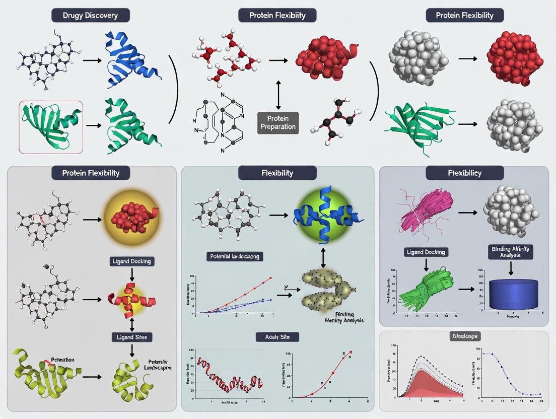
Beyond Rigid Docking: Mastering Protein Flexibility with Deep Learning and Ensemble Methods
Molecular docking, a cornerstone of structure-based drug design, has long been hampered by the challenge of protein flexibility.
Beyond Rigid Docking: Mastering Protein Flexibility with Deep Learning and Ensemble Methods
Abstract
Molecular docking, a cornerstone of structure-based drug design, has long been hampered by the challenge of protein flexibility. Traditional rigid docking methods offer incomplete representations of biological reality, often failing to predict accurate binding modes. This article provides a comprehensive overview for researchers and drug development professionals on the critical evolution toward flexible docking. We explore the foundational concepts of induced fit and conformational selection, detail the latest methodological advances including deep learning diffusion models and ensemble docking, and offer a comparative analysis of their performance in pose prediction, physical plausibility, and virtual screening. Finally, we present troubleshooting strategies for common pitfalls and discuss future directions, highlighting how integrating flexibility is transforming computational predictions into biomedical breakthroughs.
The Protein Flexibility Imperative: From Lock-and-Key to Induced Fit
FAQs: Understanding Rigid Body Docking and Its Challenges
FAQ 1: What is the fundamental limitation of traditional rigid body docking? The core limitation is the treatment of proteins as static, unmoving structures. In reality, proteins are dynamic, and their side chains, loops, and sometimes even secondary structures shift and move upon binding. Rigid body docking, which uses Fast Fourier Transform (FFT) algorithms for computational efficiency, cannot account for these conformational changes. This "rigid body assumption" introduces clear limitations on accuracy and reliability [1].
FAQ 2: What specific errors can this limitation cause in my results? This limitation can lead to several common issues:
- Scoring Failures: Docked conformations close to the native structure may not have the lowest energies, while incorrect, low-energy conformations are ranked highly [1].
- Pose Prediction Inaccuracy: The method's reduced sensitivity to shape complementarity can result in the failure to identify the correct binding pose, especially when side chains or backbone atoms must rearrange to accommodate the ligand [2].
- Cross-Docking Failures: A protein structure crystallized with one ligand may have an active site biased toward that specific molecule. Attempting to dock a different ligand into this rigid structure often fails if critical conformational shifts are not accounted for [2].
FAQ 3: My docking run completed, but the top-ranked pose looks wrong. What should I do? This is a classic symptom of scoring failure due to rigidity. Your next steps should be:
- Do not trust a single top pose. Rigid body methods must retain a large set of low-energy docked structures for further processing. Always analyze multiple top-ranked clusters, not just the first one [1].
- Inspect the interface. Check for steric clashes that a slightly flexible side chain could resolve. Poor electrostatic or desolvation energy terms at the interface can also explain why a correct pose was poorly ranked [1].
- Move to refinement. Use a method that incorporates flexibility—even if just for side chains—to refine the top clusters from your rigid body docking experiment.
FAQ 4: Are there specific types of complexes where rigid docking is known to fail? Yes. Performance is strongly linked to the conformational change between the unbound and bound states.
- Easy: Complexes with minimal backbone and side-chain movement (151 of 230 in the BM5 benchmark) [1].
- Medium/Difficult: Complexes involving enzymes, antibodies, and other proteins that undergo "induced fit" upon binding. The performance drops significantly for these 79 targets in the BM5 benchmark [1].
Troubleshooting Guide: Addressing Rigid Docking Failures
Problem: Inaccurate Poses Due to Protein Flexibility
Symptoms:
- High root-mean-square deviation (RMSD) of the ligand pose compared to a known experimental structure.
- Obvious steric clashes at the binding interface in the top-ranked models.
- Failure to recover key protein-ligand interactions (e.g., hydrogen bonds, hydrophobic contacts).
Solutions: 1. Employ Flexible Refinement Protocols
- Procedure: Use the output from a fast, rigid-body global docking server (like ClusPro) as the input for a more computationally intensive, flexible local refinement tool. This hybrid approach first identifies a broad region of the protein surface that might be the binding site and then allows for side-chain or limited backbone movement to optimize the fit.
- Rationale: This combines the exhaustive sampling speed of FFT-based docking with the accuracy of methods that can model induced fit [1].
2. Utilize Ensemble Docking
- Procedure: Instead of a single rigid protein structure, dock your ligand against an ensemble of multiple receptor conformations. This ensemble can be derived from:
- Multiple crystal structures of the same protein.
- NMR models.
- Snapshots from a Molecular Dynamics (MD) simulation [2].
- Rationale: This technique accounts for protein flexibility by allowing the ligand to select its optimal binding partner from a set of pre-generated conformations, a mechanism known as "conformational selection" [2].
3. Leverage Alignment-Based Docking
- Procedure: If a structure of your target protein bound to a similar ligand is available, use a method like CSAlign-Dock. This aligns your new ligand to the reference ligand within the binding pocket, considering full ligand flexibility, to predict the new complex structure [3].
- Rationale: This bypasses the ab initio sampling problem by using the known protein conformation from a related complex as a template, which can be superior to rigid-body docking, especially in cross-docking scenarios [3].
Problem: Physically Implausible or Clashing Poses
Symptoms:
- Ligand atoms intersecting with protein atoms in the predicted binding pose.
- Distorted ligand geometry (e.g., abnormal bond lengths or angles).
Solutions: 1. Check and Optimize Ligand Preparation
- Procedure:
- Ensure your ligand is in the correct PDBQT format, which includes atomic partial charges and atom types [4].
- Before docking, perform energy minimization of the ligand structure to resolve any unrealistic geometry from source libraries [5].
- Carefully define rotatable bonds. Lock bonds in rings or double bonds to preserve critical chemical geometry [5].
- Rationale: Many docking failures originate from poorly prepared input files. A chemically sensible and well-minimized ligand is crucial for successful pose prediction.
2. Evaluate the Scoring Function
- Procedure: Be aware that different scoring functions have varying tolerances for steric clashes. Some modern deep learning methods, for example, may generate poses with good RMSD but high steric clash [6]. Use a tool like PoseBusters to check the physical validity of your output poses [6].
- Rationale: A pose with a favorable score but obvious clashes is likely a scoring artifact and not a biologically relevant model.
Performance Data: Rigid Body Docking Success Rates
The following table summarizes the performance of a leading rigid-body docking server (ClusPro) on a standard benchmark (BM5), categorized by the difficulty level of the complex [1].
Table 1: Performance of Rigid Body Docking Across Complex Types
| Complex Category | Number of Targets | DockQ Score Range | CAPRI Accuracy Rating |
|---|---|---|---|
| Rigid-Body (Easy) | 151 | > 0.49 | Medium to High |
| Medium Difficulty | 45 | 0.23 - 0.49 | Acceptable to Medium |
| Difficult | 34 | < 0.23 | Incorrect |
The table below compares the general performance characteristics of different docking methodologies, highlighting the trade-offs involved.
Table 2: Comparison of Docking Methodologies
| Methodology | Typical Pose Accuracy* | Key Strength | Key Limitation |
|---|---|---|---|
| Traditional Rigid-Body | ~50-75% [2] | Computational speed, global sampling | Cannot handle protein flexibility |
| Fully Flexible Docking | ~80-95% [2] | High accuracy for induced fit | Computationally expensive |
| Deep Learning (Generative) | ~75-90% RMSD ≤ 2Å [6] | High pose accuracy, speed | May produce physically invalid poses [6] |
| Hybrid (AI + Search) | Balanced performance [6] | Good balance of accuracy and physical validity | Search efficiency can be an issue [6] |
*Pose accuracy rates are highly dependent on the specific target and benchmark used.
Experimental Workflow: From Rigid Body Docking to Refined Models
The following diagram illustrates a robust experimental strategy that uses rigid-body docking as a starting point and incorporates methods to overcome its limitations.
The Scientist's Toolkit: Key Research Reagents and Computational Tools
Table 3: Essential Resources for Advanced Docking Studies
| Tool / Resource | Type | Primary Function | Relevance to Flexibility |
|---|---|---|---|
| ClusPro Server [1] | Rigid-Body Docking Server | FFT-based global sampling and clustering. | Provides a fast starting point; top clusters are inputs for flexible refinement. |
| AutoDock Vina [6] | Docking Software | Traditional physics-based docking with stochastic search. | Widely used; a standard for comparative studies. |
| Molecular Dynamics (MD) [7] | Simulation Software | Simulates physical movements of atoms over time. | Generates ensembles of protein conformations for ensemble docking. |
| CSAlign-Dock [3] | Alignment-Based Docking | Docks a ligand using a known reference complex. | Accounts for protein conformational changes by leveraging template structures. |
| PoseBusters [6] | Validation Toolkit | Checks docking poses for physical and chemical plausibility. | Critical for identifying failures, especially from AI models that may ignore steric clashes. |
| Rosetta Software Suite [8] | Modeling Suite | Provides flexible backbone and high-resolution refinement protocols. | Used to model and analyze flexible regions in protein structures and assemblies. |
For decades, the understanding of molecular binding was dominated by the rigid "lock and key" model. However, advanced structural biology has revealed that proteins are highly dynamic macromolecules. This dynamism is crucial for function and is described by two primary, and often complementary, binding mechanisms: Induced Fit and Conformational Selection [2] [9].
Traditionally, these mechanisms were viewed as mutually exclusive. Induced Fit (IF) proposes that a ligand first binds to the protein's predominant state, inducing a conformational change to a stable bound complex. In contrast, Conformational Selection (CS) posits that the protein exists in an equilibrium of multiple conformations, and the ligand selectively binds to and stabilizes a pre-existing, minor population [10] [11]. Modern research, supported by binding flux analysis and advanced kinetics, now recognizes that IF and CS are not a strict dichotomy but can operate alongside each other within a thermodynamic cycle to produce the final ligand-target complex [10].
Understanding which mechanism dominates is critical in drug discovery, as it influences the selectivity, duration of action, and residence time of a drug on its target [10] [12]. This guide provides troubleshooting support for researchers grappling with the practical challenges of distinguishing these mechanisms within molecular docking studies.
Conceptual Framework & Key Differences
The core challenge is to correctly identify the temporal order of binding and conformational change. The following diagram illustrates the pathways and their interplay within a thermodynamic cycle.
Quantitative Comparison of Mechanisms
The table below summarizes the key characteristics that experimentally distinguish these mechanisms.
| Feature | Induced Fit (IF) | Conformational Selection (CS) |
|---|---|---|
| Temporal Order | Conformational change occurs after initial ligand binding [11]. | Conformational change occurs before ligand binding [11]. |
| Key Intermediate | A transient, initial encounter complex (P:L) [10]. | A pre-existing, excited protein state (P*) [10]. |
| Dominance at Low [Ligand] | Lower contribution; increases with ligand concentration [10]. | Typically dominates at low ligand concentrations [10]. |
| Observed Rate (kₒₑₛ) vs. [L] | Symmetric U-shape: kₒₑₛ has a minimum and is symmetric around [L]₀ = [P]₀ + K𝒹 [11]. | Asymmetric or monotonic: kₒₑₛ decreases monotonically for kₑ < k₋; has an asymmetric minimum for kₑ > k₋ [11]. |
| Ligand Specificity | Binds a broader population, inducing the "correct" fit. | Highly selective for a specific, pre-formed conformation. |
| Role in Drug Design | Often associated with achieving a long residence time on the target [10]. | Can be exploited to target specific, potentially inactive, protein states [12]. |
Successful experimental analysis requires a suite of specialized reagents and computational tools.
| Tool / Reagent | Function / Description | Relevance to Binding Mechanisms |
|---|---|---|
| Site-Directed Spin Labeling (SDSL) | Covalent attachment of spin labels (e.g., MTSSL) to engineered cysteine residues [12]. | Enables EPR distance measurements to probe conformational states and dynamics. |
| p38α MAP Kinase Constructs | Panel of double-cysteine mutants for distance mapping (e.g., p38α-119, 251) [12]. | Model system for studying A-loop conformational equilibrium (DFG-in/out). |
| Type I & II Kinase Inhibitors | Small molecules that bind distinct kinase conformations (e.g., SB203580, Sorafenib) [12]. | Tool compounds to selectively stabilize specific sub-states (CS vs. IF). |
| MMM Software Toolbox | Multiscale Modeling of Macromolecules for spin label multilateration [12]. | Converts EPR distance data into 3D probabilistic maps of flexible regions. |
| Structural Alphabets (SAs) | Libraries of small protein fragments for precise backbone conformation analysis [9]. | Analyzes backbone deformability and conformational changes from structural data. |
| Normal Modes Analysis (NMA) | Computational method to calculate a protein's collective motions [13]. | Predicts low-energy conformational changes for ensemble generation in docking. |
Troubleshooting FAQs & Experimental Guides
FAQ 1: How can I determine if my ligand binds via Induced Fit or Conformational Selection?
Answer: Distinguishing the mechanism requires a combination of kinetic and structural experiments. A critical first step is to analyze the chemical relaxation rate (kₒₑₛ) as a function of both ligand and protein concentration.
Problem: Under pseudo-first-order conditions (high ligand concentration), an increase in kₒₑₛ with [L] can be misinterpreted, as it is possible in both IF and CS mechanisms [11].
Solution: Perform relaxation experiments (e.g., temperature jump) across a wide range of ligand and protein concentrations. Plot kₒₑₛ versus the total ligand concentration [L]₀.
- If the plot is symmetric (see Diagram A below), the mechanism is Induced Fit.
- If the plot is asymmetric or shows a monotonic decrease (see Diagram B below), the mechanism is Conformational Selection [11].
This general method works for all concentrations and avoids the ambiguity of pseudo-first-order approximations.
Protocol: Chemical Relaxation Kinetics
- Prepare Solutions: Prepare a series of samples with a fixed total protein concentration ([P]₀) and varying total ligand concentrations ([L]₀). Ensure [L]₀ covers a range both below and above [P]₀.
- Perturb Equilibrium: Rapidly perturb the equilibrium of each sample using a fast technique such as a temperature or pressure jump.
- Monitor Relaxation: Use a stopped-flow apparatus or a similar fast-detection method to monitor the system's relaxation back to the new equilibrium. A spectroscopic signal (e.g., fluorescence) that reports on binding is typically used.
- Fit Data: Fit the relaxation trajectory to a single or multi-exponential function to extract the dominant relaxation rate, kₒₑₛ.
- Plot and Analyze: Plot kₒₑₛ as a function of [L]₀ for different fixed values of [P]₀. Analyze the shape of the curves as described above [11].
FAQ 2: My docking simulations are failing. How can I account for protein flexibility in my model?
Problem: Standard rigid-body docking fails when the protein's binding site undergoes conformational changes upon ligand binding. This can result in low docking scores for true binders and an inability to predict the correct binding pose [2] [13].
Solution: Implement a flexible docking strategy that moves beyond a single, static protein structure. The general workflow is outlined below.
Protocol: Flexible Docking Workflow
- Preprocessing & Ensemble Generation:
- Source: Collect multiple experimental structures (X-ray, NMR) of the target protein in different conformational states (e.g., apo, holo, with different ligands) [13].
- Generate: Use computational methods like Molecular Dynamics (MD) simulations or Normal Modes Analysis (NMA) to generate an ensemble of plausible conformations [13]. This ensemble simulates the Conformational Selection model.
- Cross-Docking: Perform rigid-body docking of your ligand against each conformation in the generated ensemble.
- Refinement (Induced Fit): Take the top poses from the cross-docking step and subject them to a refinement stage. This stage allows for small-scale movements of the protein backbone and side-chains, as well as rigid-body adjustments of the ligand, to optimize the fit. This step models the Induced Fit mechanism [13].
- Scoring: Re-score the refined complexes using a scoring function that accounts for the energy cost of protein deformation. This helps to identify the most biologically plausible complex [13].
FAQ 3: How can I directly observe the conformational states of a flexible protein region?
Problem: Highly flexible regions, like the activation loop in kinases, are often poorly resolved or missing in X-ray crystal structures, making it difficult to characterize their conformational landscape [12].
Solution: Employ Electron Paramagnetic Resonance (EPR) spectroscopy with site-directed spin labeling to measure distances and probe conformational distributions directly.
Protocol: EPR with SLiK (Spin Labels in Kinases)
- Design Double Mutants: Generate a panel of protein constructs (e.g., p38α MAPK) with two cysteine mutations each. One label is placed in the flexible region of interest (e.g., the A-loop at residue 172), and the other is placed in a stable, rigid reference region [12].
- Spin Labeling: Label the cysteine residues with a stable spin label like MTSSL.
- DEER/PELDOR Measurements: Perform pulsed EPR distance measurements (DEER/PELDOR) on the apo protein and in the presence of saturating ligand concentrations. This technique yields distance distributions between the two spin labels.
- Data Analysis:
- Apo State: A broad, multimodal distance distribution indicates a dynamic equilibrium between multiple conformational sub-states of the flexible loop.
- Ligand-Bound State: A shift to a narrow, unimodal distribution indicates that the ligand has selected and stabilized a specific sub-state (Conformational Selection). A change in the distribution shape not present in the apo state may suggest an Induced Fit mechanism [12].
- Multilateration: Use software like the MMM toolbox to combine distance distributions from multiple double mutants and compute a 3D probability map for the position of the flexible loop, providing a spatial model of its conformational ensemble [12].
Molecular docking techniques are essential for predicting how a small molecule (ligand) interacts with a biological target. The table below summarizes the key techniques for handling protein flexibility [14] [15].
Table 1: Key Molecular Docking Techniques for Handling Protein Flexibility
| Technique Name | Primary Objective | Key Advantage | Consideration for Protein Flexibility |
|---|---|---|---|
| Re-docking [14] | Validate docking protocol accuracy by re-docking a known ligand. | Provides a straightforward control to test computational settings. | Treats the protein as a rigid body; sensitive to minor conformational changes from the original crystal structure. |
| Cross-docking [14] | Test a docking protocol's ability to handle different ligands by docking multiple ligands into a single protein structure. | Assesses the robustness of a chosen protein conformation for docking diverse compounds. | Uses a single, rigid protein conformation; may fail if ligands induce different conformational changes. |
| Ensemble Docking [14] | Account for inherent protein flexibility by docking against multiple protein conformations. | Provides a more realistic representation of ligand binding by sampling different protein states. | Explicitly incorporates protein flexibility by using an ensemble of structures (e.g., from MD simulations or multiple crystals). |
| Blind Docking [14] | Identify novel binding sites on a protein without prior knowledge of their location. | Unbiased exploration of the entire protein surface. | Scans a rigid protein structure; can identify alternative binding pockets but may miss induced-fit effects. |
Frequently Asked Questions: Technique Selection & Results
Q: How do I validate my molecular docking protocol? A: Re-docking is the primary method for validation [14]. The co-crystallized ligand is extracted and re-docked into its original binding site. The predicted pose is compared to the experimental one, typically by calculating the Root-Mean-Square Deviation (RMSD). An RMSD value below 2.0 Å is generally considered a successful prediction, indicating your protocol can reproduce the known binding mode [14].
Q: My re-docking worked well, but cross-docking fails for ligands with different scaffolds. Why? A: This is a common challenge rooted in protein flexibility [14] [15]. A single, rigid protein structure used in cross-docking may be optimized for its native ligand but not accommodate others that induce different conformational changes. To address this, consider using ensemble docking, which uses multiple protein structures to account for flexibility [14].
Q: What is the difference between AutoDock Vina and AutoDock 4? A: While both come from the same lab, AutoDock Vina is a new "generation" with a completely new scoring function and search algorithm [16]. On average, Vina offers better speed and accuracy, though the best program can be target-dependent [16].
Q: Why are my docking results non-deterministic (different each time)? A: The docking algorithm in tools like AutoDock Vina is a stochastic (random) global optimization process [16]. Even with identical inputs, starting from different random seeds can lead to different results. It is good practice to perform multiple runs and analyze the statistical properties of the outcomes [16].
Troubleshooting Common Docking Problems
Table 2: Troubleshooting Guide for Molecular Docking Experiments
| Problem | Possible Cause | Solution |
|---|---|---|
| High RMSD in Re-docking | Incorrect protonation states of ligand or receptor [16]. | Check and correct the protonation states of key residues and the ligand for the physiological pH of interest. |
| The search space is defined incorrectly [16]. | Ensure the search space center and size are correct. Remember, in AutoDock Vina, size is in Ångstroms, not grid points [16]. | |
| Poor Cross-docking Performance | The chosen rigid protein structure cannot accommodate the new ligand due to induced fit [14]. | Switch to ensemble docking using multiple protein conformations to account for flexibility [14] [15]. |
| Inaccurate Binding Energy Prediction | The scoring function is inexact and has inherent limitations [16]. | Use docking scores for relative ranking, not absolute binding energy prediction. Correlate results with experimental data. |
| Docked conformation is unreasonable | Ligand or receptor was not prepared correctly (e.g., 2D ligand input, missing atoms) [16]. | Ensure proper 3D ligand geometry and that all missing side chains/atoms in the receptor have been modeled. |
| Warning about large search space volume | The defined search space is very large (e.g., >27,000 ų) [16]. | Reduce the search space size if possible. If a large space is necessary, increase the exhaustiveness parameter to improve the search [16]. |
Experimental Protocols
Protocol 1: Standard Re-docking Validation
This protocol is used to validate a molecular docking setup by reproducing a known experimental result [14].
- Obtain the Structure: Download a protein-ligand complex structure from the Protein Data Bank (PDB).
- Prepare Structures:
- Protein: Remove the ligand, all water molecules, and any irrelevant cofactors. Add hydrogen atoms and assign partial charges using your chosen molecular graphics or docking software.
- Ligand: Extract the bound, co-crystallized ligand from the PDB file. Ensure it is correctly protonated for the relevant pH.
- Define the Search Space: Center the docking search box on the original ligand's binding site. A typical box size is 20x20x20 Å to give the ligand some room to move.
- Perform Docking: Dock the prepared ligand into the prepared protein structure using your selected docking program.
- Analyze Results: Calculate the RMSD between the top-ranked docked pose and the original co-crystallized ligand pose. An RMSD of less than 2.0 Å is considered a successful validation [14].
Protocol 2: Ensemble Docking for Flexible Receptors
This protocol is used when protein flexibility is a major concern, such as when docking against a protein with known multiple conformations or when cross-docking fails [14] [15].
- Assemble the Ensemble: Collect multiple structures of the target protein. These can come from:
- Multiple PDB entries of the same protein (e.g., with different ligands bound).
- A single PDB entry, using models from an NMR ensemble.
- Molecular Dynamics (MD) Simulations: Snapshots from an MD simulation trajectory, such as those available from the ATLAS database, provide a robust set of conformations [17].
- Prepare Structures: Prepare each protein conformation in the ensemble as in Protocol 1.
- Dock: Perform a separate docking run for each ligand against every protein conformation in the ensemble.
- Analyze Results: Combine the results from all docking runs. The best-scoring pose across the entire ensemble is typically selected as the final prediction, providing a model that accounts for protein flexibility.
Workflow Visualization
The Scientist's Toolkit
Table 3: Essential Research Reagents & Computational Tools
| Item / Resource | Function / Description | Relevance to Docking Experiment |
|---|---|---|
| Protein Data Bank (PDB) | A repository for 3D structural data of proteins and nucleic acids. | The primary source for initial protein and protein-ligand complex structures for re-docking and cross-docking studies. |
| AutoDock Vina | A widely used molecular docking program for predicting ligand binding modes and affinities. | The core engine for performing the docking simulation itself. Known for its speed and user-friendliness [16]. |
| ATLAS Database | A database of standardized all-atom molecular dynamics (MD) simulations for a representative set of proteins [17]. | A key source for obtaining multiple protein conformations (an ensemble) to use in ensemble docking, directly addressing protein flexibility [17]. |
| PDBQT File Format | The required input file format for AutoDock Vina. Contains atomic coordinates, partial charges, and atom types. | The prepared protein and ligand files must be in this format. Preparation typically involves adding polar hydrogens and assigning atom types. |
| Molecular Graphics System (e.g., PyMOL) | Software for visualizing molecular structures, surfaces, and docking results. | Critical for analyzing input structures, defining docking search boxes, and visually inspecting the final docked poses. |
| Molecular Dynamics (MD) Software (e.g., GROMACS) | Software for simulating the physical movements of atoms and molecules over time. | Used to generate alternative protein conformations for ensemble docking, capturing the dynamic behavior of the protein in solution [17]. |
FAQs: Understanding Protein Flexibility in Drug Discovery
Q1: Why is considering protein flexibility so critical in virtual screening?
Accounting for protein flexibility is crucial because using a single, rigid protein structure provides an incomplete representation of its native state. Experimental studies clearly show conformational differences between a protein's unbound (apo) and bound (holo) states [2]. When a rigid receptor structure is used for docking ligands that require different binding site geometries (a problem known as cross-docking), the active site is often biased toward its original ligand, leading to failed docking attempts and missed hits [2]. Typical rigid docking shows best performance rates between only 50% and 75%, while methods incorporating full protein flexibility can enhance pose prediction accuracy to 80–95% [2].
Q2: What are the fundamental mechanisms of protein flexibility upon ligand binding?
Two primary models explain the conformational changes:
- Induced Fit: The ligand binding event itself induces a conformational change in the protein [2].
- Conformational Selection: The protein exists in an equilibrium of multiple conformations in solution. The ligand selectively binds to and stabilizes a pre-existing complementary conformation, shifting the population distribution [2]. Research suggests these are not mutually exclusive, and a mixed binding mechanism is likely for many systems [2].
Q3: What are the main technical challenges in implementing flexible docking?
The primary challenge is the immense computational cost associated with the large number of degrees of freedom a protein possesses. Directly modeling binding site flexibility is difficult due to the vast conformational space that must be sampled and the difficulties in formulating a perfectly accurate energy function to score these conformations [18]. This creates a trade-off between computational efficiency and biological accuracy that researchers must navigate.
Q4: How does structural simplification in lead optimization relate to protein flexibility?
Structural simplification is a lead optimization strategy that reduces molecular complexity and "molecular obesity" by removing unnecessary rings or chiral centers, often improving pharmacokinetic properties [19]. A simplified, more rigid ligand may have fewer degrees of freedom to accommodate, potentially reducing the conformational demands on the protein. However, the simplified ligand must still retain the key pharmacophores necessary for binding to the flexible protein target [19].
Troubleshooting Common Experimental Issues
Problem 1: Poor Pose Prediction and Enrichment in Virtual Screening
- Symptoms: Docked ligand poses have high Root-Mean-Square Deviation (RMSD) from known crystallographic poses; inability to distinguish true binders from decoys (poor enrichment).
- Solutions:
- Use Ensemble Docking: Instead of a single structure, dock into an ensemble of protein conformations. This ensemble can be built from multiple experimental structures (e.g., from the PDB), conformations from molecular dynamics simulations, or homology models [2] [20]. Tools like FlexE create a "united protein description" from a structural ensemble, allowing combinatorial joining of different conformations during docking [21].
- Employ Advanced Docking Software: Utilize modern docking programs that explicitly model protein flexibility. For example, the RosettaVS protocol incorporates full receptor side-chain flexibility and limited backbone movement, which has been shown to be critical for accurate predictions on standard benchmarks [22].
- Validate with Cross-Docking: Test your docking protocol by cross-docking various known ligands from different crystal structures into a single receptor structure. If performance is poor, it indicates a strong need for a flexible docking approach [2].
Problem 2: Inaccurate Binding Affinity Predictions Due to Rigid Receptors
- Symptoms: Scoring functions fail to correctly rank compounds by their experimental binding affinity, even when pose prediction is reasonable.
- Solutions:
- Incorporate Entropic Considerations: Standard scoring functions often fail to adequately estimate entropy changes (∆S) upon binding. Advanced methods, like the improved RosettaGenFF-VS, combine enthalpy calculations with an entropy model, leading to superior performance in affinity ranking [22].
- Model Key Water Molecules: The presence and displacement of active site water molecules significantly impact binding affinity. Use algorithms that can classify water molecules and simulate their contribution to multi-part interactions. Decide on a strategy for handling displaceable water molecules during the docking simulation [20].
Problem 3: High Computational Cost of Flexible Docking
- Symptoms: Screening a large compound library is prohibitively slow or computationally infeasible.
- Solutions:
- Adopt a Hierarchical Screening Protocol: Use a fast, initial screening mode to filter millions of compounds, followed by a high-precision mode for top hits. For instance, RosettaVS offers a Virtual Screening Express (VSX) mode for rapid screening and a Virtual Screening High-precision (VSH) mode for final ranking [22].
- Leverage Active Learning: Integrate your workflow with an AI-accelerated platform that uses active learning. This involves training a target-specific neural network during docking to intelligently select the most promising compounds for full, expensive docking calculations, drastically reducing the number of required simulations [22].
- Focus Flexibility: Restrict flexibility considerations to key binding site residues identified through prior knowledge or computational analysis, rather than treating the entire protein as flexible.
Performance Data for Flexible Docking Methods
The tables below summarize quantitative performance data from evaluations of flexible docking methods, providing a benchmark for expectations.
Table 1: Virtual Screening Performance on the DUD Dataset
| Method / Metric | AUC (Area Under Curve) | ROC Enrichment | Notes |
|---|---|---|---|
| RosettaVS (VSH Mode) | State-of-the-art | State-of-the-art | Incorporates receptor flexibility and an entropy model [22]. |
| Other Physics-Based Methods | Variable, generally lower | Variable, generally lower | Performance depends on the specific method and target [22]. |
Table 2: Pose Prediction Accuracy of FlexE on 105 PDB Structures
| Performance Metric | Success Rate | Threshold |
|---|---|---|
| Overall Placement Success | 83% (50/60 ligands) | RMSD < 2.0 Å [21] |
| Comparison to Rigid Cross-Docking | Similar quality to best single-structure result | - [21] |
Experimental Protocols
Protocol 1: Ensemble-Based Flexible Docking with FlexE
Objective: To dock a flexible ligand into a protein binding site that exhibits structural variations. Materials: Protein structure ensemble (e.g., from PDB or MD simulation), ligand structure, FlexE software. Methodology:
- Prepare the Ensemble: Superimpose all protein structures in the ensemble onto a common reference frame.
- Generate United Protein Description: FlexE internally processes the superimposed structures, merging similar regions and treating dissimilar areas (e.g., different side-chain rotamers or loop conformations) as separate, discrete alternatives [21].
- Define Incompatibilities: The software builds an incompatibility graph to manage combinations of alternatives that are geometrically or logically exclusive.
- Dock the Ligand: Using an incremental construction algorithm (derived from FlexX), FlexE places the flexible ligand into the united protein description. During placement, it selects the optimal combination of protein conformations that best fits the ligand, as determined by the scoring function [21].
- Analyze Results: Examine the top-ranked poses and the specific protein conformation selected for each.
Protocol 2: AI-Accelerated Virtual Screening with the OpenVS Platform
Objective: To efficiently screen an ultra-large chemical library (billions of compounds) against a flexible target. Materials: Target protein structure(s), multi-billion compound library (e.g., in SDF format), OpenVS platform, HPC cluster. Methodology:
- Target Preparation: Prepare the protein structure, specifying the binding site and allowing for side-chain and limited backbone flexibility in the protocol [22].
- Library Preparation: Curate the compound library, generating relevant tautomers, protomers, and stereoisomers.
- Configure Active Learning: Set up the active learning loop where a neural network is continuously trained on the fly to predict the docking scores of unscreened compounds based on those already docked [22].
- Run Hierarchical Docking:
- Stage 1 (VSX): The platform uses the RosettaVS express mode to rapidly screen compounds prioritized by the active learning model.
- Stage 2 (VSH): The top hits from VSX are re-docked using the high-precision mode of RosettaVS, which includes full receptor flexibility, for final ranking [22].
- Post-Screening Analysis: Chemically cluster the final hit-list, examine key interaction patterns, and select compounds for experimental testing.
Workflow Visualization
AI-Accelerated Flexible Docking Workflow
The Scientist's Toolkit: Essential Research Reagents & Software
Table 3: Key Resources for Flexible Docking Research
| Tool / Resource | Type | Primary Function in Flexible Docking |
|---|---|---|
| FlexE | Software | Docks flexible ligands into an ensemble of protein structures by creating a united protein description with combinatorial conformations [21]. |
| RosettaVS | Software Suite | A state-of-the-art physics-based docking protocol that allows for full side-chain and limited backbone flexibility during virtual screening [22]. |
| Protein Data Bank (PDB) | Database | Source for experimentally determined protein structures to build conformational ensembles for docking [2] [18]. |
| ZINC / PubChem | Database | Public repositories of purchasable and virtual compounds for building screening libraries [18]. |
| Homology Models | Computational Model | Provides a 3D protein model when an experimental structure is unavailable, though flexibility considerations become even more critical [23] [20]. |
| Molecular Dynamics (MD) | Simulation Method | Generates an ensemble of protein conformations through simulation of physical movements, useful for capturing flexibility beyond crystal structures [20]. |
A Practical Toolkit: From Ensemble Docking to Deep Learning Co-folding
Traditional molecular docking often treats the protein target as a rigid structure, which is an incomplete representation of reality. Experimental studies have clearly demonstrated conformational differences between a receptor's unbound (apo) and bound (holo) states [2]. When docking is performed against a single, rigid protein structure, the results can be biased toward the specific ligand that was co-crystallized, a problem known as the cross-docking problem [2]. This can lead to high rates of false positives and false negatives in virtual screening.
Ensemble docking addresses this limitation by using multiple protein conformations to represent the dynamic, flexible nature of the target. This approach is grounded in the conformational selection model of ligand binding, where the ligand selects its preferred binding partner from an ensemble of available protein states [24] [2]. By docking candidate ligands into a diverse set of protein conformations, researchers can more accurately model the biologically relevant binding process and improve the prediction of binding modes and affinities.
An effective ensemble docking study relies on a representative set of protein conformations. The two primary sources for these structures are experimental data and computer simulations.
Experimental Structures from the Protein Data Bank (PDB)
For many pharmaceutically relevant targets, the PDB contains numerous X-ray structures solved in complex with different ligands.
- Process: Curate all available structures for your target, then remove redundant conformations to create a manageable and diverse set.
- Advantage: Experimentally determined structures provide atomic-level detail of physically realistic conformations.
- Challenge: The pattern of disordered segments can differ significantly between structures, especially when comparing apo and holo forms or structures with and without binding partners like cyclin [25].
A graph-based redundancy removal method has been shown to be more efficient and less subjective for selecting representative structures than traditional clustering-based methods [25].
Computational Conformations from Molecular Dynamics (MD)
Molecular Dynamics simulations generate a trajectory of protein movement by simulating its physical motions over time.
- Process: After running an MD simulation (e.g., 4 ns after equilibration), the trajectory is clustered based on the Root Mean Square Deviation (RMSD) of atoms in the binding site. Representative snapshots (medoids) from each cluster form the docking ensemble [26] [27].
- Advantage: MD can sample conformational states that are not captured by crystallography but may be biologically relevant for ligand binding.
- Consideration: The sampling may be limited by the timescale of the simulation, as slow conformational changes can occur over timescales beyond what is typically practical to simulate [24].
Table: Comparison of Methods for Generating Protein Conformational Ensembles
| Method | Key Features | Advantages | Limitations |
|---|---|---|---|
| Experimental PDB Structures | Uses multiple X-ray or NMR structures from the PDB. | High-resolution, experimentally validated conformations. | May be biased toward specific ligand-bound states; limited conformational diversity. |
| Molecular Dynamics (MD) | Computational simulation of protein movement; snapshots are clustered. | Can discover novel, druggable states not seen in crystals [24]. | Computationally expensive; force field inaccuracies; limited sampling of slow motions. |
The following diagram illustrates a typical workflow for creating and using an ensemble from Molecular Dynamics simulations:
Troubleshooting Common Issues in Ensemble Docking
FAQ: How many protein conformations should I use in my ensemble?
There is a trade-off between computational cost and accuracy. Using more conformations can better represent flexibility but increases cost and the risk of false-positive pose predictions [25]. Machine learning can help select the most important conformations. For example, one study on CDK2 showed that a few of the most important conformations were sufficient to achieve high accuracy in affinity prediction, greatly reducing the necessary ensemble size [25]. When using MD, studies suggest that 6-8 clusters can be sufficient to make an ensemble, though some protocols use more (e.g., 20) for broader sampling [26] [27].
FAQ: My ensemble docking results are poor, with incorrect poses and zero energies. What went wrong?
This specific error, where results show only one structure per cluster and zero energies, was reported in a HADDOCK forum. The solution was to check the residue numbering in the input files. If the residue numbering in your ensemble PDB files does not match the numbering used in your restraint definitions, the docking calculation will fail because the restraints are not applied correctly [28]. Always verify the consistency of your input files.
FAQ: How do I choose the best docking program for my target?
The performance of docking programs and scoring functions can be highly target-dependent [25]. A general benchmarking study found that AutoDock Vina tends to reproduce more accurate binding poses, while AutoDock4 gives binding affinities that correlate better with experimental values [25]. However, the authors emphasize that for a specific target, a receptor-specific benchmarking is desirable to decide on the best tool. If possible, test multiple programs against a set of known actives and decoys for your target.
FAQ: Can ensemble docking handle large conformational changes like "DFG-flip" in kinases?
Yes, this is a key strength of the method. Kinases are a classic example where the DFG-loop can adopt at least two distinct conformations (DFG-in and DFG-out) depending on the bound inhibitor. If a rigid docking protocol uses a DFG-in structure, it will fail to correctly dock a compound that requires the DFG-out conformation. Ensemble docking that includes both states can successfully handle such cases by providing the correct protein conformation for each ligand type [27].
Enhancing Ensemble Docking with Machine Learning
A powerful advancement is combining ensemble docking with machine learning (ML) to improve the prediction of drug binding. The process generally involves:
- Feature Generation: Perform ensemble docking to generate a set of features for each ligand, such as individual docking scores and energy terms from multiple protein conformations [26].
- Model Training: Use these features, along with labels identifying active versus decoy compounds, to train a machine learning classifier (e.g., Random Forest or K-Nearest Neighbors) [26] [25].
- Conformation Selection: ML can rank the importance of different protein conformations in the ensemble for correctly classifying actives. This identifies a small subset of the most informative structures [25].
- Prediction: The trained ML model is used to predict new active compounds, often with much higher accuracy than using docking scores alone. One study reported over 99% classification accuracy when using features from a well-correlated protein conformation [26].
This integrated approach tackles the "optimum ensemble size" problem by identifying a minimal set of critical conformations, reducing computational cost while maintaining, or even improving, predictive accuracy [25].
Table: Key Research Reagents and Software Solutions
| Tool / Reagent | Type | Primary Function in Ensemble Docking |
|---|---|---|
| AutoDock Vina [26] | Docking Software | Performs the core docking calculation, scoring ligand poses for a given protein conformation. |
| AMBER14ffsb [27] | Force Field | Provides parameters for atoms during Molecular Dynamics simulations to generate ensembles. |
| Lead Finder [27] | Docking Software | Docking algorithm used in the Flare software for pose generation and scoring. |
| Scikit-learn [26] | Machine Learning Library | Provides algorithms (e.g., Random Forest) for analyzing docking results and classifying active compounds. |
| Dragon Software [26] | Descriptor Calculator | Calculates molecular descriptors for drugs, which can be used as features in machine learning models. |
| Directory of Useful Decoys (DUD-e) [26] | Database | Provides known active and decoy compounds for a target, essential for training and validating models. |
The relationship between ensemble docking and machine learning can be summarized in the following workflow, which leads to improved prediction of drug binding:
Molecular docking is a cornerstone of modern, structure-based drug design. A significant challenge in this field is accounting for the inherent flexibility of protein targets, as side-chain or even backbone adjustments frequently occur upon ligand binding, a phenomenon known as induced fit [29] [21]. Traditional docking tools often treat the protein receptor as a single, rigid structure, which can lead to failures in predicting correct binding modes for ligands that require conformational changes in the protein [2] [18].
FlexE is a software tool specifically designed to address the problem of protein structure variations during docking calculations [29] [21]. Its core innovation is the unified protein description approach. FlexE takes an ensemble of protein structures—which could represent flexibility, point mutations, or alternative homology models—and superimposes them to create a single, unified representation [21]. In this model, similar parts of the structures are merged, while dissimilar regions, such as alternative side-chain conformations or varying loops, are treated as discrete alternatives. During the docking process, FlexE can combinatorially join these alternative conformations to create new, valid protein structures that best fit the flexible ligand being docked [29]. This method directly incorporates protein flexibility during the ligand placement phase, rather than as a post-optimization step, leading to more accurate and reliable docking outcomes [21].
Experimental Protocols & Workflows
Key Workflow of FlexE
The following diagram illustrates the core process of creating a unified protein description and docking a flexible ligand.
Step-by-Step Protocol for a FlexE Docking Experiment
This protocol provides a detailed methodology for running a standard docking calculation with FlexE, using an ensemble of protein structures to account for flexibility.
Objective: To dock a flexible ligand into a protein target, considering protein structure variations present in a given ensemble. Primary Software: FlexE. Note that FlexE is derived from FlexX and utilizes its incremental construction algorithm and scoring function, adapted for the ensemble approach [21].
Procedure:
Preparation of the Protein Structure Ensemble:
- Source: Collect an ensemble of protein structures from the Protein Data Bank (PDB). This ensemble should represent the conformational variability of your target (e.g., apo/holo forms, structures bound to different ligands, NMR ensembles) [21].
- Selection Criteria: Structures in the ensemble must have a highly similar backbone trace. FlexE is designed to handle side-chain variations and slight loop movements, not large-scale domain rearrangements [21].
- Preprocessing: Superimpose all structures of the ensemble onto a common reference frame. This is a prerequisite for generating the unified protein description [21].
Preparation of the Ligand:
- Source: Obtain the 3D structure of the ligand from a database like PubChem or ZINC, or draw it using a molecular building tool [18].
- Energy Minimization: Use molecular mechanics software (e.g., Gaussian) with an appropriate force field to optimize the ligand's geometry and minimize its internal energy before docking [18].
Generation of the United Protein Description:
- Input: Provide the superimposed ensemble of protein structures to FlexE.
- Process: FlexE automatically generates the unified description by merging identical regions and identifying discrete alternative conformations for varying parts [21]. This step creates the combinatorial search space for protein conformations.
Execution of the Docking Calculation:
- Input: Provide the united protein description and the prepared, flexible ligand to FlexE.
- Algorithm: FlexE uses an incremental construction algorithm to build the ligand within the flexible active site. For each partial ligand placement, it determines the optimal combination of protein conformations with respect to the scoring function [21].
- Internal Handling: The software manages dependencies between alternative protein conformations (incompatibilities) using a graph representation, ensuring only valid, combined protein structures are considered [21].
Analysis of Results:
- Output: FlexE produces a ranked list of ligand poses.
- Validation: The quality of the top-ranked pose is typically assessed by calculating the Root Mean Square Deviation (RMSD) between the predicted ligand pose and its known position in an experimental crystal structure [29] [21]. A successful docking typically has an RMSD below 2.0 Å.
- Inspection: Visually analyze the top poses using molecular visualization tools (e.g., PyMOL, Chimera) to check for sensible intermolecular interactions.
Protocol for Cross-Docking Benchmarking
This methodology is used to evaluate the performance of FlexE against traditional rigid-receptor docking, as described in its validation studies [21].
Objective: To compare the performance of FlexE (flexible receptor) against sequential docking into single, rigid receptor structures (cross-docking). Application: Used for method validation and performance assessment.
Procedure:
- Dataset Curation: Select several protein structure ensembles from the PDB. Each ensemble must contain multiple crystal structures of the same protein with different ligands bound [21].
- Ligand Preparation: Extract the ligands from each complex in the ensemble. These will be re-docked for testing.
- FlexE Run: Dock each ligand into the unified protein description created from its entire parent ensemble.
- Cross-Docking Run: For the same ligand, perform a separate docking calculation into each individual protein structure within the ensemble using a standard rigid-receptor docking tool (e.g., FlexX). Merge the results from all these individual runs into a single ranked list.
- Performance Analysis:
- For both methods, calculate the RMSD of the predicted ligand pose against the experimental pose for each test case.
- Determine the success rate for each method, typically defined as the percentage of ligands docked with an RMSD below 2.0 Å [29] [21].
- Compare the computational time required by FlexE versus the accumulated time for all cross-docking runs.
Troubleshooting Guides and FAQs
Frequently Asked Questions
Q1: What are the main advantages of using FlexE over standard rigid-receptor docking? A1: FlexE significantly improves the ability to find correct ligand binding modes when protein flexibility is a critical factor. It prevents the failure to dock potential inhibitors that would be missed using a single, rigid protein structure [21]. While the quality of its top solutions is similar to the best outcome from exhaustive cross-docking, its computing time is often significantly lower because it avoids the need to dock into every single structure sequentially [29] [21].
Q2: My protein undergoes large domain movements upon ligand binding. Can FlexE handle this? A2: No. FlexE is designed for proteins where the "overall structure and the general shape of the active site are conserved." It explicitly handles side-chain flexibility and slight loop movements, but large main-chain variations, such as domain movements, are beyond its scope [21].
Q3: Where do I source the protein ensemble for a FlexE calculation? A3: The primary source is the Protein Data Bank (PDB), using multiple experimentally determined structures (e.g., from X-ray crystallography) of the same protein [21]. The ensemble is not limited to experimental structures; you can also use structures from molecular dynamics simulations, models generated with rotamer libraries, or ambiguous homology models, provided they are structurally superimposed [21].
Q4: What does the "unified protein description" actually mean? A4: It is a computational model created from the superimposed input structures. In this model, parts of the protein that are identical across all structures are represented once. Parts that differ (e.g., a side-chain with multiple conformations) are stored as explicit alternatives. During docking, FlexE can pick and choose from these alternatives to "assemble" a protein conformation that best complements the ligand [21].
Common Error Scenarios and Solutions
| Problem | Possible Cause | Solution |
|---|---|---|
| Docking fails to produce a pose with low RMSD for a known ligand. | The input protein ensemble may lack a conformation critical for binding the specific ligand. | Expand the ensemble by including more relevant structures from the PDB or by generating new conformations using computational methods like molecular dynamics. |
| The docking calculation is taking an excessively long time. | The combinatorial space of protein conformations might be too large due to many variable regions in the ensemble. | Check the size and diversity of your input ensemble. Consider curating a more focused ensemble with only the most relevant conformational states. |
| FlexE cannot read my input protein files. | The PDB file format may be non-standard or missing critical information like atom types or residues. | Use standard protein preparation steps: add missing hydrogen atoms, assign correct protonation states, and remove water molecules and heteroatoms unless critical [18]. Ensure all structures in the ensemble are correctly superimposed. |
| The top docking pose has steric clashes with the protein. | The scoring function's balance between different energy terms (van der Waals, hydrogen bonding, etc.) may be suboptimal for your system. | Inspect more than just the top-ranked pose. The correct binding mode might be present but ranked lower. Consider post-docking refinement with energy minimization [2]. |
Performance Data & Technical Specifications
Quantitative Performance of FlexE
The following table summarizes the key performance metrics for FlexE as reported in its foundational evaluation study [21].
| Metric | Value / Finding | Context |
|---|---|---|
| Success Rate (RMSD < 2.0 Å) | 83% (50 out of 60 ligands) | Evaluation across 10 protein ensembles (105 PDB structures + 1 model) [29] [21]. |
| Comparison to Cross-Docking | Results of "similar quality" to the best solution from sequential rigid docking. | FlexE achieves comparable pose prediction accuracy without requiring prior knowledge of the best single structure to use [21]. |
| Average Computing Time | ~5.5 minutes per ligand | Measured on a common workstation for placing one ligand into the united protein description [29] [21]. |
| Time vs. Cross-Docking | "Significantly lower than accumulated run times for single structures." | Avoids the linear time increase of docking a ligand into every single structure in the ensemble [21]. |
Essential Research Reagent Solutions
This table details the key materials and computational resources required for conducting experiments with FlexE.
| Item / Reagent | Function in the Experiment | Notes & Specifications |
|---|---|---|
| Protein Structure Ensemble | Provides the set of conformations to model protein flexibility, point mutations, or alternative models. | Typically derived from the PDB. Structures must be superimposed and have a conserved backbone [21]. |
| Ligand Database | Source of small molecules to be docked. Used for virtual screening or specific pose prediction. | Common sources: ZINC, PubChem, NCI. Ligands should be prepared (energy-minimized, correct tautomers) [18]. |
| Molecular Visualization Tool (e.g., PyMOL) | For preparing input structures, analyzing docking results, and visualizing predicted binding poses and protein-ligand interactions. | Essential for qualitative validation and interpreting the structural basis of docking scores. |
| Scoring Function | Evaluates the binding energetics of the predicted ligand-receptor complexes to rank potential poses. | FlexE uses a force field that includes evaluations of van der Waals, hydrogen bonding, electrostatic, and torsional energies, among others [21] [18]. |
Conceptual Diagrams
The Cross-Docking Problem
The diagram below illustrates the fundamental challenge that FlexE is designed to solve: a ligand may not dock correctly into a single rigid protein structure if the protein's binding site conformation is incompatible.
Troubleshooting Common Technical Issues
FAQ 1: My model produces ligand poses with physically unrealistic bond lengths or angles. How can I correct this?
This is a common issue, particularly with some early deep learning docking models. The solution depends on the tool you are using.
- If using EquiBind: The model includes a dedicated post-prediction step to correct for physical constraints. This step finds a set of coordinates that maximizes the similarity to the initial prediction while preserving bond lengths and angles via a closed-form, differentiable solution. Ensure this correction step is enabled in your pipeline [30].
- If using a model without built-in correction: Consider using a separate energy minimization tool (e.g., from molecular dynamics suites like OpenMM or GROMACS) to refine the predicted pose. This applies physical force fields to relax the structure and fix unrealistic geometries [31].
- General Advice: Newer models like DiffDock are explicitly designed to reduce such errors. If you frequently encounter this problem, switching to a diffusion-based generative model like DiffDock, which produces fewer steric clashes, is recommended [32].
FAQ 2: How should I interpret the confidence score from DiffDock for my predicted complex?
DiffDock provides a confidence score for its top-predicted pose. According to the developers, this score indicates the model's confidence in the structural quality of the prediction, not the binding affinity. A rough guideline for interpretation is [33]:
- c > 0: High confidence
- -1.5 < c < 0: Moderate confidence
- c < -1.5: Low confidence
The developers note that these thresholds assume the complex is similar to those in the training data (e.g., a drug-like molecule and a medium-sized protein). For large ligands, large protein complexes, or unbound protein conformations, you should shift these intervals downward [33].
FAQ 3: Can I use DiffDock for protein-peptide docking or to predict binding affinity?
- Protein-Peptide Docking: The standard DiffDock model was designed, trained, and tested for small-molecule docking to proteins. While it may run with peptides as input, its performance on larger biomolecules is not guaranteed. For protein-peptide interactions, specialized tools like RAPiDock are recommended [34]. For rigid protein-protein docking, you can explore DiffDock-PP [33].
- Binding Affinity Prediction: No, DiffDock does not predict binding affinity. It predicts the 3D structure of the complex and outputs a confidence score for that structure's quality. To estimate affinity, you should combine DiffDock's structural prediction with other tools like docking scoring functions (e.g., GNINA), MM/GBSA, or absolute binding free energy calculations, often after a relaxation step of the predicted pose [33].
FAQ 4: My docking performance is poor when using an unbound (apo) protein structure. How can I account for protein flexibility?
Handling unbound protein structures is a major challenge because proteins often undergo conformational changes (induced fit) upon ligand binding [2]. Here are several strategies:
- Use Flexible Docking Models: Newer models are being developed to handle protein flexibility. FlexPose enables end-to-end flexible modeling of protein-ligand complexes. DynamicBind uses equivariant geometric diffusion networks to model protein backbone and sidechain flexibility, which can help reveal cryptic pockets [31].
- Ensemble Docking: If your model is rigid, a traditional but effective method is to dock against an ensemble of multiple protein conformations. These can be obtained from experimental structures (e.g., from the PDB) of the same protein with different ligands or from computational methods like molecular dynamics (MD) simulations [2] [35].
- Leverage AlphaFold-Generated Structures: If experimental structures are unavailable, you can use predicted structures from AlphaFold. However, note that these often represent a single, ground state conformation and may not capture the flexibility needed for binding. Performance can be improved by using Alphafold to generate models with different random seeds or by using the recently released NeuralPlexer, which can predict structural differences between apo and bound forms [32].
Quantitative Performance Comparison
The table below summarizes the key performance metrics of leading deep learning docking tools as reported in the literature, providing a basis for method selection.
Table 1: Performance Comparison of Deep Learning Docking Tools
| Tool | Core Methodology | Reported Performance | Key Advantages / Limitations |
|---|---|---|---|
| EquiBind [30] [31] | Geometric Deep Learning (Equivariant Graph Neural Network) | ~100x faster than next fastest method; Mean RMSD nearly half of next most accurate method (on its benchmark). | Extreme speed; Direct, one-shot prediction. Limitations: Can produce physically unrealistic poses (26% with steric clashes); Does not model protein flexibility [30] [32]. |
| DiffDock [31] [32] [36] | Generative Diffusion Model | 38% of top predictions with RMSD < 2Å (PDBBind); DiffDock-L improves this to 50%. < 3% of predictions had steric clashes [32]. | High accuracy; Few steric clashes; Confidence estimation; Better generalization to unbound structures (22% success vs. ~10% for others) [31] [36]. |
| RAPiDock [34] | Diffusion Generative Model (for peptides) | 93.7% success rate at top-25 predictions; ~270x faster than AlphaFold2-Multimer. | Specialized for protein-peptide docking; Handles post-translational modifications; High speed and accuracy for its domain [34]. |
| Traditional Tools (e.g., VINA, GLIDE) [31] [36] | Search-and-Score | Performance varies widely; Often outperformed by DL in blind docking but can be strong with known pockets. | Well-established; Interpretable scoring functions. Limitations: Computationally demanding; Struggle with protein flexibility [31] [36]. |
Essential Experimental Protocols
Protocol: Running a Basic Docking Prediction with DiffDock
This protocol provides a step-by-step guide for using DiffDock, a state-of-the-art tool for small molecule docking.
Environment Setup
- Clone the official DiffDock repository:
git clone https://github.com/gcorso/DiffDock.git. - Navigate to the root directory and set up the Conda environment as described in the
README.mdfile. A Docker container is also available for easier deployment [33].
- Clone the official DiffDock repository:
Input Preparation
Running the Docking Calculation
Output Interpretation
- DiffDock will output several predicted poses ranked by its confidence score. Refer to the confidence score guidelines in FAQ 2 to assess the reliability of the top prediction [33].
- Visually inspect the top poses in a molecular viewer to check for reasonable binding interactions.
Protocol: Assessing Performance with Cross-Docking
Cross-docking is a rigorous method to evaluate a docking protocol's ability to handle realistic protein conformational changes.
Objective: To simulate a real-world scenario where a ligand is docked into a protein conformation that was solved with a different ligand or in its apo (unbound) state [31] [2].
Dataset Curation
- Identify a protein target with multiple experimentally determined structures in the Protein Data Bank (PDB). The set should include [31] [2]:
- Holo structures: Structures bound to different ligands.
- Apo structure: The unbound structure, if available.
- A classic example is PDB entry 1ANF, which has both apo and maltose-bound structures [30].
- Identify a protein target with multiple experimentally determined structures in the Protein Data Bank (PDB). The set should include [31] [2]:
Experimental Setup
- For each holo structure, extract the ligand. Then, try to re-dock this ligand into all other holo structures and the apo structure of the same protein [2].
- This tests the model's robustness to the natural conformational variability of the protein's binding site.
Analysis
- Calculate the Root-Mean-Square Deviation (RMSD) between the predicted ligand pose and the experimentally observed pose.
- A prediction is typically considered successful if the heavy-atom RMSD is less than 2.0 Å [31] [36].
- Compare the success rates across different protein conformations to gauge the impact of protein flexibility on your method.
Workflow Visualization: DiffDock Architecture
The following diagram illustrates the key stages of the DiffDock algorithm, which uses a diffusion process to predict ligand poses.
DiffDock's Diffusion-Based Docking Process
The Scientist's Toolkit: Research Reagent Solutions
Table 2: Essential Computational Resources for Deep Learning Docking
| Resource / Tool | Type | Function in Research |
|---|---|---|
| PDBBind [31] [33] | Database | A comprehensive, curated database of protein-ligand complexes with binding affinity data. Used for training and benchmarking docking models. |
| ESMFold [33] | Software | A protein language model that can predict protein structures from sequences. Integrated into DiffDock to fold proteins when only a sequence is provided. |
| RDKit [33] | Software Cheminformatics Library | Handles ligand input, processing SMILES strings or file formats (.sdf, .mol2), and calculates molecular features for the model. |
| AlphaFold2/3 [34] [32] | Software | Provides highly accurate protein structure predictions for targets without experimental structures. Crucial for expanding the scope of docking studies. |
| Molecular Dynamics (MD) Suites (e.g., GROMACS, OpenMM) | Software | Used for post-docking refinement of predicted poses (energy minimization) and for generating conformational ensembles for flexible docking. |
Frequently Asked Questions (FAQs)
Q1: Why is it important to account for backbone flexibility in molecular docking? Traditional docking methods often treat the protein receptor as a rigid structure, which is an incomplete representation. Experimental data shows that proteins exist as ensembles of conformations, and ligands can bind by selecting from these pre-existing states or inducing new ones [2]. Accounting for backbone flexibility is crucial for accurate pose prediction, understanding allosteric regulation, and overcoming drug resistance, as it more accurately reflects the true biological process of binding [2] [37].
Q2: What are the main challenges in modeling large backbone conformational changes? The primary challenge is the vast computational resources required to sample the protein's many degrees of freedom. Other significant challenges include:
- Sampling Failure: Inability to adequately explore the vast conformational space to find the correct binding pose.
- Scoring Failure: Difficulty in accurately ranking the correct, flexible protein-ligand complex among many decoy conformations [2].
- Data Shortage: A lack of large-scale training data that captures the structural information along transition pathways, which is a bottleneck for developing deep learning models [38].
Q3: My steered molecular dynamics (SMD) simulation is causing the entire protein-ligand complex to drift. How can I prevent this? A common practice in SMD is to apply a harmonic restraint to the protein backbone to prevent drift. Instead of restraining all heavy atoms or all Cα atoms—which can overly restrict natural protein motion—a more effective method is to restrain only the Cα atoms of residues located at a distance greater than 1.2 nm from the ligand. This approach minimizes unrealistic constraints on the active site while effectively preventing global rotation [39].
Q4: Are some types of residues more important for mediating conformational changes? Yes, statistical analyses of proteins with multiple states show that residue contacts involving amino acids with long, flexible side chains—such as ARG-GLU, GLN-GLU, and GLN-GLN—are more abundant in proteins undergoing conformational changes. These residues facilitate the formation and breakage of specific interactions, like ionic locks or hydrogen bonds, which trigger movements of domains or secondary structures [38].
Troubleshooting Common Experimental Issues
Table 1: Common Problems and Solutions in Flexibility Simulations
| Problem Symptom | Potential Cause | Recommended Solution |
|---|---|---|
| Low docking accuracy or failure to predict known binding mode. | Rigid receptor approximation; inability of the binding site to adapt to the ligand. | Use an ensemble of protein structures (e.g., from experiments or simulations) for docking [2] [37]. |
| Unphysical drift of the entire protein-ligand complex during SMD simulations. | Insufficient or inappropriate restraint of the protein backbone. | Apply harmonic restraints to Cα atoms located >1.2 nm from the ligand instead of restraining all atoms [39]. |
| Inaccurate side-chain flexibility predictions in fixed-backbone design. | The fixed backbone is too restrictive and doesn't allow for correlated movements. | Incorporate a simple model of backbone flexibility, such as Backrub motions, into Monte Carlo simulations [40]. |
| Inability to predict complex conformational changes like fold-switching. | Standard models struggle with global topological changes. | Employ a specialized deep learning model trained on a large-scale database of protein transition pathways [38]. |
Quantitative Data on Method Performance
Table 2: Performance Comparison of Flexibility Modeling Methods
| Method Category | Key Metric | Result / Performance | Context & Notes |
|---|---|---|---|
| Fixed-Backbone Model (Side-chain sampling only) | RMSD of predicted vs. experimental NMR order parameters | 0.26 [40] | Baseline performance for side-chain flexibility prediction. |
| Flexible-Backbone Model (Incorporating Backrub motions) | RMSD of predicted vs. experimental NMR order parameters | Significant improvement for 10 of 17 proteins [40] | More accurately models coupled side-chain/backbone motion. |
| Rigid Receptor Docking | Success rate for pose prediction | 50-75% [2] | Performance ceiling for rigid docking protocols. |
| Fully Flexible Docking | Success rate for pose prediction | 80-95% [2] | Highlights the benefit of incorporating protein flexibility. |
Detailed Experimental Protocols
Protocol 1: Incorporating Backbone Flexibility with Backrub Motions for Improved Side-Chain Modeling
This protocol uses Monte Carlo simulations with Backrub motions to more accurately model side-chain conformational variability, validated against NMR data [40].
- Initial Structure Preparation: Start with a high-resolution protein crystal structure.
- Generate Backbone Ensemble: Use a Backrub motion algorithm to generate an ensemble of backbone conformations. These motions are small, local shifts inspired by conformational changes observed in ultra-high-resolution crystal structures.
- Sample Side-Chains: For each backbone conformation in the ensemble, perform Metropolis Monte Carlo simulations to sample side-chain conformations from multiple rotameric states.
- Calculate Order Parameters: Compute the side-chain order parameters (S²) from the resulting conformational ensemble.
- Validation: Compare the computed S² values with experimental NMR methyl relaxation order parameters to validate the model's accuracy.
Protocol 2: A Balanced Restraint Strategy for Steered Molecular Dynamics (SMD)
This protocol outlines a method for applying backbone restraints in SMD simulations that prevents global drift without overly restricting relevant protein flexibility [39].
- System Setup: Prepare the protein-ligand complex using standard molecular dynamics procedures (solvation, ionization, energy minimization).
- Identify Restraint Atoms: Calculate the distance from every Cα atom in the protein to the ligand. Select all Cα atoms where this distance is greater than 1.2 nm.
- Apply Restraints: Apply a harmonic restraint potential to the selected subset of Cα atoms during the SMD simulation.
- Pulling Simulation: Perform the SMD simulation by applying a constant velocity or force to the ligand, pulling it away from the binding site.
- Analysis: Monitor the force-displacement profile and analyze the breaking of specific ligand-protein interactions along the unbinding pathway.
Essential Research Reagent Solutions
| Item Name | Function / Application | Reference |
|---|---|---|
| Backrub Motion Model | Models small, correlated backbone-side-chain motions to improve flexibility predictions in protein design. | [40] |
| Steered Molecular Dynamics (SMD) | Simulates the forced unbinding of a ligand from a protein, useful for studying dissociation pathways and kinetics. | [39] |
| Multi-State (MS) Protein Dataset | A large-scale database of 2,635 proteins with simulated transition pathways between two conformational states; useful for training and validating new models. | [38] |
| Molecular Dynamics with Enhanced Sampling | Combines MD with methods like metadynamics to calculate free energy landscapes and identify transition pathways for complex conformational changes. | [38] |
| Ensemble Docking | Docks a ligand into multiple pre-generated protein conformations to simulate conformational selection; a practical way to incorporate flexibility. | [2] [37] |
Workflow and Pathway Visualizations
Flexible Docking Workflow
Conformational Change Triggers
Identifying Cryptic Pockets with DynamicBind and Other Advanced Tools
Frequently Asked Questions (FAQs)
FAQ 1: What is a cryptic pocket, and why is it important in drug discovery?
Cryptic pockets are binding sites that are not present in a protein's static, unbound (apo) structure but become available upon ligand binding. These pockets are often revealed through protein conformational changes, such as side-chain rearrangements or large-scale backbone motions [41] [31]. They are critically important because they open up new avenues for targeting proteins previously considered "undruggable," thereby significantly expanding the potential scope of structure-based drug discovery [42].
FAQ 2: How does DynamicBind fundamentally differ from traditional molecular docking tools?
Traditional docking methods typically treat the protein receptor as a rigid body, allowing only the ligand to be flexible. This often leads to poor performance when the actual binding-competent (holo) state differs substantially from the available apo structure [31]. DynamicBind is a "dynamic docking" tool that uses a deep equivariant generative model to jointly adjust the protein's conformation and the ligand's pose [42] [43]. It employs an equivariant geometric diffusion network to create a smoothed energy landscape, enabling efficient sampling of large-scale conformational changes—like DFG-in to DFG-out transitions in kinases—that are computationally prohibitive for methods like Molecular Dynamics (MD) simulations [42].
FAQ 3: What are the minimum input requirements to run a DynamicBind experiment?
To use DynamicBind, you need to provide two essential inputs [44]:
- Protein Structure: A PDB file containing your receptor protein structure. AlphaFold-predicted structures are explicitly supported and are a common use case.
- Ligand Information: The small molecule ligand you wish to dock, provided in a standard format like SMILES or SDF.
FAQ 4: My DynamicBind job is taking a long time or failing. What steps can I take to troubleshoot this?
If you encounter performance issues, consider the following adjustments to your configuration on the Neurosnap webserver [44]:
- Reduce Sampling: Lower the "Number of samples/predictions" to decrease computational load.
- Adjust Inference Steps: Tweak the "Number of coordinate updates or inference steps."
- Disable Relaxation: Uncheck the "post-prediction relaxation" option, as this can sometimes cause failures.
- Reproducibility: Set a fixed "random seed" to ensure reproducible results and aid in debugging.
FAQ 5: How can I assess the quality and reliability of a DynamicBind prediction?
The model provides an internal confidence metric called the contact-LDDT (cLDDT) score, which is inspired by AlphaFold's LDDT [42]. This score correlates well with the accuracy of the predicted ligand pose (ligand RMSD). A higher cLDDT score indicates a more reliable prediction. It is recommended to generate multiple predictions and use this score to select the most plausible complex structure for further analysis.
Troubleshooting Guide
This section addresses common experimental challenges and provides targeted solutions.
Problem 1: Inability to Identify Cryptic Pockets in AlphaFold-Predicted Structures
- Symptoms: Docking runs complete, but the ligand fails to bind in a plausible pose, shows high root-mean-square deviation (RMSD) compared to experimental data, or has a high clash score. The predicted binding site remains occluded.
- Background: AlphaFold-predicted structures often represent ground-state apo conformations and may not display the side-chain rotamers or backbone rearrangements necessary for ligand binding, making cryptic pockets inaccessible [42].
- Solution:
- Verify Input Conformation: Ensure your input protein structure is in an apo-like state. DynamicBind is specifically designed to work from such starting points.
- Leverage Full Model Capacity: Confirm that your run is configured to allow protein flexibility. In DynamicBind, this is the default behavior after the initial ligand placement steps, where the model translates and rotates protein residues and modifies side-chain chi angles [42].
- Prioritize High-Confidence Outputs: Use the built-in cLDDT scoring module to filter results. Select the complex structure with the highest cLDDT score, as this is most likely to represent the correctly formed cryptic pocket and accurate ligand pose [42].
Problem 2: Handling Excessive Clashes in the Final Protein-Ligand Complex
- Symptoms: The predicted ligand pose has a low RMSD but exhibits significant steric clashes with the protein, making the structure physically unrealistic and unsuitable for downstream design.
- Background: Deep learning-based models can sometimes be more tolerant of clashes than force field-based methods. A successful prediction must balance low ligand RMSD with a low clash score [42].
- Solution:
- Quantify the Problem: Calculate the clash score for your predicted complex. DynamicBind evaluations often use a clash score below 0.35 as a stringent criterion for success [42].
- Enable Post-Processing: If available, activate the post-prediction relaxation option in the webserver. This can help refine the structure and minimize steric clashes [44].
- Utilize Multi-Stage Workflows: For critical cases, consider a hybrid approach. Use the DynamicBind-predicted complex as a starting point and perform a short, constrained energy minimization using a molecular mechanics force field to relieve clashes while preserving the overall binding mode.
Problem 3: Poor Performance in Virtual Screening Benchmarks
- Symptoms: DynamicBind fails to correctly rank active molecules above inactives in a virtual screening (VS) campaign.
- Background: The primary output of DynamicBind is a predicted structure, not a binding affinity. Its utility in VS comes from its ability to generate more accurate, ligand-specific holo structures for subsequent scoring [42].
- Solution:
- Refine Input Structures: Use DynamicBind to generate a consensus holo conformation of your target protein by docking a known active ligand.
- Re-dock with Refined Structure: Use this improved, ligand-induced protein structure as a more accurate rigid receptor for high-throughput virtual screening with traditional docking programs that have fast scoring functions.
- Develop a Custom Scoring Protocol: Extract structural features from the DynamicBind-generated complexes (e.g., specific interaction distances, pocket volumes) and use them to inform or build a machine learning-based scoring function for ranking.
Performance Data & Experimental Protocols
Quantitative Performance Benchmarks
The table below summarizes the performance of DynamicBind compared to other state-of-the-art docking methods on standard test sets. Notably, these tests use the more challenging scenario of starting from AlphaFold-predicted (apo-like) structures, not the holo structures [42].
Table 1: Ligand Pose Prediction Accuracy (Success Rate)
| Method | PDBbind Test Set (RMSD < 2Å) | PDBbind Test Set (RMSD < 5Å) | MDT Test Set (RMSD < 2Å) | MDT Test Set (RMSD < 5Å) |
|---|---|---|---|---|
| DynamicBind | 33% | 65% | 39% | 68% |
| DiffDock | 19% (Stringent) | 65% (Relaxed) | Information Missing | Information Missing |
| Traditional Docking (Vina, etc.) | Lower than DL methods | Lower than DL methods | Information Missing | Information Missing |
Table 2: Success Rate with Clash Consideration (PDBbind Test Set)
| Method | Success Rate (RMSD < 2Å & Clash < 0.35) | Success Rate (RMSD < 5Å & Clash < 0.50) |
|---|---|---|
| DynamicBind | 0.33 | Information Missing |
| DiffDock | 0.19 | Information Missing |
Standard Experimental Protocol for Cryptic Pocket Identification
This protocol outlines the key steps for using DynamicBind to identify and validate a cryptic pocket.
Step 1: Input Preparation
- Protein Structure: Obtain an apo structure of your target protein, ideally an AlphaFold2 prediction or an experimentally determined unbound structure. Save it in PDB format [44].
- Ligand Selection: Choose a ligand known or suspected to bind to the cryptic pocket. Generate its 3D conformation from a SMILES string using a tool like RDKit [42].
Step 2: Job Configuration on Neurosnap Webserver
- Upload Inputs: Provide your protein PDB file and ligand SMILES string.
- Advanced Settings:
- Set the number of samples to at least 10 to ensure diverse sampling of the conformational landscape.
- Use the default number of inference steps (20) for a thorough refinement.
- Select the latest model version ("Version 2" for improved accuracy).
- Enable post-prediction relaxation to minimize clashes, unless the job fails.
- Set a random seed for reproducibility [44].
Step 3: Output Analysis and Validation
- Pose Selection: From the list of output complexes, select the one with the highest cLDDT confidence score as your top prediction [42].
- Structural Analysis: Visually inspect the top model in molecular visualization software (e.g., PyMOL, Chimera). Compare the predicted protein conformation to the initial input. Look for:
- Pocket Opening: Significant side-chain movements or backbone shifts that create a new cavity.
- Ligand Complementarity: Good shape and electrostatic fit between the ligand and the newly formed pocket.
- Plausible Interactions: The presence of specific hydrogen bonds, hydrophobic contacts, and other favorable interactions.
- Experimental Validation: The ultimate validation involves experimental techniques such as X-ray crystallography or Cryo-EM to solve the structure of the protein-ligand complex and confirm the existence of the predicted cryptic pocket.
Workflow Visualization
The Scientist's Toolkit: Essential Research Reagents & Materials
Table 3: Key Computational Tools and Resources
| Item Name | Function / Purpose | Relevance to Cryptic Pocket Research |
|---|---|---|
| AlphaFold2 | Protein structure prediction from amino acid sequence. | Provides high-quality, readily available apo protein structures, which are the standard input for probing conformational changes with DynamicBind [42]. |
| RDKit | Open-source cheminformatics toolkit. | Used to generate initial 3D conformations of small molecule ligands from SMILES strings, a required input for DynamicBind [42]. |
| DynamicBind Webserver | Online platform for running the DynamicBind model. | Makes the tool accessible without local installation; handles the computationally intensive task of flexible docking and cryptic pocket prediction [44]. |
| PDBbind Database | Curated database of protein-ligand complexes with binding affinity data. | Serves as a primary source of training and benchmarking data for docking methods, allowing for performance validation [42]. |
| Molecular Visualization Software (e.g., PyMOL) | 3D visualization and analysis of molecular structures. | Critical for visually inspecting and analyzing the predicted cryptic pockets and protein conformational changes generated by DynamicBind. |
Navigating Pitfalls and Enhancing Performance in Flexible Docking
Troubleshooting Guide: Common Physical Implausibility Errors
Table 1: Common Physical Implausibility Issues and Diagnostic Strategies
| Error Type | Root Cause | Diagnostic Checks | Recommended Solutions |
|---|---|---|---|
| Steric Clashes [31] | Model prioritizes binding pose accuracy over physical constraints; limitations in training data (e.g., PDBBind) on holo structures [31]. | Calculate inter-atomic distances; check for atoms within Van der Waals radii [31]. | Use DL models with physics-informed training (e.g., DiffDock) [31]; post-docking refinement with MD/energy minimization [45]. |
| Improper Bond Angles/Lengths [31] | Search algorithms and scoring functions in early DL models (e.g., EquiBind) don't enforce molecular geometry rules [31]. | Validate bond lengths and angles against standard chemical geometry libraries [31]. | Employ diffusion models (e.g., DiffDock) that iteratively refine poses [31]; use models that incorporate energy-based terms (PIGNet) [31]. |
| Poor Cross-docking Performance [31] | Model trained on holo structures fails to generalize to apo or alternative conformations; inability to handle protein flexibility/induced fit [31]. | Perform redocking vs. cross-docking benchmarks; analyze root-mean-square deviation (RMSD) of ligand poses [31]. | Implement flexible docking methods (e.g., FlexPose, CABS-dock) [31] [45]; use DL for pocket prediction then refine with traditional docking [31]. |
| Unrealistic Protein Sidechain Conformations [31] | Treating the protein receptor as rigid during docking, ignoring sidechain adjustments upon ligand binding [31]. | Inspect chi-angle distributions of binding site residues in predicted complexes [31]. | Apply methods that model sidechain flexibility (e.g., FlexPose, DynamicBind) [31]; use MD simulations for sidechain repacking [45]. |
Frequently Asked Questions (FAQs)
Q1: Our deep learning docking predictions consistently show severe steric clashes. Why does this happen, and how can we fix it?
Early DL docking models, such as EquiBind, were primarily designed to predict binding location and orientation quickly but often lacked explicit terms in their loss functions to penalize physical violations like steric clashes [31]. To address this:
- Use Modern Diffusion Models: Later approaches like DiffDock incorporate diffusion models that progressively refine a noisy ligand pose towards a realistic one, inherently reducing steric clashes and producing more physically plausible structures [31].
- Post-Prediction Refinement: A common and practical strategy is to use the DL-predicted pose as an initial guess and refine it using classical molecular mechanics methods. This can involve short molecular dynamics (MD) simulations or energy minimization steps to relax the structure and resolve clashes [45].
Q2: Our model was trained on PDBBind but performs poorly when docking to unbound (apo) protein structures. What is the reason, and what are the solutions?
This is a classic challenge rooted in protein flexibility. The PDBBind database primarily contains ligand-bound (holo) protein structures. Models trained on this data learn to associate ligands with these specific conformations and struggle when the input protein is in a different, unbound state—a phenomenon known as the induced fit effect [31]. Solutions include:
- Incorporate Protein Flexibility: Use next-generation DL docking methods like FlexPose that enable end-to-end flexible modeling of both the ligand and the protein, making them less sensitive to the input protein conformation (apo or holo) [31].
- Data Augmentation and Specialized Training: Train models on ensembles of protein conformations, including apo structures and MD simulation snapshots, to teach the model about natural protein dynamics [31] [46].
- Hybrid Approach: Use a DL model to first identify the potential binding site, and then use a traditional, flexible docking algorithm to refine the pose within that pocket [31].
Q3: How can we quantitatively validate the physical realism of a predicted protein-ligand complex beyond binding pose accuracy?
While low root-mean-square deviation (RMSD) of the ligand is crucial, it does not guarantee physical realism. A comprehensive validation should include checks for:
- Steric Clashes: As described in the troubleshooting guide.
- Bond Geometry: Validate all bond lengths and angles against standard values.
- Torsion Angles: Check for unfavorable torsion angles in the ligand.
- Energetic Favorability: Perform a brief energy minimization and ensure the final structure has a reasonable potential energy and favorable protein-ligand interaction energy. Tools like molecular dynamics can be used to assess the stability of the pose [45].
Q4: Are there methods to predict protein flexibility from sequence to improve docking preparations?
Yes, this is an emerging area. Tools like PEGASUS use protein Language Models (pLMs) to predict molecular dynamics (MD)-derived metrics of flexibility, such as residue-wise root mean square fluctuation (RMSF), directly from the protein sequence [47]. This information can help identify rigid and flexible regions before docking, allowing researchers to decide which protein residues should be treated as flexible during the docking process.
Experimental Protocols for Validation and Improvement
Protocol 1: Validating Pose Physical Realism Using Molecular Dynamics
This protocol uses short MD simulations to assess the stability of a docked pose [45].
- System Preparation: Place the DL-predicted protein-ligand complex in a simulation box with explicit water molecules and ions to neutralize the system.
- Energy Minimization: Run a steepest descent or conjugate gradient energy minimization to remove any residual steric clashes introduced during the modeling process.
- Equilibration: Perform a short (50-100 ps) equilibration in the NVT (constant Number of particles, Volume, and Temperature) and NPT (constant Number of particles, Pressure, and Temperature) ensembles to stabilize the temperature and pressure of the system.
- Production Run: Execute a short, unrestrained MD simulation (1-10 ns).
- Analysis:
- Pose Stability: Calculate the backbone RMSD of the protein and the heavy-atom RMSD of the ligand over time. A stable or fluctuating around a low value indicates a stable pose.
- Interaction Persistence: Monitor key protein-ligand interactions (hydrogen bonds, hydrophobic contacts) throughout the trajectory to see if they are maintained.
Protocol 2: Benchmarking Performance Across Docking Tasks
To evaluate a model's robustness to protein flexibility, benchmark it on different docking tasks as defined in [31].
Table 2: Standardized Docking Benchmark Tasks
| Task Name | Protein Structure Type | Ligand Source | Key Evaluation Metric | Purpose |
|---|---|---|---|---|
| Re-docking | Holo (bound) | Native ligand from the same complex | Ligand RMSD | Tests basic pose reproduction in an ideal, known binding site. |
| Flexible Re-docking | Holo with randomized binding-site sidechains | Native ligand | Ligand RMSD | Evaluates model robustness to minor conformational changes. |
| Cross-docking | Holo from a different ligand complex | Non-native ligand from a related complex | Ligand RMSD | Simulates docking to a protein in an alternative conformational state. |
| Apo-docking | Apo (unbound) structure | Ligand from a holo structure | Ligand RMSD | Most realistic test for drug discovery; evaluates handling of induced fit. |
Workflow:
- Prepare datasets for each of the above tasks for your target protein(s).
- Run your DL docking model on each task.
- Calculate the success rate based on the percentage of predictions with a ligand RMSD below 2.0 Å.
- Analyze the results: A model that performs well on re-docking but poorly on cross-docking and apo-docking is likely overfitted to holo structures and fails to account for necessary protein flexibility [31].
The Scientist's Toolkit
Table 3: Essential Research Reagents and Computational Tools
| Item Name | Function/Benefit | Example Use Case |
|---|---|---|
| DiffDock [31] | A deep learning docking method that uses diffusion models to generate more physically plausible ligand poses with improved accuracy. | State-of-the-art pose prediction for a known binding pocket. |
| FlexPose [31] | A DL model enabling end-to-end flexible modeling of protein-ligand complexes, handling both apo and holo protein inputs. | Docking to unbound protein structures or proteins with significant flexibility. |
| CABS-dock [45] | A tool for flexible protein-peptide docking that does not require pre-defined binding site knowledge and allows for full flexibility of the peptide and protein side-chains. | Searching for binding sites and docking flexible peptides to a protein surface. |
| PEGASUS [47] | A sequence-based predictor of MD-derived protein flexibility (e.g., RMSF), helping to identify rigid and flexible regions from sequence alone. | Pre-docking analysis to decide which protein residues to treat as flexible. |
| MD Simulation Software (e.g., GROMACS, NAMD) | Used for post-docking refinement and validation via energy minimization and molecular dynamics, resolving clashes and assessing pose stability [45] [46]. | Relaxing a DL-predicted complex and validating its stability over a short simulation. |
Workflow Visualization
Frequently Asked Questions
FAQ 1: Why do homology-based methods fail to predict the structure of novel protein pockets? Homology-based methods, including secondary structure predictors, rely on evolutionary information from known structures in databases like the Protein Data Bank (PDB). They produce a single "best-guess" prediction for a given amino acid sequence [48]. When a protein pocket adopts a novel fold not well-represented in the PDB, these methods have no template to draw from, leading to inaccurate predictions. This is particularly problematic for fold-switching proteins, where a single sequence can adopt multiple distinct secondary structures. The underrepresented conformer in the PDB is often predicted inaccurately [48].
FAQ 2: How does protein flexibility create challenges for molecular docking? Most traditional docking methods treat the protein receptor as a single rigid structure [2]. In reality, proteins are flexible and can undergo significant conformational changes upon ligand binding (induced fit) or exist in an ensemble of states (conformational selection) [2]. When a novel ligand is docked into a rigid protein structure that is biased toward a different ligand, it results in cross-docking failure [2]. This static representation is an incomplete model of the binding process, limiting the accuracy of binding mode and affinity predictions.
FAQ 3: What is the trade-off between incorporating protein flexibility and computational cost? Accounting for full protein flexibility during docking involves exploring a massive number of degrees of freedom, which is computationally intractable for most large-scale applications [2] [15]. While methods like molecular dynamics can provide a more physically realistic representation, they are too slow for virtual screening. This creates a fundamental trade-off: more sophisticated and accurate methods that account for flexibility often sacrifice speed and scalability [15].
FAQ 4: Can modern AI-based structure prediction tools like AlphaFold handle novel pockets? Deep learning tools like AlphaFold have revolutionized structure prediction by achieving high accuracy without relying solely on close homologs [49] [50]. However, their performance can be influenced by the depth and diversity of the multiple sequence alignments (MSAs) used during training and inference. For a truly novel pocket with few evolutionary relatives, the model may have insufficient information to make a high-confidence prediction. Furthermore, these models typically predict a single static structure, which may not capture the ensemble of conformations a flexible pocket can adopt [49].
FAQ 5: What strategies can improve docking performance for proteins with flexible or novel pockets? Several strategies have been developed to address these challenges:
- Ensemble Docking: Docking against a collection of multiple rigid protein conformations (e.g., from NMR, MD simulations, or multiple crystal structures) to account for flexibility implicitly [15].
- Two-Stage Docking: Methods like PPDock first predict the binding pocket location and then perform localized docking, which improves accuracy and efficiency for blind docking [51].
- End-to-End Co-Design: Advanced generative models, such as PocketGen, simultaneously design the pocket's sequence and structure conditioned on the ligand, promoting consistency between the two and generating high-affinity pockets [52].
Troubleshooting Guides
Problem: Poor docking pose prediction for a known ligand into a novel protein structure. This often indicates a cross-docking problem, where the protein's active site conformation is incompatible with your ligand.
| Troubleshooting Step | Detailed Protocol & Metrics |
|---|---|
| 1. Confirm Structural Bias | Methodology: Perform a pair-wise structural alignment between your protein (the docking target) and a structure co-crystallized with a similar ligand. Calculate the root-mean-square deviation (RMSD) specifically for the binding site residues.Metrics: A binding site Cα RMSD > 1.0–1.5 Å suggests significant conformational differences that could hinder rigid docking [2]. |
| 2. Generate a Conformational Ensemble | Methodology: Use molecular dynamics (MD) simulations or normal mode analysis (NMA) to generate an ensemble of protein conformations. Alternatively, mine the PDB for different structures of the same protein bound to various ligands.Metrics: Aim for an ensemble of 10-50 structures that capture the range of pocket side-chain and backbone movements [2] [15]. |
| 3. Perform Ensemble Docking | Methodology: Dock your ligand against each structure in your conformational ensemble. Use a docking program capable of batch processing.Metrics: Analyze the consensus across the ensemble. The correct pose often appears consistently with a favorable score. Report the variance in predicted binding affinity (Vina score) across the ensemble [2]. |
Problem: Low accuracy in predicting the structure of a novel protein pocket. This occurs when the target pocket has a fold or sequence not well-represented in training data.
| Troubleshooting Step | Detailed Protocol & Metrics |
|---|---|
| 1. Assess Prediction Confidence | Methodology: When using AI predictors like AlphaFold or ESMFold, examine the per-residue confidence score (pLDDT).Metrics: pLDDT scores below 70 indicate low confidence predictions that should be treated with caution. For the overall structure, a predicted TM-score < 0.7 suggests an incorrect fold [49]. |
| 2. Leverage Protein Language Models | Methodology: Integrate a protein language model (pLM) into the prediction or design pipeline. Modern pocket generators like PocketGen use a structural adapter to align sequence features from pLMs with structural information.Metrics: This can improve the Amino Acid Recovery (AAR) rate, a key metric for sequence-structure consistency. State-of-the-art models achieve AAR >63% [52]. |
| 3. Validate with Experimental Data | Methodology: If possible, use mutagenesis data or biochemical assays to test the predicted pocket. computationally, use a method like PocketGen to generate multiple candidate pockets and evaluate them with affinity scoring functions.Metrics: Evaluate generated pockets using the AutoDock Vina score for affinity and scRMSD (self-consistent RMSD < 2 Å) for structural validity [52]. |
Quantitative Data on Prediction Challenges
Table 1: Secondary Structure Prediction Inaccuracy as a Marker of Fold-Switching This table compares the secondary structure prediction accuracy (Q3 score) between Fold-Switching Regions (FSRs) and non-fold-switching regions (NFSRs), highlighting the challenge for conventional predictors [48].
| Protein Region Type | JPred Mean Q3 Score | PSIPRED Mean Q3 Score | SPIDER2 Mean Q3 Score |
|---|---|---|---|
| Fold-Switching Regions (FSRs) | 0.67 | 0.68 | 0.67 |
| Non-Fold-Switching Regions (NFSRs) | 0.85 | 0.89 | 0.87 |
Table 2: Performance Comparison of Protein Pocket Generation Methods This table benchmarks modern pocket generation methods on the CrossDocked dataset, evaluating binding affinity and structural validity [52].
| Method | Vina Score (Top-1) ↑ | AAR (%) ↑ | Success Rate (%) ↑ |
|---|---|---|---|
| PocketGen | -9.655 | 63.40 | 97 |
| RFdiffusion All-Atom (RFAA) | -8.120 | 58.91 | 81 |
| FAIR | -8.521 | 60.25 | 85 |
| dyMEAN | -8.335 | 59.70 | 83 |
The Scientist's Toolkit: Research Reagent Solutions
Table 3: Essential Tools for Studying Flexible Protein Pockets
| Tool / Reagent | Function & Application |
|---|---|
| PocketGen | A deep generative model for end-to-end sequence and structure generation of protein pockets. It ensures sequence-structure consistency and is optimized for high binding affinity [52]. |
| AlphaFold2/3 | A neural network-based model for highly accurate protein structure prediction from sequence. Useful for generating initial structural hypotheses, though it may not fully capture conformational ensembles [49]. |
| AutoDock Vina | A widely used molecular docking program for predicting binding modes and estimating binding affinities. It is a standard tool for virtual screening and pose prediction [2]. |
| ProteinMPNN | A protein sequence design tool based on a neural network. It is often used in tandem with structure prediction tools to design sequences that fold into a desired structure [52]. |
| Conformational Ensemble (from MD/NMR) | A collection of protein structures representing its dynamic states. Used in ensemble docking to implicitly account for protein flexibility and overcome cross-docking problems [2] [15]. |
Workflow and Pathway Visualizations
AlphaFold2 Prediction Workflow
Two-Stage Blind Docking (PPDock)
Conformational Selection Model
Your Questions Answered
This FAQ addresses common challenges researchers face when integrating deep learning-based pocket prediction with conventional molecular docking software.
FAQ 1: Why should I use a deep learning-based pocket finder instead of a classical algorithm for my docking workflow?
Answer: Deep learning (DL) pocket finders, like RAPID-Net, are designed to achieve a better balance between precision and recall compared to classical methods. Classical algorithms or DL tools focused solely on geometric precision may generate overly conservative predictions, potentially missing viable binding sites (low recall). In contrast, modern DL approaches are trained with downstream docking performance in mind. They improve the coverage of potential binding sites, including secondary or allosteric pockets, which is crucial for blind docking scenarios where prior site information is unavailable [53]. This leads to higher docking success rates.
FAQ 2: My docking results contain many poses that are chemically unrealistic or far from the true binding site, even when using a predicted pocket. What could be wrong?
Answer: This is a common issue where the primary bottleneck is often pose ranking, not pose sampling. A study on the PoseBusters benchmark revealed that when guided by a DL pocket predictor, the docking software could sample a correct pose (RMSD < 2 Å) in over 92% of cases, but the top-ranked pose was correct only about 55% of the time [54]. This indicates that the scoring function, not the pocket definition, is likely the problem. Troubleshooting Steps:
- Pose Ensemble Analysis: Do not rely solely on the top-ranked pose. Manually inspect the entire ensemble of generated poses (e.g., the top 5 or 10) for chemically sensible geometry and location within the predicted pocket.
- Rescore Poses: Use an independent scoring method or a more sophisticated energy function to re-rank the generated poses.
- Check Pocket Definition: Ensure the predicted pocket forms a compact, well-defined region. Some classical methods can produce fragmented or overly large search grids that hinder accurate docking [53].
FAQ 3: How can I handle cases where the protein structure is very large, or I am working with a predicted structure from a tool like AlphaFold?
Answer: DL pocket predictors like RAPID-Net are particularly suited for this. Large protein systems (e.g., over 5,120 tokens) can be too computationally expensive for end-to-end DL docking platforms like AlphaFold 3 to process as a whole [53]. A hybrid strategy mitigates this:
- Workflow: First, use the DL pocket finder to identify specific, high-probability binding pockets on the large or predicted structure. Then, perform conventional docking only within these localized regions. This drastically reduces the computational search space and focuses resources on the most promising areas [53]. Furthermore, docking on high-quality AlphaFold models has been shown to achieve success rates approaching those of cross-docking on experimental structures [55].
FAQ 4: What does the "ensemble-based" model in a tool like RAPID-Net mean for my docking experiment?
Answer: An ensemble model runs multiple independent neural networks on the same input protein structure and aggregates the results. This is a strategy to improve prediction stability and coverage. You will typically get two types of outputs:
- High-Confidence Pockets: Regions identified by the majority of models (e.g., 3 out of 5). These are your primary targets for docking.
- High-Coverage Pockets: Regions reported by at least one model. These might be shallower or secondary sites [53]. Actionable Advice: Start your docking experiments with the high-confidence pockets. If these do not yield satisfactory results, expand your search to include the high-coverage pockets to explore potential allosteric or secondary binding sites.
FAQ 5: The ligand keeps docking outside the predicted pocket. How can I fix this?
Answer: This can occur due to several setup errors in the conventional docking software [56]:
- Incorrect Grid Center: The docking grid must be centered on the predicted pocket. Double-check that the coordinates (
center_x, center_y, center_z) used in your docking command (e.g., in AutoDock Vina) match the geometric center of the DL-predicted pocket. - Insufficient Grid Size: The box defining the search space might be too small. Ensure the
size_x, size_y, size_zparameters are large enough to fully encompass the predicted pocket with a margin for ligand rotation. - Probe Misplacement: Some docking software uses an initial probe position. Verify that this probe was not accidentally placed outside the region of interest during receptor setup [56].
Performance Comparison of Pocket Identification Strategies
The table below summarizes quantitative data on the performance of different pocket identification and docking strategies, highlighting the effectiveness of hybrid approaches.
| Method / Tool | Strategy Type | Key Performance Metric | Result | Dataset / Context |
|---|---|---|---|---|
| RAPID-Net + Vina [54] | Hybrid DL + Conventional Docking | Top-1 Pose Accuracy (RMSD < 2Å & Chemically Valid) | 54.9% | PoseBusters Benchmark |
| DiffBindFR [54] | Deep Learning (End-to-End) | Top-1 Pose Accuracy (RMSD < 2Å & Chemically Valid) | 49.1% | PoseBusters Benchmark |
| RAPID-Net + Vina [54] | Hybrid DL + Conventional Docking | Pose Sampling Capability (≥1 pose with RMSD < 2Å) | 92.2% | PoseBusters Benchmark |
| AlphaFold 3 [53] | Deep Learning (End-to-End) | Docking Accuracy | Could not process large protein (8F4J) as a whole | PoseBusters Benchmark (Specific PDB: 8F4J) |
| Pre-DL Protocols [55] | Classical Modeling & Docking | Docking Success Rate | Baseline | GPCR Complexes |
| DL-Based Models + Docking [55] | Hybrid DL + Conventional Docking | Docking Success Rate | ~30% Improvement over pre-DL | GPCR Complexes |
Experimental Protocol: Implementing a Hybrid Docking Workflow
This section provides a detailed methodology for a typical hybrid docking experiment using a DL-based pocket predictor and conventional docking software, based on protocols cited in the literature [53] [57].
Objective: To accurately predict the binding pose and affinity of a small molecule ligand to a protein target without prior knowledge of the binding site.
Required Materials & Software:
- Protein Structure: A 3D structure of your target protein in PDB or PDBQT format. Sources include the RCSB PDB or predicted models from AlphaFold [58] [57].
- Ligand Structure: A 3D chemical structure of the small molecule in a format like MOL2 or SDF, which can be obtained from databases like PubChem [57].
- Deep Learning Pocket Predictor: Software such as RAPID-Net [53] [54].
- Molecular Docking Software: Conventional docking tools like AutoDock Vina [53] [57].
- Visualization Software: PyMOL or UCSF Chimera for analyzing results [57].
Step-by-Step Procedure:
Protein Preparation:
- Obtain your protein structure file.
- Remove Heteroatoms: Delete all water molecules and non-essential ions or cofactors that are not part of the binding site of interest [57].
- Add Hydrogen Atoms: Use the docking software's preparation tools to add polar hydrogen atoms. Proper protonation states are critical for accurate electrostatics [57].
Pocket Identification with Deep Learning:
- Input the prepared protein structure into the DL pocket prediction tool (e.g., RAPID-Net).
- Run the prediction. The output will typically be a set of 3D coordinates defining one or more potential binding pockets.
- Identify the Pocket of Interest: Analyze the output. Most tools rank pockets by confidence or size. For initial docking, select the top-ranked, high-confidence pocket. Note its geometric center and spatial extent.
Ligand Preparation:
- Obtain the 3D structure of your ligand.
- Energy Minimization: Pre-optimize the ligand's geometry to ensure it has reasonable bond lengths and angles.
- Generate Tautomers/Conformers: If applicable, consider generating possible tautomers or low-energy conformers of the ligand to account for flexibility [56].
- Convert the ligand to the required format for docking (e.g., PDBQT for Vina).
Docking Box Setup:
- Define the search space for the conventional docking software based on the DL-predicted pocket.
- Center the Box: Set the grid center coordinates (
center_x,center_y,center_z) to the geometric center of the predicted pocket. - Box Size: Set the box dimensions (
size_x,size_y,size_z) to be large enough to encompass the entire predicted pocket, allowing the ligand to rotate freely. A common default is 25Å × 25Å × 25Å, but this should be adjusted to fit your specific pocket [57].
Run Docking Simulation:
- Execute the docking run with your chosen software (e.g., AutoDock Vina) using the prepared receptor, ligand, and the defined docking box [57].
- The software will generate an ensemble of predicted binding poses, each with a corresponding scoring function value (e.g., in kcal/mol).
Analysis of Results:
- Binding Affinity: Lower (more negative) scores generally indicate stronger predicted binding [57].
- Pose Validation: Critically examine the top poses. Check for:
- Chemical Realism: Correct bond lengths, angles, and lack of steric clashes.
- Pose Location: Ensure the pose is located within the predicted pocket.
- Interaction Patterns: Look for sensible non-covalent interactions with the protein, such as hydrogen bonds, ionic bonds, and hydrophobic contacts [58].
- Ensemble Inspection: Remember to inspect multiple top-ranked poses, not just the first one, due to potential ranking errors [54].
Workflow Diagram
The following diagram illustrates the logical sequence and decision points in a hybrid docking workflow.
The Scientist's Toolkit: Essential Research Reagents & Software
This table lists key computational tools and their roles in conducting hybrid docking studies.
| Item Name | Function / Role in Hybrid Docking |
|---|---|
| AlphaFold 2/3 [53] [55] | Provides high-accuracy protein structure predictions when experimental structures are unavailable, serving as the input for pocket prediction. |
| RAPID-Net [53] [54] | A deep learning algorithm for accurate identification of druggable pockets on protein structures, designed for seamless integration with docking workflows. |
| AutoDock Vina [53] [57] | A conventional, widely-used molecular docking program that performs the pose sampling and scoring within the pockets identified by the DL tool. |
| PyMOL / Chimera [57] | Visualization software used for preparing structures, analyzing predicted pockets, and inspecting final docking poses for chemical and spatial validity. |
| PoseBusters Benchmark [53] [54] | A standard benchmark dataset and toolset used to validate the chemical and geometric realism of docking poses, enabling performance evaluation. |
| GPCR Complex Datasets [55] | Specialized datasets for a key drug target family, used to test and validate the hybrid docking strategy's performance on pharmaceutically relevant targets. |
Frequently Asked Questions
What is the main trade-off in traditional molecular docking methods? Traditional docking methods primarily rely on search-and-score algorithms, which are computationally demanding. To be viable for virtual screening applications, these methods often sacrifice accuracy for speed by simplifying their search algorithms and scoring functions [31].
How do Deep Learning (DL) docking methods differ from traditional ones? DL-based docking methods directly utilize the 2D chemical information of ligands and the 1D sequence or 3D structural data of proteins as inputs. This approach bypasses computationally intensive conformational searches by leveraging the parallel computing power of DL models, enabling efficient analysis of large datasets and accelerated docking [6].
What is a major challenge for DL-based docking methods? DL models often struggle to generalize beyond their training data and frequently mispredict key molecular properties, such as stereochemistry, bond lengths, and steric interactions, leading to physically unrealistic predictions [31] [6].
Why is accounting for protein flexibility so important? Proteins are inherently flexible and can undergo substantial conformational changes upon ligand binding—a phenomenon known as the induced fit effect. Without accounting for these effects, docking methods struggle to accurately predict binding poses, especially when docking to unbound (apo) protein conformations [31].
What is the difference between re-docking and cross-docking? Re-docking involves docking a ligand back into the bound (holo) conformation of the receptor. Cross-docking involves docking ligands to alternative receptor conformations from different ligand complexes, which better simulates real-world cases where proteins are in unknown conformational states [31].
Troubleshooting Guides
Problem: Docking predictions are physically implausible.
- Description: Predicted ligand poses have improper bond lengths, bond angles, or steric clashes with the protein [31] [6].
- Solution:
- Use a Pose Validation Tool: Employ toolkits like PoseBusters to systematically evaluate docking predictions against chemical and geometric consistency criteria [6].
- Consider a Hybrid Method: If using a DL method like a regression-based model (which is often prone to such errors), switch to a generative diffusion model (e.g., SurfDock) or a traditional method (e.g., Glide SP) that generally produces more physically valid structures [6].
- Post-Processing: Use energy minimization or short molecular dynamics simulations to refine the docked pose and relieve steric clashes.
Problem: Poor performance when docking to a novel protein structure.
- Description: The docking method fails to identify the correct binding pose when the protein target has low sequence or structural similarity to those in the method's training set [6].
- Solution:
- Leverage Traditional Methods: Traditional physics-based methods like Glide SP and AutoDock Vina often show better generalization to novel protein binding pockets compared to some DL methods [6].
- Use Flexible Docking Models: If using a DL approach, select a model specifically designed for flexible docking or blind docking, such as FlexPose or DynamicBind, which are better equipped to handle variations in protein conformation [31].
- Identify the Binding Site: If performing blind docking, use a DL model to predict the binding site first, then refine the poses with a conventional docking method in the identified pocket [31].
Problem: Model fails to recover critical protein-ligand interactions.
- Description: Even when the ligand pose has an acceptable Root-Mean-Square Deviation (RMSD), key molecular interactions like hydrogen bonds or hydrophobic contacts are missing [6].
- Solution:
- Interaction Analysis: After docking, manually inspect the predicted pose using molecular visualization software to check for key interactions known from literature or similar complexes.
- Prioritize High-Validity Models: Choose docking methods that score high on both RMSD accuracy and physical validity (PB-valid rate), as they are more likely to recapitulate true interactions [6].
- Incorporate Interaction Constraints: Some advanced docking protocols allow you to define distance restraints for specific interactions during the docking process to guide the pose prediction.
Problem: High computational cost for large-scale virtual screening.
- Description: The docking process is too slow to efficiently screen large libraries of compounds.
- Solution:
- Utilize DL Docking: Deep learning docking methods often operate at a fraction of the computational cost compared to traditional methods [31] [6].
- Optimize Protocol: For traditional methods, consider reducing the exhaustiveness of the search or pre-defining a rigid binding pocket to speed up calculations.
- Hybrid Screening: Implement a two-tier screening approach: use a fast DL method for initial screening of the entire library, then re-dock the top hits with a more accurate (but slower) traditional or hybrid method.
Performance Comparison of Docking Method Types
The table below summarizes a multidimensional evaluation of different molecular docking paradigms, highlighting the inherent trade-offs between accuracy, physical realism, and computational cost [6].
| Method Type | Examples | Pose Accuracy (RMSD ≤ 2Å) | Physical Validity (PB-Valid) | Key Strengths | Key Limitations | Ideal Use Case |
|---|---|---|---|---|---|---|
| Traditional | Glide SP, AutoDock Vina | Moderate to High | Very High (>94%) | High physical realism, excellent generalization | Computationally intensive, slower for VS | High-accuracy pose prediction on known pockets |
| Generative Diffusion | SurfDock, DiffBindFR | Very High (>70-90%) | Moderate | State-of-the-art pose accuracy, fast | Can produce steric clashes, lower validity | Fast, accurate pose generation when physical checks are used |
| Regression-Based | KarmaDock, QuickBind | Variable, often lower | Low | Very fast prediction speed | Often produces invalid structures, poor steric handling | Initial, rapid sampling where speed is critical |
| Hybrid (AI Scoring) | Interformer | High | High | Good balance of accuracy and physical realism | Search efficiency can be a limitation | Virtual screening requiring a balance of speed and accuracy |
Experimental Protocol: Evaluating Docking Methods
This protocol provides a framework for benchmarking docking methods to select the right tool for a specific research question.
1. Objective: To systematically evaluate the performance of different molecular docking methods in predicting protein-ligand binding poses, with a focus on handling protein flexibility.
2. Materials and Reagents:
- Research Reagent Solutions:
Item Function PDBBind Database A curated database of protein-ligand complexes with experimentally determined structures and binding data, used for training and testing [31]. Astex Diverse Set A benchmark set of high-quality protein-ligand complexes for validating docking accuracy on known complexes [6]. PoseBusters Benchmark A set of complexes for evaluating the physical plausibility and chemical correctness of docked poses [6]. DockGen Dataset A dataset containing novel protein binding pockets, used to test method generalization [6]. Molecular Visualization Software Tools for visually inspecting docked poses and protein-ligand interactions.
3. Methodology:
- Step 1: Dataset Curation. Select appropriate benchmark sets for your evaluation. Include a mix of:
- Re-docking: Tests ability to reproduce a pose from a holo structure.
- Cross-docking/Apo-docking: Tests performance on alternative or unbound conformations, crucial for assessing flexibility handling [31].
- Step 2: Protein and Ligand Preparation. Prepare protein structures (e.g., add hydrogens, assign protonation states) and ligand structures (e.g., generate 3D conformations, assign correct tautomers) using a standardized workflow for all methods.
- Step 3: Docking Execution. Run the selected docking methods (e.g., one traditional, one DL-based, one hybrid) on the prepared datasets. Use default parameters unless specified by the study design.
- Step 4: Performance Analysis. Evaluate the results using multiple metrics:
- Pose Accuracy: Calculate the RMSD between the docked pose and the experimental structure. A success rate of RMSD ≤ 2Å is typically used [59] [6].
- Physical Validity: Use a tool like PoseBusters to check for steric clashes, improper bond lengths/angles, and correct stereochemistry [6].
- Interaction Recovery: Manually or automatically check if key interactions (H-bonds, pi-stacking) from the crystal structure are recapitulated.
- Computational Cost: Measure the wall-clock time taken for each docking prediction.
4. Data Interpretation:
- Compare the success rates across different datasets. A method that performs well on re-docking but poorly on cross-docking may not handle protein flexibility well.
- A method with high pose accuracy but low physical validity may require post-processing.
- Choose the method that offers the best compromise between accuracy, physical realism, and speed for your specific application (e.g., high-accuracy pose prediction vs. large-scale virtual screening).
Workflow for Selecting a Docking Method
The diagram below outlines a logical decision process for selecting the most appropriate molecular docking method based on research goals and constraints.
Incorporating Protein Flexibility in Docking
This diagram illustrates a computational workflow for integrating protein flexibility into molecular docking predictions, moving beyond rigid structures.
Benchmarking Success: A Multi-Dimensional Evaluation of Docking Paradigms
Molecular docking is a cornerstone of modern computational drug discovery, used to predict how small molecules interact with protein targets. For decades, the primary metric for evaluating docking accuracy has been the Root-Mean-Square Deviation (RMSD). However, as computational methods advance—especially with the rise of deep learning—researchers now recognize that RMSD alone is insufficient. This guide decodes three critical performance metrics—RMSD, PB-Valid rate, and Interaction Recovery—within the essential context of handling protein flexibility, a major challenge in achieving biologically relevant docking results [2] [31].
Why Protein Flexibility Demands Better Metrics
Proteins are dynamic entities that undergo conformational changes upon ligand binding, a phenomenon known as induced fit [2]. Traditional rigid docking often fails in real-world scenarios like cross-docking (docking a ligand to a protein structure crystallized with a different ligand) or apo-docking (docking to a protein's unbound structure) [31]. These challenges necessitate docking methods that account for protein flexibility and metrics that can validate the physical and biological plausibility of the predicted poses beyond mere atomic proximity [35].
The Metric Decoder: Definitions and Significance
The following table summarizes the core metrics you will encounter in modern docking literature and benchmarking.
Table 1: Key Performance Metrics in Molecular Docking
| Metric | Full Name | What It Measures | Interpretation & Ideal Value |
|---|---|---|---|
| RMSD [2] [31] | Root-Mean-Square Deviation | The average distance between the atoms of a predicted ligand pose and a reference crystal structure. | Lower is better. A pose with RMSD ≤ 2.0 Å is typically considered a successful prediction [31]. |
| PB-Valid Rate [6] | PoseBusters Valid Rate | The percentage of predicted poses that are physically and chemically plausible, checking for steric clashes, bond lengths, angles, and stereochemistry [60]. | Higher is better. A 100% rate means all poses are physically realistic. Complements RMSD to avoid "correct but impossible" poses. |
| Interaction Recovery [61] | Protein-Ligand Interaction Fingerprint Recovery | The ability of a predicted pose to recapitulate key molecular interactions (e.g., hydrogen bonds, halogen bonds, ionic interactions) from the crystal structure. | Higher is better. Measures biological relevance. A pose with low RMSD can still have poor interaction recovery if key functional groups are misaligned [61]. |
Performance in Practice: A Comparative Analysis
Different docking methodologies have distinct strengths and weaknesses across these metrics. The table below synthesizes benchmarking data from recent literature to guide your method selection.
Table 2: Comparative Performance of Docking Methodologies (Summary of Benchmarking Data)
| Docking Methodology | RMSD Performance | PB-Valid Rate Performance | Interaction Recovery Performance | Overall Profile |
|---|---|---|---|---|
| Traditional Methods (e.g., Glide SP, GOLD) [61] [6] | Good to High | Consistently High (e.g., >94% for Glide SP) [6] | Excellent. Scoring functions are explicitly designed to seek favorable interactions [61]. | High physical plausibility and reliable interaction recovery. The robust benchmark. |
| Generative Diffusion Models (e.g., SurfDock, DiffDock) [6] | State-of-the-Art (e.g., >75% success on diverse sets) [6] | Moderate to Low. Often generate steric clashes or incorrect bond angles [60] [6]. | Variable to Poor. May miss key interactions like halogen bonds despite good RMSD [61]. | Superior pose accuracy but can lack physical/biological realism. Requires careful validation. |
| Regression-based DL Models (e.g., EquiBind, KarmaDock) [31] [6] | Moderate, but often the lowest among DL approaches [6]. | Lowest. Frequently produce physically implausible structures [6]. | Not well documented, but presumed poor due to low physical validity. | Fast but often unreliable for producing realistic complexes. |
| Hybrid Methods (AI scoring with traditional search) [6] | High | High | Good, leveraging the strengths of traditional conformational sampling [6]. | A balanced approach, offering a good trade-off between accuracy and physical validity. |
Essential Experimental Protocols
Protocol 1: Running a Standard Pose Prediction Benchmark
This protocol allows you to evaluate the RMSD and PB-Valid rate for your chosen docking tool.
- Curate a Test Set: Obtain a set of high-quality protein-ligand complex structures from the PDB. For a rigorous test, use a time-split or cluster-split set (e.g., PoseBusters benchmark) to ensure complexes are not in the training data of ML methods [61].
- Prepare Structures:
- Protein: Remove the native ligand, add hydrogens, and assign protonation states using a tool like PDB2PQR or commercial suites [61]. For flexibility, consider using an ensemble of multiple conformations [2] [35].
- Ligand: Extract the ligand from the complex and prepare a 3D structure file, ensuring correct protonation.
- Run Docking: Execute your docking software to generate multiple poses for each ligand into the prepared protein structure.
- Analyze Results:
- Calculate RMSD: Superimpose the protein backbone of the docking receptor onto the crystal structure receptor. Then, calculate the RMSD between the heavy atoms of the docked ligand pose and the crystal ligand.
- Calculate PB-Valid Rate: Use the PoseBusters Python package to check all generated poses for physical and chemical constraints. The percentage of poses that pass all checks is the PB-Valid rate [60] [6].
Protocol 2: Assessing Interaction Fingerprint Recovery
This protocol is crucial for validating the biological relevance of a predicted pose [61].
- Identify Reference Interactions: For your benchmark complex, calculate the reference Protein-Ligand Interaction Fingerprint (PLIF) from the crystal structure.
- Calculate Predicted Pose PLIF: Generate the interaction fingerprint for your top-ranked docked pose using the same tool and parameters.
- Compute Recovery: Compare the predicted PLIF to the reference PLIF. Interaction Recovery is typically reported as the percentage of reference interactions that are successfully reproduced in the predicted pose [61].
The Scientist's Toolkit: Key Research Reagents & Software
Table 3: Essential Tools for Docking Validation
| Tool Name | Type | Primary Function in Validation | Key Reference |
|---|---|---|---|
| PoseBusters | Python Package | Automatically checks docking poses for physical and chemical plausibility (steric clashes, bond lengths, etc.). | Buttenschoen et al. (as cited in [6]) |
| ProLIF | Python Package | Generates Protein-Ligand Interaction Fingerprints (PLIFs) to quantify interaction recovery. | [61] |
| PDB2PQR | Standalone Tool | Prepares protein structures by adding hydrogens and optimizing protonation states for accurate interaction analysis. | [61] |
| DOCK3.7 / AutoDock Vina | Traditional Docking Software | Represents robust, traditional methods useful for benchmarking and generating physically valid poses. | [62] [6] |
| DiffDock / SurfDock | Deep Learning Docking | Represents state-of-the-art DL methods; useful for testing against high RMSD accuracy benchmarks. | [31] [6] |
Frequently Asked Questions (FAQs)
Q: My docking tool produces a pose with a great RMSD (<2.0 Å) but fails the PoseBusters check. Should I trust this pose? A: No, you should not trust it blindly. A low RMSD confirms the pose is close to the experimental structure, but a failed PB check means it contains physical impossibilities like severe steric clashes or incorrect chemistry. This pose is not a realistic representation of a binding mode and should be rejected or heavily scrutinized [60] [6].
Q: Why would a pose with acceptable RMSD and PB-Valid score still have poor interaction recovery? A: This occurs when the overall ligand position is correct, but the orientation of key functional groups is wrong. The ligand might be in the right pocket but flipped, causing critical hydrogen bonds or halogen bonds to be missed. This highlights why interaction recovery is a non-redundant metric for confirming biological relevance [61].
Q: How can I improve interaction recovery when using deep learning docking methods? A: Since DL methods often lack explicit terms for interactions in their loss functions, a practical solution is a hybrid approach. Use the fast DL method to generate candidate poses, then refine the top candidates using a traditional docking/scoring function or short molecular dynamics (MD) simulations, which are better at optimizing specific interactions [63] [61].
Q: What is the most robust docking strategy in the context of protein flexibility? A: For flexible targets, the most reliable strategy is ensemble docking, where you dock against multiple experimentally determined or computationally generated conformations of the protein [2] [35]. This simulates the process of "conformational selection." When analyzing results, prioritize poses that are not only low in RMSD but also high in PB-Valid rate and interaction recovery across multiple protein conformations.
Molecular docking, a cornerstone of computational drug discovery, aims to predict how a small molecule (ligand) binds to a protein target. A long-standing critical challenge in this field is accounting for protein flexibility. Proteins are dynamic entities that can undergo conformational changes upon ligand binding, a phenomenon often described as "induced fit" [2] [35]. Traditional docking methods often treat the protein as a rigid body, which is an incomplete representation and can lead to inaccurate predictions. Studies have shown that rigid receptor docking typically achieves success rates between 50 and 75%, while methods that incorporate protein flexibility can enhance pose prediction accuracy to 80–95% [2].
The advent of deep learning (DL) has transformed the molecular docking landscape, introducing new paradigms that move beyond the traditional "search-and-score" framework [31]. These new approaches can be broadly categorized into generative diffusion models, regression-based architectures, and hybrid frameworks [6]. This technical analysis provides a tiered performance comparison of these models, focusing on their efficacy in handling the critical issue of protein flexibility, and offers practical guidance for researchers navigating these tools.
Performance Tiers: A Quantitative Breakdown
A comprehensive 2025 benchmark study evaluated multiple docking methods across several critical dimensions, including pose prediction accuracy and physical validity, on datasets like Astex Diverse Set, PoseBusters, and the challenging DockGen set which features novel protein binding pockets [6]. The results reveal a clear performance hierarchy.
Table 1: Tiered Performance Analysis of Docking Model Types
| Performance Tier | Model Type | Representative Methods | Pose Accuracy (RMSD ≤ 2Å) | Physical Validity (PB-Valid Rate) | Key Characteristics |
|---|---|---|---|---|---|
| Tier 1 (Best) | Traditional & Hybrid | Glide SP, Interformer | High & Consistent (e.g., Glide: >70% across datasets) | Excellent (e.g., Glide: >94% across datasets) | Best balance of accuracy and physical plausibility; Combines AI scoring with traditional search |
| Tier 2 | Generative Diffusion | SurfDock, DiffBindFR | Superior (e.g., SurfDock: >75% across datasets) | Moderate to Low (e.g., SurfDock: ~40-63%) | Excellent pose generation but often produces steric clashes or improper bonds |
| Tier 3 | Regression-Based | KarmaDock, GAABind, QuickBind | Low to Moderate | Lowest | Fast but often fail to produce physically valid poses; High steric tolerance |
This tiered analysis demonstrates that no single model type currently dominates all performance metrics. The choice of tool involves a fundamental trade-off between the superior pose accuracy of generative models and the exceptional physical realism provided by traditional and hybrid methods [6].
FAQs: Troubleshooting Model Selection and Application
Q1: My DL model achieves a good RMSD (< 2Å) but the predicted pose looks physically unrealistic with strained bonds or clashes. What is happening and how can I fix this?
This is a common limitation identified in several DL docking methods, particularly regression-based and some generative models [6]. The high RMSD accuracy indicates the ligand's position is close to the native pose, but the model's loss function may not sufficiently penalize violations of physical chemistry.
Troubleshooting Guide:
- Root Cause: The model may prioritize heavy-atom positioning over modeling precise chemical geometries like bond lengths, angles, and steric interactions [31] [6].
- Solution:
- Incorpose a Refinement Step: Pass the top-scoring DL pose through a physics-based energy minimization tool using a force field like AMBER [18]. This can resolve clashes and correct bond geometries without significantly altering the overall pose.
- Use a PoseBuster-like Tool: Employ validation software (e.g., PoseBusters) to check for physical and chemical sanity before proceeding with further analysis [6].
- Switch Model Types: Consider using a hybrid model (Tier 1) like Interformer, which integrates AI with physical constraints, or a traditional method like Glide SP for final pose refinement [6].
Q2: When docking to a novel protein structure or a conformation not in my training set, my model's performance drops significantly. How can I improve generalization?
This is a key challenge in DL-based docking, as models can overfit to the conformational states present in their training data (often holo structures from the PDBbind database) [31] [6].
Troubleshooting Guide:
- Root Cause: The model has not learned the full spectrum of protein flexibility and struggles with the "cross-docking" or "apo-docking" scenario, where the input protein is in an unbound or different ligand-bound state [31] [35].
- Solution:
- Employ Flexible DL Models: Use next-generation DL tools explicitly designed for protein flexibility, such as FlexPose or DynamicBind [31]. These models aim to handle apo, holo, and cross-docking tasks end-to-end.
- Adopt a Hybrid Pipeline: Use a DL model for initial binding site identification or pose prediction, and then refine the pose with a traditional flexible docking algorithm within that pocket [31] [6]. This leverages the strengths of both approaches.
- Leverage Ensemble Docking: If multiple conformations of your target protein are available (e.g., from NMR or molecular dynamics simulations), dock against an ensemble of structures and consensus-score the results [2] [35].
Q3: For a virtual screening campaign aiming to discover new hits, which type of model should I prioritize for both speed and accuracy?
Virtual screening (VS) demands not only accurate pose prediction but also the ability to correctly rank compounds by binding affinity across diverse chemotypes [6].
Troubleshooting Guide:
- Recommendation: A Hybrid Model (Tier 1) often provides the best balance for VS [6].
- Rationale:
- Hybrid models use traditional conformational search algorithms, which are robust and well-understood, paired with AI-driven scoring functions that can capture complex interaction patterns more effectively than classical scoring functions [6] [64].
- This combination generally yields physically plausible poses (reducing the time spent manually checking invalid outputs) and demonstrates strong performance in affinity ranking, which is critical for VS enrichment [6].
- Alternative: If you are screening an extremely large library (billions of molecules) and speed is the primary constraint, a fast regression-based model (Tier 3) could be used for initial filtering, with the understanding that its outputs require rigorous physical validation [6].
Experimental Protocols for Benchmarking Docking Performance
To objectively evaluate and compare different docking models for your specific target, follow this standardized experimental protocol.
Protocol 1: Assessing Pose Prediction Accuracy
Objective: To measure a model's ability to predict the correct ligand binding geometry.
Materials:
- A curated set of protein-ligand complexes with high-resolution crystal structures (e.g., from the PDB).
- The docking software to be evaluated (Generative, Regression, Hybrid, Traditional).
- Analysis tools: RMSD calculation script (e.g., in PyMOL or Schrodinger), PoseBusters [6].
Methodology:
- Protein and Ligand Preparation:
- Prepare the protein structure by removing water molecules and heteroatoms, adding hydrogen atoms, and assigning partial charges using a tool like Chimera [18] or MOE [65].
- Extract the native ligand from the crystal structure and minimize its geometry using a quantum chemistry package like Gaussian [18].
- Docking Execution:
- For each complex, dock the ligand back into its native protein structure (re-docking).
- Ensure the binding site is defined based on the native ligand's position.
- Run the docking simulation and output the top-ranked pose.
- Analysis:
Protocol 2: Evaluating Performance in Flexible Docking Scenarios
Objective: To test a model's robustness to protein conformational changes, a key aspect of handling flexibility.
Materials:
- Paired datasets for cross-docking: protein structures of the same target solved with different ligands [31] [2].
- Apo protein structures for apo-docking [31].
Methodology:
- Cross-Docking:
- Dock ligand A from complex A into the protein structure from complex B.
- Repeat for multiple ligand/protein pairs across the same target.
- Calculate the success rate (RMSD ≤ 2.0 Å) across all cross-docking experiments.
- Apo-Docking:
- Dock a ligand into the unbound (apo) conformation of its target protein.
- Compare the predicted pose against the holo (bound) crystal structure to compute RMSD.
- Analysis:
Visualization of Model Workflows and Performance Trade-offs
Diagram: Workflow and Key Characteristics of Docking Model Types. This diagram illustrates the fundamental processes of each model type and their primary performance trade-offs, with Hybrid/Traditional models (Tier 1) offering the most reliable balance.
The Scientist's Toolkit: Essential Research Reagents and Software
Table 2: Key Software and Resources for Molecular Docking Research
| Tool Name | Type / Category | Primary Function in Docking Research |
|---|---|---|
| PDBbind [31] | Database | Curated database of protein-ligand complexes with binding affinity data; used for training and benchmarking. |
| PoseBusters [6] | Validation Tool | Checks docking poses for physical and chemical plausibility (bond lengths, angles, steric clashes). |
| AutoDock Vina [6] [64] | Traditional Docking Software | Widely used, open-source traditional docking program for flexible ligand docking. |
| Glide (Schrödinger) [6] [65] | Traditional Docking Software | High-performance commercial docking software known for its robust scoring function. |
| MOE [65] | Integrated Software Suite | All-in-one platform for molecular modeling, simulation, and cheminformatics, including docking. |
| Chimera [18] | Visualization & Analysis | Tool for interactive visualization and analysis of molecular structures, including docking results. |
| DiffDock [31] | Generative Model (Diffusion) | A diffusion-based generative model for molecular docking showing high pose accuracy. |
| FlexPose [31] | Flexible DL Docking | A deep learning model designed for end-to-end flexible modeling of protein-ligand complexes. |
| Interformer [6] | Hybrid Model | Integrates traditional conformational searches with AI-driven scoring functions. |
| DynamicBind [31] | Flexible DL Docking | Equivariant geometric diffusion network for modeling backbone and sidechain flexibility. |
Frequently Asked Questions
1. My deep learning docking prediction has a good RMSD value, but the bond lengths and angles look wrong. Is this a common issue? Yes, this is a documented challenge. Despite achieving favorable RMSD scores, many deep learning models, particularly regression-based architectures, often produce physically implausible structures. They can mispredict key molecular properties like stereochemistry, bond lengths, and angles, leading to high steric clashes. It is recommended to always validate the physical validity of DL-predicted poses using tools like the PoseBusters toolkit [6].
2. When docking to a protein structure with no known ligand-bound (holo) structure available, why do my results seem inaccurate? This scenario, known as apo-docking, is challenging because proteins are flexible and can undergo conformational changes upon ligand binding (induced fit). Most docking methods, both traditional and DL-based, are trained primarily on holo structures and struggle to generalize to unbound (apo) conformations. For such cases, consider using the newer generation of DL models like FlexPose that are designed for end-to-end flexible modeling, or hybrid strategies that use DL to predict binding sites followed by pose refinement with traditional methods [31].
3. For a virtual screening campaign on a novel protein target, should I use a traditional or a deep learning method? The choice depends on your priority. Traditional methods like Glide SP consistently demonstrate high physical validity and robust generalization to novel proteins, making them a reliable, "off-the-shelf" choice. Deep learning methods, especially generative diffusion models, can offer superior pose accuracy and speed but may exhibit a significant performance drop on novel protein binding pockets not represented in their training data. A prudent approach is to use a hybrid method, which integrates AI-driven scoring with traditional conformational searches, offering a good balance of accuracy and physical plausibility for virtual screening [6].
4. What does "blind docking" mean, and what are its primary use cases? Blind docking predicts binding interactions without prior knowledge of the binding site, exploring the entire protein surface. It is widely used in early-stage drug discovery for identifying allosteric sites, for drug repurposing, and for target fishing, especially when analyzing poorly characterized proteins. Both traditional physics-based and ML-based approaches exist for blind docking [66].
Troubleshooting Guides
Problem: High Rate of Physically Implausible Poses from Deep Learning Model
- Possible Cause: The regression-based or generative DL model has prioritized pose accuracy (low RMSD) over fundamental chemical and physical constraints [6].
- Solution:
- Integrate a post-prediction validation step using a tool like PoseBusters to check for bond length validity, proper stereochemistry, and the absence of severe protein-ligand clashes [6].
- If the poses are inaccurate but physically plausible, consider refining them with a physics-based energy minimization protocol.
- For future runs, switch to a DL method known for better physical validity, such as a generative diffusion model (e.g., DiffDock) or a hybrid method [31] [6].
Problem: Poor Pose Prediction when Docking to an Unbound (Apo) Protein Structure
- Possible Cause: The docking method cannot account for the protein's sidechain or backbone flexibility and the induced fit effect that occurs upon ligand binding [31].
- Solution:
- If resources allow, use molecular dynamics (MD) to generate an ensemble of protein conformations for docking.
- Employ a flexible docking method. Among DL models, FlexPose or DynamicBind are designed to handle protein flexibility. Among traditional/hybrid methods, ReplicaDock or AlphaRED (which combines AlphaFold with physics-based docking) are designed for this purpose [31] [67].
- If using a rigid method, consider a hybrid approach: use a DL model to predict the likely binding site and conformational changes, then perform local, high-accuracy docking into that predicted pocket [31].
Problem: Model Fails to Generalize to a Novel Protein Target
- Possible Cause: The deep learning model has overfitted to the structural patterns in its training data (often derived from PDBBind) and performs poorly on unseen protein folds or binding pockets [31] [6].
- Solution:
- Use a traditional docking method like Glide SP or AutoDock Vina, which are less dependent on training data and generally show more robust generalization to novel proteins [6].
- If using a DL approach, select a model that has been specifically benchmarked on challenging datasets like DockGen, which contains novel binding pockets [6].
- Leverage a hybrid method (e.g., Interformer) that uses AI to enhance a physics-based scoring function, potentially offering the best of both worlds [6].
Method Performance Comparison
The table below summarizes the comparative performance of different docking paradigms across critical dimensions for drug discovery, based on a comprehensive multi-dimensional evaluation [6].
Table 1: Comparative Strengths and Weaknesses of Docking Methodologies
| Method Paradigm | Pose Accuracy | Physical Plausibility | Handling Protein Flexibility | Generalization to Novel Targets | Ideal Use Case |
|---|---|---|---|---|---|
| Traditional (e.g., Glide SP, AutoDock Vina) | Moderate | High | Limited (rigid or side-chain only) | Robust | High-throughput virtual screening on novel targets; ensuring physically valid poses [6]. |
| DL: Generative Diffusion (e.g., SurfDock, DiffDock) | High | Moderate | Early stages (coarse) | Moderate | Rapid, high-accuracy pose prediction when binding site is known; large-scale screening [31] [6]. |
| DL: Regression-Based (e.g., EquiBind, KarmaDock) | Variable, often lower | Low | Limited | Poor | Fast, initial pose generation, but requires rigorous physical validation [31] [6]. |
| Hybrid (e.g., Interformer, AlphaRED) | High | Good | Good (physics-informed) | Good | Challenging targets requiring balance of accuracy and physical realism; integrating flexibility [6] [67]. |
Table 2: Quantitative Performance Across Docking Tasks (Success Rates %)
| Method | Re-docking (RMSD ≤ 2Å) | Cross-docking (RMSD ≤ 2Å) | Apo-docking (RMSD ≤ 2Å) | Physical Validity (PB-Valid) |
|---|---|---|---|---|
| Glide SP (Traditional) | High | Moderate | Lower | >94% [6] |
| SurfDock (Generative DL) | >90% [6] | ~77% [6] | ~76% [6] | ~40-64% [6] |
| Regression-Based DL | Lower | Low | Lowest | Often <50% [6] |
Experimental Protocols for Key Docking Scenarios
Protocol 1: Standard Re-docking and Validation Workflow
- Objective: To validate a docking pipeline by re-docking a known ligand into its original protein structure and evaluate pose accuracy and physical plausibility.
- Materials:
- Software: Docking program of choice (e.g., Glide, Vina, DiffDock), PoseBusters toolkit [6].
- Data: A protein-ligand complex crystal structure from the PDB.
- Procedure:
- Prepare Structures: Remove the native ligand from the protein structure. Prepare both the protein and ligand files according to your software's requirements (adding hydrogens, assigning charges, etc.).
- Define the Binding Site: Use the native ligand's coordinates to define the search space for traditional methods. For blind DL methods, this step may be omitted.
- Run Docking: Execute the docking simulation to generate multiple ligand poses.
- Analyze Results:
- Calculate the RMSD of the top-ranked predicted pose against the native crystal structure pose. A value ≤ 2.0 Å is typically considered a successful prediction.
- Run the top-ranked pose through PoseBusters to confirm its physical and chemical validity [6].
Protocol 2: Cross-Docking to Assess Sensitivity to Protein Conformation
- Objective: To test a method's performance in a more realistic scenario where the protein structure comes from a different complex.
- Materials:
- Software: As in Protocol 1.
- Data: A set of multiple crystal structures of the same protein bound to different ligands.
- Procedure:
- Select Structures: Choose one protein structure to be the receptor.
- Select Ligands: Extract ligands from other complexes of the same protein.
- Run Docking: Dock each ligand into the selected receptor structure without modifying the protein conformation.
- Evaluate Performance: Calculate the RMSD of the predicted pose against the native pose of that ligand from its own original complex. This tests the method's ability to handle slight conformational variations in the binding site [31].
Protocol 3: Flexible Docking for Apo Structures
- Objective: To predict a ligand binding pose when only an unbound (apo) protein structure is available.
- Materials:
- Procedure:
- Input Structure: Use the apo protein structure as input.
- Configure for Flexibility: Ensure the method is configured to allow for protein side-chain and/or backbone flexibility.
- Execute and Validate: Run the docking prediction. If a holo structure is available for validation, compare the predicted pose and the predicted protein conformation changes to the actual holo structure [31].
Workflow Visualization: Choosing Your Docking Strategy
This diagram outlines a logical decision process for selecting the most appropriate molecular docking method based on your research objectives and constraints.
Decision Workflow for Molecular Docking Methods
Table 3: Essential Resources for Molecular Docking Experiments
| Resource Name | Type | Function and Application |
|---|---|---|
| PDBBind Database [31] [68] | Dataset | A comprehensive collection of protein-ligand complex structures with binding affinity data, used for training and benchmarking docking methods. |
| Docking Benchmark 5.5 (DB5.5) [67] | Dataset | A curated set of protein complexes with both unbound and bound structures, essential for testing docking accuracy and handling of flexibility. |
| PoseBusters [6] | Software Toolkit | A validation suite that checks the physical and chemical plausibility of molecular docking predictions, critical for auditing DL model outputs. |
| AlphaFold-Multimer (AFm) [69] [67] | Software Tool | A deep learning system for predicting protein complex structures; can be used to generate starting structures or integrated into hybrid pipelines like AlphaRED. |
| Glide SP [6] | Software Tool | A traditional, physics-based docking algorithm known for high physical validity and reliability in virtual screening. |
| DiffDock [31] | Software Tool | A deep learning-based docking method using diffusion models, recognized for high pose prediction accuracy. |
Frequently Asked Questions (FAQs)
FAQ 1: Why does my docking software successfully predict the binding pose but fail to correctly rank the binding affinity of my compound series?
Pose prediction and affinity ranking are distinct challenges governed by different aspects of the scoring function. Successful pose prediction primarily requires a scoring function that can identify the native-like geometry, which depends on the accurate description of short-range interactions like hydrogen bonds and van der Waals contacts. In contrast, accurate affinity ranking requires the scoring function to precisely calculate the free energy of binding (ΔG_bind), which involves not only these enthalpic components but also critical entropic and solvation/desolvation effects that are notoriously difficult to model. Many scoring functions are parameterized specifically for pose prediction and lack the necessary terms to capture the subtle free energy differences between related compounds. Furthermore, most conventional docking programs use a single, rigid receptor structure, which ignores the contribution of protein flexibility to binding thermodynamics and can lead to inaccurate rankings for ligands that induce different conformational states.
FAQ 2: How significant is protein flexibility for achieving high success rates in virtual screening?
Protein flexibility is a critical factor. While rigid docking can show performance rates between 50% and 75%, methods that incorporate full protein flexibility can enhance pose prediction success to 80–95% [2]. This improvement is vital because binding is often accompanied by conformational changes in the receptor, ranging from side-chain rearrangements to larger backbone movements. Ignoring this flexibility leads to the "cross-docking problem," where a protein structure crystallized with one ligand may be biased and unable to accommodate a different ligand, resulting in false negatives during virtual screening. The energy required for these conformational changes also impacts the calculated binding affinity, making its inclusion essential for accurate screening.
FAQ 3: What are the key metrics for evaluating the performance of a virtual screening campaign, beyond pose prediction?
The primary metrics focus on a method's ability to distinguish true binders (actives) from non-binders (decoys) in a large library:
- Enrichment Factor (EF): This measures the concentration of active compounds found in the top X% of the ranked screening library compared to a random selection. For example, an EF1% of 16.72 means that the top 1% of the list is enriched with actives by a factor of 16.72 over random [22].
- Area Under the Curve (AUC) of the ROC Curve: The Receiver Operating Characteristic (ROC) curve plots the true positive rate against the false positive rate at various ranking thresholds. The AUC provides a single value for overall screening performance, with a higher AUC indicating better discrimination.
- Success Rate: This metric reports the percentage of targets for which the best binder is successfully placed within the top 1%, 5%, or 10% of the ranked list [22].
FAQ 4: My project involves an antibody-antigen target, a known challenge for AI models. What strategies can improve my results?
Antibody-antigen complexes are particularly difficult for some AI-based prediction tools due to a lack of evolutionary information across the interface. A promising strategy is to integrate deep learning with physics-based sampling. For instance, one study combined AlphaFold-multimer (AFm) with a physics-based replica exchange docking algorithm (ReplicaDock 2.0) in a pipeline called AlphaRED. While AFm alone had a success rate of only about 20% on antibody-antigen targets, the AlphaRED pipeline improved the success rate to 43% by using AFm as a structural template generator and then employing physics-based methods to better sample conformational changes [70].
Troubleshooting Guides
Issue 1: Low Early Enrichment in Virtual Screening
Problem: Your virtual screen successfully identifies active compounds, but they are spread throughout the ranked list instead of being concentrated at the top, leading to a low enrichment factor.
Solution: This issue often stems from a scoring function that is insufficiently accurate for the specific target or compound class.
- Step 1: Re-evaluate your scoring function. Consider using a consensus of multiple scoring functions or switching to a method demonstrated to have high screening power on independent benchmarks.
- Step 2: Incorporate receptor flexibility. If your protocol uses a single rigid receptor, try using an ensemble of receptor conformations derived from molecular dynamics (MD) simulations, NMR models, or multiple crystal structures. A study showed that using ensembles from experimental and MD data significantly improves a model's understanding of local dynamics and binding [71].
- Step 3: Re-check the preparation of your ligand library. Ensure that tautomeric, protomeric, and stereochemical states are correctly enumerated, as errors here can lead to incorrect scoring [72].
- Step 4: Verify the quality of your active and decoy compound set. Ensure that the actives are true binders and that the decoys are physically similar but chemically distinct to avoid artificial enrichment.
Issue 2: Inaccurate Binding Affinity Ranking for Lead Optimization
Problem: During lead optimization, your computational model fails to correctly predict the relative binding affinities of a congeneric series of compounds, providing a poor correlation with experimental data.
Solution: Affinity ranking requires high precision in estimating free energy differences.
- Step 1: Implement advanced free energy perturbation (FEP) methods if computational resources allow. These are considered the gold standard for affinity prediction but are computationally expensive [71].
- Step 2: For a faster alternative, consider modern AI foundation models that are beginning to approach the accuracy of FEP. For example, the Boltz-2 model claims to approach FEP performance while being over 1000 times more computationally efficient [71].
- Step 3: Analyze energy components. Use a scoring function that allows for the decomposition of the binding energy into individual terms (e.g., van der Waals, electrostatic, solvation, hydrogen bonding, torsional entropy). This can help identify if a specific physical interaction is being miscalculated for your compound series [22].
- Step 4: Ensure the docking protocol allows for sufficient sampling of both ligand and protein side-chains. Inadequate sampling can result in the model missing the true lowest-energy conformation, leading to an inaccurate affinity estimate.
Table 1: Performance Comparison of Selected Docking and Affinity Prediction Methods
| Method / Tool | Type | Key Strength | Reported Performance Metric | Value |
|---|---|---|---|---|
| Boltz-2 [71] | AI Foundation Model | Binding affinity prediction | Correlation with experiment / Computational speed-up vs. FEP | Approaches FEP / >1000x faster |
| RosettaVS (RosettaGenFF-VS) [22] | Physics-based (Flexible) | Virtual screening accuracy | Top 1% Enrichment Factor (EF1%) on CASF2016 | 16.72 |
| AlphaRED [70] | Hybrid (AI + Physics) | Docking with flexibility for difficult targets | Success rate on antibody-antigen complexes | 43% |
| Fully Flexible Docking [2] | Conceptual | Pose prediction | Success rate for pose prediction | 80-95% |
Table 2: Essential Research Reagent Solutions
| Reagent / Resource | Function in Research | Key Consideration |
|---|---|---|
| Structural Ensembles (from MD, NMR, PDB) [71] | Provides multiple conformations of the target protein for flexible docking. | Crucial for modeling proteins that undergo significant conformational changes upon ligand binding. |
| Curated Affinity Datasets (e.g., PDBbind, CASF, DUD) [68] | Standardized benchmarks for training and validating scoring functions. | Quality and bias in the data are critical for model generalizability. |
| Free Energy Perturbation (FEP) | High-accuracy binding affinity calculation for lead optimization. | Considered a gold standard but is computationally prohibitive for large-scale screening. |
| Tautomer/Protomer Enumeration Tools [72] | Generates chemically plausible states for each ligand prior to docking. | Essential for accurate ligand representation; incorrect states are a major source of docking error. |
Experimental Protocols
Protocol 1: A Hybrid AI-Physics Workflow for Challenging Flexible Targets (e.g., AlphaRED)
This protocol is designed for cases where deep learning models like AlphaFold-multimer (AFm) struggle, such as with antibody-antigen complexes or targets with large conformational changes [70].
Workflow Diagram: Hybrid AI-Physics Docking
Detailed Methodology:
- Input & Template Generation: Provide the amino acid sequences of the protein subunits to AlphaFold-multimer (AFm) to generate a preliminary three-dimensional complex structure.
- Flexibility Analysis: Extract the residue-specific confidence metrics (pLDDT) from the AFm output. Regions with low pLDDT scores are identified as potentially flexible and are flagged for enhanced sampling in the subsequent physics-based step.
- Physics-Based Docking Initiation: The AFm-generated structure is used as the starting template for the ReplicaDock 2.0 protocol. The flexibility information from the previous step is used to guide backbone and side-chain moves.
- Enhanced Sampling: The ReplicaDock 2.0 protocol, which uses a temperature replica exchange algorithm, performs extensive conformational sampling. This biophysics-driven process explores binding-induced conformational changes that the static AFm model may have missed.
- Output Analysis: The result is an ensemble of docked complexes. These can be clustered and analyzed to identify the most probable binding modes and to assess the dynamics of the interaction.
Protocol 2: A Multi-Stage Virtual Screening Protocol for Ultra-Large Libraries (e.g., RosettaVS)
This protocol is designed to efficiently and accurately screen billions of compounds by balancing speed and precision [22].
Workflow Diagram: Multi-Stage Virtual Screening
Detailed Methodology:
- Stage 1 - AI-Accelerated Triage (VSX Mode):
- Objective: Rapidly reduce the billion-compound library to a manageable subset.
- Procedure: Use a fast docking mode like RosettaVS's Virtual Screening Express (VSX). This mode typically involves limited conformational sampling and may keep the receptor rigid or partially flexible. This step is often accelerated by an active learning framework, where a target-specific neural network is trained on-the-fly to predict which compounds are worth docking, avoiding unnecessary calculations on likely non-binders.
- Output: A subset (e.g., 1-5%) of the original library containing the most promising candidates.
- Stage 2 - High-Precision Docking (VSH Mode):
- Objective: Accurately rank the top candidates from Stage 1.
- Procedure: Subject the reduced compound subset to a high-precision docking protocol like RosettaVS's Virtual Screening High-precision (VSH). This mode employs more exhaustive conformational sampling and incorporates full receptor flexibility (side-chains and limited backbone) to model induced fit effects more accurately. The scoring function in this stage includes both enthalpy (∆H) and entropy (∆S) components for a more reliable estimate of the binding free energy.
- Output: A final, rigorously ranked list of high-confidence hit compounds for experimental validation.
Conclusion
The journey from rigid to flexible docking represents a paradigm shift in computational drug discovery, moving simulations closer to biological truth. The key takeaway is that no single method is universally superior; traditional physics-based methods like Glide SP excel in physical plausibility, deep learning generative models like SurfDock lead in pose accuracy, and hybrid approaches offer a promising balance. Success hinges on selecting the right tool for the specific docking task—be it re-docking, cross-docking, or blind docking—while acknowledging current limitations in generalization and physical realism. The future lies in integrating these approaches, developing models that more naturally incorporate full protein flexibility, and leveraging ever-larger and more diverse training datasets. This continued evolution promises to significantly enhance the reliability of virtual screening, accelerate the identification of novel therapeutics, and ultimately bridge the gap between in silico predictions and successful clinical outcomes.
References
- 1. Performance and its limits in rigid body protein ... [https://pmc.ncbi.nlm.nih.gov/articles/PMC7484347/]
- 2. Protein Flexibility in Docking and Surface Mapping - PMC [https://pmc.ncbi.nlm.nih.gov/articles/PMC4272345/]
- 3. Structure alignment of ligands considering full flexibility ... [https://www.sciencedirect.com/science/article/pii/S2001037022005414]
- 4. How to Fix AutoDock4 Errors on Windows 10 [https://parssilico.com/blogs/69-how-to-fix-autodock4-errors-on-windows-10]
- 5. Avoid Common Docking Issues: Preparing Ligands Properly ... [https://blog.samson-connect.net/avoid-common-docking-issues-preparing-ligands-properly-in-samson]
- 6. Decoding the limits of deep learning in molecular docking for ... [https://pubs.rsc.org/en/content/articlehtml/2025/sc/d5sc05395a]
- 7. The key role of the dynamics and flexibility of proteins in ... [https://pubmed.ncbi.nlm.nih.gov/41101376/]
- 8. Local structural flexibility drives oligomorphism in ... [https://www.nature.com/articles/s41594-025-01490-z]
- 9. Frontiers | Protein in the light of structural alphabets flexibility [https://www.frontiersin.org/journals/molecular-biosciences/articles/10.3389/fmolb.2015.00020/full]
- 10. : From rate constants to... Induced fit versus conformational selection [https://pmc.ncbi.nlm.nih.gov/articles/PMC8404059/]
- 11. How to Distinguish Conformational and Selection Based... Induced Fit [https://journals.plos.org/ploscompbiol/article?id=10.1371/journal.pcbi.1005067]
- 12. . Conformational : insights into the binding... selection vs induced fit [https://pubs.rsc.org/en/content/articlehtml/2020/cc/d0cc02539a]
- 13. Principles of Flexible - Protein Docking - PMC Protein [https://pmc.ncbi.nlm.nih.gov/articles/PMC2574623/]
- 14. Molecular Docking Techniques: An Overview of The Most ... [https://drugmarvel.wordpress.com/2023/10/13/molecular-docking-techniques-an-overview-of-the-most-commonly-used-jargon/]
- 15. Understanding the challenges of protein flexibility in drug design [https://pubmed.ncbi.nlm.nih.gov/26414598/]
- 16. Frequently Asked Questions - AutoDock Vina - Read the Docs [https://autodock-vina.readthedocs.io/en/latest/faq.html]
- 17. ATLAS: protein flexibility description from atomistic ... [https://pmc.ncbi.nlm.nih.gov/articles/PMC10767941/]
- 18. Molecular Docking Simulation with Special Reference to ... [https://www.jscimedcentral.com/jounal-article-info/JSM-Chemistry/Molecular-Docking-Simulation--with-Special-Reference-to-Flexible-Docking-Approach-8918]
- 19. Structural simplification: an efficient strategy in lead optimization [https://pmc.ncbi.nlm.nih.gov/articles/PMC6804494/]
- 20. Open challenges in structure-based virtual screening [https://www.sciencedirect.com/science/article/pii/S000398611530028X]
- 21. FlexE: efficient molecular docking considering protein ... [https://www.sciencedirect.com/science/article/abs/pii/S0022283601945516]
- 22. An artificial intelligence accelerated virtual screening ... [https://www.nature.com/articles/s41467-024-52061-7]
- 23. Open challenges in structure-based virtual screening [https://pubmed.ncbi.nlm.nih.gov/26271444/]
- 24. Ensemble Docking in Drug Discovery - PMC [https://pmc.ncbi.nlm.nih.gov/articles/PMC6129458/]
- 25. Ensemble learning from ensemble docking: revisiting the ... [https://www.nature.com/articles/s41598-021-04448-5]
- 26. Incorporating Protein Dynamics Through Ensemble ... [https://pmc.ncbi.nlm.nih.gov/articles/PMC5961778/]
- 27. Using molecular dynamics to produce an ensemble of ... [https://www.massbio.org/news/member-news/using-molecular-dynamics-to-produce-an-ensemble-of-protein-conformations-for-more-biologically-relevant-docking-experiments/]
- 28. Problems with ensemble docking - HADDOCK - BioExcel [https://ask.bioexcel.eu/t/problems-with-ensemble-docking/4252]
- 29. FlexE: efficient molecular docking considering protein ... [https://pubmed.ncbi.nlm.nih.gov/11327774/]
- 30. Drug-Protein Docking with EquiBind Geometric Deep ... [https://zontal.io/blazing-fast-high-accuracy-drug-protein-docking-with-equibind-geometric-deep-learning/]
- 31. Beyond rigid docking: deep learning approaches for fully ... [https://pmc.ncbi.nlm.nih.gov/articles/PMC12406700/]
- 32. Alternatives to AlphaFold 3— how they work and when to use... | Medium [https://medium.com/@falk_hoffmann/promising-alternatives-to-alphafold-3-how-they-work-and-when-to-use-them-ccbcde490b14]
- 33. Implementation of DiffDock: Diffusion Steps, Twists, and ... [https://github.com/gcorso/DiffDock]
- 34. Protein–peptide docking with a rational and accurate ... [https://www.nature.com/articles/s42256-025-01077-9]
- 35. Managing protein flexibility in docking and its applications [https://www.sciencedirect.com/science/article/abs/pii/S1359644609000063]
- 36. Speeding up drug discovery with diffusion generative models [https://news.mit.edu/2023/speeding-drug-discovery-with-diffusion-generative-models-diffdock-0331]
- 37. Managing protein flexibility in docking and its applications [https://www.sciencedirect.com/science/article/pii/S1359644609000063]
- 38. Exploring Protein Conformational Changes Using a Large ... [https://pmc.ncbi.nlm.nih.gov/articles/PMC11600214/]
- 39. Treatment of flexibility of protein backbone in simulations ... [https://www.nature.com/articles/s41598-024-59899-3]
- 40. A simple model of backbone flexibility improves ... [https://pmc.ncbi.nlm.nih.gov/articles/PMC3574579/]
- 41. Efficient incorporation of protein flexibility and dynamics into molecular docking simulations - PMC [https://pmc.ncbi.nlm.nih.gov/articles/PMC3172316/]
- 42. DynamicBind: predicting ligand-specific protein ... [https://www.nature.com/articles/s41467-024-45461-2]
- 43. DynamicBind: Predicting ligand-specific protein- ... [https://openreview.net/forum?id=bh8dw3guwY]
- 44. Use DynamicBind Online [https://neurosnap.ai/service/DynamicBind]
- 45. Modeling of protein–peptide interactions using the CABS ... [https://www.sciencedirect.com/science/article/pii/S1046202315300207]
- 46. Characterization of conformational flexibility in protein ... [https://www.sciencedirect.com/science/article/abs/pii/S1047847725000395]
- 47. Prediction of MD‐derived protein flexibility from sequence [https://pmc.ncbi.nlm.nih.gov/articles/PMC12267886/]
- 48. Inaccurate secondary structure predictions often indicate ... [https://pmc.ncbi.nlm.nih.gov/articles/PMC6635839/]
- 49. Highly accurate protein structure prediction with AlphaFold [https://www.nature.com/articles/s41586-021-03819-2]
- 50. Toward the solution of the protein structure prediction ... [https://www.sciencedirect.com/science/article/pii/S0021925821006700]
- 51. Pocket Prediction-Based Protein-Ligand Blind Docking [https://pubmed.ncbi.nlm.nih.gov/39814581/]
- 52. Efficient generation of protein pockets with PocketGen [https://www.nature.com/articles/s42256-024-00920-9]
- 53. Accurate Pocket Identification for Binding-Site-Agnostic ... [https://arxiv.org/html/2502.02371v1]
- 54. Accurate Pocket Identification for Binding-Site-Agnostic ... [https://pubmed.ncbi.nlm.nih.gov/39975446/]
- 55. Evaluating GPCR modeling and docking strategies in the ... [https://www.sciencedirect.com/science/article/pii/S2001037022005517]
- 56. ICM User's Guide: FAQ-Docking [https://www.molsoft.com/icmpro/faq-docking.html]
- 57. Molecular Docking Tutorial: A Step-by-Step Guide for ... [https://www.chemcopilot.com/blog/molecular-docking]
- 58. Docking strategies for predicting protein-ligand interactions ... [https://pmc.ncbi.nlm.nih.gov/articles/PMC11583305/]
- 59. Benchmarking different docking protocols for predicting the ... [https://pmc.ncbi.nlm.nih.gov/articles/PMC10945300/]
- 60. PocketVina Enables Scalable and Highly Accurate ... [https://arxiv.org/html/2506.20043v1]
- 61. Assessing interaction recovery of predicted protein-ligand poses [https://jcheminf.biomedcentral.com/articles/10.1186/s13321-025-01011-6]
- 62. A practical guide to large-scale docking [https://www.nature.com/articles/s41596-021-00597-z]
- 63. Ten quick tips to perform meaningful and reproducible ... [https://pmc.ncbi.nlm.nih.gov/articles/PMC12064015/]
- 64. A beginner's approach to deep learning applied to VS and MD ... [https://jcheminf.biomedcentral.com/articles/10.1186/s13321-025-00985-7]
- 65. Top Drug Discovery Software Solutions to Watch in 2025 [https://www.deepmirror.ai/post/top-drug-discovery-software-solutions-to-watch-in-2025]
- 66. Docking in the Dark: Insights into Protein ... [https://www.mdpi.com/1424-8247/18/12/1777]
- 67. Reliable protein-protein docking with AlphaFold, Rosetta, ... [https://elifesciences.org/reviewed-preprints/94029]
- 68. Binding Affinity Prediction: From Conventional to Machine ... [https://arxiv.org/html/2410.00709v2]
- 69. Topological deep learning for enhancing peptide-protein ... [https://www.nature.com/articles/s42004-025-01727-4]
- 70. Reliable protein–protein docking with AlphaFold, Rosetta, ... [https://elifesciences.org/articles/94029]
- 71. Boltz-2: Towards Accurate and Efficient Binding Affinity ... [https://pmc.ncbi.nlm.nih.gov/articles/PMC12262699/]
- 72. Challenges in Docking: Mini Review [https://www.jscimedcentral.com/jounal-article-info/JSM-Chemistry/Challenges-in-Docking%3A-Mini-Review-8879]