Advanced Strategies for Molecular Docking with Homology-Modeled Protein Structures
This article provides a comprehensive guide for researchers and drug development professionals on performing successful molecular docking studies using homology-modeled protein targets.
Advanced Strategies for Molecular Docking with Homology-Modeled Protein Structures
Abstract
This article provides a comprehensive guide for researchers and drug development professionals on performing successful molecular docking studies using homology-modeled protein targets. With experimental protein structures unavailable for many drug targets, homology modeling has become indispensable. The article covers foundational principles, detailing the interplay between template selection, model quality, and docking algorithm physics. It presents a step-by-step methodological workflow for preparing models and executing docking simulations with tools like AutoDock Vina and DOCK. Crucially, it addresses common pitfalls and optimization strategies for handling the inherent flexibility and potential inaccuracies of modeled structures. Finally, it outlines rigorous validation protocols and comparative analysis techniques to assess docking reliability, empowering scientists to confidently integrate computational predictions into their structure-based drug discovery pipelines.
Understanding the Foundation: From Sequence to Docking-Ready Homology Models
Technical Support Center: Troubleshooting Homology Modeling & Docking
Frequently Asked Questions (FAQs)
Q1: My homology model has poor stereo-chemical quality despite a good template sequence alignment. What are the most common causes? A: This is often due to errors in loop modeling or side-chain packing for regions with low template similarity. First, verify your alignment in these variable regions; consider using multiple templates. Use explicit loop modeling protocols with longer sampling times. For side-chains, ensure you are using a robust rotamer library and consider using a combined scoring function (knowledge-based + physics-based) for refinement.
Q2: After docking into a homology model, I get unrealistic binding poses with ligands buried in the protein core, not in the active site. How can I fix this? A: This typically indicates inaccuracies in the binding pocket geometry or over-reliance on docking grid center coordinates. Define your docking search space using the predicted position of key catalytic/residue side chains from the template, not just a geometric center. Perform induced-fit docking or a quick MD relaxation of the binding site residues with constraints on the protein backbone before the final docking run.
Q3: How do I choose the best template when sequence identity is between 30-50%, the "twilight zone" for homology modeling? A: Do not rely on sequence identity alone. Prioritize templates based on:
- Query Coverage: Favor templates covering the entire target, especially the functional domains.
- Resolution & R-free: Choose the highest resolution crystal structure (<2.2 Å is ideal).
- Ligand Presence: A template co-crystallized with a relevant ligand (even a small molecule) provides critical binding site information.
- Overall Fold Assessment: Use fold assessment servers (like PDBsum) to check the template's quality.
Q4: My virtual screening against a homology model yields an extremely high hit rate in experimental testing, suggesting many false positives. What went wrong? A: This often points to an overly open or lipophilic binding pocket in the model, attracting too many promiscuous binders. Apply strict cavity definition filters during docking. Post-docking, use consensus scoring from at least three different scoring functions. Implement a pharmacophore model based on conserved interactions in the template family to filter poses.
Q5: Should I use my homology model for molecular dynamics (MD) simulations, and what are the key precautions? A: Yes, but with caution. Homology models require careful equilibration. Always perform a multi-step minimization and equilibration protocol, with strong positional restraints on the protein backbone initially, gradually releasing them. Run replicates. Pay close attention to the stability of loop regions and the binding site geometry throughout the simulation.
Troubleshooting Guides
Issue: Low Confidence in Modeled Binding Site Residues
- Symptoms: Docking results are inconsistent, key interacting residues are in unlikely rotamer conformations.
- Diagnostic Steps:
- Run model quality assessment tools specifically on the binding site (e.g., using
MolProbityclash score,QMEANDisColocal score). - Compare the backbone dihedral angles (Ramachandran plot) of binding site residues to the template.
- Run model quality assessment tools specifically on the binding site (e.g., using
- Resolution Protocol:
- Perform in silico mutagenesis to align binding site residues with the template if the sequence differs, using SCWRL4 or similar.
- Run a short, constrained MD simulation (50-100 ns) with restraints on the protein backbone outside the binding site to allow side-chain relaxation.
- Use the resulting ensemble of structures for ensemble docking.
Issue: Template Selection Ambiguity for a Novel Target
- Symptoms: Multiple templates with similar sequence identity but different conformations (e.g., open vs. closed state).
- Diagnostic Steps: Perform a phylogenetic analysis to understand evolutionary distance. Check literature for known conformational states relevant to your drug mechanism.
- Resolution Protocol:
- Build models using all plausible templates.
- Conduct a consensus active site analysis to identify structurally conserved residues.
- Dock a known reference ligand (if any) into all models.
- Select the model where the reference ligand docks with a pose that best recapitulates the template's native interactions.
Table 1: Expected Model Accuracy vs. Template Sequence Identity
| Template-Target Sequence Identity | Expected RMSD (Å) of Core Backbone | Recommended Use in Drug Discovery |
|---|---|---|
| >50% | <1.5 Å | High-confidence docking, SBDD |
| 30% - 50% | 1.5 - 3.0 Å | Careful docking, ensemble methods |
| <30% | >3.0 Å | Low confidence; avoid for docking |
Table 2: Performance of Different Model Refinement Protocols
| Refinement Protocol | Typical Δ in GDT-HA* | Computational Cost | Best For |
|---|---|---|---|
| Molecular Dynamics (Explicit Solvent) | 2.0 - 5.0 | Very High | Final model optimization |
| Moderate MD (Implicit Solvent) | 1.0 - 3.0 | High | Binding site relaxation |
| Side-chain Repacking & Minimization | 0.5 - 2.0 | Low | Initial correction after modeling |
*GDT-HA: Global Distance Test-High Accuracy; Δ represents potential improvement.
Experimental Protocols
Protocol 1: Building a Restrained Homology Model for Docking
- Target-Template Alignment: Use
HMMERorPSI-BLASTto identify templates. Perform multiple sequence alignment withClustalOmegaorMUSCLE, manually curate loop regions. - Model Building: Use
MODELLERorSWISS-MODEL. Generate 50 models. Apply symmetry restraints if the target is a homo-oligomer. - Initial Refinement: Subject all models to a brief energy minimization in
GROMACSorRosetta(500 steps steepest descent) with restraints on Cα atoms. - Model Selection: Rank models using
QMEAN,MolProbity, andPROSA-web. Select the top 5. - Binding Site Refinement: For the top 5 models, run a fast (5ns) implicit solvent MD simulation focusing on the binding site region (10Å around the catalytic residue).
Protocol 2: Ensemble Docking into a Homology Model
- Ensemble Generation: From Protocol 1, take the refined top 5 models. Optionally, add snapshots from the binding site MD trajectory (cluster to 10 representative structures).
- Binding Site Preparation: For each structure in the ensemble, prepare the protein (add hydrogens, assign charges) using
PDB2PQRor theProtein Preparation Wizard(Schrödinger). - Grid Generation: Define the docking grid centered on the geometric center of the conserved residues from your alignment. Use a large enough box size (e.g., 25Å) to account for model uncertainty.
- Consensus Docking: Perform docking with
AutoDock VinaorGLIDEagainst each grid. Use a consensus scoring scheme: rank compounds by their average score across all ensemble members, penalizing poses with high score variance.
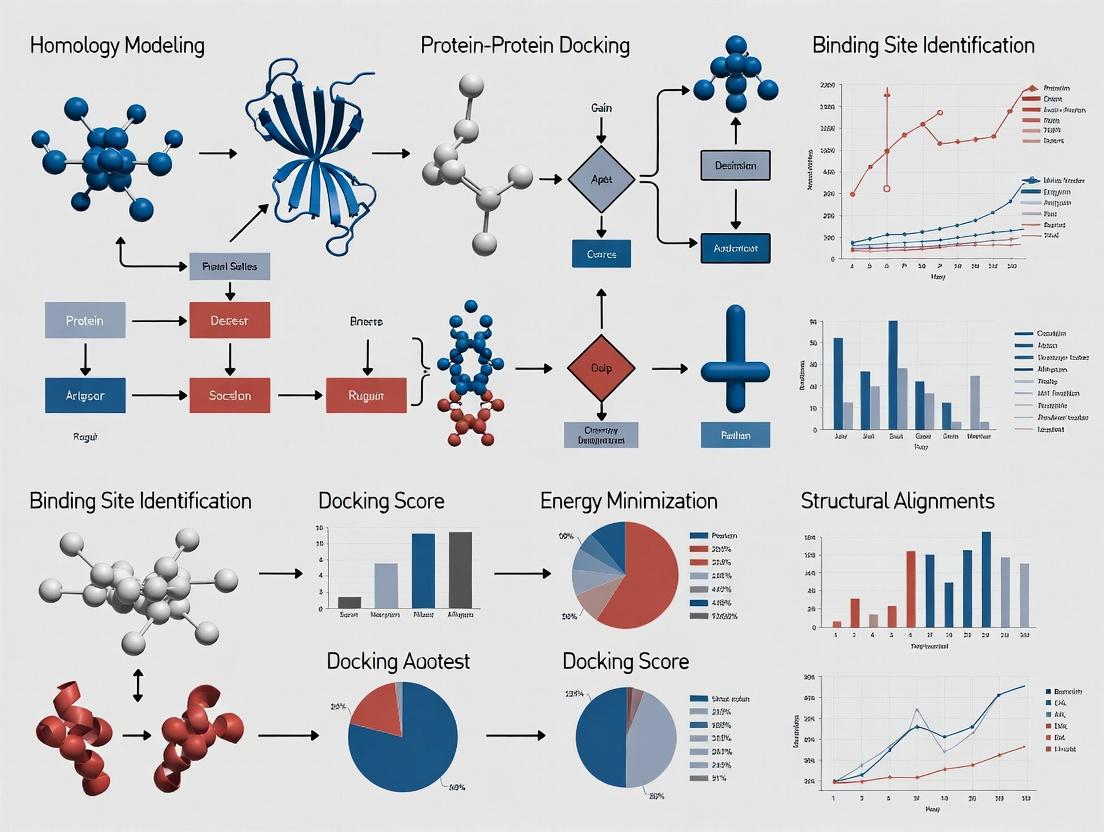
Visualization
Title: Homology Modeling to Docking Workflow
Title: Docking Troubleshooting Decision Tree
The Scientist's Toolkit: Research Reagent Solutions
Table 3: Essential Computational Tools for Homology Modeling & Docking
| Tool/Solution Name | Primary Function | Key Consideration for Homology Models |
|---|---|---|
| MODELER / SWISS-MODEL | Core homology model generation. | Use multiple templates and loop refinement options. |
| RosettaCM | Integrative modeling, especially useful for low-homology targets. | Computationally intensive but can yield superior models. |
| GROMACS / AMBER | Molecular Dynamics for model refinement and stability assessment. | Requires careful parameterization and extended equilibration. |
| AutoDock Vina / GLIDE | Molecular docking into prepared protein structures. | Use softened potentials or larger search boxes for model ambiguity. |
| QMEAN / MolProbity | Model quality assessment (global and local). | Critical for selecting the most physically plausible model. |
| Pymol / ChimeraX | Visualization and analysis of models, alignments, and docking poses. | Essential for manual inspection of binding site geometry. |
| Consensus Scoring Scripts | Combine scores from multiple docking runs or scoring functions. | Mitigates bias from any single function's limitations. |
Technical Support Center
Troubleshooting Guides & FAQs
Q1: During template selection, my target sequence shows high homology (>50%) to multiple templates. Which one should I choose, and why does my model fail validation despite high sequence identity?
A: High sequence identity does not guarantee a suitable template for your specific research question. Follow this decision protocol:
- Prioritize Functional Relevance: Select the template co-crystallized with a ligand or under experimental conditions (e.g., pH) most similar to your study, even if its sequence identity is 2-3% lower.
- Check for Global vs. Local Issues: Use the alignment table below to diagnose. Model failures often stem from incorrect alignment in active site or flexible loop regions, not overall fold.
- Actionable Step: Realign using a method sensitive to local features (e.g., HHsearch) and manually inspect the alignment over your residue of interest (e.g., the binding pocket).
Table 1: Template Selection Decision Matrix
| Criterion | Optimal Choice | Risk if Ignored | Tool for Evaluation |
|---|---|---|---|
| Sequence Identity | >30% for core docking | Increased backbone errors | BLAST, HHblits |
| Resolution (Å) | <2.0 Å | Poor side-chain packing | PDB header |
| Ligand Presence | Co-crystallized with similar ligand | Incorrect binding site conformation | PDBsum |
| Coverage | Covers >90% of target length | Model contains large gaps | Alignment viewer |
Q2: After building the model, the active site geometry is distorted, leading to failed docking poses. How can I refine this region specifically?
A: This is a common issue in homology modeling for docking. Implement a protocol for local active site refinement:
- Extract the active site: Isolate residues within 10Å of your catalytic center or putative binding site.
- Apply stronger restraints: In your modeling software (e.g., MODELLER, RosettaCM), increase restraint weights on the backbone atoms of the active site to preserve template geometry.
- Perform targeted loop modeling: Use a dedicated protocol (e.g., Rosetta loopmodel, MODELLER DOPE assessment) only on the misfiled loops surrounding the pocket.
- Validate with geometry checkers: Post-refinement, analyze only the active site with MolProbity to ensure Ramachandran outliers and steric clashes are resolved.
Q3: During model evaluation, different metrics (DOPE, MolProbity, QMEAN) give conflicting results. Which metrics are most critical for downstream docking studies?
A: For docking applications, prioritize metrics that correlate with binding site accuracy over global fold metrics. Use this tiered validation protocol:
Table 2: Tiered Model Evaluation Protocol for Docking
| Tier | Metric Category | Target Value for Docking | Rationale |
|---|---|---|---|
| Tier 1 (Critical) | Stereochemical Quality (MolProbity) | Clashscore < 10, Ramachandran Outliers < 2% | Ensures physically plausible side chains for ligand interaction. |
| Tier 2 (Essential) | Local Geometry (3D) | DOPE score per-residue low in binding site | Direct indicator of binding region stability. |
| Tier 3 (Contextual) | Global Fold (QMEAN, GA341) | QMEAN Z-score > -4.0 | Confirms overall fold is correct; poor scores can indicate template misalignment. |
| Tier 4 (Functional) | Conservation Check (Verify3D) | >80% of residues have 3D-1D score > 0.2 | Ensures the model's environment is compatible with its sequence. |
Q4: My final model has a good global RMSD to the template but poor ligand docking scores compared to a crystal structure control. What specific steps can I take to improve the model's utility for virtual screening?
A: This indicates accurate backbone but inaccurate side-chain conformations (rotamers) in the binding pocket. Implement a binding site rotamer optimization protocol:
- Fix the backbone of the binding site residues based on your best template.
- Use a rotamer library (e.g., SCWRL4, Rosetta fixbb) to systematically sample and optimize side-chain conformations.
- Score with a docking-specific function: Minimize the energy of the binding site using a simplified version of your intended docking score function (e.g., Vinardo, ChemPLP).
- Validate by re-docking a known native ligand (if any) and monitor the improvement in RMSD of the docked pose.
Experimental Protocols
Protocol: Template Selection and Alignment for Docking-Ready Models Objective: Generate a target-template alignment optimized for binding site accuracy.
- Input: Target sequence, PDB database.
- Primary Search: Execute PSI-BLAST with an E-value cutoff of 0.001 over 3 iterations against the PDB.
- Profile-Profile Alignment: For top hits (>30% identity), perform a profile-based alignment using HHsearch. Manually inspect the alignment in the binding site region using Jalview.
- Template Prioritization: Rank templates by a composite score: (0.4 * Sequence Identity) + (0.3 * (1/Resolution)) + (0.3 * Binding Site Coverage). Select the top 3 templates.
- Output: A curated multiple sequence alignment file (.aln) for model building.
Protocol: Model Building with MODELLER for Docking Studies Objective: Build a model with emphasis on binding site geometry.
- Software: MODELLER v10.4.
- Script Modification: In the MODELLER Python script, increase the
special_restraintsweight for residues within 8Å of the template's ligand or catalytic site to 5.0. - Generation: Build 100 models using the
automodelclass. - Initial Scoring: Rank models by the MODELLER objective function and the
DOPEassessment score. - Output: Top 10 models for rigorous evaluation.
Visualizations
Four-Step Template Modeling Workflow
Model Evaluation & Troubleshooting Logic
The Scientist's Toolkit: Research Reagent Solutions
Table 3: Essential Toolkit for Template-Based Modeling & Docking
| Tool / Reagent | Category | Primary Function | Example / Source |
|---|---|---|---|
| MODELER | Software Suite | Integrates all four steps: alignment, model building, loop modeling, scoring. | https://salilab.org/modeller/ |
| RosettaCM | Software Suite | Robust comparative modeling with integrative loop and domain modeling. | Rosetta Commons |
| Swiss-Model | Web Server | Fully automated, user-friendly pipeline for standard homology modeling. | https://swissmodel.expasy.org/ |
| MolProbity | Validation Server | Comprehensive structure validation for stereochemistry and clashes. | http://molprobity.biochem.duke.edu/ |
| UCSF Chimera | Visualization | Interactive visualization for alignment inspection, model analysis, and figure generation. | https://www.cgl.ucsf.edu/chimera/ |
| PDB Database | Data Repository | Source for all experimental protein structure templates. | https://www.rcsb.org/ |
| HH-suite | Search/Alignment | Sensitive profile-based methods (HHblits, HHsearch) for remote homology detection. | https://github.com/soedinglab/hh-suite |
| SCWRL4 | Software Tool | Fast and accurate side-chain conformation prediction for final model refinement. | http://dunbrack.fccc.edu/scwrl4/ |
Within strategies for molecular docking with homology modeled protein structures, the reliability of docking outcomes is intrinsically linked to the quality of the initial protein model. This technical support center focuses on the critical evaluation of model quality through specific metrics, with emphasis on how template selection—specifically its sequence identity to the target and structural coverage—impacts downstream virtual screening and drug discovery efforts.
Troubleshooting Guides & FAQs
Q1: My docking poses with a homology model show poor affinity despite high scoring function values. What could be wrong?
A: This discrepancy often originates in the model's structural quality, not the docking algorithm itself.
- Primary Check: Verify your model's Global Distance Test (GDT) score and Root Mean Square Deviation (RMSD) of the binding site residues. A high global GDT can mask local errors in the active site.
- Actionable Protocol:
- Align your homology model to the experimental structure (if available) or a high-quality reference using PyMOL or UCSF Chimera.
- Calculate the RMSD specifically for the residues within 5Å of the expected binding pocket.
- If the local RMSD > 2.0 Å, the binding site geometry is likely unreliable. Re-model using a different template with higher identity in that region or consider loop modeling techniques.
Q2: How do I interpret Template Identity and Coverage metrics when selecting a template for homology modeling?
A: These are pre-modeling indicators of potential quality.
- Template Identity: The percentage of identical amino acids between the target and template sequences. Higher identity generally yields more accurate models.
- Template Coverage: The percentage of the target sequence length that can be aligned to the template. Low coverage indicates large missing loops or domains.
- Decision Guide: Prefer templates with >30% identity for docking studies. For coverage, aim for >90%. If coverage is low (e.g., 70%), anticipate that unmodeled loops may border the binding site and require specialized modeling.
Q3: What are the key quantitative metrics to validate a homology model before proceeding to docking?
A: Post-modeling, use a combination of stereochemical and statistical potential checks. The following table summarizes the key metrics and their ideal thresholds:
Table 1: Key Model Quality Validation Metrics
| Metric | Tool Example | Ideal Threshold | Indicates |
|---|---|---|---|
| Ramachandran Favored | MolProbity, PROCHECK | >95% | Stereochemical quality of backbone dihedral angles. |
| Rotamer Outliers | MolProbity | <1% | Proper side-chain conformations. |
| Clashscore | MolProbity | <10 | Number of severe atomic steric overlaps per 100 atoms. |
| Cβ Deviations | WHAT-IF | 0 | Abnormal backbone conformation. |
| DOPE/Z-Score | MODELLER | Negative (Lower is better) | Statistical potential of mean force; overall model fitness. |
| Local Quality Estimate | QMEANDisCo | >0.7 per residue | Per-residue model reliability, critical for binding sites. |
Q4: I have multiple templates with varying identity and coverage. How do I design an experiment to choose the best for docking?
A: Implement a comparative modeling and validation pipeline.
Experimental Protocol: Comparative Template Assessment
- Template Selection: From your database (e.g., PDB), select 3-5 templates covering a range of identities (e.g., 25%, 40%, 60%) to your target.
- Model Building: Use a standard tool like MODELLER or SWISS-MODEL to generate one homology model per template, using the same parameters.
- Model Validation: For each model, run the validation suite in MolProbity and calculate the QMEANDisCo score. Compile results in a table.
- Binding Site Analysis: Superimpose the models and visually inspect the geometry of the predicted catalytic/binding residues.
- Decision Point: The optimal model is not always from the highest-identity template. Choose the model with the best combination of high local quality scores in the binding site, good stereochemistry (Ramachandran, Clashscore), and high overall coverage.
Table 2: Example Results from a Comparative Template Experiment
| Template PDB | Identity | Coverage | Model GDT (est.) | Clashscore | Binding Site Local QMEAN |
|---|---|---|---|---|---|
| 1A0B | 65% | 98% | 0.88 | 5.2 | 0.85 |
| 2X4F | 42% | 95% | 0.79 | 8.7 | 0.80 |
| 3KJ9 | 28% | 78% | 0.65 | 15.3 | 0.55 |
In this example, 1A0B is the clear choice. 2X4F may be a contender if 1A0B is unavailable, but 3KJ9's low coverage and poor local score disqualify it for docking.
Workflow & Relationship Diagrams
Title: Homology Model Quality Assessment Workflow for Docking
Title: Thesis: How Model Quality Factors Impact Docking Results
The Scientist's Toolkit: Research Reagent Solutions
Table 3: Essential Resources for Homology Modeling & Validation
| Item | Function & Purpose |
|---|---|
| SWISS-MODEL Server | Fully automated, web-based homology modeling pipeline. Ideal for quick model generation and initial quality estimates. |
| MODELLER Software | A highly flexible scripting platform for comparative modeling, allowing fine-grained control over the modeling process. |
| MolProbity Web Service | Integrative validation server providing Ramachandran, clashscore, rotamer, and Cβ deviation analysis. |
| UCSF Chimera / PyMOL | Molecular visualization software critical for visualizing model-template alignment, binding site geometry, and validation outliers. |
| PDB (Protein Data Bank) | Primary repository of experimentally determined 3D structures used as templates for homology modeling. |
| MMseqs2 / HMMER | Sensitive sequence search and alignment tools for identifying distant homologs as potential templates. |
| QMEANDisCo Server | Provides global and local (per-residue) quality estimates based on consensus methods, highlighting unreliable regions. |
| RosettaCM | An advanced, fragment-integrated comparative modeling suite for challenging targets with low template identity. |
Troubleshooting Guide & FAQs
Frequently Asked Questions
Q1: My docking poses with a homology model show good shape complementarity but are consistently ranked poorly by the scoring function. What could be the cause? A: This is a common issue when docking against homology models. The scoring function heavily depends on the precise geometry of the receptor's binding site. Inaccuracies in side-chain packing or loop modeling within the model can create artificial steric clashes or incorrect distances for optimal non-covalent interactions. The scoring function penalizes these, even if the overall pose seems correct. Focus on refining the binding site region of your model through loop modeling and side-chain rotamer optimization before docking.
Q2: How do I decide which scoring function to use for virtual screening on a novel homology model target? A: There is no single best function. The performance is target-dependent. The recommended protocol is to conduct a small-scale validation test. If you have known active and inactive compounds for your target (even a few), dock them against your model using multiple functions (e.g., Vina, Glide SP/XP, ChemPLP). Evaluate which function best separates actives from inactives. In the absence of known actives, use consensus scoring—selecting poses ranked highly by multiple, chemically diverse functions—to increase confidence.
Q3: Why does a small change in a ligand's torsion angle lead to a dramatic drop in the computed binding score? A: Scoring functions are highly sensitive to the geometry of non-covalent interactions. A change of a few degrees can break a critical hydrogen bond, moving the donor-acceptor distance outside the optimal range (typically ~2.5-3.2 Å) or misaligning dipoles. Similarly, it can disrupt favorable pi-stacking or cation-pi interactions. The functions use steep potential wells for these terms, so minor deviations result in large energy penalties, reflecting the precise nature of molecular recognition.
Q4: During ensemble docking with multiple homology model conformations, how should I interpret and combine the results? A: Docking against an ensemble accounts for model uncertainty and flexibility. Score normalization across different receptor conformations is crucial. First, dock your ligand library against each model conformation separately. Then, for each ligand, use the best score across all conformations (pose-based consensus) or calculate the average score. This approach identifies ligands that can bind favorably to at least one plausible state of the model. Present results as a ranked list based on this combined metric.
Q5: The hydrophobic contribution in my scoring function seems counterintuitive—sometimes burying a hydrophobic group lowers the score. Why? A: Most modern scoring functions evaluate hydrophobic interactions via contact terms or surface area burial, not simple "more is better." The issue may be desolvation penalty. If a hydrophobic group is not fully buried and remains partially exposed to solvent, it loses favorable van der Waals contacts with water without gaining sufficient protein contacts, resulting in a net energy cost. The scoring function is telling you the placement is suboptimal—the group may need to be more completely buried or positioned in a tighter hydrophobic pocket.
Experimental Protocols
Protocol 1: Validation of Docking Poses from a Homology Model Using Known Ligands
- Objective: To assess the predictive accuracy of a docking protocol by reproducing known ligand binding modes.
- Method:
- Preparation: Prepare your homology model and a known co-crystallized ligand from a related structure using standard software (e.g., UCSF Chimera, Schrödinger Maestro). Add hydrogens, assign partial charges, and define receptor grids.
- Docking: Perform flexible-ligand docking of the known ligand into your model. Use a high exhaustiveness setting to ensure thorough sampling.
- Analysis: Calculate the Root-Mean-Square Deviation (RMSD) between the top-ranked docked pose and the ligand's original conformation. A successful prediction typically has a heavy-atom RMSD < 2.0 Å.
- Scoring Function Audit: Manually inspect the top poses. Verify that key hydrogen bonds, salt bridges, and hydrophobic contacts predicted by the scoring function align with expected interactions from the related structure.
Protocol 2: Consensus Scoring to Prioritize Hits from Virtual Screening
- Objective: To improve the reliability of virtual screening hits by combining multiple scoring functions.
- Method:
- Docking Campaign: Dock your compound library against the prepared homology model using at least three distinct scoring functions (e.g., one empirical, one force-field-based, one knowledge-based).
- Rank Normalization: For each scoring function, rank all compounds from best (1) to worst (N).
- Consensus Calculation: For each compound, calculate its average rank across all functions. Re-sort the list based on this average rank.
- Hit Selection: Prioritize compounds with the lowest (best) average ranks. Optionally, apply a filter to select only compounds that appear in the top 10% of at least two individual lists.
Data Presentation: Common Non-Covalent Interaction Parameters in Scoring Functions
Table 1: Typical Geometric and Energetic Parameters for Key Non-Covalent Interactions
| Interaction Type | Optimal Distance (Å) | Optimal Angle (°) | Typical Energy Contribution (kcal/mol) | Functional Form in Scoring |
|---|---|---|---|---|
| Hydrogen Bond | Donor-Acceptor: 2.5-3.2 | D-H...A: ~180 | -1 to -5 (strong) | 12-10 Lennard-Jones, Angular term |
| Salt Bridge | Between charged groups: <4.0 | N/A | -3 to -6 | Coulombic electrostatics with distance-dependent dielectric |
| Van der Waals | Sum of vdW radii | N/A | -0.1 to -0.2 per contact | 6-12 Lennard-Jones potential |
| Pi-Pi Stacking | Aromatic ring centroids: 3.5-4.5 | Parallel or T-shaped | -0.5 to -2 | Special planar interaction terms |
| Cation-Pi | Cation to ring centroid: 3.0-4.5 | Cation over ring face | -2 to -5 | Combination of electrostatic and vdW terms |
| Hydrophobic | <4.0 from nonpolar atoms | N/A | ~-0.03 per Ų buried | Surface Area (SA) burial model |
The Scientist's Toolkit: Research Reagent Solutions
Table 2: Essential Tools for Docking with Homology Models
| Item | Function in Research |
|---|---|
| Homology Modeling Suite (e.g., MODELLER, SWISS-MODEL) | Generates the initial 3D protein structure from the target sequence using a related template structure. |
| Loop Modeling Tool (e.g., Rosetta, FREAD) | Refines uncertain loop regions in the model, which are often near binding sites and critical for accurate docking. |
| Side-Chain Prediction Software (e.g., SCWRL4, ROSETTA) | Optimizes the rotameric states of amino acid side chains to minimize steric clashes and optimize packing. |
| Molecular Dynamics (MD) Simulation Package (e.g., GROMACS, AMBER) | Generates an ensemble of flexible receptor conformations for ensemble docking, capturing backbone and side-chain dynamics. |
| Docking Software with Multiple Functions (e.g., AutoDock Vina, Schrödinger Glide, GOLD) | Performs the ligand sampling and scoring, offering different algorithmic approaches to evaluate non-covalent interactions. |
| Consensus Scoring Script (e.g., Custom Python/R Script) | Combines results from multiple docking runs to improve hit identification and reduce scoring function bias. |
| Visualization & Analysis Software (e.g., PyMOL, UCSF Chimera) | Used for manual inspection of poses, auditing scoring function predictions, and analyzing interaction networks. |
Visualizations
Troubleshooting Guides & FAQs
Q1: Why do my ligands consistently dock to an unrealistic, solvent-exposed location on my homology model instead of the predicted binding pocket? A: This is often due to an overestimation of pocket hydrophobicity or incorrect side-chain rotamers in the model, creating a false favorable spot. First, recalculate and visualize the electrostatic potential surface of your model using tools like PyMOL or ChimeraX. Compare it to a known high-resolution structure of a close homolog. Manually inspect the side-chain packing in the true binding site; problematic residues may need optimization with a tool like SCWRL4 or RosettaFixBB before redocking.
Q2: After docking into a homology model, my pose rankings show no correlation with experimental activity. What's wrong? A: The geometry of the modeled active site may be too distorted for reliable scoring. Implement a two-step verification: 1) Perform a control docking of a known native ligand (or a close analog) from a co-crystal structure of the template. If this fails to reproduce the correct pose, your model's "dockability" is low. 2) Use a consensus scoring approach across multiple docking programs (AutoDock Vina, Glide, GOLD) to identify consistently ranked poses, as outlier rankings often arise from model artifacts.
Q3: How can I assess the local backbone reliability of my modeled binding site before investing in large-scale virtual screening? A: Utilize local model quality estimation tools. Run your model through the QMEANDisCo server or use ModFOLDclust2. These provide per-residue confidence scores. Focus on the binding site residues: a cluster of low scores (e.g., below 0.6) indicates a problematic region. A practical protocol is to generate an ensemble of models (e.g., 5-10) and only proceed with docking if the backbone atoms of key binding residues (e.g., catalytic triads, binding motifs) are consistent across the ensemble (Cα RMSD < 1.5 Å).
Q4: My homology model has a large, flexible loop near the binding site that is poorly aligned in the template. How should I handle it for docking? A: Indiscriminate docking into a flexible, poorly modeled loop region will generate false positives. Implement a loop modeling and clustering protocol:
- Excise and remodel the loop using RosettaCM or MODELLER's loop refinement.
- Generate multiple loop conformations (e.g., 100).
- Cluster the conformations based on loop backbone RMSD.
- Select representative models from the top 3-5 clusters for docking.
- Perform docking against each representative and compare results. Ligands that only dock well to one rare conformation are less reliable.
Q5: What are the definitive signs that a homology model is simply not suitable for docking-based studies? A: Red flags that critically compromise model "dockability" include:
- Sequence identity to the best template is below 30% for the binding site region.
- Key functional residues (e.g., catalytic residues, binding motifs) are misaligned or missing.
- Steric clashes exist in the core of the binding pocket that cannot be relieved by side-chain optimization.
- Consensus evaluation scores (like GA341 score in MODELLER < 0.7, or MolProbity clashscore > 30) indicate globally poor model quality.
Experimental Protocols
Protocol 1: Binding Site Geometry Validation via Native Ligand Docking
- Objective: To test the ability of a homology model to recapitulate a known binding mode.
- Materials: Homology model, template co-crystal structure with native ligand, docking software (e.g., AutoDock Vina), molecular visualization software.
- Method:
- Prepare the homology model and the template structure: add hydrogens, assign charges (e.g., using Gasteiger charges).
- Extract the native ligand from the template co-crystal structure.
- Define a docking grid/box. For the homology model, center it on the predicted binding site. For the template, center it on the crystallographic pose of the ligand.
- Perform docking of the native ligand into both the homology model and its template structure using identical parameters.
- Measure the Root-Mean-Square Deviation (RMSD) of the top-ranked docked pose from the crystallographic pose.
- Success Criteria: A pose RMSD ≤ 2.0 Å in the template control confirms the docking protocol. An RMSD ≤ 3.0 Å in the homology model suggests acceptable binding site geometry.
Protocol 2: Ensemble Docking to Account for Binding Site Flexibility
- Objective: To improve docking outcomes by accommodating structural uncertainty in the model.
- Materials: Main homology model, loop-modeled variants, or MD simulation snapshots.
- Method:
- Generate the Ensemble: Create multiple receptor structures. This can be done via:
- Sampling alternative side-chain rotamers (using SCWRL4).
- Modeling flexible loops in distinct conformations (using MODELLER).
- Running short, restrained molecular dynamics (MD) simulations and clustering snapshots.
- Prepare Structures: Prepare all ensemble members identically for docking (add charges, etc.).
- Perform Docking: Dock the ligand library against each ensemble member using the same grid center but potentially larger dimensions.
- Analyze Results: Consolidate results. Rank ligands by their best score across the ensemble or by their average score. Analyze pose consistency across the ensemble.
- Generate the Ensemble: Create multiple receptor structures. This can be done via:
- Note: This protocol is computationally intensive but crucial for models with mobile binding site elements.
Data Presentation
Table 1: Correlation Between Model Quality Metrics and Docking Success Rate
| Model Quality Metric | Threshold for "Dockable" Model | Impact on Virtual Screening (VS) Performance |
|---|---|---|
| Global Model Score (QMEAN) | > -4.0 | High score correlates with better enrichment in VS. |
| Binding Site Cα RMSD (vs. Native) | < 1.5 Å | Directly determines ability to reproduce native ligand pose (RMSD < 2.5 Å). |
| MolProbity Clashscore | < 20 | Lower clashscores reduce false favorable docking pockets. |
| Sequence Identity in Binding Site | > 40% | Higher identity dramatically increases probability of successful docking. |
| Per-Residue Confidence (pLDDT) in Site | Average > 70 | Ensures local backbone reliability for scoring function accuracy. |
Table 2: Troubleshooting Summary: Problematic Features vs. Remedial Actions
| Problematic Feature in Model | Symptom During Docking | Recommended Remedial Action |
|---|---|---|
| Overpacked Hydrophobic Cave | Ligands dock to non-physiological, deep hydrophobic spots. | Remodel side-chains with constraints; solvate model and re-calc. surface. |
| Mis-oriented Hydrogen Bond Donor/Acceptor | Loss of critical polar interaction; incorrect pose ranking. | Manual rotamer adjustment or use of H-bond network prediction tools. |
| Poorly Modeled Flexible Loop | Inconsistent poses; high score variance for similar ligands. | Ensemble docking with multiple loop conformations (see Protocol 2). |
| Global Backbone Distortion in Site | Native control docking fails (RMSD > 3.5 Å). | Consider alternative template or refine with rigid-body MSA. |
Visualization
Title: Workflow for Docking with Homology Models
Title: Model Features Directly Impact Docking Outcome
The Scientist's Toolkit: Research Reagent Solutions
| Item/Reagent | Function in Docking with Homology Models |
|---|---|
| MODELLER | Software for homology modeling; generates 3D coordinates from alignments. |
| SWISS-MODEL Server | Automated web-based homology modeling pipeline for quick initial models. |
| PyMOL/ChimeraX | Molecular visualization software for analyzing model quality, surface properties, and docking poses. |
| AutoDock Vina | Widely used, open-source docking program for pose prediction and scoring. |
| Rosetta Software Suite | For advanced model refinement (RosettaRelax), loop modeling, and ensemble generation. |
| SCWRL4 | Algorithm for accurate side-chain conformation prediction and optimization. |
| QMEANDisCo Server | Online tool for local model quality estimation, crucial for binding site assessment. |
| MolProbity | Service for structure validation, identifying steric clashes, and rotamer outliers. |
| PDBbind Database | Curated database of protein-ligand complexes for native ligand docking controls. |
| ZINC20 Database | Public library of commercially available compounds for virtual screening. |
A Practical Workflow: Preparing Models and Executing Docking Simulations
Technical Support Center
Troubleshooting Guides & FAQs
Q1: My docking results are nonsensical, with ligands buried in non-physiological pockets or showing extreme energies. What went wrong in the model preparation step? A: This is often due to incorrect protonation states of key residues or missing critical hydrogens. Histidine tautomerization (HID, HIE, HIP) is a frequent culprit. Use a rigorous pKa prediction tool (e.g., PROPKA) to determine the correct protonation states at your target pH (typically 7.4). Re-run the hydrogen addition and charge assignment with these parameters.
Q2: After adding hydrogens and charges, the homology model shows severe steric clashes or distorted geometry. How should I proceed? A: Homology models, especially in loop regions, often contain local strain. Before docking, perform a restrained energy minimization. This relaxes the structure while keeping it close to the original model. Use a force field (e.g., AMBER, CHARMM) with restraints on the protein backbone heavy atoms (RMSD restraint of 0.3-0.5 Å). This resolves clashes without altering the overall fold.
Q3: How do I handle ambiguous side-chain rotamers in my model, particularly for surface residues not in the active site? A: For non-critical residues, use a fast side-chain optimization algorithm (e.g., SCWRL4, FASPR). For active site residues, a more careful approach is needed. Utilize a conformational search using molecular mechanics (MM) or short molecular dynamics (MD) simulations with implicit solvent, keeping the backbone fixed. Select the lowest energy rotamer consistent with known catalytic mechanisms or ligand binding data.
Q4: Which force field and charge set should I use for preparing a homology model for docking with AutoDock Vina or similar software? A: Consistency is key. Docking programs have internal scoring functions. For preparation, use a standard molecular mechanics force field.
- For general preparation/minimization: AMBER ff14SB or CHARMM36.
- For assigning Gasteiger charges (common for Vina): Use tools in Open Babel or MGLTools.
- For assigning AM1-BCC charges (often more accurate for ligands): Use Antechamber (from AMBER) or OpenEye toolkits. See the table below for a quantitative comparison.
Table 1: Comparison of Common Charge Assignment Methods for Docking Preparation
| Charge Method | Computational Speed | Typical Use Case | Recommended For |
|---|---|---|---|
| Gasteiger | Very Fast | High-throughput screening, large ligand libraries | Protein & ligand in AutoDock Vina/Znk |
| AM1-BCC | Moderate | Accurate ligand charge derivation, lead optimization | Ligand parameterization for more rigorous docking |
| RESP (HF/6-31G*) | Slow | Benchmarking, QM-derived accuracy for key complexes | Small, critical ligand sets in validated studies |
Q5: The prepared model has gaps or missing atoms in incomplete loops. Can I still use it for docking? A: It depends on the loop's location. If it's far from (>15 Å) the binding site, you may proceed. If it's near the site, you must model the loop. Use a dedicated loop modeling tool (e.g., ModLoop, Rosetta loop modeling, or the loop refinement protocol in your homology modeling software). Follow this protocol:
Experimental Protocol: Loop Refinement for Binding Site Integrity
- Identify: Isolate the incomplete loop region (residues with missing atoms).
- Sample: Generate 100-500 decoy conformations using a kinematic closure (KIC) or MD-based algorithm.
- Score: Rank decoys using a composite score (e.g., Rosetta's full-atom energy, DOPE score in MODELLER).
- Select & Minimize: Choose the top 5-10 lowest-energy models. Perform a restrained minimization of the selected loop with the surrounding protein (5 Å shell) to relieve clashes.
- Validate: Check the refined loop's geometry with MolProbity. Ensure it does not occlude the intended binding pocket.
Q6: How do I validate that my prepared model is "docking-ready"? A: Perform a post-preparation validation suite. Compare the prepared model to the initial model using RMSD (should be < 2.0 Å overall backbone). Specifically check:
- Steric Quality: Ramachandran plot (≥90% in favored regions via MolProbity).
- Charge Distribution: Visualize electrostatic surface (e.g., with PyMOL/APBS) for a physically plausible pattern.
- Active Site Integrity: Confirm catalytic residues are properly oriented and protonated.
Diagram Title: Workflow for Protein Model Preparation for Docking
The Scientist's Toolkit: Research Reagent Solutions
Table 2: Essential Software Tools for Model Preparation
| Tool / Resource | Primary Function | Key Application in Preparation |
|---|---|---|
| PDB2PQR / PROPKA | Adds hydrogens, assigns protonation states based on pKa prediction. | Determining correct His, Asp, Glu, Lys states at physiological pH. |
| MGLTools (AutoDock Tools) | Prepares PDBQT files, assigns Gasteiger charges, merges non-polar hydrogens. | Standardized input generation for AutoDock Vina/Znk. |
| AmberTools (tleap, antechamber) | Force field parameterization, charge assignment (AM1-BCC, RESP), system building. | Creating high-quality parameters for ligands and restrained minimization. |
| SCWRL4 / FASPR | Fast and accurate side-chain conformation prediction. | Optimizing rotamers for non-active site residues. |
| Rosetta (relax protocol) | All-atom refinement and side-chain packing. | High-resolution optimization of loops and binding site residues. |
| UCSF Chimera / PyMOL | Visualization, structure analysis, and model manipulation. | Visual validation of charges, protonation, and steric fit. |
| MolProbity | All-atom structure validation server. | Final check of stereochemistry, clashes, and rotamer quality. |
Technical Support & Troubleshooting
Q1: After modeling my protein, the active site predicted by different servers (e.g., CASTp, COACH) shows significant variation. How do I define a reliable binding site for grid generation?
A: Discrepancy is common in homology models due to loop flexibility and side-chain packing errors. Follow this protocol:
- Consensus Analysis: Use at least three prediction tools (e.g., CASTp, SiteHound, Metapocket 2.0). Identify residues present in ≥70% of predictions.
- Template Alignment: Superimpose your model with the template structure(s) used for modeling. If the template has a known ligand (e.g., from PDB: 1XYZ), transfer that binding site footprint.
- Literature Mining: Use PDBsum to analyze known binding sites in homologous proteins (≥40% sequence identity).
- Define the Site: Use the union of residues from consensus and template alignment. Treat residues from literature as validation.
| Tool | Type | Key Output | Recommended Threshold |
|---|---|---|---|
| CASTp 3.0 | Geometry-based | Pockets, volume, area | Top 3 pockets by volume |
| COACH | Template-based | Ligand binding residues | Confidence score >0.7 |
| DeepSite | Deep Learning | Binding propensity grid | Probability >0.8 |
Q2: When generating a grid box around my defined site, what dimensions and center should I use to ensure it captures relevant pharmacophore space without being computationally prohibitive?
A: The optimal grid balances coverage and efficiency. Use this quantitative guide:
| Parameter | Recommended Value | Rationale & Adjustment Rule |
|---|---|---|
| Box Center | Centroid of residues defining the binding site. | Avoid using a single atom; the centroid captures the site's geometric center. |
| Box Dimensions | Start at 20Å x 20Å x 20Å. | For most drug-like ligands (<500 Da). |
| Dimension Adjustment | Increase by 1.5x if the native ligand (from template) is not fully enclosed. | Check using "Grid Box Validation" workflow below. |
| Grid Point Spacing | 0.375 Å to 0.5 Å. | Higher resolution (0.375Å) for precise scoring; 0.5Å for initial screening. |
Experimental Protocol: Grid Box Validation
- Dock the cognate ligand (from the highest-identity template) into the generated grid.
- Calculate the RMSD between the docked pose and the ligand's original coordinates (transferred from the template).
- Success Criteria: RMSD < 2.0 Å. If RMSD > 2.0 Å, systematically increase grid dimensions by 2Å increments and re-dock until criteria is met.
Q3: My homology model has a poorly defined, flexible loop near the suspected binding site. Should I include it in the grid, and how?
A: Flexible loops can lead to false positives/negatives. Implement a two-stage strategy:
- Stage 1 (Rigid): Generate a grid with the loop excluded. Perform initial docking to identify top candidate ligands.
- Stage 2 (Induced-Fit): For the top 10-20 ligands, use a flexible loop protocol (e.g., in RosettaFlex or Schrödinger's Prime) to refine the model, then re-dock into a new grid that includes the refined loop.
Q4: For a blind docking search on a model with no known site, what are the optimal global grid parameters?
A: Use an ensemble grid strategy to cover the protein surface efficiently.
| Strategy | Grid Center | Grid Dimensions | Use-Case |
|---|---|---|---|
| Single Global Box | Protein centroid. | Encompass entire protein. | Small proteins (<250 residues). |
| Multiple Sub-grids | Centers of largest 3 pockets from CASTp. | 25Å x 25Å x 25Å each. | Larger proteins, to prioritize likely pockets. |
Frequently Asked Questions (FAQs)
Q: Can I use the grid parameters from my template's crystal structure directly on my model? A: Not directly. While a good starting point, the binding cavity volume can differ by 10-15% in models. Always calculate the centroid based on your model's aligned residues and validate (see Q2 Protocol).
Q: Which software is most tolerant to the structural imperfections of a homology model during grid generation? A: AutoDock-GPU and LeDock are generally robust. For more advanced models, Schrödinger's Glide allows protein flexibility scaling during grid generation. See toolkit below.
Q: How do I report grid parameters for reproducibility? A: Always report: Software & Version, Box Center (x, y, z), Box Dimensions (Å), Grid Spacing (Å), and the list of residues used to define the center.
Experimental Workflow: From Model to Grid
Title: Workflow for Defining and Validating a Docking Grid on a Homology Model
The Scientist's Toolkit: Research Reagent Solutions
| Item / Software | Function in Binding Site/Grid Workflow | Key Consideration for Models |
|---|---|---|
| UCSF ChimeraX | Visualization, structural alignment, and centroid calculation. | Essential for visually inspecting model quality and superposing templates. |
| AutoDockTools | Generation of grid parameter files (GPF) for AutoDock Vina/GPU. | Robust to minor steric clashes; widely used benchmark. |
| Schrödinger (Glide) | High-throughput grid generation with protein flexibility options. | "Scaled van der Waals radii" setting can soften potential from modeling errors. |
| PyMOL (with APBS) | Electrostatic potential surface calculation and visualization. | Critical for defining grids in charged binding sites (e.g., kinases). |
| MetaPocket 2.0 | Consensus binding site prediction server. | Integrates 8 methods; improves reliability on models. |
| PDBsum | Database of ligand binding sites in known structures. | Source for template-based site definition. |
| REFINED | Web server for model refinement focused on binding sites. | Can improve local geometry before grid generation. |
Troubleshooting Guide & FAQs
Q1: During conformational sampling, my ligand exhibits unrealistic ring puckering or strained geometries. How can I resolve this? A: This is often caused by improper initial geometry or inadequate sampling parameters. Use the following protocol:
- Initial Optimization: Always perform a preliminary geometry optimization using quantum mechanical methods (e.g., HF/6-31G*) or a reliable force field (MMFF94) before conformational search.
- Sampling Method: For flexible rings, use a systematic torsional search or molecular dynamics (MD)-based sampling (e.g., 100-500ps in implicit solvent) instead of only stochastic methods.
- Restraints: Apply ring conformational restraints if the ligand's crystallographic data is available.
Q2: How do I determine the correct protonation and tautomeric state for my ligand at physiological pH (7.4) when docking into a homology model with uncertain electrostatic environment? A: The uncertainty of the model's binding site necessitates a multi-state docking approach.
- Protocol:
- Generate potential protonation/tautomeric states using tools like
Epik,MOE, orChemAxonCalculator Plugins at pH 7.4 ± 2.0 (range: 5.4 - 9.4). - Perform a quick docking of all states (low precision) into your homology model.
- Also, dock all states into a high-resolution reference crystal structure of a related protein, if available.
- Compare the consensus poses and rankings across both models. States that perform well consistently are more reliable.
- Select the top 2-3 states for final, high-precision docking.
- Generate potential protonation/tautomeric states using tools like
Q3: What is the impact of partial charge assignment methods on docking accuracy into homology models, and which should I choose? A: Homology models often have imprecise electrostatics, making charge choice critical. See Table 1 for a quantitative summary from recent benchmarks.
Table 1: Impact of Ligand Charge Assignment Methods on Docking to Homology Models
| Charge Method | Basis | Computational Cost | Typical Use Case for Homology Models | Reported RMSD Impact* |
|---|---|---|---|---|
| Gasteiger-Marsili | Empirical | Very Low | Initial high-throughput screening, very large libraries | Higher variability (± 2.0 Å) |
| MMFF94 | Force Field | Low | Standard protocol for organic molecules; good balance | Moderate reliability (± 1.5 Å) |
| AM1-BCC | Semi-Empirical QM | Medium | Recommended for final docking poses; better polarity | Improved accuracy (± 1.2 Å) |
| RESP (HF/6-31G*) | Ab Initio QM | High | Gold standard for key lead compounds; small libraries | Best theoretical accuracy (± 1.0 Å) |
*Reported RMSD (Root Mean Square Deviation) impact range relative to crystal ligand pose in benchmark studies.
Q4: I have a metal-coordinating ligand. How should I handle its charges and geometry? A: Standard force fields often fail. Follow this protocol:
- Geometry Optimization: Optimize the ligand-metal complex (if metal is from the protein) using DFT (e.g., B3LYP/6-31G*) with appropriate basis set for the metal.
- Charge Assignment: Use quantum mechanically derived charges (e.g., CHELPG, Merz-Kollman) for the ligand atoms involved in coordination.
- Docking Consideration: Treat coordinating bonds as potentially flexible constraints during docking if the metal ion is present in the protein model.
Q5: After preparing multiple conformers and states, my docking library is too large. How do I filter it? A: Apply a hierarchical filtering protocol:
- Energy Filter: Remove conformers with high steric strain (> 10 kcal/mol from MMFF94).
- Cluster by RMSD: Cluster remaining conformers using a 1.0-1.5 Å RMSD cutoff and select the centroid of each major cluster.
- State Priority: For each unique ligand, keep a maximum of the lowest-energy protonation state and the top 2-3 highest-population tautomers.
Experimental Protocols
Protocol 1: Comprehensive Ligand State Preparation for Homology Model Docking
- Objective: Generate an ensemble of ligand conformations and protonation/tautomeric states suitable for docking into a homology model with uncertain active site electrostatics.
- Software: Schrödinger Suite (LigPrep, Epik, ConfGen), Open Babel, RDKit, Gaussian.
- Steps:
- Input & Desalting: Provide ligand in SMILES or 2D/3D SDF format. Remove counterions and salts.
- Tautomer/Protomer Generation: Use Epik at pH 7.4 ± 2.0 to generate likely states. Set energy window to 10-20 kcal/mol for maximum coverage.
- Conformational Sampling: For each state, generate conformers using a mixed method:
- Systematic Rotamer Search: For all rotatable bonds (< 10).
- Monte Carlo Multiple Minimum (MCMM): For molecules with >10 rotatable bonds. Generate 1000-5000 conformations per state.
- Geometry Optimization & Minimization: Optimize all generated conformers using the OPLS4 or MMFF94 force field with implicit solvent (GBSA).
- Charge Assignment: Assign partial charges using the AM1-BCC method for the final library. For critical ligands, compute RESP charges via Gaussian (HF/6-31G* optimization & population analysis).
- Output: Produce a multi-conformer, multi-state library in Maestro (.maegz) or MDL SDF format.
Protocol 2: Benchmarking Ligand Preparation Protocols
- Objective: Validate your ligand preparation protocol by redocking ligands to their native crystal structures and to homology models.
- Steps:
- Curate a test set of 50-100 protein-ligand complexes from PDB.
- For each ligand, prepare it using your protocol (Protocol 1), generating an ensemble.
- Dock the ensemble back into the native crystal structure and calculate RMSD to the crystal pose.
- Create homology models for the same targets (using related templates).
- Dock the prepared ligand ensembles into the homology models.
- Compare the success rates (RMSD < 2.0 Å) between native and model structures to quantify preparation impact.
Workflow Diagram
Title: Ligand Prep Workflow for Homology Model Docking
The Scientist's Toolkit: Research Reagent Solutions
Table 2: Essential Software & Tools for Ligand Preparation
| Item (Software/Tool) | Category | Primary Function in Ligand Prep |
|---|---|---|
| Schrödinger LigPrep/Epik | Commercial Suite | Integrated pipeline for generating 3D structures, tautomers, protonation states, and conformers. |
| Open Babel | Open-Source Tool | Format conversion, hydrogen addition/removal, basic conformer generation and charge assignment. |
| RDKit | Open-Source Cheminfo | Python-based toolkit for molecule manipulation, descriptor calculation, and rule-based conformer generation. |
| Omega (OpenEye) | Commercial Conformer Generator | High-speed, rule-based conformer generation for large libraries. |
| Gaussian/GAMESS | Quantum Chemistry Software | Ab initio geometry optimization and electrostatic potential calculation for deriving high-accuracy charges (RESP). |
| Antechamber (AmberTools) | Utility | Assigns AM1-BCC charges and converts between molecular file formats, often used for preparing ligands for MD. |
| MOE (Molecular Operating Environment) | Commercial Suite | Comprehensive ligand preparation, including protonation, conformational search, and charge assignment (MMFF94, etc.). |
| CYP450 Metabolism Prediction Modules | Specialized Plugin | Predicts likely sites of metabolism to guide protonation/tautomer state consideration for specific targets. |
Troubleshooting Guides & FAQs for Molecular Docking Experiments
FAQ 1: In my homology modeled receptor, DOCK systematically fails to find any poses with a favorable score. What could be the primary issue?
Answer: This is a frequent challenge when docking to homology models. The primary cause is often an improperly defined binding site due to inaccuracies in loop modeling or side-chain packing. Systematic search algorithms like DOCK are highly sensitive to the precise geometric definition of the search grid. A small deviation in the active site conformation can cause the grid to be misaligned, resulting in no favorable poses. First, verify your binding site definition using a known co-crystallized ligand from your template structure. Second, consider using a softer scoring potential or expanding the grid dimensions by 5-10 Å to account for model uncertainty.
FAQ 2: AutoDock Vina yields highly variable results (different top poses) across consecutive runs on the same homology model. Is this normal, and how should I interpret the output?
Answer: Yes, this is expected behavior due to Vina's stochastic search method (Monte Carlo). Variability indicates that the energy landscape of your homology model's binding site may be relatively flat or have multiple shallow minima. Best practice is to perform multiple runs (e.g., 20-50) with different random seeds and analyze the clustering of output poses. Consistent clustering around a similar pose conformation, despite the variability, increases confidence in the prediction. Use the --exhaustiveness parameter (increase to 32 or higher) to improve search depth and reproducibility.
FAQ 3: When validating my docking protocol on a crystal structure, both algorithms work well. But on my homology model, the predicted binding mode is radically different. How should I proceed?
Answer: This discrepancy highlights the intrinsic uncertainty of docking to modeled structures. Implement a consensus docking strategy:
- Perform docking with both DOCK (systematic) and Vina (stochastic).
- Use an ensemble of the top homology models (not just a single model).
- Apply a post-docking scoring or re-scoring step using a different, knowledge-based function. The consensus pose that appears across multiple models and/or algorithms is more likely to be reliable than any single top-scoring result.
Quantitative Data Comparison
Table 1: Algorithmic Comparison for Homology Model Docking
| Feature | DOCK (Systematic) | AutoDock Vina (Stochastic) |
|---|---|---|
| Search Method | Anchor-and-grow, systematic sampling | Monte Carlo with local gradient optimization |
| Speed (Typical Ligand) | Slower (minutes to hours) | Faster (seconds to minutes) |
| Determinism | Fully deterministic (same output for same input) | Non-deterministic (output varies per run) |
| Handling of Model Uncertainty | Low; requires precise grid definition | Moderate; stochastic search can sample imperfect pockets |
| Key Parameter for Homology Models | Grid spacing and box size | exhaustiveness and search space center/box |
| Optimal Use Case | Well-defined, rigid binding sites from high-quality models | Flexible search in potentially inaccurate or soft binding sites |
Experimental Protocol for Consensus Docking on Homology Models
Protocol: Validated Docking Workflow for Modeled Protein Structures
- Model Preparation: Refine the homology model's binding site loops and side chains using a tool like SCWRL4 or MODELLER's loop refinement.
- Binding Site Definition:
- Use the CASTp server or a known ligand from the template to define the pocket.
- For DOCK: Generate grids using
gridprogram with a box extending 8-10 Å beyond the defined site. Use a grid spacing of 0.3 Å. - For Vina: Define the center (x, y, z) and box size (sizex, sizey, size_z) from the same coordinates.
- Ligand Preparation: Generate 3D conformers and assign charges (e.g., using Open Babel or MOE). Convert to PDBQT format for Vina or mol2 for DOCK.
- Parallel Docking Execution:
- Run DOCK with the standard anchor-and-grow parameters.
- Run AutoDock Vina with an exhaustiveness value of 32, performing at least 20 independent runs.
- Post-Processing & Consensus Analysis:
- Cluster all output poses (from both programs) using an RMSD cutoff of 2.0 Å (e.g., using
clusterrmsdin DOCK or SciPy). - Rank clusters by both average docking score and population size.
- Visually inspect the top 3 consensus poses within the model's binding site context.
- Cluster all output poses (from both programs) using an RMSD cutoff of 2.0 Å (e.g., using
Visualization: Algorithm Workflow & Selection Logic
Title: Algorithm Selection Logic for Modeled Structures
Title: Consensus Docking Experimental Workflow
The Scientist's Toolkit: Key Research Reagent Solutions
Table 2: Essential Resources for Docking to Homology Models
| Item | Function & Relevance |
|---|---|
| Homology Modeling Suite (e.g., MODELLER, SWISS-MODEL) | Generates the initial 3D protein model from the template structure; critical first step. |
| Model Refinement Tool (e.g., GalaxyRefine, Rosetta) | Improves side-chain packing and loop regions of the model, directly impacting docking accuracy. |
| Protein Preparation Software (e.g., Chimera, MOE, Maestro) | Adds hydrogens, assigns charges, and optimizes H-bond networks for the model prior to docking. |
| Ligand Preparation Tool (e.g., Open Babel, LigPrep) | Generates correct 3D conformations, tautomers, and protonation states for small molecule ligands. |
Grid Generation Utility (DOCK grid, AutoDockTools) |
Defines the 3D search space for the docking algorithm; crucial parameter for homology models. |
| Pose Clustering & Analysis Scripts (e.g., in-house Python/R) | Post-processes multiple docking outputs to identify consensus poses and analyze variability. |
| Visualization Platform (e.g., PyMOL, UCSF ChimeraX) | Enables critical visual inspection of predicted poses within the context of the modeled binding site. |
Troubleshooting Guides & FAQs
Exhaustiveness
Q1: My docking results show poor ligand pose reproducibility. How can I improve consistency? A: Low reproducibility is often due to insufficient exhaustiveness. This parameter controls the number of Monte Carlo runs performed. For homology models, which have higher uncertainty, a higher value is required. Increase the exhaustiveness value to at least 16-24 for initial screens and 32-64 for final pose prediction. This allows for more thorough sampling of the conformational space.
Q2: Is there a quantitative guideline for setting exhaustiveness relative to the binding site size? A: Yes. While the binding site volume is a key factor, a practical guideline based on recent benchmarks is summarized below:
| Binding Site Volume (ų) | Recommended Exhaustiveness | Expected Computation Time Increase* |
|---|---|---|
| < 500 | 8 - 16 | 1x (Baseline) |
| 500 - 1000 | 16 - 32 | 2x - 4x |
| > 1000 | 32 - 64 | 4x - 8x |
*Time increase relative to an exhaustiveness of 8.
Protocol for Determining Optimal Exhaustiveness:
- Prepare your homology model and ligand using standard preparation tools (e.g., AutoDockTools, MGLTools, or Schrodinger's Protein Preparation Wizard).
- Define a docking grid box that fully encompasses the predicted binding site.
- Perform a series of docking runs on a known reference ligand (if available) with exhaustiveness values: 8, 16, 32, 64.
- For each run, repeat the docking 5-10 times with different random seeds.
- Calculate the Root-Mean-Square Deviation (RMSD) between the top-ranked poses from each repeat. Lower inter-run RMSD indicates higher reproducibility.
- Plot the mean top-pose RMSD (from a reference crystal pose) and the reproducibility RMSD against exhaustiveness. The optimal value is where both metrics plateau.
Flexibility
Q3: How should I handle side-chain flexibility in a homology-modeled binding site? A: Incorporate selective flexibility for key residues. Identify residues within 5-6 Å of the docked ligand that are predicted to have high B-factors or are in flexible loops from your model validation. You can treat these side chains as flexible during docking using methods like:
- Specified Flexible Residues: In tools like AutoDockFR or Vina, you can define side chains to be treated as flexible torsions.
- Ensemble Docking: Generate multiple conformations of the receptor (e.g., from molecular dynamics snapshots of the model) and dock against each.
Q4: What is the recommended protocol for identifying which residues to set as flexible? A:
- Run a short molecular dynamics (MD) simulation (50-100 ns) of the solvated homology model.
- Analyze the simulation trajectory and calculate the Root Mean Square Fluctuation (RMSF) for each residue.
- Cluster the frames from the MD trajectory to obtain 5-10 representative receptor conformations.
- Perform ensemble docking against this set of conformations. Residues that show significant conformational variation across the ensemble and are near the binding site are prime candidates for explicit flexibility.
- Alternatively, use computational alanine scanning or computational mutagenesis tools to predict hotspot residues whose flexibility impacts binding.
Handling of Co-factors/Ions
Q5: My homology model includes a critical catalytic metal ion (e.g., Zn²⁺). How do I parameterize it for docking? A: Metal ions require special force field parameters. The protocol involves:
- Retain the ion in the structure. Do not remove it.
- Assign correct charges and parameters. Use specialized tools to generate parameters:
- MCPB.py: (Metal Center Parameter Builder) for AMBER/GAFF force fields.
- MCPB.py: Can be used to generate parameters for AutoDock/ AutoDock Vina via conversion.
- Manual Parameterization: Define the ion's charge (e.g., +2 for Zn²⁺) and create a parameter file specifying its van der Waals radius and well depth. Consult the CHARMM or AMBER parameter database for standard values.
- Include the ion in the grid map generation so the scoring function accounts for its presence and interaction with the ligand.
Q6: An essential co-factor (e.g., NAD, HEM) was present in the template but is missing in my model. How do I reintroduce it? A:
- Structural Alignment: Superimpose your homology model onto the template structure that contains the co-factor.
- Co-factor Transfer: Extract the coordinates of the co-factor (and any coordinating residues) from the template and align them onto your model.
- Geometry Optimization: Perform a constrained energy minimization of the co-factor and its immediate protein environment within your model to relieve any steric clashes introduced during transfer, keeping the overall protein backbone restrained.
- Parameterization: Ensure you have the correct force field parameters and partial charges for the co-factor. Libraries such as the PRODRG2 server or the R.E.D.D.B. database can be used to obtain these.
Docking Workflow for Homology Models
The Scientist's Toolkit: Research Reagent Solutions
| Item | Function in Docking with Homology Models |
|---|---|
| Homology Modeling Suite (e.g., MODELLER, SWISS-MODEL) | Generates the initial 3D protein structure from a related template. The foundation for all subsequent steps. |
| Structure Validation Tool (e.g., MolProbity, PROCHECK) | Evaluates the stereochemical quality of the model to identify problematic regions (e.g., Ramachandran outliers, clashes) before docking. |
| Force Field Parameter Database (e.g., R.E.D.D.B., AMBER parameter DB) | Provides accurate partial charges and van der Waals parameters for non-standard residues, metal ions, and co-factors essential for scoring. |
| Molecular Dynamics Software (e.g., GROMACS, NAMD) | Used to sample the flexibility and generate an ensemble of conformations for the homology model, crucial for assessing dynamics. |
| Docking Software with Flexibility Support (e.g., AutoDockFR, Vina) | The core engine that performs the ligand sampling and scoring, preferably with options for side-chain or backbone flexibility. |
| Pose Clustering & Analysis Scripts (e.g., RDKit, MDAnalysis) | Custom or community scripts to analyze docking outputs, calculate RMSD, cluster poses, and visualize results. |
| High-Performance Computing (HPC) Cluster Access | Essential for running exhaustive docking searches, ensemble docking, and any preceding MD simulations within a practical timeframe. |
Navigating Challenges: Troubleshooting Docking Failures and Refining Strategies
Troubleshooting Guides & FAQs
FAQ 1: Why does my virtual screen against a homology model yield a high hit rate in vitro, but the compounds show no activity in functional assays?
- Likely Cause: Scoring bias favoring compounds that complement errors in the modeled binding pocket, rather than the true native structure.
- Diagnosis: Perform a control docking run into the native template structure (if available). Compare the score distributions and ligand poses between the model and the template. A significant score improvement in the model suggests the scoring function is exploiting cavity artifacts.
- Solution: Re-score all top hits using a more rigorous, consensus scoring protocol or molecular mechanics-based methods (e.g., MM/PBSA). Prioritize compounds whose poses are conserved between the model and the template structure.
FAQ 2: My docking results show all top hits clustering in one pose, but it seems chemically unreasonable. What went wrong?
- Likely Cause: Poor conformational sampling due to incorrect side-chain rotamer placement in the binding site, creating a steric or electrostatic artifact that traps the docking algorithm.
- Diagnosis: Visually inspect the binding site of your homology model. Compare key side-chain orientations with those in the template and related structures. Run a short molecular dynamics (MD) simulation or side-chain repacking to assess the stability of the suspect residues.
- Solution: Generate an ensemble of homology models (using different loop modeling protocols or backbone perturbations) and dock into the ensemble. Use clustering analysis across models to identify poses that are robust despite local structural variations.
FAQ 3: How can I determine if failed experiments are due to poor sampling or an inherent scoring bias?
- Diagnosis: Conduct a "reverse decoy" test. Dock known active ligands and known inactive/decoy molecules into your model.
- Protocol:
- Curate a small test set of 5-10 known binders and 50-100 property-matched decoys.
- Perform high-throughput, conformational-expansive docking (e.g., high exhaustiveness in Vina-type software).
- Analyze the Receiver Operating Characteristic (ROC) curve and Enrichment Factors (EFs).
- Protocol:
- Interpretation: Poor early enrichment (EF1%) suggests scoring cannot distinguish actives. Good enrichment but poor pose prediction suggests sampling is inadequate to find the correct binding mode.
FAQ 4: The top-ranked compounds from docking are all chemically similar and have poor drug-like properties. Is this a bias?
- Likely Cause: Yes. This is often a combination of scoring bias (e.g., over-penalizing certain functional groups common in drugs) and dataset bias in your screening library.
- Solution: Apply property-based filters (e.g., Lipinski's Rule of Five, PAINS filters) before docking to pre-process the library. Post-docking, use a diversity-based selection from the top-scoring cluster representatives, not just the absolute top scores.
Experimental Protocol for Diagnosing Sampling & Scoring Issues
Protocol: Ensemble Docking and Consensus Scoring for Homology Models
- Model Generation Ensemble: Generate 5-10 alternative homology models using different software (e.g., MODELLER, Rosetta, SWISS-MODEL) or by sampling different loop conformations.
- Preparation: Prepare all protein models and ligands uniformly (same protonation states, force field parameters).
- Docking Execution: Dock the entire ligand library into each model structure using a standard protocol. Use a high sampling parameter (e.g., exhaustiveness=32 in AutoDock Vina).
- Pose Clustering: Pool all poses from all models. Cluster poses by ligand structural similarity and binding pose (RMSD < 2.0 Å).
- Consensus Scoring: Apply 2-3 distinct scoring functions (e.g., one empirical, one knowledge-based, one force-field based) to the top representative poses from each major cluster.
- Hit Prioritization: Rank compounds based on a consensus score (e.g., average rank across scorers) and the frequency of the pose appearing across the model ensemble.
Table 1: Diagnostic Test Results for a Sample Homology Model Docking Run
| Diagnostic Test | Metric | Value (Observed) | Target Threshold | Interpretation |
|---|---|---|---|---|
| Reverse Decoy | EF1% (Early Enrichment) | 8.5% | >10% | Marginal early enrichment. |
| Reverse Decoy | AUC-ROC (Area Under Curve) | 0.72 | >0.7 | Acceptable overall discrimination. |
| Template Comparison | Pose RMSD (Model vs. Template) | 4.2 Å | <2.0 Å | Poor pose conservation; model artifact likely. |
| Scoring Bias Check | Score Improvement (Model vs. Template) | +3.5 kcal/mol | ~0 kcal/mol | Strong bias, exploiting model errors. |
| Clustering Diversity | # of Unique Poses (Top 100 hits) | 3 | >10 | Extremely poor sampling diversity. |
Table 2: Key Research Reagent Solutions
| Item | Function in Diagnosis/Experiment |
|---|---|
| Homology Modeling Suite (e.g., MODELLER, RosettaCM) | Generates the initial 3D protein structure from the target sequence using a known template. |
| Molecular Docking Software (e.g., AutoDock Vina, Glide, rDock) | Computationally predicts the binding pose and affinity of small molecules to the protein model. |
| Decoy Dataset Generator (e.g., DUD-E, DEKOIS) | Provides property-matched inactive molecules to validate scoring function enrichment. |
| Consensus Scoring Script/Tool (e.g., VinaCarb, SIEVE-Score) | Combines results from multiple scoring functions to reduce individual method bias. |
| Molecular Dynamics Software (e.g., GROMACS, NAMD) | Assesses local stability of the homology model's binding site and refines docked poses. |
| Chemical Filtering Library (e.g., RDKit, Open Babel Pan-assay interference compounds (PAINS) filters) | Removes compounds with undesirable or promiscuous chemical motifs prior to docking. |
Diagrams
Troubleshooting Guides & FAQs
Q1: During docking with my homology model, the ligand consistently fails to make key interactions known from mutagenesis studies. The binding site region has a poorly modeled loop. What are my first steps?
A: This is a classic symptom of local structural ambiguity. First, assess the model's quality in that region. Check the per-residue confidence score (e.g., pLDDT from AlphaFold2) for the problematic loop and binding site. If scores are low (<70), consider these actions:
- Use flexible loop remodeling: Employ tools like Rosetta relax or MODELLER's loop modeling to generate an ensemble of alternative loop conformations.
- Apply soft restraints: In your docking software, use soft positional restraints on the backbone atoms of well-modeled regions (high confidence scores) while allowing the problematic loop and side chains to be flexible.
- Utilize known biological constraints: Incorporate the mutagenesis data directly by defining ambiguous interaction restraints (e.g., in HADDOCK) that tether the ligand to the residues known to be critical.
Q2: What strategies can I use to sample conformational flexibility in both the receptor and the ligand during docking to a low-confidence model?
A: Employ ensemble docking and induced-fit protocols.
- Receptor Ensemble: Generate multiple receptor conformations (an ensemble) from:
- Alternate loop models.
- Molecular Dynamics (MD) simulation snapshots.
- Different homology model templates.
- Ligand Conformers: Use a database of pre-generated ligand conformers or allow full ligand flexibility during docking.
- Protocol: Dock your flexible ligand library against each receptor conformation in the ensemble. Clustering the results across ensembles helps identify poses robust to receptor flexibility.
Experimental Protocol: Generating and Validating a Loop Ensemble for Docking
- Input: Homology model with poor loop region (residues 55-65).
- Remodeling: Use Rosetta's LoopModel application with the looprelax protocol. Run 100-200 independent modeling trajectories.
- Clustering: Cluster the output decoys based on loop backbone RMSD. Select the top 5 centroid models with the lowest Rosetta energy scores.
- Validation: Check each loop model for steric clashes, favorable Ramachandran distributions, and agreement with any sequence-based conservation or propensity data.
- Docking Ensemble: Use these 5 models, plus the original, as your receptor ensemble for docking.
Q3: How do I decide between using a fully flexible peptide docking approach versus constraining certain interactions when my binding site is ambiguous?
A: The decision is based on the strength of prior experimental data.
| Prior Knowledge Strength | Recommended Docking Strategy | Rationale |
|---|---|---|
| Strong (e.g., specific cross-linking residues, unambiguous NMR contacts) | Constrained or guided docking. Define specific distance restraints between protein and ligand atoms. | Maximizes the chance of finding poses consistent with experimental data, reducing false positives in ambiguous regions. |
| Moderate/Weak (e.g., alanine scan showing importance, but no structural detail) | Flexible docking with ambiguous restraints. Use ambiguous interaction restraints (AIRs) to target the ligand to a broader binding region. | Balances data incorporation with necessary conformational sampling in poorly modeled areas. |
| None (only binding affinity known) | Fully flexible, ensemble-based docking. | Maximizes conformational sampling. Post-docking, filter poses by energy and cluster analysis to propose hypotheses. |
Research Reagent Solutions Toolkit
| Item | Function in Context |
|---|---|
| Rosetta Software Suite | For de novo loop modeling, protein relaxation, and generating conformational ensembles. |
| HADDOCK | Docking platform specializing in integrating ambiguous experimental restraints (e.g., from mutagenesis) to guide calculations. |
| MODELER | Homology modeling tool with integrated loop optimization routines. |
| GROMACS/AMBER | Molecular Dynamics packages to generate dynamic conformational ensembles via simulation. |
| AlphaFold2/ColabFold | Provides high-accuracy initial models and crucial per-residue confidence metrics (pLDDT) to identify ambiguous regions. |
| PyMOL/Molecular Operating Environment (MOE) | Visualization and analysis software for inspecting models, loops, and docking poses. |
| ClusPro/PATCHDOCK | Fast, rigid-body ensemble docking servers for initial pose sampling. |
Diagrams
Title: Workflow for Docking to Models with Ambiguous Regions
Title: Three Core Strategies for Addressing Structural Ambiguity
Technical Support Center
Troubleshooting Guides & FAQs
Q1: My homology model has a poorly scored side-chain rotamer in the active site. During docking, the ligand clashes with it, producing unrealistic poses and poor scores. How should I handle this?
- A: This is a primary reason to incorporate side-chain flexibility. Do not rely on a single rigid conformation.
- Step-by-Step Protocol:
- Identify Flexible Residues: Using your modeling software (e.g., MODELLER, Rosetta) or a structural analysis tool (e.g., UCSF Chimera), list all residues within 5-10 Å of the expected binding pocket.
- Generate Rotamer Ensemble: Use a tool like
SCWRL4,RosettaFixbb, or theRotamersampling inMOE. For each target side-chain, generate multiple likely rotameric states based on the Dunbrack library or conformational sampling. - Create an Ensemble of Structures: Produce multiple PDB files, each representing a unique combination of these sampled rotamers for the key residues. This is your "rotamer ensemble."
- Perform Ensemble Docking: Dock your ligand library against each member of this ensemble using your chosen software (e.g., AutoDock Vina, Glide, GOLD). Use consistent grid/box parameters centered on the binding site.
- Analyze Consensus Poses: Cluster the top-ranked poses across all ensemble members. Poses that occur consistently despite rotamer differences are more reliable.
- Data Consideration: See Table 1 for software options.
- Step-by-Step Protocol:
- A: This is a primary reason to incorporate side-chain flexibility. Do not rely on a single rigid conformation.
Q2: When creating an ensemble for docking, how do I choose between sampling side-chain rotamers vs. sampling different backbone conformations from molecular dynamics (MD)?
- A: The choice depends on the model's quality and computational resources. Use the following workflow for decision-making.
Title: Decision Flow for Ensemble Type Selection
- Q3: After ensemble docking, I get widely varying docking scores (ΔG) for the same ligand across different receptor conformers. How do I interpret the final score?
- A: This is expected. Do not take the absolute minimum score; instead, use a statistical or consensus approach.
- Protocol for Score Interpretation:
- For each ligand, collect all docking scores from the ensemble.
- Calculate the minimum score, the mean score, and the score range.
- Rank ligands based on their minimum score AND their mean score. A good candidate should rank well by both metrics.
- Apply a consensus ranking method (e.g., rank-by-vote or rank-by-median) across the top N poses from each ensemble member.
- Data Presentation: See Table 2 for an example.
- Protocol for Score Interpretation:
- A: This is expected. Do not take the absolute minimum score; instead, use a statistical or consensus approach.
Table 1: Software for Flexibility & Ensemble Docking
| Software/Tool | Primary Use in Pipeline | Key Function for Flexibility | License Type |
|---|---|---|---|
| SCWRL4 | Pre-processing | Predicts optimal side-chain rotamers onto a fixed backbone. | Academic Free |
| Rosetta | Pre-processing/ Sampling | Extensive conformational sampling of both backbone and side-chains via Monte Carlo. | Academic Free |
| AutoDock Vina | Docking | Limited side-chain flexibility via "flexible residues" (requires pre-definition). | Open Source |
| AutoDock FR | Docking | Docks while explicitly sampling side-chain rotamers and ligand torsion. | Open Source |
| Schrödinger Glide | Docking | SP or XP modes handle receptor flexibility via softened potentials; Induced Fit Docking (IFD) allows full side-chain movement. | Commercial |
| GOLD | Docking | Can define flexible protein side-chain torsions during genetic algorithm search. | Commercial |
Table 2: Example Ligand Ranking from Ensemble Docking Results
| Ligand ID | Min Score (kcal/mol) | Mean Score (kcal/mol) | Score Std. Dev. | Rank by Min | Rank by Mean | Final Consensus Rank |
|---|---|---|---|---|---|---|
| LIG-234 | -10.2 | -9.5 | 0.4 | 1 | 2 | 1 |
| LIG-589 | -9.8 | -9.6 | 0.6 | 2 | 1 | 2 |
| LIG-117 | -9.7 | -8.1 | 1.1 | 3 | 5 | 4 |
| LIG-742 | -9.1 | -8.9 | 0.3 | 7 | 3 | 3 |
Experimental Protocol: Integrated Side-Chain & Ensemble Docking Workflow
Title: Protocol for Enhanced Docking to a Homology Model Using Rotamer and MD Ensembles.
Input Preparation:
- Generate your best homology model using MODELLER or SWISS-MODEL.
- Prepare the protein structure: add hydrogens, assign charges (e.g., using
pdb4amberorPROPKAat pH 7.4). - Prepare ligand library in 3D format with optimized geometries and charges (e.g., using
Open BabelorLigPrep).
Conformational Ensemble Generation (Two-Pronged):
- Path A: Side-Chain Rotamer Ensemble:
- Script the use of
SCWRL4to generate 10-20 alternative models with different rotamer combinations for binding site residues (list specific residues, e.g., ASP129, TYR205).
- Script the use of
- Path B: Backbone Ensemble from MD:
- Perform a short (50-100 ns) explicit solvent MD simulation using
GROMACSorAMBER. - Cluster the trajectory (e.g., using
gmx clusteron backbone RMSD). - Extract 10-20 representative cluster centroid structures.
- Perform a short (50-100 ns) explicit solvent MD simulation using
- Path A: Side-Chain Rotamer Ensemble:
Consensus Binding Site Definition:
- Align all ensemble members.
- Define a docking grid box that encompasses the union of all predicted binding site residues across all members. Use
AutoGridor a similar tool.
Parallelized Ensemble Docking:
- Use a job scheduler to run
AutoDock VinaorFRconcurrently on each ensemble member with the same grid parameters and ligand set.
- Use a job scheduler to run
Post-Docking Analysis:
- Extract top poses (e.g., top 3 per ligand per ensemble member).
- Cluster all poses by ligand structural RMSD.
- Apply the consensus scoring and ranking method described in FAQ Q3.
The Scientist's Toolkit: Research Reagent Solutions
| Item | Function in Context |
|---|---|
| Homology Modeling Suite (e.g., MODELLER) | Generates the initial 3D protein structure from the target sequence using a related template. |
| Rotamer Library (e.g., Dunbrack 2011) | A statistical database of preferred side-chain conformations used to sample realistic states during model building and flexibility incorporation. |
| Molecular Dynamics Software (e.g., GROMACS) | Simulates physical protein movements to generate a thermodynamically informed ensemble of backbone conformations. |
| Docking Software with Ensemble Support (e.g., AutoDock FR) | Executes the actual docking calculations against multiple protein conformations, allowing for specified flexible residues. |
| Pose Clustering Tool (e.g., UCSF Chimera 'MD & Ensemble Analysis') | Analyzes and clusters thousands of output docking poses to identify consensus binding modes. |
| Consensus Scoring Script (Custom Python/Perl) | Automates the aggregation and statistical analysis of docking scores across an ensemble to produce a final ligand ranking. |
Technical Support Center
Troubleshooting Guide & FAQs
Q1: During energy minimization of a homology model, I encounter fatal errors stating "cannot find atom type" or "unknown atom type." What is the root cause and how can I resolve it? A: This error indicates a mismatch between the residue/atom naming conventions in your homology model's PDB file and the force field's parameter library. This is common with non-standard residues (e.g., phosphorylated amino acids, unique ligands) or modeled loops with unusual geometry. To resolve:
- Identify the problematic residue/atom: Check the error log for the residue number and atom name.
- Cross-reference with force field files: Compare the atom names in your PDB file with the
.rtp(residue topology) or.prepfiles of your force field (e.g., CHARMM, AMBER). - Remediation Protocol: Use a tool like
pdb2gmx(GROMACS) ortleap(AMBER) with the-interflag to interactively assign protonation states and types. For non-standard residues, you may need to generate parameters using tools likeCGenFF(for CHARMM) orACPYPE(for AMBER/GAFF) and manually merge them into your topology.
Q2: My docking results into a homology model are physically implausible, with ligands buried in non-polar regions or forming unrealistic clashes. Could force field incompatibility be the cause? A: Yes. Inaccurate partial charges, van der Waals radii, or bond parameters for modeled residue side chains can create artificial energy wells. This is particularly critical for binding site residues.
- Diagnostic Protocol: Perform a short molecular dynamics (MD) simulation (1-2 ns) of the apo model. Analyze the root-mean-square fluctuation (RMSF) of binding site residues. Excessively high fluctuations (>2.5 Å) may indicate poor parameterization.
- Solution: Use a consistent parameterization strategy. For the entire protein, use a standard protein force field (e.g., ff19SB). For any modeled non-standard motifs (e.g., metal-binding sites), derive parameters at the same level of theory (e.g., HF/6-31G*) using the RESP method to ensure charge compatibility.
Q3: How do I handle a modeled catalytic site containing metal ions (e.g., Zn²⁺, Mg²⁺) or modified cofactors for which my docking software has no parameters? A: This requires building and validating custom parameters. Experimental Protocol for Metal Ion Parameterization:
- Obtain initial parameters: Get bonded (ionic bonds) and non-bonded (charge, LJ parameters) terms from published force field supplements (e.g.,
MCPB.pyfor AMBER) or literature. - Validate via geometry optimization: In your modeling software, fix the protein backbone and optimize only the metal ion and its coordinating residues using the new parameters. Compare the optimized metal-ligand distances to high-resolution crystal structures (typically within ±0.1 Å).
- Validate via interaction energy: Perform a QM/MM single-point energy calculation on the modeled site and compare it to a QM-only calculation of a small model cluster. The difference should be within chemical accuracy (~1-2 kcal/mol).
Q4: Are there standardized benchmarks for assessing force field compatibility in homology models before proceeding to docking? A: Yes. The following quantitative benchmarks are recommended pre-docking checks.
Table 1: Pre-Docking Model and Force Field Validation Benchmarks
| Validation Metric | Target Value | Tool/Method | Interpretation |
|---|---|---|---|
| MolProbity Clashscore | < 10 | MolProbity Server | Indicates steric conflicts from bad parameters. |
| Rotamer Outliers | < 1% | MolProbity / PROCHECK | Side chain parameter quality. |
| QM/MM Energy Difference | < 2 kcal/mol | Gaussian/ORCA + AMBER | Validates custom ligand/metal parameters. |
| Backbone Torsion RMSE (vs. MD ensemble) | < 30° | Pymol / VMD | Stability of fold under the force field. |
| Ligand Binding Site RMSD (after short MD) | < 1.5 Å | GROMACS / NAMD | Checks binding site integrity. |
Q5: What is a robust workflow to ensure force field consistency from homology modeling through to docking and scoring? A: Follow this integrated protocol to maintain parameter integrity.
Protocol: Integrated Force Field-Consistent Modeling-to-Docking Workflow
- Model Building: Build homology model using MODELLER or SWISS-MODEL. Output:
model_init.pdb - Conscious Parameterization:
a. For standard protein residues: Use a standard force field tool (
pdb2gmx,tleap). b. For non-standard components: Generate parameters using the designated method (see Q3). c. Merge topology files carefully, ensuring no duplicate atom type definitions. - Validation & Relaxation: Solvate the system, run a restrained energy minimization (5000 steps), followed by a short, restrained MD simulation (2 ns, 310K, NPT) to relax side chains. Output:
model_relaxed.pdb - Docking Preparation: Extract the
model_relaxed.pdbstructure. Prepare the docking grid using the same partial charges for receptor atoms as used in the MD force field to maintain energy landscape consistency. - Docking & Post-Processing: Perform docking. Re-score top poses using MM/GBSA or MM/PBSA using the identical force field from step 2 to ensure scoring consistency.
Title: Force Field Consistent Modeling to Docking Workflow
The Scientist's Toolkit: Research Reagent Solutions
Table 2: Essential Tools for Parameter Management
| Tool / Resource | Primary Function | Key Application in This Context |
|---|---|---|
| CGenFF / MATCH | CHARMM General Force Field parameter generator. | Adds parameters for drug-like small molecules & ligands. |
| ACPYPE (AnteChamber PYthon Parser) | Interface between ANTECHAMBER (GAFF) and MD engines. | Automates GAFF parameter generation for GROMACS/AMBER. |
| MCPB.py (AMBER) | Metal Center Parameter Builder. | Develops parameters for metal ions & coordinating residues. |
| RESP ESP charge Derive (R.E.D.) | Derives electrostatic potential (ESP) charges. | Ensures QM-derived partial charges are compatible with target FF. |
| PDB2PQR / PROPKA | Assigns protonation states at given pH. | Critical for correct charge assignment on Asp, Glu, His, etc. |
| MolProbity | All-atom structure validation server. | Identifies clashes, rotamer outliers post-parameterization. |
AMBER/CHARMM Force Field Distribution Files (*.dat, *.rtp, *.prep) |
Libraries of residue templates & parameters. | Source for standard residue definitions; template for custom ones. |
| VMD / PyMOL | Molecular visualization & analysis. | Visual inspection of parameterization errors (bond lengths, angles). |
Troubleshooting Guide & FAQs
Q1: After post-docking minimization, my ligand pose has moved far from the original binding site. What are the primary causes and solutions?
A1: This is often caused by incorrect force field parameters or an unstable initial homology model.
- Cause 1: Missing or incorrect partial charges on the ligand. The MMFF94 or GAFF force field may not correctly parameterize uncommon functional groups.
- Solution: Use a tool like
ANTECHAMBER(from AmberTools) or theParameterizemodule in Schrodinger to generate accurate charges. Manually inspect and correct charges if necessary.
- Solution: Use a tool like
- Cause 2: High-energy clashes in the initial docked pose. The minimization algorithm aggressively resolves these.
- Solution: Apply constraints. Restrain protein backbone atoms (Cα) with a harmonic potential (force constant: 2.0-5.0 kcal/mol·Å²). This maintains the binding pocket geometry.
- Cause 3: Incomplete binding site in the homology model. Missing loops or side chains create artificial cavities.
- Solution: Before docking, use loop modeling (e.g., MODELLER, Rosetta) and side-chain repacking to refine the binding site region.
Q2: Consensus scoring produces conflicting results. How do I decide which poses are truly improved?
A2: Conflicting scores indicate the need for a defined consensus strategy and validation.
- Solution 1: Implement a tiered voting system. Assign a pose a "vote" from each scoring function if it ranks in the top 10%. Poses with the most votes proceed.
- Solution 2: Use a normalized score sum. Normalize each score (Z-score or 0-1 range) across all poses, then sum them. Rank by total normalized score.
- Solution 3: Integrate knowledge-based filters. Overlay top consensus poses and discard those that violate key interaction patterns (e.g., losing a critical hydrogen bond identified in related crystal structures).
Q3: My refined poses show excellent scores but poor interaction complementarity in visualization. Which metric failed?
A3: Empirical scoring functions can be biased by ligand size or specific atom types. Always complement with interaction analysis.
- Diagnosis: Calculate intermolecular interaction energies per residue (using MM/GBSA decomposition) and visualize with PyMOL/LigPlot+.
- Protocol:
- Take the top 5 consensus poses.
- Perform a brief MD simulation (100 ps) or implicit solvent minimization.
- Run MM/GBSA free energy decomposition (e.g., using
MMPBSA.pyin AMBER). - Identify residues contributing >1 kcal/mol to binding. The correct pose should have favorable contributions from key binding site residues.
Q4: For homology models, which energy minimization parameters are most critical to avoid model distortion?
A4: Use a restrained, multi-stage minimization protocol focused on the binding site.
Experimental Protocol: Restrained Minimization for Homology Model Refinement
- Fix the Backbone: Minimize only hydrogen atoms (500 steps, steepest descent).
- Restrain the Backbone: Minimize side chains with heavy backbone restraints (force constant: 5.0 kcal/mol·Å²) (1000 steps, conjugate gradient).
- Refine the Binding Site: Define residues within 8Å of the ligand. Release restraints on these residues, keeping restraints on the rest of the protein (2000 steps, conjugate gradient).
- Final Gentle Minimization: Minimize the entire system with very weak restraints (0.1 kcal/mol·Å²) on all protein heavy atoms (500 steps).
Q5: How many scoring functions should be included in a consensus for it to be reliable without overcomplication?
A5: Research indicates diminishing returns beyond 5-7 diverse functions. Use functions based on different physical principles.
Table 1: Recommended Consensus Scoring Functions for Homology Models
| Scoring Function Class | Example Algorithms | Strengths | Weaknesses with Models |
|---|---|---|---|
| Force Field-Based | AutoDock Vina, DOCK6 | Good physics; handles flexibility. | Sensitive to small coordinate errors. |
| Empirical | Glide SP, ChemPLP | Fast; trained on PDB data. | May overfit to crystal structure details. |
| Knowledge-Based | DrugScore, PMF | Captures statistical preferences. | Dependent on the quality of the training set. |
| Descriptor-Based | X-Score (HPScore, HMScore, HSScore) | Combines multiple terms; robust. | Can be less accurate for novel scaffolds. |
- Recommended Set: Vina (FF), Glide SP (Empirical), ChemPLP (Empirical), X-Score (Descriptor), and either a knowledge-based function or MM/GBSA post-scoring.
Research Reagent Solutions Toolkit
Table 2: Essential Tools for Post-Docking Refinement Experiments
| Item / Software | Function in Experiment | Key Consideration |
|---|---|---|
| Schrodinger Suite (Maestro, Glide, Prime) | Integrated workflow for docking, MM-GBSA minimization, and scoring. | Commercial; industry standard. Use "Protein Preparation Wizard" for model optimization. |
| AutoDock Vina & ADT | Docking and basic scoring. Open-source foundation for pipeline scripting. | Parameter file (vina.conf) must be carefully set for homology models (increase search space). |
| UCSF Chimera / PyMOL | Visualization and pose comparison. Critical for diagnosing poor minimizations. | Use cmd.align in PyMOL to superimpose poses pre- and post-minimization. |
| GNINA (AutoDock Vina CNN) | Docking with deep learning scoring. Useful as a novel consensus function. | The CNN score can be used alongside traditional Vina score for consensus. |
| MODELLER / Rosetta | Prerequisite: Building and refining the initial homology model. | Model quality dictates refinement ceiling. Always validate with PROCHECK/QMEAN. |
| AmberTools (sander) | Performing explicit or implicit solvent minimization with AMBER force fields. | Use tleap to correctly parameterize the homology model system (FF14SB, GAFF2). |
| RDKit | Scripting ligand preparation (tautomers, protonation states, conformers). | Essential for automating pre-processing for large virtual screens. |
Experimental Workflow Diagram
Consensus Scoring Decision Logic Diagram
Ensuring Reliability: Validation Protocols and Comparative Performance Analysis
Technical Support Center
Troubleshooting Guides & FAQs
Q1: After generating my homology model, I used it for docking with a benchmark set. My docking program fails to rank any known active compounds from DUD-E in the top ranks. What could be the cause?
A1: This is a common issue with modeled targets. First, validate your model's binding site geometry. Use a tool like MolProbity to check for steric clashes and unrealistic side-chain rotamers in the pocket. Incorrect side-chain packing is a major culprit. Second, ensure you have performed a robust binding site refinement and minimization protocol before docking. A quick diagnostic is to re-dock the native ligand (if known) from the template structure; failure to do so indicates a fundamental problem with the prepared model.
Q2: When preparing decoys from DUD-E for my homology model, should I use the provided decoys directly?
A2: Use caution. DUD-E decoys are property-matched to actives for the original, experimental target structure. Your homology model may have a binding pocket with slightly different physicochemical properties. It is recommended to verify the property matching (e.g., molecular weight, logP) of the actives and decoys in the context of your model's binding site. Consider using the Database of Useful Decoys: Enhanced (DUD-E) generation protocol tailored to your model if you have a large set of known actives.
Q3: My validation metrics (e.g., AUC, EF) are significantly worse when docking against my model compared to the crystal structure. How do I determine if this is due to model error or my docking protocol? A3: Systematically isolate the variables. Follow this protocol:
- Control Experiment: Run the identical docking protocol (same software, parameters, ligand preparation) on the high-resolution template/crystal structure. Record the AUC and enrichment factor at 1% (EF1%).
- Model Experiment: Run the protocol on your refined homology model.
- Comparative Analysis: A large drop (>0.2 in AUC) suggests model-induced error. A small drop (<0.1) may be protocol-related. Additionally, analyze the docking poses of top-ranked actives in the model versus the crystal structure. Systematic pose divergence points to binding site inaccuracies.
Q4: What are the critical statistical metrics I should report when using DUD-E for benchmark validation, and what are acceptable thresholds? A4: At a minimum, report the following metrics in a table. Thresholds for a "good" model in a well-validated protocol are suggested below.
Table 1: Key Validation Metrics and Target Thresholds for Docking Benchmarks
| Metric | Formula/Description | Target Threshold (for a competent model/protocol) |
|---|---|---|
| AUC-ROC | Area Under the Receiver Operating Characteristic curve. | >0.7 |
| Enrichment Factor at 1% (EF1%) | (Fraction of actives in top 1%) / (Fraction of actives in database). | >10 |
| LogAUC | AUC with a logarithmic weighting of the early portion of the curve. | >10 |
| Boltzmann-Enhanced Discrimination of ROC (BEDROC) | Weighted metric emphasizing early enrichment (α=20, α=80). | α=20: >0.5 |
Q5: How can I visually diagnose enrichment performance during benchmark analysis? A5: Generate two standard plots: the ROC curve (plotting True Positive Rate vs. False Positive Rate) and the Enrichment Plot (plotting Fraction of Actives Found vs. Fraction of Database Screened). A curve that rises steeply early indicates good early enrichment, crucial for virtual screening.
Title: Validation Benchmark Workflow for Homology Model Docking
The Scientist's Toolkit: Research Reagent Solutions
Table 2: Essential Resources for Docking Benchmark Validation
| Item | Function & Description |
|---|---|
| DUD-E Database | Primary source for known actives and property-matched decoys for over 100 protein targets. Provides the gold standard for unbiased benchmarking. |
| Homology Modeling Software (e.g., MODELLER, SWISS-MODEL, RosettaCM) | Generates the 3D protein structure model from a related template. Critical first step determining model quality. |
| Protein Structure Analysis Suite (e.g., MolProbity, PROCHECK) | Validates the geometric quality of the homology model, identifying clashes, poor rotamers, and backbone irregularities. |
| Molecular Docking Software (e.g., AutoDock Vina, Glide, GOLD) | Performs the virtual screening of actives and decoys against the protein model to generate poses and scores. |
| Scripting Toolkit (e.g., RDKit, Open Babel, Python/Bash Scripts) | For automating the preparation of ligands (protonation, format conversion), splitting database files, and parsing docking outputs. |
| Metrics Calculation Library (e.g., scikit-learn, DOCKET) | Used to compute AUC, EF, BEDROC, and generate ROC/Enrichment plots from ranked docking lists. |
| High-Performance Computing (HPC) Cluster | Essential for docking large benchmark libraries (often 10,000+ compounds) in a reasonable timeframe. |
Technical Support Center: Troubleshooting Docking with Homology Models
Frequently Asked Questions (FAQs)
Q1: After docking into my homology model, I get an acceptable ligand RMSD (<2.0 Å) when compared to the native co-crystal structure, but the binding affinity predictions are poor. What could be the cause? A: This is a classic sign of a locally accurate pose within a globally inaccurate binding site. Your model's overall fold may be correct, but the side-chain conformations (rotamers) in the binding pocket could be mis-modeled. Validate using:
- Critical Interaction Analysis: Check if key hydrogen bonds or pi-stacking interactions from the native structure are conserved in your docked pose, regardless of RMSD.
- Binding Site RMSD: Calculate RMSD specifically for the residues lining the binding pocket (e.g., within 5Å of the native ligand). This may be high even if overall pose RMSD is low.
Q2: My homology model has a loop region near the active site that was poorly templated. How should I handle docking to avoid artifacts? A: Poorly modeled loops introduce high uncertainty. Implement a multi-protocol strategy:
- Loop Refinement: Use explicit loop modeling tools (e.g., in MODELLER, Rosetta) to generate multiple conformations.
- Ensemble Docking: Dock your ligand library against an ensemble of models that sample different loop conformations.
- Interaction-Focused Scoring: Prioritize poses that form conserved interactions with stable parts of the binding site (e.g., secondary structure elements), not the flexible loop.
Q3: What is a "good" RMSD threshold for validating docking poses against a homology model, given the model's inherent inaccuracies? A: The threshold is more lenient than for crystal structures. See Table 1 for guidance. Reliance on interaction conservation metrics becomes more critical.
Table 1: Pose Validation Metrics Interpretation for Homology Models
| Metric | Target (vs. X-ray) | Target (vs. Homology Model) | Interpretation Tip |
|---|---|---|---|
| Ligand RMSD | ≤ 2.0 Å | ≤ 2.5 - 3.0 Å | Higher tolerance needed due to model backbone/side-chain uncertainty. |
| Pocket RMSD | N/A | Calculate separately | If > 2.5 Å, ligand RMSD may be misleading. Focus on interactions. |
| Critical H-bonds Conserved | 100% | ≥ 80% | Prioritize conservation of interactions with catalytic residues or known pharmacophore anchors. |
| Conserved Hydrophobic Contacts | High | Moderate to High | Look for conservation of core burial, even if side-chain orientations differ. |
Q4: How can I validate my docking protocol is robust for use with homology models before running a large virtual screen? A: Perform a control "decoy" experiment:
- Re-docking Test: If a known ligand exists, remove it, re-dock it, and check RMSD to its original position.
- Cross-docking Test: Use a ligand from a related protein complex (if available). This tests model selectivity.
- Consensus Scoring: Use at least two different scoring functions. Only trust poses ranked highly by multiple methods.
Detailed Experimental Protocols
Protocol 1: Calculating RMSD with Binding Site Alignment Purpose: To isolate the accuracy of the binding site and the ligand pose independently of global model errors. Methodology:
- Prepare Structures: Align your homology model (HM) to the reference crystal structure (REF) using all Cα atoms.
- Calculate Global Cα RMSD: Record this as model quality metric (e.g., 1.5 Å).
- Define Binding Site: Select residues in REF within 5.0 Å of the reference ligand. Extract the equivalent residues in HM.
- Re-align: Superpose HM to REF using only the Cα atoms of these binding site residues.
- Calculate Pocket Cα RMSD: This measures local active site accuracy.
- Calculate Ligand RMSD: On the alignment from Step 4, calculate the RMSD of the docked ligand's heavy atoms to the reference ligand. This measures pose accuracy relative to the binding site geometry.
Protocol 2: Analyzing Critical Interaction Conservation Purpose: To evaluate if a docked pose, regardless of RMSD, recapitulates the essential chemical interactions of the native complex. Methodology:
- Identify Critical Interactions: From the REF structure, list all non-covalent interactions (e.g., specific H-bonds, salt bridges, pi-pi stacking) between the ligand and protein.
- Define Conservation Criteria: Set geometric criteria (e.g., H-bond: donor-acceptor distance ≤ 3.5 Å, angle ≥ 120°).
- Analyze Docked Pose: Using your docked pose in the HM, check each interaction from Step 1 against the criteria in Step 2.
- Quantify Conservation: Calculate the percentage of critical interactions that are successfully reproduced. A pose conserving >70-80% of key interactions is often more biologically relevant than a low-RMSD pose that misses them.
Visualization of Workflows
Title: Pose Validation Workflow for Homology Model Docking
Title: Troubleshooting Decision Tree for Docking Problems
The Scientist's Toolkit: Key Research Reagent Solutions
Table 2: Essential Tools for Docking & Validation with Homology Models
| Tool/Reagent Category | Specific Example(s) | Function in Context |
|---|---|---|
| Modeling Software | MODELLER, SWISS-MODEL, Rosetta, I-TASSER | Generates the 3D homology model from the target sequence using a template. |
| Loop Modeling Tool | MODELLER Loop Refinement, RosettaLoops, FALC-loop | Samples conformations for regions with no template (indels), critical for binding sites. |
| Structure Preparation Suite | UCSF Chimera, Schrödinger Protein Prep Wizard, MOE | Adds hydrogen atoms, assigns protonation states, fixes steric clashes, optimizes H-bond networks. |
| Molecular Docking Suite | AutoDock Vina, Glide, GOLD, rDock | Performs the computational placement (docking) of small molecules into the prepared protein model. |
| Interaction Analysis Tool | PLIP, LigPlot+, UCSF Chimera (H-bond analysis) | Identifies and visualizes non-covalent interactions between the protein and docked ligand. |
| Scripting & Analysis Language | Python (with MDAnalysis, RDKit, BioPython), R | Enables custom analysis scripts for RMSD, interaction conservation, and batch processing. |
| Consensus Scoring Platform | Vina, DSX, NNScore; or custom scripts | Combines scores from multiple scoring functions to improve pose ranking reliability. |
Technical Support Center: Troubleshooting Virtual Screening Validation
This technical support center addresses common issues encountered when validating virtual screening (VS) campaigns against homology modeled protein structures, a critical step in ensuring the reliability of your research thesis.
FAQs & Troubleshooting Guides
Q1: My calculated Enrichment Factor (EF) is anomalously high (>100). What could be the cause? A: This typically indicates an error in the definition of your active compound database or the total number of compounds screened.
- Check: Ensure your "known active" list is not inadvertently included within your decoy library, creating artificial enrichment. Verify the
N(total compounds screened) andN_active(total known actives in library) values in your EF calculation formula:EF = (Hit_actives / N) / (N_active / N_total).
Q2: During ROC curve generation, I get a perfect AUC of 1.0 even with a poor-looking pose ranking. What's wrong? A: This is often caused by incorrectly formatted input files for the analysis tool.
- Troubleshoot Protocol:
- Verify Active/Decoy Labels: Ensure the class labels (e.g., "active" vs "decoy" or "1" vs "0") in your score file are correct and consistently formatted (case-sensitive, no extra spaces).
- Check for Duplicate Entries: Remove duplicate molecule entries, as they can skew statistics.
- Manual Sanity Check: Manually calculate the ROC for the top 5% of your list to confirm the trend.
Q3: The early recognition performance (e.g., EF₁%) varies drastically between different homology models of the same target. How do I determine which model is best? A: This is a core challenge in VS with homology models.
- Validation Protocol:
- Perform a retrospective VS validation with a consistent benchmark dataset for all models.
- Compare not just EF₁% but also the BEDROC (α=80.5) and AUAC (Area Under the Accumulation Curve).
- Prioritize the model that shows consistently robust early enrichment across multiple metrics, not the one with a single high but potentially sporadic value.
Q4: My negative control (decoy) set shows unexpected "enrichment." How can I diagnose this? A: This suggests your decoys are not property-matched adequately or your docking protocol is biased.
- Diagnostic Guide:
- Decoy Set Analysis: Use tools like
DUD-EorDEKOISto generate property-matched decoys. Manually check key physicochemical properties (MW, logP, #HBD/HBA) vs. your actives using a simple table:
- Decoy Set Analysis: Use tools like
Experimental Protocols for Key Validation Metrics
Protocol 1: Calculating Robust Enrichment Factors (EFs)
- Rank your library: After docking, rank all compounds (actives + decoys) by their docking score (best to worst).
- Define the fraction: Select the top X% of the ranked list (common X values are 1%, 5%, 10%).
- Count actives: Count the number of known active compounds (
Hit_actives) within this top fraction. - Calculate: Apply the EF formula:
EFₓ% = (Hit_actives / (N * X%)) / (N_active / N_total). An EF of 1 indicates random enrichment.
Protocol 2: Generating and Interpreting the ROC Curve and AUC
- Prepare a truth table: Create a two-column file. Column 1: docking score. Column 2: true class label (1 for active, 0 for decoy).
- Sort and calculate: Sort the list by score (descending). For each threshold, calculate the True Positive Rate (
TPR = TP / (TP + FN)) and False Positive Rate (FPR = FP / (FP + TN)). - Plot and integrate: Plot FPR (x-axis) vs. TPR (y-axis). Calculate the Area Under this Curve (AUC) using the trapezoidal rule. AUC = 0.5 indicates random performance.
Protocol 3: Calculating the BEDROC Metric for Early Recognition
- Rank your library: Same as Protocol 1.
- Define parameter α: Choose the α parameter, which emphasizes early recognition. α=80.5 focuses roughly on the top 1-5% of the list.
- Calculate BEDROC: Use the established formula:
BEDROC = (Σᵢ exp(-α rᵢ/N) / (N_active)) / ( (1 - exp(-α)) / (exp(α/N_total) - 1) )whererᵢis the rank of the i-th active, andNisN_total. Use available scripts (e.g., in RDKit orenrichmentPython libraries) for reliable calculation.
Visualization of Workflows
Title: Virtual Screening Validation Workflow
Title: Model Refinement & VS Validation Cycle
The Scientist's Toolkit: Research Reagent Solutions
| Item | Function in VS Validation |
|---|---|
| Benchmark Dataset (e.g., DUD-E, DEKOIS 2.0) | Provides pre-compiled sets of known actives and property-matched decoys for specific targets, enabling standardized validation. |
| Homology Modeling Suite (e.g., MODELLER, SWISS-MODEL) | Generates the initial 3D protein structure from a related template, which is the subject of the VS validation. |
| Molecular Docking Software (e.g., AutoDock Vina, Glide, GOLD) | Performs the virtual screening by predicting how small molecules bind to the protein model and assigning a score. |
| Analysis Toolkit (e.g., RDKit, scikit-learn, Enrichment.py) | Used to calculate EF, ROC AUC, BEDROC, and generate plots from raw docking output files. |
| Molecular Dynamics Software (e.g., GROMACS, NAMD) | Refines and assesses the stability of homology models prior to docking, a critical pre-validation step. |
| Consensus Scoring Script | A custom script to combine scores from multiple scoring functions, reducing noise and improving enrichment. |
Technical Support Center
Troubleshooting Guides & FAQs
Q1: Why does my docking program (AutoDock Vina) fail to generate any poses for a homology model, while it works on a crystal structure of a similar target?
A: This is commonly due to clashes or unrealistic steric hindrance in the homology model's binding site. The modeled side chains may be in conformations that physically block the ligand's entry.
- Step-by-Step Protocol:
- Validate Model Geometry: Use MolProbity or PROCHECK to analyze ramachandran plots and identify rotamer outliers in the binding site residues.
- Perform Side-Chain Refinement: Use SCWRL4 or RosettaFixBB to repack side chains around the predicted binding pocket, keeping the backbone fixed.
- Apply Constrained Minimization: Run a short energy minimization (e.g., using GROMACS or OpenMM) with harmonic restraints on the protein backbone to relieve clashes while maintaining the overall fold.
- Re-dock with Flexible Side Chains: Configure your docking run to allow specified binding site side chains (e.g., within 5Å of the predicted ligand) to be flexible during the docking simulation.
Q2: How should I handle the lack of a co-crystallized ligand or defined binding site in my homology model for docking studies?
A: Binding site prediction is a critical pre-docking step for novel models.
- Step-by-Step Protocol:
- Run Multiple Prediction Tools: Use at least three complementary tools:
- Geometry-Based: FPocket or CASTp to identify surface pockets.
- Template-Based: Use the binding site coordinates from your template structure(s) if the alignment is confident.
- Sequence/Evolution-Based: Use ConSurf to identify evolutionarily conserved patches on the model's surface.
- Generate a Consensus Site: Overlap the top predicted pockets from each method. The consensus region is your highest-confidence putative binding site.
- Define the Search Space: In your docking software, set the grid box or search volume to fully encompass this consensus site. Ensure the box dimensions are large enough to account for prediction uncertainty (typically >20Å per side).
- Run Multiple Prediction Tools: Use at least three complementary tools:
Q3: My docking results on a homology model show poor correlation with experimental activity data (IC50) for a congeneric series. What systematic checks should I perform?
A: This often points to issues with the model's electrostatic potential or the scoring function's compatibility with model imperfections.
- Step-by-Step Protocol:
- Assess Pocket Electrostatics: Use APBS via PDB2PQR to calculate and visualize the electrostatic potential surface of your model's binding site. Compare it qualitatively to that of a high-quality template structure. Significant deviations may require re-evaluating protonation states with H++ or PROPKA.
- Benchmark Scoring Functions: Re-dock a set of known actives and decoys (if available) using different scoring functions within your docking suite (e.g., in Schrodinger: Glide SP, XP; in UCSF DOCK: Chemgauss4, Vinardo).
- Perform Consensus Scoring: Rank your compounds by the average rank across multiple docking programs (e.g., AutoDock Vina, LeDock, rDock) applied to the same prepared model. Consensus scoring often improves robustness against model errors.
Q4: When comparing multiple docking programs, what is the best practice for preparing the homology model to ensure a fair comparison?
A: A standardized, rigorous model preparation protocol is essential.
- Step-by-Step Protocol:
- Initial Processing: Add hydrogens, assign protonation states at pH 7.4 (using tools like
pdbfixerorreduce), and optimize H-bond networks. - Restrained Minimization: Perform 1000 steps of steepest descent minimization using the AMBERff14SB or CHARMM36 force field in an implicit solvent model (GBSA), with heavy atom positional restraints (force constant of 10 kcal/mol/Ų). This relaxes clashes without distorting the homology-derived fold.
- Generate Unified Input Files: Convert the final, prepared model to formats required by each docking program (e.g., .pdbqt for Vina, .mol2 for DOCK) from the same minimized structure to ensure consistency.
- Initial Processing: Add hydrogens, assign protonation states at pH 7.4 (using tools like
Table 1: Performance of Docking Programs on High-Quality vs. Modeled Targets (CASF-2016 Benchmark Derivatives)
| Docking Program | RMSD ≤ 2.0Å Success Rate (Crystal Structure) | RMSD ≤ 2.0Å Success Rate (Homology Model, GDT_HA > 80) | Typical Processing Time per Ligand (s) | Recommended Use Case for Models |
|---|---|---|---|---|
| AutoDock Vina | 78% | 52% | 30-60 | Initial screening, balanced speed/accuracy |
| GNINA (CNN scoring) | 82% | 65% | 45-90 | Improved pose ranking on models |
| rDock | 75% | 58% | 20-40 | High-throughput screening, SFD protocols |
| LeDock | 80% | 50% | 10-30 | Ultrafast large library screening |
| UCSF DOCK3.7 | 85% | 55% | 60-120 | Detailed grid-based scoring, site analysis |
Table 2: Impact of Model Quality on Docking Performance (Consolidated Metrics)
| Model Quality Metric (GDT_HA) | Average Ligand RMSD (Å) | Enrichment Factor (EF1%) | Required Binding Site Flexibility |
|---|---|---|---|
| > 85 (High) | 1.8 - 3.5 | 18 - 25 | Side-chain flexibility sufficient |
| 70 - 85 (Medium) | 3.5 - 6.0 | 8 - 17 | Critical: Side-chain + backbone loop flexibility |
| < 70 (Low) | > 6.0, often fails | < 8 | Not recommended for structure-based design |
Experimental Protocols
Protocol 1: Standardized Benchmarking of Docking Programs on a Homology Model Objective: To quantitatively compare the pose prediction accuracy of multiple docking programs against a homology model with known experimental ligand poses.
- Model & Ligand Set: Select a target with a high-quality crystal structure (to be used as "native" control) and a suitable template for homology modeling (sequence identity 30-50%). Curate a set of 10-20 diverse ligands with publicly available co-crystal structures.
- Model Generation: Build 5 homology models using MODELLER, RosettaCM, or I-TASSER, employing different alignment strategies. Select the model with the best MolProbity score and binding site residue similarity to the native.
- System Preparation: Prepare the native crystal structure and the selected homology model using a strict, unified protocol (see FAQ Q4). Prepare all ligands using
obabelor MOE: generate 3D coordinates, assign charges (GAFF), and minimize. - Docking Execution: Dock each ligand into both the native structure and the homology model using AutoDock Vina, GNINA, and rDock. Use the native crystal ligand coordinates to define the grid/search box center. Employ identical box dimensions for all programs.
- Analysis: For each program, calculate the RMSD of the top-ranked pose to the experimental ligand pose. Compute success rates (RMSD ≤ 2.0Å) for the native and model targets. Generate tables and graphs comparing performance degradation.
Protocol 2: Consensus Scoring Strategy to Mitigate Model Uncertainty Objective: To improve virtual screening enrichment against a homology model by combining results from multiple docking programs.
- Library & Model: Prepare a virtual screening library containing known active compounds and decoys (e.g., from DUD-E). Prepare the homology model as in Protocol 1.
- Multi-Program Docking: Dock the entire library using three distinct docking engines (e.g., Vina—empirical scoring, GNINA—deep learning scoring, rDock—knowledge-based scoring). Record the docking score and rank for each compound from each program.
- Rank Fusion: For each compound, calculate its average rank across the three programs. Re-sort the entire compound library based on this average rank to create a new consensus ranking.
- Evaluation: Plot the enrichment curves for the individual program rankings and the consensus ranking. Calculate the enrichment factor at 1% (EF1%) for each. The consensus method should demonstrate superior early enrichment.
Visualizations
Title: Workflow for Docking to Homology Models
Title: Consensus Scoring Strategy Workflow
The Scientist's Toolkit: Research Reagent Solutions
Table 3: Essential Tools for Docking to Homology Models
| Item Name | Category | Function/Benefit |
|---|---|---|
| MODELLER | Homology Modeling | Generates 3D protein models from alignments. Provides automation and satisfaction of spatial restraints. |
| RosettaCM | Homology Modeling | Comparative modeling with Rosetta's high-resolution energy function. Excellent for difficult alignments. |
| MolProbity | Model Validation | Provides comprehensive geometric quality scores (clashscore, rotamer, ramachandran) critical for docking readiness. |
| PDB2PQR / APBS | Electrostatics | Prepares structures for and calculates electrostatic potentials, vital for assessing binding site physics. |
| Open Babel / obabel | Ligand Preparation | Converts ligand formats, adds hydrogens, assigns charges (essential for multi-program workflows). |
| AutoDock Vina | Docking Engine | Fast, widely-used open-source program. Good baseline for empirical scoring on models. |
| GNINA | Docking Engine | Utilizes convolutional neural networks for scoring; often shows improved performance on imperfect structures. |
| RDKit | Cheminformatics | Python toolkit for analyzing docking results, generating consensus rankings, and managing compound libraries. |
| AMBER or CHARMM | Force Field | Used for the critical restrained minimization step to refine homology models pre-docking. |
Leveraging AI-Driven Quality Assessment (QA) Tools for Model and Complex Validation
Technical Support Center: Troubleshooting AI-QA in Homology Modeling for Docking
FAQ & Troubleshooting Guides
Q1: After generating my homology model, my AI-QA tool (e.g., QMEAN, ModFold) gives it a low global score. What are my first steps to diagnose the issue? A: A low global score indicates potential structural flaws. Follow this diagnostic protocol:
- Check the Alignment: Re-inspect your target-template sequence alignment. Over 80% of poor models originate from alignment errors. Use multiple alignment tools (Clustal Omega, MUSCLE) and verify conserved motif positions.
- Analyze Per-Residue/Local Scores: Identify regions (loops, termini) with poor local confidence scores. These are likely mis-modeled.
- Cross-Validate with Physics: Run a basic molecular dynamics (MD) minimization (e.g., 1000 steps in GROMACS). Rapid energy escalation or large atom displacements in specific regions confirm the AI-QA alert.
- Protocol - Iterative Refinement Loop: Target low-scoring regions for loop remodeling using a dedicated server (e.g., MODELLER's loop refinement, RosettaLoop). Re-submit the refined model to the AI-QA tool. Iterate until scores plateau.
Q2: My AI-QA tool reports good overall model quality, but subsequent protein-ligand docking yields unrealistic poses or extremely high energy. What could be wrong? A: This suggests a local, functionally critical error in the binding site. The AI-QA global score may be averaged over the entire structure, masking a pocket-specific issue.
- Perform Binding Site-Specific QA: Use tools like DeepSite or PUResNet to predict the binding pocket from sequence/structure. Then, apply a local quality check:
- Extract the pocket residues (e.g., 8Å around the predicted center).
- Calculate the local QMEAN or pLDDT score specifically for this subset.
- Compare the local score to the global average. A deviation >15% is a red flag.
- Protocol - Binding Site Verification: Use ConSurf to map evolutionary conservation onto your model's surface. A predicted binding pocket with low conservation may be incorrect. Re-evaluate template selection if the true active site is not conserved in your template.
Q3: How do I reconcile conflicting quality scores from different AI-QA tools (e.g., one tool labels a region as poor, another as acceptable)? A: Conflict often arises from different training datasets and objectives. A systematic comparison is required.
- Create a Consensus View: Tabulate scores from at least three diverse tools (e.g., one based on physical potentials, one on evolutionary information, one on deep learning).
- Focus on Agreement: Regions flagged as low-quality by multiple independent methods are high-priority targets for refinement.
- Protocol - Consensus Refinement Workflow: a. Generate 5-10 alternative models for the conflicted region using different loop modeling protocols. b. Score each alternative with the same suite of AI-QA tools. c. Select the alternative that maximizes the consensus score.
Q4: I am validating a docked protein-ligand complex. Which AI-driven metrics are most relevant beyond typical docking scores (like Vina score)? A: Traditional docking scores often correlate poorly with affinity. Integrate these AI-driven complex validation metrics:
Table: AI-Driven Metrics for Protein-Ligand Complex Validation
| Metric/Tool | Principle | Interpretation for Validation | Optimal Range/Value |
|---|---|---|---|
| ΔΔG Prediction (e.g., MM/PBSA, ΔVina RF20) | Estimates binding free energy change. | More reliable than docking score. Compare to known actives/decoys. | Lower (more negative) = better. Significant difference (>1.5 kcal/mol) from decoys. |
| Pose Confidence Score (e.g., PoseBusters, RMSD prediction NN) | AI trained to identify physically implausible poses. | Flags steric clashes, incorrect chirality, poor torsion angles. | Pass/Fail or score >0.7 for high confidence. |
| Interaction Fingerprint Similarity | Compares predicted pose interactions to a reference crystal structure. | Ensures key H-bonds, hydrophobic contacts are reproduced. | Tanimoto similarity >0.8 to a known active pose. |
| Consensus Scoring (e.g., AutoDock-GPU, Vinardo, Glide) | Aggregates scores from multiple scoring functions. | Reduces bias from any single function. | Rank poses by consensus, not a single score. |
Experimental Protocol: Integrated AI-QA Workflow for Docking with Homology Models
- Model Generation: Generate an ensemble of 10 homology models using MODELLER or SWISS-MODEL with varying alignment parameters.
- Primary AI-QA Filter: Submit all models to QMEANDisCo and AlphaFold2's pLDDT per-residue scoring. Discard models with global Z-score < -2.0 or average pLDDT < 70.
- Binding Site Refinement: For surviving models, isolate the predicted binding pocket (using DeepSite). Remodel pockets with local pLDDT < 60 using RosettaCM.
- Complex Validation: Dock a known ligand (from a related template) into the refined models. Select the top 5 poses per model.
- AI Complex QA: Pass each pose through PoseBusters (filter) and score with ΔVina RF20 (rank). The final validated complex is the pose with the highest RF20 score from a model that passes PoseBusters and originates from a high-quality parent model (Step 2).
Visualizations
Diagram 1: AI-QA Supported Homology Modeling & Docking Workflow
Diagram 2: AI-QA Metrics Consensus for Model Decision
The Scientist's Toolkit: Research Reagent Solutions
Table: Essential Resources for AI-QA in Homology Model Docking
| Tool/Resource Name | Category | Primary Function in Workflow |
|---|---|---|
| SWISS-MODEL / MODELLER | Homology Modeling Server/Suite | Generates initial 3D protein models from target-template alignment. |
| AlphaFold2 (ColabFold) | Deep Learning Structure Prediction | Provides a high-accuracy reference model and per-residue confidence (pLDDT) scores for quality assessment. |
| QMEANDisCo / MolProbity | AI/Physics-Based QA | Provides global and local quality scores, identifying steric clashes and improbable geometries. |
| DeepSite / PUResNet | Binding Site Prediction | Uses convolutional neural networks to predict binding pocket location from structure or sequence, enabling local QA. |
| AutoDock-GPU / Vina | Molecular Docking Engine | Performs the protein-ligand docking simulation to generate putative binding poses. |
| ΔVina RF20 / PoseBusters | AI-Driven Complex Validation | RF20 predicts binding affinity more accurately than classical scores. PoseBusters checks for physical plausibility of poses. |
| GROMACS / AMBER | Molecular Dynamics Suite | Used for short minimization or relaxation to test model stability and refine geometries post-modeling/docking. |
| Consurf | Evolutionary Analysis Server | Maps residue conservation onto models, validating the functional relevance of predicted binding sites. |
Conclusion
Successful docking with homology models hinges on a meticulous, iterative process that integrates careful model construction, informed methodological choices, systematic troubleshooting, and rigorous validation. While modeled structures introduce uncertainty, the strategies outlined—from multi-template modeling and binding site refinement to ensemble docking and consensus scoring—can significantly mitigate these risks. The convergence of more accurate AI-based structure prediction tools like AlphaFold and sophisticated, flexible docking algorithms promises to further close the gap between computational prediction and experimental reality. For researchers, mastering these strategies is no longer optional but essential, enabling the confident exploration of novel biological targets and accelerating the discovery of new therapeutic candidates in the era of computational structural biology.