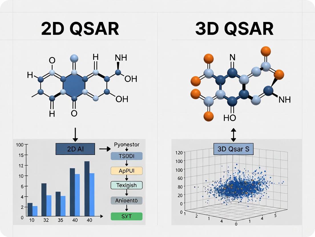
2D vs 3D QSAR: A Comprehensive Comparison for Modern Drug Discovery
This article provides a thorough analytical comparison between 2D and 3D Quantitative Structure-Activity Relationship (QSAR) methodologies, addressing key considerations for researchers and drug development professionals.
2D vs 3D QSAR: A Comprehensive Comparison for Modern Drug Discovery
Abstract
This article provides a thorough analytical comparison between 2D and 3D Quantitative Structure-Activity Relationship (QSAR) methodologies, addressing key considerations for researchers and drug development professionals. We explore the foundational principles distinguishing descriptor-based and spatial modeling approaches, examine their methodological implementations across therapeutic areas including cancer, infectious diseases, and neurological disorders, and analyze practical optimization strategies integrating machine learning and AI. The content delivers critical validation frameworks and performance comparisons to guide method selection, supported by recent case studies demonstrating successful applications in lead optimization and overcoming drug resistance. This resource aims to equip scientists with the knowledge to strategically implement QSAR approaches that accelerate and enhance their drug discovery pipelines.
Understanding QSAR Dimensions: From Molecular Descriptors to 3D Fields
Quantitative Structure-Activity Relationship (QSAR) modeling represents a cornerstone computational approach in chemical and pharmaceutical research that mathematically correlates molecular structural features with biological activity or chemical properties. Among QSAR methodologies, 2D-QSAR stands as a fundamental approach that utilizes two-dimensional molecular descriptors to build predictive models without requiring three-dimensional structural information. This guide examines the historical context, core principles, and methodological framework of 2D-QSAR modeling while objectively comparing its performance capabilities against contemporary 3D-QSAR approaches. Through systematic analysis of experimental protocols, validation metrics, and case studies across therapeutic domains, we provide researchers with a comprehensive reference for selecting and implementing appropriate QSAR strategies in drug discovery pipelines.
Historical Development and Theoretical Foundations
The conceptual foundation of QSAR emerged in the 1960s with the pioneering work of Corwin Hansch and Toshio Fujita, who established the first systematic approach to correlate biological activity with physicochemical parameters through linear free-energy relationships. This Hansch analysis paradigm represented the genesis of modern 2D-QSAR by demonstrating that substituent effects on biological activity could be quantified using parameters such as hydrophobicity (logP) and electronic properties (Hammett constants) [1]. The subsequent decades witnessed substantial methodological evolution, with the introduction of topological descriptors encoding molecular connectivity patterns and the development of increasingly sophisticated statistical approaches for model building [1].
The core theoretical premise of 2D-QSAR rests upon the fundamental principle that biological activity (or chemical properties) of compounds can be expressed as a mathematical function of their structural features, formally represented as:
Activity = f(physicochemical properties and/or structural properties) + error [1]
Unlike its 3D counterpart, 2D-QSAR does not require molecular alignment or conformational analysis, instead relying exclusively on descriptors derived from molecular graph representations that are invariant to rotation and translation [1] [2]. This descriptor-based approach calculates molecular features directly from the 2D molecular structure, encompassing a wide spectrum of physicochemical, topological, and electronic parameters that collectively encode information critical for biological interactions [3] [2].
Core Principles and Methodological Framework of 2D-QSAR
Essential Molecular Descriptors in 2D-QSAR
2D-QSAR modeling employs an extensive array of molecular descriptors that quantitatively encode structural information. These descriptors are computationally derived directly from the 2D molecular structure without spatial conformation and can be categorized into several distinct classes based on the structural properties they represent [3] [1] [2].
Table 1: Classification of Fundamental 2D-QSAR Molecular Descriptors
| Descriptor Category | Specific Examples | Structural Interpretation | Biological Relevance |
|---|---|---|---|
| Physicochemical Properties | LogP, molar refractivity, polar surface area, molecular weight | Hydrophobicity, bulkiness, polarity, size | Membrane permeability, solubility, absorption |
| Topological Descriptors | Connectivity indices (order 0, 1, 2), Wiener index, Zagreb index | Molecular branching, shape, atom connectivity | Molecular recognition, binding affinity |
| Electrostatic Descriptors | Partial atomic charges, HOMO/LUMO energies, ionization potential, electron affinity | Electron density distribution, nucleophilicity/electro-philicity | Electronic interactions, reaction mechanisms |
| Geometrical Descriptors | Molecular volume, surface area, shadow indices | Molecular size and shape | Steric complementarity to biological targets |
| Quantum Chemical Descriptors | Heat of formation, dipole moment, dipole energy [3] | Electronic state, energy, reactivity | Binding interactions, reaction rates |
The 2D-QSAR Model Development Workflow
The construction of a robust 2D-QSAR model follows a systematic, iterative process comprising distinct stages from data collection through model validation. The following diagram illustrates this standardized workflow:
Figure 1: Standardized workflow for developing 2D-QSAR models, illustrating the sequential stages from data collection to model application.
Data Collection and Preprocessing
The initial phase involves assembling a structurally diverse dataset of compounds with corresponding biological activity values, typically expressed as half-maximal inhibitory concentration (IC₅₀), minimum inhibitory concentration (MIC), or other potency measures [3] [4]. These experimental values are converted to logarithmic scale (pIC₅₀ = -logIC₅₀) to linearize the relationship with free energy changes [4]. The dataset is then partitioned into training and test sets, typically following an 70:30 to 80:20 ratio, to ensure sufficient compounds for model development while retaining an external validation subset [4] [5].
Molecular Optimization and Descriptor Calculation
Molecular structures undergo computational optimization using molecular mechanics (MM2/MM3) or semi-empirical quantum chemical methods (AM1, PM3) to achieve minimum energy conformations [3] [5]. Subsequently, specialized software platforms calculate a comprehensive set of 2D descriptors encompassing constitutional, topological, geometrical, and quantum chemical parameters [3] [2]. Descriptor redundancy is addressed through elimination of constant or near-constant variables and correlation analysis to remove highly intercorrelated descriptors that can lead to model overfitting [2].
Model Development and Validation
Statistical techniques ranging from multiple linear regression (MLR) to advanced machine learning methods (Random Forest, Support Vector Machines, Neural Networks) are employed to establish mathematical relationships between descriptors and biological activity [6] [2] [7]. Model robustness is evaluated using internal validation techniques such as leave-one-out (LOO) or leave-group-out (LGO) cross-validation, while predictive ability is assessed using the external test set [3] [6] [4]. Additional validation through Y-scrambling ensures the absence of chance correlations [1].
Experimental Protocols and Validation Metrics
Case Study: 2D-QSAR Model for Anti-Tuberculosis Compounds
A representative 2D-QSAR investigation was conducted on natural products and derivatives with anti-tuberculosis activity against Mycobacterium tuberculosis [3]. The study compiled 79 compounds with reported minimum inhibitory concentration (MIC) values, which were converted to -log(MIC) to serve as the dependent variable. Molecular structures were optimized using the MO-G computational application with augmented Molecular Mechanics (MM2/MM3) parameters, with minimization continuing until energy changes fell below 0.001 kcal/mol or after 300 iterations [3].
The researchers calculated 42 different physicochemical descriptors using the QSAR module of Scigress Explorer software, followed by forward feed multiple linear regression with leave-one-out cross-validation to identify the most significant descriptors [3]. The resulting model demonstrated a relationship correlating measure of 74% (R² = 0.74) and predictive accuracy of 72% (R²CV = 0.72), with dipole energy and heat of formation identified as the most significant descriptors correlating with anti-tubercular activity [3].
Case Study: 2D-QSAR Model for NSCLC Therapeutic Agents
In a separate study focusing on non-small cell lung cancer (NSCLC), researchers developed a 2D-QSAR model using 45 tetrahydropyrazolo-quinazoline and tetrahydropyrazolo-pyrimidocarbazole derivatives [4]. Antiproliferative activities against A549 NSCLC cell lines (IC₅₀ values) were converted to pIC₅₀ using the relationship pIC₅₀ = -log(IC₅₀ × 10⁻⁶) [4].
The resulting model exhibited strong predictive performance with assessment parameters R² = 0.798, adjusted R² = 0.754, cross-validated Q² = 0.673, and external test set R² = 0.531 [4]. These validation metrics comprehensively demonstrate model robustness and predictive capability for identifying novel NSCLC therapeutic agents.
Comparative Analysis: 2D-QSAR versus 3D-QSAR
Performance Metrics Across Methodologies
Direct comparison of 2D and 3D-QSAR approaches reveals distinct advantages and limitations for each methodology. The following table synthesizes performance data from multiple studies to facilitate objective comparison:
Table 2: Comparative Performance Analysis of 2D-QSAR and 3D-QSAR Approaches Across Multiple Studies
| Study Context | QSAR Method | Statistical Performance | Key Advantages | Methodological Limitations |
|---|---|---|---|---|
| SARS-CoV-2 Mpro Inhibitors [7] | 2D-QSAR (MLP with Morgan fingerprints) | R² training=1.00, q² CV=0.80, R² test=0.72 | No molecular alignment required, rapid virtual screening | Limited insight into 3D structural requirements |
| 3D-QSAR (Field QSAR) | R² training=0.96, q² CV=0.81, R² test=0.71 | Visualizes 3D pharmacophoric requirements | Alignment-sensitive, computationally intensive | |
| Histamine H3 Receptor Antagonists [6] | 2D-QSAR (MLR) | MAPE=2.9-3.6, SDEP=0.31-0.36 | Simpler implementation, comparable to advanced methods | Limited to QSAR prediction only |
| 2D-QSAR (ANN) | MAPE=2.9-3.6, SDEP=0.31-0.36 | Captures non-linear relationships | "Black box" interpretation challenges | |
| 3D-QSAR (HASL) | Inferior to 2D methods | - | Lower predictive accuracy in this application | |
| Organic Compound Degradation [8] | 2D-QSAR | R²=0.898, q²=0.841, Q²ext=0.968 | Excellent predictive power for reaction rates | Limited 3D interaction insights |
| 3D-QSAR (CoMSIA) | R²=0.952, q²=0.951, Q²ext=0.970 | Superior statistical fit, electrostatic field analysis | Requires conformer generation and alignment | |
| Dipeptide-Alkylated Nitrogen-Mustard Compounds [5] | 2D-QSAR (Linear HM) | R²=0.798 (from other study [4]) | Identifies key electronic descriptors | Limited to linear relationships |
| 2D-QSAR (Non-linear GEP) | Training R²=0.95, Test R²=0.87 | Captures complex non-linear patterns | Complex model interpretation | |
| 3D-QSAR (CoMSIA) | N/A provided | Visual guidance for molecular modification | Requires reliable alignment rules |
Methodological Differentiation and Selection Criteria
The comparative analysis reveals that 2D-QSAR approaches generally excel in predictive accuracy for biological activity when 3D structural requirements are either well-encoded in 2D descriptors or when the dataset contains congeneric series with conserved binding modes [6] [7]. The strength of 2D-QSAR lies in its computational efficiency, minimal pre-processing requirements, and exceptional suitability for high-throughput virtual screening of large chemical libraries [2].
Conversely, 3D-QSAR methodologies provide superior structural insights and stereochemical guidance for molecular modification, particularly valuable during lead optimization phases when detailed understanding of steric and electrostatic requirements is necessary [8] [7]. However, this enhanced structural insight comes with increased computational complexity, alignment sensitivity, and conformational dependence that can introduce variability in model performance [8] [5].
Selection between these approaches should be guided by specific research objectives: 2D-QSAR is recommended for rapid activity prediction and virtual screening applications, while 3D-QSAR is more appropriate for molecular design and optimization tasks requiring spatial understanding of structure-activity relationships [7].
Essential Research Reagents and Computational Tools
Successful implementation of 2D-QSAR modeling requires both computational tools and cheminformatics resources. The following table details essential solutions for conducting 2D-QSAR research:
Table 3: Essential Research Reagent Solutions for 2D-QSAR Modeling
| Tool Category | Specific Solutions | Application Function | Key Features |
|---|---|---|---|
| Molecular Modeling Platforms | Scigress Explorer [3], HyperChem [5] | Molecular structure optimization and descriptor calculation | MM2/MM3 force fields, semi-empirical methods (AM1, PM3) |
| Descriptor Calculation Software | CODESSA [5], RDKit [2] [7] | Comprehensive descriptor calculation | Constitutional, topological, electrostatic, quantum chemical descriptors |
| Statistical Analysis Environments | Flare Python API [2], Scikit-learn | Model development and validation | Multiple Linear Regression, Random Forest, SVM, Neural Networks |
| Cheminformatics Toolkits | RDKit [7], OpenBabel | Molecular representation and manipulation | SMILES parsing, fingerprint generation, substructure search |
| Validation Tools | Custom Python/R scripts [2], QSAR model validation scripts | Model robustness assessment | Cross-validation, Y-scrambling, applicability domain analysis |
2D-QSAR maintains a fundamental position in the computational chemistry toolbox, offering robust predictive capability for biological activity and chemical properties through efficient descriptor-based modeling. Its historical development reflects continuous methodological refinement, while its core principles remain grounded in establishing quantitative relationships between structural features and biological responses. The comparative analysis presented herein demonstrates that 2D-QSAR delivers predictive performance comparable to, and in some cases superior to, 3D-QSAR approaches while requiring fewer computational resources and avoiding complex molecular alignment procedures.
For research applications prioritizing high-throughput screening and rapid activity prediction, 2D-QSAR represents an optimal methodology, particularly during early discovery phases. The integration of modern machine learning approaches with traditional 2D descriptors continues to expand the capabilities of this established paradigm, ensuring its ongoing relevance in contemporary drug discovery workflows. As computational power increases and descriptor sets become more sophisticated, 2D-QSAR methodology will continue to evolve, maintaining its essential role in rational drug design and chemical property prediction.
Theoretical Foundations: Beyond Flatland - The Shift from 2D to 3D Descriptors
Quantitative Structure-Activity Relationship (QSAR) methodologies are foundational to contemporary drug design, enabling the prediction of a molecule's biological activity based on its chemical and physical characteristics. While classical 2D-QSAR correlates one- or two-dimensional molecular descriptors (e.g., molecular weight, logP) with biological activity, 3D-QSAR represents a natural extension that exploits the three-dimensional properties of the ligands to build more predictive and mechanistically insightful models [9] [10]. This paradigm shift is crucial because drug interactions occur in three-dimensional space; a molecule's biological effect is determined not just by its constituent atoms, but by their precise spatial arrangement and the resulting molecular fields [10].
The core principle of 3D-QSAR is that the difference in the three-dimensional structural properties of molecules is responsible for variations in their biological activities [9]. These methods apply empirical force field calculations on three-dimensionally aligned ligand structures, allowing for the analysis of steric, electrostatic, hydrophobic, and hydrogen-bonding fields surrounding the molecules [11] [12]. This provides a superior level of insight compared to 2D methods, as it helps identify specific regions in space where particular molecular features enhance or diminish biological activity.
A Comparative Framework: 2D-QSAR vs. 3D-QSAR
The choice between 2D and 3D approaches hinges on the research question, available data, and desired outcome. The table below summarizes their core distinctions.
| Feature | 2D-QSAR | 3D-QSAR |
|---|---|---|
| Molecular Representation | 1D and 2D descriptors (e.g., logP, molecular weight, topological indices) [10]. | 3D structure and conformation-dependent fields (e.g., steric, electrostatic) [10] [12]. |
| Descriptor Basis | Physicochemical, electronic, and topological properties derived from molecular formula or graph [10]. | Interaction energies (steric, electrostatic) calculated between a probe and the molecule at grid points [12]. |
| Core Requirement | A set of compounds with known activity [10]. | A set of compounds with known activity and a valid spatial alignment rule [12]. |
| Key Advantage | Computationally fast; does not require molecular conformation or alignment [10]. | Provides visual, interpretable maps of favorable/unfavorable chemical regions [13] [12]. |
| Primary Limitation | Lacks spatial context, limiting mechanistic insight for receptor-based design [9]. | Highly dependent on the correctness of molecular alignment and conformation choice [12]. |
| Typical Applications | Early-stage profiling for ADMET properties, rapid virtual screening [10] [14]. | Lead optimization, understanding structure-activity relationships, and designing novel analogs [9] [12]. |
Performance and Predictive Power: Experimental Comparisons
Direct comparisons in scientific studies reveal that the performance of 2D and 3D methods can be context-dependent.
Case Study 1: Predicting H3 Receptor Antagonist Binding A study on arylbenzofuran-derived histamine H3 receptor antagonists compared Multiple Linear Regression (MLR- a 2D method), Artificial Neural Networks (ANN- a more complex 2D method), and HASL (a 3D-QSAR method). The results demonstrated that the simpler 2D method was highly competitive [6].
- MLR (2D): Mean Absolute Percentage Error (MAPE) = 2.9-3.6, Standard Deviation of Error of Prediction (SDEP) = 0.31-0.36 [6].
- ANN (2D): MAPE = 2.9-3.6, SDEP = 0.31-0.36 [6].
- HASL (3D): Results "were not as good as those obtained by 2D methods" [6].
The study concluded that "simple traditional approaches such as MLR method can be as reliable as... more advanced and sophisticated methods like ANN and 3D-QSAR analyses" [6].
Case Study 2: Corrosion Inhibition by Pyrazole Derivatives In contrast, research on pyrazole corrosion inhibitors showed that modern machine learning models applied to 3D descriptors can achieve excellent predictive performance, sometimes surpassing their 2D counterparts [15].
- XGBoost with 2D Descriptors: Training set R² = 0.96; Test set R² = 0.75 [15].
- XGBoost with 3D Descriptors: Training set R² = 0.94; Test set R² = 0.85 [15].
This indicates that for this specific dataset, the 3D descriptor model generalized better to the test set, showing higher predictive R².
The 3D-QSAR Workflow: From Structures to Predictive Models
The development of a robust 3D-QSAR model follows a meticulous, multi-stage process. The workflow below outlines the critical steps from initial data preparation to final model application.
Detailed Experimental Protocols
Step 1: Conformational Analysis and Selection The first critical step is generating a biologically relevant 3D conformation for each molecule. This often involves energy minimization using molecular mechanics (MM) force fields (e.g., MM3, MM+) or semi-empirical methods (e.g., AM1) to find low-energy conformers [11] [12]. For molecules with flexible rotatable bonds, a systematic scan of dihedral angles may be performed to reflect energetically preferred conformations, as the choice of conformation can profoundly impact the model's quality [12].
Step 2: Molecular Alignment This is the most crucial step that defines the model's frame of reference. Molecules must be superimposed in 3D space based on a common hypothesis. Common protocols include:
- Common Substructure Alignment: A shared core structure (e.g., a pyridinium ring in MNVP+ analogs) is used for fitting atoms [12].
- Pharmacophore-Based Alignment: Molecules are aligned based on their shared pharmacophoric features (e.g., hydrogen bond donors/acceptors, aromatic rings).
- Database Mining: Using the
Select KBestapproach or Genetic Algorithm-Partial Least Squares (GA-PLS) to select the most relevant descriptors from a large pool (e.g., >1000 descriptors from software like Dragon) for model building [15] [11].
Step 3: Field Calculation and Model Building The aligned molecules are placed in a 3D grid. A probe atom (e.g., an sp³ carbon with a +1 charge) is used to calculate steric (Lennard-Jones) and electrostatic (Coulombic) interaction energies at each grid point. These interaction energies become the independent variables (descriptors) for the model [12]. Statistical methods like Partial Least Squares (PLS) are then used to correlate these field variables with the biological activity data [10] [12]. Modern implementations may also use advanced machine learning techniques like Gaussian Process Regression (GPR) or similarity descriptors based on molecular shape and electrostatics to build consensus models [13].
The Scientist's Toolkit: Essential Reagents and Software
A range of specialized software is available to execute the complex workflow of 3D-QSAR modeling.
| Tool Name | Type | Primary Function in 3D-QSAR |
|---|---|---|
| OpenEye's 3D-QSAR [13] | Commercial Software | Creates consensus models using shape/electrostatic similarity descriptors (ROCS, EON) and machine learning (kPLS, GPR). Valued for interpretable visual results. |
| 3D-QSAR.com [16] | Web Platform | Offers user-friendly, web-based tools for developing ligand-based and structure-based 3D-QSAR models and managing molecular datasets. |
| SYBYL (with CoMFA/CoMSIA) [12] | Commercial Suite | The classic industry platform for performing Comparative Molecular Field Analysis (CoMFA) and Comparative Molecular Similarity Indices Analysis (CoMSIA). |
| QSAR Toolbox [17] | Freemium Software | A data-rich platform primarily for chemical hazard assessment, supporting category formation, read-across, and the ability to run external QSAR models. |
| Open3DQSAR [14] | Open-Source Tool | Provides transparent 3D-QSAR analysis for scientists who prefer an open-source and flexible environment for their research. |
The exploration of 3D-QSAR underscores its unique value in drug discovery: it translates abstract chemical structures into visual, three-dimensional maps that guide medicinal chemists toward more potent compounds. While 2D-QSAR remains a powerful tool for rapid property prediction, 3D-QSAR provides superior mechanistic insight during lead optimization by highlighting the spatial and electronic features critical for binding [12].
The future of 3D-QSAR is being shaped by integration with advanced artificial intelligence. Modern implementations are moving beyond traditional PLS, incorporating machine learning algorithms like XGBoost and CatBoost to handle molecular field data [15]. Furthermore, the drive for interpretability is being addressed by techniques like SHAP analysis, which identifies key descriptors and validates the relevance of selected variables, strengthening the model's reliability [15]. As these tools become more accessible and integrated into user-friendly web platforms and automated workflows, 3D-QSAR will continue to be a cornerstone of rational drug design.
Quantitative Structure-Activity Relationship (QSAR) modeling represents a cornerstone of modern computational chemistry and drug discovery, providing crucial mathematical frameworks that connect a molecule's chemical structure to its biological activity or physicochemical properties. At the heart of every QSAR model lie molecular descriptors—quantitative representations of structural features that enable this predictive capability. These descriptors are broadly categorized into four fundamental classes that form the foundation of structure-activity analysis: electronic, steric, hydrophobic, and quantum-chemical parameters.
The evolution of QSAR from its beginnings over 40 years ago has dramatically expanded both the breadth and depth of descriptor utilization. According to historical context found in the search results, traditional QSAR modeling was initially viewed strictly as an analytical physical chemical approach applicable only to small congeneric series of molecules [18]. The technique was first introduced by Hansch et al. based on implications from linear free-energy relationships and the Hammett equation, operating on the fundamental assumption that differences in physicochemical properties account for differences in biological activities of compounds [18]. These core structural properties remain essential to modern QSAR practices, though their calculation and application have grown increasingly sophisticated.
This guide examines these key descriptor categories within the broader research framework comparing 2D versus 3D QSAR approaches—two distinct paradigms that utilize molecular descriptors in fundamentally different ways to predict chemical behavior and biological activity. Understanding these descriptor categories and their implementation across QSAR methodologies provides researchers with critical insights for selecting appropriate modeling strategies in drug development and chemical property prediction.
Theoretical Foundations: The Four Key Descriptor Categories
Electronic Parameters
Electronic parameters quantify the distribution of electrons within a molecule, directly influencing intermolecular interactions, binding affinity, and chemical reactivity. These descriptors capture a molecule's ability to participate in electrostatic interactions, hydrogen bonding, and charge-transfer complexes—critical factors in drug-receptor recognition and binding.
The Hammett constant (σ) represents a classical electronic parameter that describes the electron-donating or withdrawing properties of substituents through their influence on ionization constants of benzoic acid derivatives [18]. Modern computational approaches have expanded this concept to include molecular dipole moments, atomic partial charges, and electrostatic potential maps derived from quantum mechanical calculations. In 3D-QSAR methods like CoMFA (Comparative Molecular Field Analysis), electronic properties are sampled using probe atoms to generate electrostatic potential fields around aligned molecules, providing rich data for activity prediction [6] [7].
Steric Parameters
Steric parameters describe the spatial occupancy and shape characteristics of molecules, encompassing size, volume, and topological dimensions that influence molecular fit into biological targets and accessibility to reaction sites. Steric effects can either facilitate or hinder biological activity through complementarity with receptor binding pockets.
Traditional steric descriptors include Verloop STERIMOL parameters [18] and Taft's steric constants, which quantify the spatial requirements of substituents. Contemporary 3D-QSAR approaches implement sophisticated steric field calculations using van der Waals probes and shape-based alignment algorithms [7]. The Cresset Field 3D-QSAR method, for instance, utilizes molecular field points from the XED force field to sample volume and shape characteristics for each molecule in the training set, generating descriptors that capture subtle steric influences on activity [7].
Hydrophobic Parameters
Hydrophobic parameters quantify the relative affinity of molecules or substituents for lipophilic versus aqueous environments, directly influencing transport properties, membrane permeability, and binding interactions driven by desolvation effects. Hydrophobicity represents a critical determinant in drug absorption, distribution, and bioavailability.
The octanol-water partition coefficient (Log P) and its pH-dependent counterpart Log D serve as the fundamental quantitative measures of hydrophobicity [18]. In classical QSAR, Hansch substituent hydrophobicity constants (π) parameterize the contribution of specific substituents to overall molecular lipophilicity [18]. Modern descriptor calculations incorporate these principles through computational methods that estimate partition coefficients and solvent-accessible surface areas, with tools like ACDlabs and Dragon software routinely generating these parameters for QSAR modeling [6] [11].
Quantum-Chemical Parameters
Quantum-chemical parameters derive from quantum mechanical calculations and describe electronic structure properties with higher theoretical rigor than classical electronic parameters. These descriptors provide insights into reactivity, stability, and intermolecular interaction capabilities that directly influence biological activity.
Key quantum-chemical descriptors include the energies of the highest occupied and lowest unoccupied molecular orbitals (HOMO and LUMO), which determine nucleophilicity and electrophilicity respectively; molecular orbital coefficients that predict reaction regioselectivity; and ionization potentials influencing electron-transfer capabilities [11]. Studies demonstrate these parameters can be calculated using semiempirical methods like AM1, with software packages such as Hyperchem enabling their computation for QSAR modeling [11]. The inclusion of quantum-chemical descriptors often enhances model interpretability by connecting observed activities to fundamental electronic structure principles.
Comparative Analysis: 2D vs. 3D QSAR Descriptor Implementation
Fundamental Methodological Differences
The distinction between 2D and 3D QSAR approaches fundamentally resides in how molecular structures are represented and compared. 2D-QSAR utilizes descriptors derived from molecular constitution and topology without explicit spatial reference, while 3D-QSAR incorporates the three-dimensional arrangement of atoms and functional groups, requiring molecular alignment to a common framework [6].
2D-QSAR methods employ numerical descriptors that can be calculated directly from molecular connection tables or 2D structural representations. These include constitutional descriptors (atom and bond counts, molecular weight), topological indices (connectivity indices, path counts), and electronic parameters calculated without spatial coordinates [11]. The relative computational simplicity of 2D descriptors enables rapid screening of large compound libraries without conformational analysis or alignment requirements.
3D-QSAR approaches extend this paradigm by incorporating spatial molecular fields and properties sampled in three dimensions. Methods such as CoMFA (Comparative Molecular Field Analysis), HASL (Hypothetical Active Site Lattice), and Field 3D-QSAR utilize probe atoms to map electrostatic and steric properties onto grid points surrounding aligned molecules [6] [7]. This generates significantly larger descriptor sets that capture shape complementarity and spatial property distributions relevant to biological recognition.
Performance Comparison: Predictive Accuracy and Applications
Direct comparative studies provide valuable insights into the relative performance of 2D and 3D QSAR methodologies across different applications. A 2012 study comparing Multiple Linear Regression (MLR), Artificial Neural Networks (ANN), and HASL 3D-QSAR for predicting histamine H3 receptor antagonist activity found that 2D methods (MLR and ANN) outperformed the 3D HASL approach in prediction accuracy [6]. The calculated values for the mean absolute percentage error (MAPE) ranged from 2.9 to 3.6 for both MLR and ANN methods, while results from 3D-QSAR studies using HASL were not as robust [6].
Conversely, a 2025 study on pyrazole corrosion inhibitors demonstrated strong predictive ability for both 2D and 3D descriptors using XGBoost machine learning models, with the XGBoost model demonstrating R² = 0.96 for the 2D training set and R² = 0.94 for the 3D training set [15]. The test set performance remained solid with R² = 0.75 for 2D descriptors and R² = 0.85 for 3D descriptors, suggesting contextual dependence of method efficacy [15].
Recent SARS-CoV-2 Mpro inhibitor studies using Cresset's Flare software showed comparable predictive performance between 2D and 3D approaches, with both methods generating models with test set r² values around 0.72 [7]. However, the 3D-QSAR approach provided additional interpretability through visualization of regions where the model predicts strong effects on activity, highlighting the value of 3D methods beyond pure predictive accuracy [7].
Table 1: Performance Comparison of 2D vs. 3D QSAR Approaches in Recent Studies
| Study Focus | 2D Method Performance | 3D Method Performance | Best Performing Algorithm | Key Findings |
|---|---|---|---|---|
| Pyrazole corrosion inhibitors [15] | Training: R² = 0.96, Test: R² = 0.75 | Training: R² = 0.94, Test: R² = 0.85 | XGBoost | 3D descriptors showed superior test set performance |
| SARS-CoV-2 Mpro inhibitors [7] | Test set r² = 0.72 (MLP with Morgan fingerprints) | Test set r² = 0.72 (MLP 3D-QSAR) | Multiple (comparable performance) | Both methods showed comparable predictive accuracy |
| Histamine H3 receptor antagonists [6] | MAPE: 2.9-3.6, SDEP: 0.31-0.36 | Inferior to 2D methods | MLR and ANN | Traditional 2D methods outperformed 3D HASL approach |
Mechanistic Interpretability and Visualization Capabilities
A critical distinction between 2D and 3D QSAR approaches lies in their capacity for mechanistic interpretation and structural insight. While 2D-QSAR models often provide excellent predictive capability, they typically function as "black boxes" with limited direct translation to structural modifications [6].
3D-QSAR methods excel in interpretability, generating visual representations of molecular regions where specific property enhancements would improve activity. For example, the Cresset Field 3D-QSAR method illustrates electrostatic and steric model coefficients superimposed on molecular structures, identifying favorable and unfavorable regions for modification [7]. In SARS-CoV-2 Mpro inhibitor studies, this approach identified specific molecular regions where less positive charge would improve activity and highlighted the 2-chlorobenzyl moiety as the optimal region for steric modification [7].
This spatial interpretability provides medicinal chemists with direct structural insights for molecular optimization—a significant advantage over the often-opaque correlation equations generated by 2D-QSAR models. The 3D-QSAR contour maps effectively serve as visual guides for rational drug design, indicating where specific steric or electronic modifications would likely enhance biological activity.
Experimental Protocols and Methodologies
Standard 2D-QSAR Development Workflow
The development of robust 2D-QSAR models follows a systematic protocol encompassing data collection, descriptor calculation, feature selection, model building, and validation [19] [20].
Step 1: Data Set Curation and Preparation
- Collect biological activity data (e.g., IC₅₀, Ki, EC₅₀) for a congeneric series of compounds
- Apply inclusion criteria based on data quality and structural diversity
- Divide compounds into training and test sets using activity stratification to ensure representative distribution [7]
Step 2: Molecular Descriptor Calculation
- Generate 2D structures and optimize using molecular mechanics (MM+ force field) or semiempirical methods (AM1) [11]
- Calculate descriptors using software packages such as Dragon, Hyperchem, or ACDlabs [11]
- Compute diverse descriptor types including constitutional, topological, geometrical, and quantum-chemical parameters [11]
Step 3: Descriptor Selection and Reduction
- Apply feature selection algorithms like Genetic Algorithm-Partial Least Squares (GA-PLS) to identify most relevant descriptors [11]
- Remove highly correlated descriptors (e.g., correlation threshold >0.95) to reduce multicollinearity [11]
- Retain approximately 10% of initially calculated descriptors for model building [11]
Step 4: Model Development and Validation
- Implement multiple algorithms: Multiple Linear Regression (MLR), Partial Least Squares (PLS), Artificial Neural Networks (ANN), Support Vector Machines (SVM), Random Forest (RF), or Deep Neural Networks (DNN) [6] [21]
- Perform internal validation using leave-one-out or leave-many-out cross-validation [20]
- Conduct external validation using the predetermined test set [19] [20]
- Apply stringent validation criteria including Golbraikh and Tropsha parameters, concordance correlation coefficient (CCC), and residual analysis [20]
Standard 3D-QSAR Development Workflow
3D-QSAR methodologies incorporate additional steps for molecular alignment and spatial field calculation, introducing complexity but enhancing interpretability [7].
Step 1: Molecular Modeling and Conformation Generation
- Build 3D molecular structures from 2D representations
- Perform conformational search within a specified energy window (typically 2.5 kcal/mol) [7]
- Select biologically relevant conformations based on experimental data or computational docking
Step 2: Molecular Alignment
- Align molecules to a common reference framework using maximum common substructure (MCS) algorithms [7]
- Utilize crystallographic ligand coordinates from protein-ligand complexes when available [7]
- Apply alternative alignment rules to assess model sensitivity to alignment method
Step 3: Molecular Field Calculation
- Sample steric and electrostatic fields using probe atoms on 3D grid points surrounding aligned molecules
- Implement standard probes: sp³ carbon atom for steric fields and H⁺ ion for electrostatic fields [7]
- Calculate interaction energies using appropriate force fields (e.g., Cresset XED force field) [7]
Step 4: Model Building and Validation
- Apply Partial Least Squares (PLS) regression to correlate field values with biological activity
- Implement machine learning approaches including k-Nearest Neighbors (kNN), SVM, Gaussian Process Regression (GPR), Random Forest (RF), and Multilayer Perceptron (MLP) for 3D-QSAR [7]
- Perform cross-validation and external validation using the same rigorous standards as 2D-QSAR
- Generate 3D coefficient contour maps for visual interpretation of results [7]
Machine Learning Integration in Modern QSAR
Contemporary QSAR practices increasingly incorporate advanced machine learning algorithms that transcend the traditional 2D/3D dichotomy. Studies demonstrate that methods like Deep Neural Networks (DNN) and Random Forest (RF) can achieve superior predictive performance compared to traditional regression-based QSAR approaches [21].
In comparative studies using the same dataset and molecular descriptors, machine learning methods (DNN and RF) exhibited higher prediction accuracy (r² ~0.90) than traditional QSAR methods (PLS and MLR with r² ~0.65) when sufficient training data was available [21]. Notably, with smaller training sets, DNN maintained higher predictive performance (r² = 0.94) compared to RF (r² = 0.84), while traditional methods showed significant performance degradation [21].
Table 2: Performance of Computational Algorithms in QSAR Modeling
| Algorithm Type | Representative Methods | Strengths | Limitations | Ideal Use Cases |
|---|---|---|---|---|
| Traditional Regression | MLR, PLS | Interpretable models, minimal computational requirements | Limited complexity handling, prone to overfitting with many descriptors | Small congeneric series, preliminary analysis |
| Machine Learning | RF, SVM, XGBoost | Handles complex nonlinear relationships, robust with large descriptor sets | "Black box" nature, extensive parameter tuning required | Diverse compound sets, large descriptor spaces |
| Deep Learning | DNN, ANN | Automatic feature detection, excels with large datasets | Extensive data requirements, computational intensity | Very large datasets, complex structure-activity relationships |
| 3D-QSAR Specific | Field 3D-QSAR, CoMFA, HASL | High interpretability, spatial visualization | Alignment sensitivity, computational expense | Lead optimization, structure-based design |
The Scientist's Toolkit: Essential Research Reagent Solutions
Successful QSAR modeling requires specialized software tools and computational resources for descriptor calculation, model development, and validation. The following table summarizes key solutions used in contemporary QSAR research, as identified from experimental protocols in the search results.
Table 3: Essential Computational Tools for QSAR Research
| Tool Category | Specific Software/Platform | Primary Function | Key Features | Applicable QSAR Type |
|---|---|---|---|---|
| Molecular Modeling | Hyperchem [11] | 3D structure generation and optimization | MM+ force field, AM1 semiempirical calculations | Both 2D and 3D QSAR |
| Descriptor Calculation | Dragon [11] | Comprehensive descriptor calculation | 1481+ 1D, 2D, and 3D molecular descriptors | Primarily 2D-QSAR |
| Descriptor Calculation | ACDlabs [11] | Physicochemical property prediction | Log D, pKa, molar volume, parachor | Primarily 2D-QSAR |
| 3D-QSAR Specific | Cresset Flare [7] | 3D-QSAR model development | Field 3D-QSAR, XED force field, molecular alignment | Primarily 3D-QSAR |
| 3D-QSAR Specific | HASL [6] | 3D-QSAR using lattice approach | Hypothetical Active Site Lattice generation | Primarily 3D-QSAR |
| Cheminformatics | RDKit [7] | Open-source cheminformatics | Molecular fingerprints, descriptor calculation | Both 2D and 3D QSAR |
| Machine Learning | Scikit-learn, TensorFlow [21] | Advanced model development | DNN, RF, SVM, and other ML algorithms | Both 2D and 3D QSAR |
| Validation Tools | Various statistical packages [20] | Model validation and assessment | External validation parameters, applicability domain | Both 2D and 3D QSAR |
The comparative analysis of electronic, steric, hydrophobic, and quantum-chemical parameters across 2D and 3D QSAR methodologies reveals a nuanced landscape where each approach offers distinct advantages depending on research objectives. 2D-QSAR provides computational efficiency, straightforward implementation, and strong predictive performance for congeneric series, making it ideal for high-throughput screening and preliminary SAR analysis. Conversely, 3D-QSAR delivers superior mechanistic interpretability through spatial visualization of molecular regions critical for activity, offering invaluable guidance for lead optimization in advanced drug discovery stages.
Contemporary research trends indicate a paradigm shift toward hybrid approaches that leverage the strengths of both methodologies. Machine learning algorithms increasingly bridge the gap between descriptor types, handling complex relationships in high-dimensional data spaces while providing robust predictive models [21]. The critical importance of rigorous validation—particularly external validation with compounds outside the training set—cannot be overstated for both approaches, as this remains the ultimate measure of model utility and reliability [19] [20].
Strategic selection between 2D and 3D QSAR approaches should be guided by specific research context: available computational resources, dataset characteristics, project timeline, and ultimately, whether the primary need is quantitative prediction or structural insight for molecular design. As computational power increases and algorithms evolve, the integration of these complementary approaches will continue to enhance our ability to translate chemical structure into predictive understanding of biological activity.
Quantitative Structure-Activity Relationship (QSAR) modeling represents a cornerstone of computational medicinal chemistry, providing critical frameworks for predicting biologically relevant properties of chemical compounds. The theoretical foundations of modern QSAR rest upon three pivotal approaches: the Hammett equation, which introduced quantitative electronic parameters; the Hansch-Fujita method, which incorporated hydrophobic and steric effects; and the Free-Wilson model, which established the concept of group additivity. These classical methodologies form the essential groundwork upon which contemporary 2D and 3D-QSAR techniques have been built [22] [23]. Understanding these foundational approaches is prerequisite to meaningful comparison of modern 2D versus 3D-QSAR strategies in current drug discovery pipelines.
The historical development of QSAR began with observations by Meyer, Overton, and Ferguson regarding correlations between lipophilicity and biological activity [22] [23]. The field formally emerged in the early 1960s through independent work by Hansch and Fujita and by Free and Wilson, building upon Hammett's earlier linear free-energy relationships for chemical reactivity [18] [23]. These approaches established the principle that molecular properties could be quantitatively correlated with biological response, enabling predictive modeling in drug design.
Theoretical Foundations and Mathematical Formulations
Hammett Electronic Constants
The Hammett equation, developed by Louis Hammett in the 1930s, represents one of the earliest applications of linear free-energy relationships (LFER). It quantitatively describes how electronic effects of substituents influence reaction rates or equilibrium constants for organic reactions [23].
Mathematical Formulation:
Where:
- K = Rate or equilibrium constant for substituted compound
- K₀ = Rate or equilibrium constant for unsubstituted compound
- ρ = Reaction constant (sensitivity to substituent effects)
- σ = Substituent constant (electron-donating/withdrawing ability)
Hammett constants (σ) provide quantitative descriptors of substituent electronic properties, with positive values indicating electron-withdrawing groups and negative values indicating electron-donating groups [18] [23]. While originally developed for chemical reactivity, these principles formed the conceptual basis for extending quantitative relationships to biological systems.
Hansch-Fujita Analysis
Hansch and Fujita revolutionized QSAR by extending Hammett's electronic parameters to include hydrophobic and steric effects, creating a multiparameter approach that could model complex biological interactions [24] [22] [23]. This approach correlates biological activity with various physicochemical properties through multiple regression analysis.
Linear Hansch Equation:
Nonlinear Hansch Equation (accounting for parabolic lipophilicity):
Where:
- C = Molar concentration producing biological effect
- log P = Lipophilicity parameter (octanol-water partition coefficient)
- σ = Hammett electronic constant
- Eₛ = Taft steric parameter
- a, b, c, d = Coefficients determined by regression analysis
The Hansch approach operates on the principle that biological activity depends on a combination of transport properties (governed largely by lipophilicity) and interactions with the target site (influenced by electronic and steric properties) [24] [22].
Free-Wilson Analysis
The Free-Wilson model, developed concurrently with the Hansch approach, provides a mathematically distinct methodology based on the concept of group contribution additivity [24] [22].
Mathematical Formulation:
Where:
- BA = Biological activity
- μ = Overall average biological activity (unsubstituted parent compound)
- aᵢⱼ = Contribution of substituent j at position i
- Σ(aᵢⱼ) = Sum of all substituent contributions
The model uses presence/absence of specific substituents at defined molecular positions as descriptor variables, which are correlated with biological activity using multiple linear regression [24] [22]. Each substituent's contribution is assumed to be constant and independent of other substituents in the molecule.
Mixed Approach
Recognizing the complementary strengths of Hansch and Free-Wilson methods, Kubinyi developed a mixed approach that integrates both methodologies [24] [22].
Mathematical Formulation:
Where:
- Σ(aᵢⱼ) = Free-Wilson component (group contributions)
- Σ(kᵢφⱼ) = Hansch component (physicochemical properties)
- k = Constant term
This hybrid approach leverages the group contribution basis of Free-Wilson analysis while incorporating the mechanistic insights provided by physicochemical parameters from the Hansch approach [24] [22].
Table 1: Comparative Analysis of Classical QSAR Approaches
| Feature | Hammett Equation | Hansch-Fujita Analysis | Free-Wilson Analysis |
|---|---|---|---|
| Fundamental Basis | Linear Free-Energy Relationships | Multiparameter Extrathermodynamic Approach | Group Contribution Additivity |
| Key Parameters | σ (electronic) | log P (lipophilic), σ (electronic), Eₛ (steric) | Indicator variables for substituents |
| Mathematical Form | Linear: log(K/K₀) = ρσ | Linear/Nonlinear regression | Multiple linear regression with indicator variables |
| Primary Application | Chemical reactivity | Complex biological systems | Congeneric series with multiple substitution sites |
| Key Assumptions | Electronic effects dominate; substituent effects are additive and constant | Biological activity depends on transport and interaction properties | Group contributions are constant and position-specific |
| Historical Context | 1930s (earliest QSAR concept) | Early 1960s (foundational biological QSAR) | Early 1960s (parallel development to Hansch) |
Comparative Experimental Validation: Classical and Modern QSAR
Case Study: Histamine H₃ Receptor Antagonists
A comprehensive comparison of QSAR methodologies was performed using 58 arylbenzofuran-derived histamine H₃ receptor antagonists [6]. This study directly compared multiple linear regression (MLR, representing classical Hansch-type approaches), artificial neural networks (ANN, representing modern machine learning), and HASL (a 3D-QSAR method).
Experimental Protocol:
- Dataset: 58 compounds with measured receptor binding affinities
- Descriptor Calculation: Molecular descriptors including E_HOMO (electronic), LogD (lipophilic), Mor19v, Mor30m, Mor18u (3D-MORSE), MAXDP, and PSA
- Variable Selection: Genetic algorithm coupled partial least square and stepwise multiple regression
- Validation: Leave-group-out cross-validation technique
- Performance Metrics: Mean absolute percentage error (MAPE), standard deviation of error of prediction (SDEP)
Results: The calculated MAPE values ranged from 2.9 to 3.6, and SDEP values ranged from 0.31 to 0.36 for both MLR and ANN methods, indicating statistically comparable performance. The 3D-QSAR HASL method performed less effectively than the 2D approaches in this specific application [6]. This demonstrates that traditional Hansch-type approaches (implemented via MLR) can perform equally well compared to more advanced computational methods for certain congeneric series.
Case Study: SARS-CoV-2 Mᴾʳᵒ Inhibitors
A recent study on SARS-CoV-2 main protease inhibitors provides insight into the comparative performance of classical and modern QSAR approaches [7].
Experimental Protocol:
- Dataset: 76 non-covalent inhibitors with different chemotypes and evenly distributed activity (pIC₅₀: 4.00-7.74)
- Data Partitioning: 56 molecules training set, 20 molecules test set (26% activity stratification)
- 2D-QSAR Descriptors: 6 physicochemical descriptors (MW, TPSA, #RB, NumHAcceptors, NumHDonors, RingCount) and fingerprints (RDKit, Morgan, MACCS keys)
- Modeling Methods: Support Vector Machine, Gaussian Process Regression, Random Forest, Multilayer Perceptron
- Validation: Training set r², cross-validated q², test set r²
Results: The best performing 2D-QSAR model (MLP with Morgan fingerprints) achieved r² training = 1.00, q² CV = 0.80, and r² test = 0.72, while the best 3D-QSAR model (MLP) achieved r² training = 1.00, q² CV = 0.82, and r² test = 0.72 [7]. This demonstrates comparable predictive ability between well-constructed 2D and 3D models, with the classical physicochemical descriptors providing robust performance.
Table 2: Performance Comparison of QSAR Methodologies Across Studies
| Study/Application | Methodology | Statistical Performance | Relative Advantages |
|---|---|---|---|
| Histamine H₃ Receptor Antagonists [6] | MLR (Hansch-type) | MAPE: 2.9-3.6; SDEP: 0.31-0.36 | Simplicity, interpretability, equal performance to advanced methods |
| ANN | MAPE: 2.9-3.6; SDEP: 0.31-0.36 | Handling nonlinear relationships | |
| 3D-QSAR (HASL) | Inferior to 2D methods | - | |
| SARS-CoV-2 Mᴾʳᵒ Inhibitors [7] | 2D-QSAR (MLP) | r² test = 0.72 | Fast calculation, no alignment needed |
| 3D-QSAR (Field) | r² test = 0.71 | Spatial interpretation, field visualization | |
| Bioactive Conformations [25] | 2D Descriptors | Variable performance | Computational efficiency |
| 3D Descriptors | Variable performance | Structural specificity | |
| 2D+3D Combined | Superior to either alone | Complementary information |
Methodological Workflows and Applications
Workflow: Classical Hansch Analysis
The standard methodology for implementing Hansch analysis follows a systematic protocol [24] [22] [23]:
- Compound Selection: Assemble a congeneric series with measured biological activities
- Descriptor Calculation:
- Calculate log P values (experimentally or computationally)
- Compile Hammett σ constants from literature
- Obtain steric parameters (Eₛ, molar refractivity)
- Model Development:
- Perform multiple linear regression analysis
- Test both linear and parabolic lipophilicity relationships
- Include interaction terms if theoretically justified
- Model Validation:
- Statistical significance testing (F-test, t-test)
- Cross-validation (leave-one-out, leave-group-out)
- External validation with test set compounds
- Model Interpretation:
- Analyze coefficient magnitudes and signs
- Relate to mechanistic understanding of transport and binding
- Generate predictions for new analogs
Workflow: Free-Wilson Analysis
The Fujita-Ban variant of Free-Wilson analysis follows this experimental protocol [24] [22]:
- Data Matrix Preparation: Create a matrix with rows representing compounds and columns representing presence/absence of specific substituents at defined positions
- Reference Compound Selection: Choose an appropriate reference structure (often the unsubstituted parent)
- Regression Analysis: Perform multiple linear regression using indicator variables (0/1) as independent variables and log(BA) as dependent variable
- Statistical Validation: Evaluate model significance and contribution of individual substituents
- Predictive Application: Use group contributions to predict activities of untested substitution patterns within the defined chemical space
Applications and Limitations
Hansch Analysis Applications:
- Enzyme inhibition studies (e.g., dihydrofolate reductase inhibitors) [24]
- Receptor binding affinity prediction (β-adrenergic, benzodiazepine, dopamine receptors) [24]
- Pharmacokinetic parameter modeling (absorption, distribution) [24]
- Multidrug resistance reversal agent design [24]
Free-Wilson Analysis Applications:
- Analgesic benzomorphans and morphinans [24]
- Antibacterial phenol and chlorophenol activities [24]
- Dihydrofolate reductase inhibition by 2,4-diaminopyrimidines [24]
Key Limitations:
- Hansch Analysis: Requires accurate physicochemical parameters; assumes linear free-energy relationships; limited to congeneric series [22]
- Free-Wilson Analysis: Cannot predict substituents not included in original analysis; requires many compounds for statistical significance; assumes strict additivity [24] [22]
Visualization of QSAR Method Relationships
Table 3: Essential Computational Tools for QSAR Research
| Tool Category | Specific Tools/Software | Key Functionality | Application in Foundational QSAR |
|---|---|---|---|
| Descriptor Calculation | RDKit [7], Dragon | Calculation of molecular descriptors | Generation of physicochemical parameters (log P, σ, Eₛ equivalents) |
| Statistical Analysis | Various ML packages [7] [26] | Multiple linear regression, machine learning | Model development for Hansch and Free-Wilson analyses |
| 3D-QSAR Platforms | Flare [7], CoMFA | Field-based alignment and analysis | Comparative methodology for validation studies |
| Validation Tools | Cross-validation algorithms [6] [7] | Model performance assessment | Statistical validation of classical QSAR models |
| Data Mining | ChEMBL [26], PDB [25] | Bioactivity and structural data | Compound series selection and bioactive conformation analysis |
The theoretical foundations established by Hammett, Hansch-Fujita, and Free-Wilson approaches continue to inform modern QSAR research, including contemporary comparisons between 2D and 3D methodologies. The classical principles of LFER, multiparameter optimization, and group additivity remain conceptually relevant in current chemoinformatic approaches [18] [23].
Recent comparative studies demonstrate that the predictive performance of classical 2D approaches often equals or surpasses more computationally intensive 3D methods for congeneric series [6] [7] [26]. However, the integration of classical principles with modern 3D structural information represents the most promising direction, as evidenced by studies showing superior performance when combining 2D and 3D descriptors [25].
The enduring relevance of these foundational approaches lies in their rigorous mathematical framework, interpretability, and demonstrated success in drug discovery programs. As QSAR continues to evolve with advances in machine learning and structural biology, the theoretical foundations established by these pioneering methodologies provide essential principles for meaningful model interpretation and application in therapeutic development.
In modern computational chemistry and drug design, the principle that structurally similar molecules exhibit similar properties or activities is foundational. The quantitative structure-activity relationship (QSAR) paradigm formalizes this principle into predictive models. A critical divergence in QSAR methodology lies in how "structure" is represented: either as two-dimensional (2D) molecular graphs or three-dimensional (3D) spatial constructs. 2D representations reduce a molecule to a set of numerical descriptors derived from its connectivity and atomic composition, independent of its shape or conformation. In contrast, 3D representations explicitly incorporate the spatial orientation of atoms and molecules, capturing steric, electrostatic, and other field-based properties that dictate molecular interactions [27]. This guide provides an objective comparison of how these two approaches capture chemical space, supported by experimental data and detailed methodologies to inform selection for specific research applications.
Core Conceptual Differences: A Theoretical Framework
The fundamental distinction between 2D and 3D methods originates from their treatment of molecular geometry.
- 2D-QSAR (Dimensionality-Invariant Descriptors): This approach uses descriptors that are invariant to the molecule's conformation or rotation in space. These are often "summary" descriptors, such as molecular weight (MW), partition coefficient (LogP), topological polar surface area (TPSA), counts of rotatable bonds (#RB), hydrogen bond donors/acceptors, and molecular fingerprints that encode substructures [7] [27]. The molecular structure is treated as a graph, and the resulting descriptors do not change with the molecule's 3D orientation.
- 3D-QSAR (Spatially-Dependent Descriptors): This approach requires a three-dimensional molecular structure and generates descriptors that are intimately tied to its shape and interaction fields in space. The process typically involves placing each molecule within a 3D grid and using probe atoms to measure steric (van der Waals) and electrostatic (Coulombic) interaction energies at thousands of grid points. Methods like Comparative Molecular Field Analysis (CoMFA) and Comparative Molecular Similarity Indices Analysis (CoMSIA) are classic examples [8] [27]. The quality of these descriptors is highly sensitive to the conformational geometry and, crucially, the correct alignment of all molecules in the dataset relative to a common 3D frame of reference [27].
The diagram below illustrates the fundamental workflow difference between these two approaches.
Performance Comparison: Quantitative Experimental Data
Direct comparisons in published studies reveal that the performance of 2D vs. 3D methods is highly context-dependent, with both demonstrating strengths in different scenarios.
Table 1: Performance Comparison of 2D- and 3D-QSAR Models on SARS-CoV-2 Mpro Inhibitors [7]
| QSAR Type | Regression Model | r² Training Set | q² Training Set (CV) | r² Test Set |
|---|---|---|---|---|
| 2D-QSAR (Descriptors & Fingerprints) | MLP (Morgan FP) | 1.00 | 0.80 | 0.72 |
| 2D-QSAR (Descriptors & Fingerprints) | SVM (MACCS keys) | 0.96 | 0.80 | 0.50 |
| 3D-QSAR | MLP | 1.00 | 0.82 | 0.72 |
| 3D-QSAR | Field QSAR | 0.96 | 0.81 | 0.71 |
Table 2: Performance of Machine Learning Models using 2D and 3D Descriptors for Pyrazole Corrosion Inhibitors [15]
| Model | Descriptor Type | Training Set R² | Test Set R² |
|---|---|---|---|
| XGBoost | 2D | 0.96 | 0.75 |
| XGBoost | 3D | 0.94 | 0.85 |
| SVR | 2D | 0.93 | 0.67 |
| SVR | 3D | 0.91 | 0.64 |
Table 3: Comparison of QSAR Methods for Predicting H3 Receptor Antagonist Binding Affinities [6]
| QSAR Method | Description | Statistical Performance (MAPE) |
|---|---|---|
| Multiple Linear Regression (MLR) | 2D Method | 2.9 - 3.6 |
| Artificial Neural Network (ANN) | 2D Method | 2.9 - 3.6 |
| HASL | 3D Method | Not as good as 2D methods |
Insights from Comparative Data
- Comparable Predictive Power: In the SARS-CoV-2 Mpro study, the best 2D and 3D models achieved an identical test set ( r^2 ) of 0.72, indicating that for this dataset, both approaches can yield models with equivalent predictive accuracy [7]. This finding is critical as it suggests that simpler 2D methods can sometimes match the predictive performance of more computationally intensive 3D methods.
- Advantage of 3D Descriptors with Advanced ML: In the corrosion inhibitor study, the XGBoost model using 3D descriptors achieved a superior test set ( R^2 ) (0.85) compared to its 2D counterpart (0.75). This highlights that for certain endpoints, 3D structural information can enhance model generalizability when leveraged by powerful machine learning algorithms [15].
- Simplicity Can Be Sufficient: The H3 receptor antagonist study concluded that simple traditional 2D methods like Multiple Linear Regression (MLR) can be as reliable as more advanced and sophisticated methods like ANN and 3D-QSAR analyses for predicting binding affinities [6].
Experimental Protocols: Methodologies for Model Construction
To ensure reproducibility and informed method selection, the following sections detail the standard protocols for building 2D- and 3D-QSAR models.
Protocol for 2D-QSAR Model Development
1. Dataset Curation: A set of molecules with experimentally determined biological activities (e.g., IC₅₀, Ki) is assembled. The activity is typically converted to a logarithmic scale (pIC₅₀ = -logIC₅₀) to ensure a normal distribution [28]. The dataset is then divided into training and test sets, often using activity stratification.
2. Molecular Descriptor Calculation: 2D molecular structures (e.g., in SMILES format) are used as input to software like Dragon [28] or RDKit [7] to calculate thousands of molecular descriptors. These can include physicochemical properties (MW, LogP, TPSA), topological indices, and fingerprint bits (e.g., RDKit, Morgan, MACCS keys) [7].
3. Data Preprocessing and Variable Selection: The initial descriptor matrix is reduced by removing constant, near-constant, and highly correlated descriptors. Feature selection algorithms like Genetic Algorithm-Partial Least Squares (GA-PLS) or Ordered Predictors Selection (OPS) are then employed to select a subset of descriptors most relevant to the biological activity [6] [28].
4. Model Building and Validation: Machine learning methods such as Support Vector Machine (SVM), Random Forest (RF), Multilayer Perceptron (MLP), or Gaussian Process Regression (GPR) are applied to the training set [15] [7]. Model performance is rigorously assessed using internal cross-validation (e.g., leave-one-out, yielding ( q^2 )) on the training set and external validation using the held-out test set (yielding ( r^2 ) test) [7].
Protocol for 3D-QSAR Model Development
1. Data Collection and 3D Structure Generation: Similar to 2D-QSAR, a dataset of compounds with known activity is compiled. 2D structures are converted into 3D models using tools like RDKit or Sybyl, followed by geometry optimization using molecular mechanics (e.g., MM+ force field) or quantum mechanical methods (e.g., AM1) to obtain low-energy conformations [27].
2. Molecular Alignment: This is a critical and often challenging step. All molecules must be superimposed in a common 3D space based on a presumed bioactive conformation. Alignment can be guided by a maximum common substructure (MCS) [7], a common scaffold (e.g., Bemis-Murcko scaffold) [27], or a known active compound used as a template.
3. Field Descriptor Calculation: The aligned molecules are placed within a 3D grid. A probe atom (e.g., an sp³ carbon with a +1 charge) is used to calculate steric (Lennard-Jones) and electrostatic (Coulombic) interaction energies at each grid point. This is the core of the CoMFA method. The CoMSIA method extends this by calculating similarity indices for steric, electrostatic, hydrophobic, and hydrogen bond donor/acceptor fields, often providing smoother and more interpretable results [8] [27].
4. Model Building, Validation, and Interpretation: Partial Least Squares (PLS) regression is the standard technique to correlate the thousands of field descriptors with biological activity [27]. The model is validated via cross-validation. A key output is 3D contour maps, which visually indicate regions where specific molecular properties (e.g., steric bulk, electronegativity) favorably or unfavorably influence activity, providing direct design insights [7] [27].
The Scientist's Toolkit: Essential Research Reagents and Software
Table 4: Key Software and Tools for QSAR Modeling
| Tool Name | Type | Primary Function in QSAR |
|---|---|---|
| Dragon [28] | Software | Calculates a vast array of >1,481 0D, 1D, 2D, and 3D molecular descriptors. |
| RDKit [7] | Open-Source Cheminformatics Library | Used for 2D descriptor and fingerprint calculation, 3D conformation generation, and maximum common substructure (MCS) alignment. |
| Gaussian 09 [8] | Software | Performs quantum mechanical calculations to optimize 3D geometries and compute electronic properties (e.g., dipole moment, HOMO/LUMO energies) for 2D-QSAR descriptors. |
| Flare/Cresset FieldQSAR [7] | Software | Implements 3D-QSAR methods using field points from the XED force field as descriptors, providing advanced visualization of model coefficients. |
| SYBYL [27] | Software Suite | A traditional computational chemistry platform containing implementations of classic 3D-QSAR methods like CoMFA and CoMSIA. |
The choice between 2D and 3D QSAR is not about one being universally superior to the other, but about selecting the right tool for the specific research question and constraints.
- Choose 2D-QSAR when: Your priority is speed and simplicity. It is excellent for high-throughput virtual screening of large chemical libraries, when the bioactive conformation is unknown, or when you need a rapid, robust predictive model without the complexities of alignment. The experimental data shows it can often match or even surpass the predictive accuracy of 3D methods [6] [7].
- Choose 3D-QSAR when: Your primary goal is mechanistic interpretation and rational design. It is indispensable when you need visual, spatial guidance on where and how to modify a molecule (e.g., "add a bulky group here," "make this region more electronegative") [7] [27]. It is most effective when a reliable alignment hypothesis is available and when the activity is strongly influenced by steric and electrostatic complementarity with a target.
In practice, a hybrid or consensus approach is often the most powerful strategy. Leveraging 2D methods for fast filtering and 3D methods for detailed lead optimization combines the strengths of both worlds, providing a comprehensive toolkit for navigating chemical space in modern drug discovery and materials science.
Implementation Strategies: When and How to Apply Each QSAR Approach
Quantitative Structure-Activity Relationship (QSAR) modeling represents a cornerstone of computational drug design, providing mathematical frameworks that correlate chemical structure with biological activity [23]. Among the diverse QSAR methodologies, classical 2D approaches utilizing Multiple Linear Regression (MLR) and Partial Least Squares (PLS) remain fundamentally important despite the emergence of more complex machine learning and 3D techniques. These methods establish quantitative relationships using molecular descriptors derived from two-dimensional structural representations, offering interpretability, computational efficiency, and proven predictive capability across diverse pharmaceutical applications [6] [11].
The foundational principle of 2D-QSAR rests on the paradigm that molecular structure encodes determinants of biological activity. MLR and PLS serve as the statistical engines that decode these relationships, transforming structural information into predictive models that guide lead optimization in drug discovery campaigns [29]. This guide provides a comprehensive technical comparison of these classical techniques, examining their theoretical bases, implementation protocols, performance characteristics, and appropriate applications within modern computational chemistry workflows.
Theoretical Foundations and Algorithmic Mechanisms
Multiple Linear Regression (MLR) in QSAR
Multiple Linear Regression operates on the principle of establishing a direct linear relationship between multiple independent variables (molecular descriptors) and a dependent variable (biological activity). The general form of an MLR equation in QSAR is expressed as:
BA = β₀ + β₁D₁ + β₂D₂ + ... + βₙDₙ + ε
Where BA represents the biological activity, β₀ is the regression constant, β₁...βₙ are regression coefficients for descriptors D₁...Dₙ, and ε denotes the error term [30]. The method requires careful descriptor selection to avoid overfitting, particularly because MLR assumes descriptor independence and lacks inherent mechanisms for handling correlated variables [6].
MLR's primary strength lies in its model interpretability; each coefficient quantitatively indicates how a unit change in a specific descriptor influences biological activity. For instance, in a QSAR study of glycogen synthase kinase-3β (GSK-3β) inhibitors, MLR yielded a highly interpretable model with standard parameters (S-value = 0.37, F-value = 37.17, r² = 0.855) that clearly indicated the contribution of specific descriptors like Verloop L and Lipole Z components [29]. However, this interpretability comes with the strict requirement of descriptor orthogonality, making MLR particularly sensitive to multicollinearity among independent variables.
Partial Least Squares (PLS) in QSAR
Partial Least Squares regression addresses the multicollinearity limitation of MLR by projecting the original descriptors into a new set of orthogonal components called latent variables. These components maximize the covariance between the descriptor matrix (X) and the activity vector (Y) [31]. The PLS algorithm essentially performs simultaneous dimensionality reduction and regression, making it particularly suited for datasets with numerous, potentially correlated descriptors.
The mathematical formulation of PLS involves decomposing both the X and Y matrices to extract latent structures:
X = TPᵀ + E Y = UQᵀ + F
Where T and U are score matrices, P and Q are loading matrices, and E and F represent residual matrices [31]. The regression is then performed using these latent variables rather than the original descriptors, effectively handling situations where the number of descriptors exceeds the number of compounds or when significant intercorrelation exists among descriptors.
PLS has demonstrated particular utility in advanced QSAR implementations such as image-based descriptor analysis. In a study of pleuromutilin derivatives, PLS successfully handled pixel-based descriptors extracted from molecular images, achieving impressive predictive performance (Q² = 0.9495, R² = 0.9586) for antibacterial activity prediction [31]. This capability to extract meaningful patterns from high-dimensional descriptor spaces makes PLS a robust choice for complex QSAR problems.
Experimental Protocols and Implementation
Standardized QSAR Workflow
The development of reliable MLR and PLS QSAR models follows a systematic workflow encompassing data preparation, descriptor calculation, model construction, and validation. Adherence to standardized protocols ensures model robustness and predictive reliability, with particular attention to validation techniques that guard against overfitting and chance correlations.
Detailed Methodological Procedures
Data Preparation and Descriptor Calculation
The initial phase involves curating a structurally diverse dataset of compounds with experimentally determined biological activities (typically IC₅₀, EC₅₀, or Kᵢ values). Following data collection, molecular structures are drawn using chemical drawing software (e.g., ChemDraw) and subjected to geometry optimization using molecular mechanics (MM+ force field) followed by semi-empirical methods (AM1 or PM3) until the root mean square gradient reaches ≤0.01 kcal/mol [5].
Molecular descriptors are then calculated using specialized software packages such as DRAGON, CODESSA, or HyperChem. These encompass constitutional, topological, geometrical, electrostatic, and quantum-chemical descriptors [11] [5]. Descriptor pre-processing eliminates constant or near-constant variables, followed by dataset division into training and test sets (typically 70-80% for training, 20-30% for testing) using random sampling or systematic approaches like Kennard-Stone algorithm.
Model Development Protocols
MLR Implementation: For MLR analysis, descriptor selection employs algorithms like Genetic Algorithm-PLS (GA-PLS) or heuristic method (HM) to identify the most relevant, minimally correlated descriptors. The heuristic approach implemented in CODESSA software begins with two-parameter correlations, progressively adding descriptors while monitoring statistical parameters (R², F-test, t-test, R²cv) until optimal model complexity is achieved [5]. This process yields a linear equation with specific coefficients for each descriptor.
PLS Implementation: PLS modeling utilizes the entire descriptor matrix after pre-processing (mean-centering and scaling). The critical step involves determining the optimal number of latent components through cross-validation, typically using leave-one-out (LOO) or leave-group-out (LGO) approaches. The optimal component count maximizes the cross-validated R² (Q²) while preventing overfitting. Implementation is facilitated by software packages like MATLAB, SIMCA, or R packages with specialized PLS modules [31].
Model Validation Procedures
Rigorous validation is essential for establishing model reliability. Internal validation employs LOO or LGO cross-validation to calculate Q². External validation uses the test set to determine predictive R² (R²pred). Additionally, Y-randomization tests (typically 100+ iterations) verify that models outperform chance correlations, with the resulting random models exhibiting significantly lower R² and Q² values [30]. Criteria such as those proposed by Golbraikh and Tropsha further validate predictive capability through parameters like |R₀² - R'₀²| < 0.3 and 0.85 ≤ k ≤ 1.15 [30].
Performance Comparison and Experimental Data
Quantitative Performance Metrics
Table 1: Comparative Performance of MLR and PLS in Published QSAR Studies
| Application Domain | Technique | Statistical Metrics | Descriptor Type | Reference |
|---|---|---|---|---|
| GSK-3β Inhibitors | MLR | R² = 0.855, Q² = 0.78, F = 37.17 | Traditional Physicochemical | [29] |
| Pleuromutilin Derivatives | PLS | R² = 0.9586, Q² = 0.9495 | Image Pixel Descriptors | [31] |
| Acetylcholinesterase Inhibitors | MLR | R² = 0.81, Q² = 0.76 | Topological & Quantum Chemical | [30] |
| Histamine H₃ Receptor Antagonists | MLR | SDEP = 0.31-0.36, MAPE = 2.9-3.6 | Mixed 1D-3D Descriptors | [6] |
| Histamine H₃ Receptor Antagonists | PLS | Comparable to MLR | Mixed 1D-3D Descriptors | [6] |
Case Study Comparisons
Direct comparative studies provide particularly valuable insights into technique performance. In a study predicting binding affinities of arylbenzofuran histamine H₃ receptor antagonists, both MLR and PLS demonstrated statistically equivalent predictive power, with standard deviation of error of prediction (SDEP) ranging from 0.31-0.36 and mean absolute percentage error (MAPE) between 2.9-3.6 for both methods [6] [11]. This suggests that for well-curated datasets with appropriate descriptor selection, MLR can perform equivalently to more sophisticated methods.
However, PLS demonstrates superior performance in specific challenging scenarios. In the pleuromutilin derivative study, PLS successfully handled high-dimensional image pixel descriptors where MLR would likely fail due to extreme multicollinearity [31]. Similarly, PLS typically outperforms MLR when descriptor-to-compound ratio is high or when working with inherently correlated descriptor sets like molecular fingerprints or spectral data.
Table 2: Technique Selection Guide Based on Dataset Characteristics
| Dataset Characteristic | Recommended Technique | Rationale | Typical Applications |
|---|---|---|---|
| Small number of orthogonal descriptors | MLR | Superior interpretability, simpler model | Congeneric series, lead optimization |
| Many correlated descriptors | PLS | Handles multicollinearity, dimensionality reduction | Complex chemotypes, diverse libraries |
| High descriptor-to-compound ratio | PLS | Prevents overfitting, latent variable extraction | Image-based QSAR, high-throughput screening |
| Mechanistic interpretation priority | MLR | Direct coefficient interpretation | Structure-activity relationship analysis |
| Prediction accuracy priority | PLS | Better generalization with complex data | Virtual screening, activity prediction |
Integration with Modern QSAR Workflows
The Scientist's Toolkit: Essential Research Reagents and Software
Table 3: Essential Computational Tools for Classical 2D-QSAR Implementation
| Tool Category | Specific Examples | Function in QSAR Workflow | Technical Specifications |
|---|---|---|---|
| Chemical Structure Drawing | ChemDraw, ChemSketch | 2D structure creation and initial optimization | Standardized representation, export formats |
| Molecular Modeling | HyperChem, Gaussian | Geometry optimization, quantum chemical calculations | MM+, AM1, PM3, DFT methods |
| Descriptor Calculation | DRAGON, CODESSA | Comprehensive descriptor generation | 1500+ descriptor types, batch processing |
| Statistical Analysis | MATLAB, XLSTAT, R | MLR/PLS implementation, model validation | Custom scripts, specialized packages |
| Model Validation | QSAR Model Validation Tools | Y-randomization, external validation | Golbraikh-Tropsha compliance checking |
Synergy with Advanced Modeling Approaches
Contemporary drug discovery often integrates classical 2D-QSAR with complementary computational techniques. MLR and PLS models frequently inform molecular docking studies by highlighting critical structural features, creating a powerful iterative design cycle [31] [32] [30]. For instance, in the development of carbamate-based acetylcholinesterase inhibitors, a 2D-QSAR model guided the design of novel compounds that were subsequently validated through molecular docking and dynamics simulations [30].
The relationship between 2D and 3D-QSAR approaches is increasingly complementary rather than competitive. While 3D-QSAR methods like CoMFA and CoMSIA provide superior spatial understanding of steric and electrostatic requirements, they require structural alignment and conformational analysis that introduce complexity [32] [5]. Classical 2D techniques offer rapid screening and straightforward interpretation for initial SAR exploration, often preceding 3D-QSAR analysis in hierarchical modeling workflows [7].
Multiple Linear Regression and Partial Least Squares regression maintain enduring relevance in quantitative structure-activity relationship modeling despite the proliferation of more complex machine learning algorithms. MLR provides unparalleled interpretability for well-behaved datasets with orthogonal descriptors, while PLS offers robust handling of correlated variables and high-dimensional descriptor spaces. The selection between these classical techniques should be guided by dataset characteristics, project objectives, and practical constraints rather than assumed superiority of either approach.
Within the broader context of 2D versus 3D-QSAR methodologies, MLR and PLS occupy a crucial niche balancing interpretability, computational efficiency, and predictive power. These techniques remain indispensable tools for medicinal chemists seeking to transform structural information into predictive models that accelerate rational drug design. Their continued evolution through integration with modern machine learning and structural biology approaches ensures their persistent utility in addressing the complex challenges of contemporary pharmaceutical development.
Quantitative Structure-Activity Relationship (QSAR) modeling represents a cornerstone of contemporary computational drug discovery, providing predictive frameworks that correlate chemical structure with biological activity. While traditional 2D-QSAR methods utilize numerical descriptors that are invariant to molecular conformation (e.g., logP, molar refractivity, dipole moment), 3D-QSAR approaches recognize that molecular binding occurs in three-dimensional space and must account for spatial orientation [27] [33]. This fundamental distinction enables 3D-QSAR to capture essential elements of molecular recognition that 2D methods cannot, particularly the steric and electrostatic complementarity between ligands and their biological targets [33].
The paradigm shift from 2D to 3D-QSAR marks a critical evolution in computational chemistry. Classical QSAR describes molecules using summary descriptors that lack spatial context, whereas 3D-QSAR represents molecules as three-dimensional objects with specific shapes and interaction potentials distributed throughout their surrounding space [27] [33]. This transition allows medicinal chemists to visualize and quantify the spatial requirements for biological activity, providing intuitive guidance for molecular optimization that aligns with how receptors actually perceive their ligands [33].
This article focuses on two foundational 3D-QSAR methodologies: Comparative Molecular Field Analysis (CoMFA) and Comparative Molecular Similarity Indices Analysis (CoMSIA). We will examine their theoretical principles, methodological workflows, comparative performance, and applications in modern drug discovery, with particular emphasis on their advantages over traditional 2D approaches and their integration with contemporary computational techniques.
Theoretical Foundations and Methodological Principles
Molecular Interaction Fields and the Probe Concept
The conceptual foundation of 3D-QSAR rests on the calculation and comparison of Molecular Interaction Fields (MIFs), which represent the potential interaction energy between a molecule and specific chemical probes at points throughout the surrounding space [33]. The underlying principle is that biological receptors do not perceive ligands as sets of atoms and bonds, but rather as shapes carrying complex force fields [33]. These fields can be quantified using appropriate "probe" atoms or groups with associated energy functions [33].
Steric Field Characterization: The steric (van der Waals) field is typically probed using an sp³ carbon atom, with energy calculated using a Lennard-Jones potential that captures both repulsive (at short distances) and attractive (dispersion) forces [33]. At long ranges, these forces are minimal, but they become critically important for determining binding complementarity at molecular contact distances [33].
Electrostatic Field Mapping: The electrostatic field is measured using a charged probe (typically +1) and calculated according to Coulomb's law, representing the interaction between point charges [33]. Unlike steric interactions, electrostatic fields operate at longer distances and can guide initial ligand approach and orientation [33].
To computationally manage these calculations, a 3D lattice of grid points is superimposed around the molecules, allowing systematic sampling of interaction energies at discrete locations in space [33]. The resulting data matrices form the descriptor sets that correlate with biological activity in 3D-QSAR models.
Comparative Molecular Field Analysis (CoMFA)
CoMFA, the first validated 3D-QSAR method, calculates steric (Lennard-Jones) and electrostatic (Coulombic) fields for a set of aligned molecules using a probe atom placed at each grid point [27] [33] [34]. The method employs an energy cutoff (typically 30 kcal/mol) to avoid unrealistically high values near atomic nuclei [34]. The resulting interaction energy matrices are analyzed using Partial Least Squares (PLS) regression to identify field regions where specific molecular properties enhance or diminish biological activity [34].
A significant limitation of CoMFA is its high sensitivity to molecular alignment and conformational selection, as minor shifts in orientation can substantially alter the calculated fields [27]. Additionally, the Lennard-Jones potential used for steric fields can produce abrupt changes near molecular surfaces, potentially complicating interpretation [33] [34].
Comparative Molecular Similarity Indices Analysis (CoMSIA)
CoMSIA extends the CoMFA approach by introducing Gaussian-type functions to evaluate similarity indices for multiple physicochemical properties [27] [34]. This methodology offers several theoretical advancements:
Broader Field Sampling: CoMSIA typically incorporates five property fields: steric, electrostatic, hydrophobic, and hydrogen bond donor and acceptor capabilities [34].
Improved Numerical Stability: The Gaussian function eliminates singularities at atomic positions, resulting in smoother potential maps and reduced sensitivity to minor alignment variations [34].
Enhanced Interpretability: The distinct contour maps for different molecular properties provide more intuitive guidance for structural optimization, particularly for hydrogen bonding and hydrophobic interactions that are not explicitly captured in standard CoMFA [27].
The Gaussian distance dependence in CoMSIA (with a default attenuation factor of 0.3) means that similarity indices approach zero at greater distances, automatically limiting the analysis to relevant spatial regions [34].
Methodological Workflows and Experimental Protocols
Molecular Data Preparation and Conformational Analysis
The initial phase of any 3D-QSAR study requires careful data curation and preparation. Researchers must assemble a set of compounds with experimentally determined biological activities (e.g., IC₅₀, Ki values) measured under consistent conditions to minimize experimental noise [27]. Subsequently, the 2D structures are converted to 3D representations using molecular modeling software such as Sybyl or RDKit, followed by geometry optimization using molecular mechanics (e.g., Tripos force field) or quantum chemical methods [27] [34].
Table 1: Essential Software Tools for 3D-QSAR Studies
| Software/Tool | Primary Function | Application in 3D-QSAR |
|---|---|---|
| Sybyl | Molecular modeling and analysis | Structure sketching, energy minimization, CoMFA/CoMSIA implementation [34] |
| RDKit | Cheminformatics | 2D to 3D structure conversion, conformation generation [27] |
| GALAHAD | Pharmacophore alignment | Generation of molecular alignments based on pharmacophore hypotheses [34] |
| Dragon | Molecular descriptor calculation | Computation of various 1D, 2D, and 3D molecular descriptors [11] |
Molecular Alignment Strategies
Molecular alignment constitutes the most critical and technically challenging step in 3D-QSAR studies [27]. Several alignment strategies have been developed, each with distinct advantages and limitations:
Common Substructure Alignment: Molecules are superimposed based on a shared structural framework, such as a Bemis-Murcko scaffold or maximum common substructure (MCS) [27]. This approach assumes consistent binding modes but may fail for structurally diverse compounds.
Pharmacophore-Based Alignment: Tools like GALAHAD use genetic algorithms to identify common pharmacophore features and generate alignments that maximize their overlap [34]. This method is particularly valuable for datasets with limited structural commonality.
Docking-Derived Alignment: When the receptor structure is available, molecular docking can predict binding conformations, which are then used for alignment [35] [36]. This approach integrates structure-based and ligand-based design principles.
A comparative study on α1A-adrenergic receptor antagonists demonstrated that pharmacophore alignment using GALAHAD produced robust CoMFA and CoMSIA models with superior predictive power compared to common substructure alignment [34].
Field Calculation and Model Building
Following alignment, steric and electrostatic fields are calculated at grid points surrounding the molecular set. In CoMFA, the probe atom (typically an sp³ carbon with +1 charge) calculates interaction energies using Lennard-Jones and Coulomb potentials [34]. CoMSIA employs similarity indices using a Gaussian function to estimate property values [34].
The resulting data matrices, containing thousands of field values, are analyzed using Partial Least Squares regression to identify relationships between field patterns and biological activity [34]. To avoid overfitting and reduce noise, column filtering is applied (typically σmin = 2.0 kcal/mol), excluding grid points with low variance [34]. The optimal number of principal components is determined through cross-validation techniques.
Model Validation and Interpretation
Robust 3D-QSAR models require rigorous validation to ensure predictive reliability [19]. Key validation approaches include:
Internal Validation: Leave-One-Out (LOO) or Leave-Many-Out cross-validation calculates the cross-validated correlation coefficient (q²), indicating model robustness [34].
External Validation: Prediction of a test set of compounds not included in model building provides the most reliable assessment of predictive power [19] [34]. A sufficient test set size (25%-33% of total compounds) is recommended [34].
Statistical Significance Testing: Techniques such as Fischer's randomization test verify that models capture true structure-activity relationships rather than chance correlations [35].
Validated models are interpreted through 3D contour maps that visualize regions where specific molecular properties enhance (favourable) or diminish (unfavourable) biological activity [27]. These maps are typically superimposed on reference molecules, providing medicinal chemists with intuitive visual guidance for structural modification.
Comparative Performance Analysis: CoMFA vs. CoMSIA
Statistical Performance in Case Studies
Direct comparisons of CoMFA and CoMSIA methodologies across diverse chemical systems reveal distinct performance patterns. The following table summarizes statistical results from representative studies:
Table 2: Comparative Performance of CoMFA and CoMSIA Across Various Targets
| Study System | Method | q² | r² | r²pred | Reference |
|---|---|---|---|---|---|
| Thiazolone derivatives as HCV NS5B polymerase inhibitors (67 compounds) | CoMFA | 0.621 | 0.950 | 0.685 | [37] |
| CoMSIA | 0.685 | 0.940 | 0.822 | [37] | |
| α1A-Adrenergic receptor antagonists (44 compounds) | CoMFA | 0.840 | - | 0.694 | [34] |
| CoMSIA | 0.840 | - | 0.671 | [34] | |
| Phenyl alkyl ketones as PDE4 inhibitors | CoMFA | 0.758 | 0.972 | 0.963 | [35] |
| CoMSIA | 0.854 | 0.961 | 0.947 | [35] | |
| 6-hydroxybenzothiazole-2-carboxamide as MAO-B inhibitors | CoMSIA | 0.569 | 0.915 | - | [38] |
Analysis of these results indicates that CoMSIA frequently produces higher cross-validated correlation coefficients (q²), suggesting superior predictive robustness for internal validation [37] [35]. However, CoMFA may demonstrate advantages in specific external prediction scenarios, as evidenced by the α1A-adrenergic receptor study [34]. Both methods consistently yield high conventional correlation coefficients (r²), confirming their ability to capture structure-activity relationships within training sets.
Field Contributions and Interpretability
The inclusion of additional molecular properties in CoMSIA provides more comprehensive insights into structure-activity relationships:
Table 3: Relative Field Contributions in CoMFA and CoMSIA Models
| Study System | Method | Steric | Electrostatic | Hydrophobic | H-Bond Donor | H-Bond Acceptor |
|---|---|---|---|---|---|---|
| Thiazolone derivatives as HCV NS5B polymerase inhibitors | CoMFA | 0.567 | 0.433 | - | - | - |
| CoMSIA | 0.204 | 0.254 | 0.219 | 0.156 | 0.167 | |
| α1A-Adrenergic receptor antagonists | CoMSIA | 0.158 | 0.204 | 0.297 | 0.173 | 0.168 |
CoMFA models are necessarily restricted to steric and electrostatic contributions, while CoMSIA incorporates additional fields that often play critical roles in molecular recognition [34]. The significant contribution of hydrophobic fields in many CoMSIA models highlights their importance for biological activity and demonstrates CoMSIA's ability to capture this essential determinant of binding affinity [34].
Alignment Sensitivity and Applicability Domain
A fundamental distinction between CoMFA and CoMSIA lies in their sensitivity to molecular alignment. CoMFA's reliance on Lennard-Jones and Coulomb potentials with sharp gradients makes it highly sensitive to alignment quality [27] [34]. In contrast, CoMSIA's Gaussian functions produce smoother fields with reduced alignment dependence, making it more suitable for datasets with greater structural diversity or when bioactive conformations are uncertain [27].
This alignment robustness extends CoMSIA's applicability domain to chemically diverse compound sets while potentially sacrificing some resolution for closely congeneric series. The choice between methods should therefore consider both the structural homogeneity of the dataset and the confidence in molecular alignment.
Integration with Contemporary Computational Methods
Synergy with Machine Learning Approaches
Recent advances demonstrate the powerful synergy between traditional 3D-QSAR and modern machine learning techniques. Studies on pyrazole corrosion inhibitors have implemented Support Vector Regression, Categorical Boosting, XGBoost, and Backpropagation Artificial Neural Networks with both 2D and 3D molecular descriptors [15]. The XGBoost model demonstrated particularly strong predictive ability for both training (R² = 0.96) and test sets (R² = 0.85 for 3D descriptors) [15].
This integration enhances predictive performance while maintaining interpretability through techniques like SHAP analysis, which identifies key descriptors influencing predictions and provides mechanistic insights into structure-activity relationships [15]. Similarly, random forest and k-nearest neighbor algorithms have been successfully applied to 3D-QSAR studies of thyroid peroxidase inhibitors, achieving high predictive accuracy for external validation sets [36].
Complementarity with Structure-Based Methods
3D-QSAR increasingly functions within integrated workflows that combine ligand-based and structure-based approaches. Molecular docking provides alignment hypotheses based on predicted binding modes, while 3D-QSAR refines these insights through quantitative field analysis [35] [36]. Subsequent molecular dynamics simulations validate the stability of proposed ligand-receptor complexes and provide insights into binding kinetics [38].
For example, a comprehensive study on MAO-B inhibitors developed a CoMSIA model that guided the design of novel derivatives, which were then evaluated through docking and molecular dynamics simulations [38]. This integrated approach confirmed the stability of key complexes (RMSD 1.0-2.0 Å) and identified specific residue contributions to binding affinity [38].
Comparative Workflow Visualization
The following diagram illustrates the integrated workflow for CoMFA and CoMSIA studies, highlighting their shared and distinct elements:
Research Reagent Solutions: Essential Tools for 3D-QSAR Implementation
Successful implementation of 3D-QSAR studies requires access to specialized software tools and computational resources:
Table 4: Essential Research Reagents and Tools for 3D-QSAR Studies
| Category | Specific Tools | Function and Application |
|---|---|---|
| Molecular Modeling Suites | SYBYL, Schrodinger Suite, MOE | Comprehensive platforms for structure preparation, minimization, and 3D-QSAR implementation [34] |
| Open-Source Cheminformatics | RDKit, Open3DALIGN | 2D to 3D structure conversion, descriptor calculation, and alignment [27] |
| Pharmacophore Modeling | GALAHAD, Phase | Generation of pharmacophore hypotheses and molecular alignments [34] |
| Molecular Dynamics | GROMACS, AMBER | Validation of binding conformations and stability assessment [38] |
| Force Fields | Tripos Force Field, MMFF94, AMBER | Energy minimization and conformational analysis [34] |
| Probe Atoms/Groups | sp³ Carbon (+1 charge), Water, Methyl Group | Calculation of molecular interaction fields [33] |
CoMFA and CoMSIA represent sophisticated evolution in QSAR methodology that effectively address fundamental limitations of traditional 2D approaches. By explicitly incorporating three-dimensional molecular characteristics, these methods provide insights that align with the spatial nature of molecular recognition, enabling more rational and efficient drug design.
The comparative analysis presented herein demonstrates that CoMSIA generally offers advantages in predictive robustness, interpretability, and alignment tolerance, while CoMFA remains valuable for congeneric series where high-precision alignment is achievable. The integration of both methods with modern machine learning algorithms and structure-based approaches creates powerful hybrid frameworks that leverage the complementary strengths of multiple methodologies.
Future developments in 3D-QSAR will likely focus on enhanced alignment-independent descriptors, improved handling of molecular flexibility, and tighter integration with structural biology through more sophisticated docking and dynamics simulations. As computational power increases and algorithms refine, 3D-QSAR methodologies will continue to evolve, maintaining their essential role in the rational design of bioactive compounds across pharmaceutical and chemical sciences.
Quantitative Structure-Activity Relationship (QSAR) modeling has undergone a profound transformation, evolving from classical statistical approaches to sophisticated machine learning (ML) frameworks that capture complex, non-linear patterns across expansive chemical spaces [39]. This paradigm shift is characterized by the integration of powerful algorithms including Random Forest (RF), eXtreme Gradient Boosting (XGBoost), and Deep Neural Networks (DNNs), which have significantly enhanced the predictive power and applicability of QSAR models in modern drug discovery and environmental toxicology [39] [40]. These ML techniques are applied to both 2D-QSAR, which utilizes molecular descriptors derived from structural connectivity, and 3D-QSAR, which incorporates spatial and electrostatic properties, providing complementary avenues for investigating structure-activity relationships [15] [11] [32].
The integration of these advanced computational techniques has established a new foundation for modern drug discovery, enabling the virtual screening of extensive chemical databases, de novo drug design, and lead optimization for specific biological targets [39]. This review provides a comprehensive comparison of RF, XGBoost, and DNNs within QSAR modeling, presenting objective performance data and detailed experimental protocols to guide researchers in selecting appropriate methodologies for their specific applications in pharmaceutical development and chemical risk assessment.
Theoretical Foundations: QSAR Descriptor Paradigms
2D vs. 3D Molecular Descriptors
The fundamental distinction between 2D and 3D-QSAR approaches lies in the type of molecular descriptors used to characterize chemical structures, which subsequently influences the choice and performance of ML algorithms.
2D descriptors encode information derived from the molecular graph structure, including topological indices, molecular weight, atom counts, and bond types. These descriptors are computationally efficient to calculate and do not require molecular alignment, making them suitable for high-throughput screening of large chemical libraries [15] [39]. Common 2D descriptors include partition coefficient (Log P), molar refractivity, and various electrotopological state indices [11].
3D descriptors capture spatial and volumetric properties of molecules, incorporating characteristics such as molecular shape, electrostatic potential fields, and surface properties. These descriptors typically require generation of three-dimensional conformations and molecular alignment, adding computational complexity but providing richer structural information relevant to biological interactions [39] [32]. Methods like Comparative Molecular Field Analysis (CoMFA) and Comparative Molecular Similarity Indices Analysis (CoMSIA) generate 3D descriptors that correlate molecular fields with biological activity [32].
Evolution of QSAR Modeling Approaches
QSAR methodology has progressively evolved from classical statistical techniques to modern machine learning approaches:
- Classical QSAR: Utilized multiple linear regression (MLR), partial least squares (PLS), and principal component regression (PCR) to correlate molecular descriptors with biological activity [39] [11]. These methods are valued for their interpretability but often struggle with complex, non-linear relationships.
- Machine Learning Revolution: Introduced non-linear algorithms including Support Vector Machines (SVM), Random Forests, and gradient boosting methods capable of capturing intricate structure-activity patterns [39] [40].
- Deep Learning Advancement: Incorporated neural networks with multiple hidden layers that can automatically learn hierarchical feature representations from raw molecular data, including SMILES strings and molecular graphs [39] [41].
Table 1: Comparison of QSAR Modeling Approaches
| Modeling Approach | Descriptor Compatibility | Key Advantages | Common Applications |
|---|---|---|---|
| Classical (MLR, PLS) | Primarily 2D | High interpretability, computational efficiency | Preliminary screening, mechanistic interpretation |
| Random Forest | 2D & 3D | Handles non-linearity, built-in feature importance | Virtual screening, toxicity prediction |
| XGBoost | 2D & 3D | High predictive accuracy, regularization prevents overfitting | Lead optimization, environmental fate prediction |
| Deep Neural Networks | 2D, 3D & raw data | Automatic feature learning, handles high-dimensional data | De novo drug design, multi-parameter optimization |
Comparative Performance Analysis of ML Algorithms in QSAR
Quantitative Performance Metrics Across Studies
Rigorous comparison of ML algorithms requires examination of standardized performance metrics across diverse QSAR applications. The following table synthesizes experimental results from recent studies investigating RF, XGBoost, and DNN performance in various predictive modeling scenarios.
Table 2: Performance Comparison of ML Algorithms in QSAR Applications
| Application Domain | Algorithm | Performance Metrics | Descriptor Type | Reference |
|---|---|---|---|---|
| Pyrazole corrosion inhibitors | XGBoost | R²(train)=0.96, R²(test)=0.75, RMSE<2.84 | 2D & 3D | [15] |
| Pyrazole corrosion inhibitors | CatBoost | Comparative performance with XGBoost | 2D & 3D | [15] |
| Kinase inhibition | XGBoost-DNN Hybrid | Accuracy improvement up to 14% vs standalone XGBoost | 2D | [41] |
| Kinase inhibition | Random Forest | Baseline for XGBoost-DNN comparison | 2D | [41] |
| Beta-lactamase inhibition | Random Forest | 30 models with accuracy and ROC-AUC scores | 2D & 3D | [42] |
| Repeat dose toxicity | Random Forest | RMSE=0.71 log10-mg/kg/day, R²=0.53 | 2D | [40] |
Algorithm-Specific Strengths and Limitations
XGBoost has demonstrated exceptional performance in multiple QSAR studies, particularly in scenarios with structured tabular data containing molecular descriptors. A study on pyrazole corrosion inhibitors reported XGBoost achieving training R² values of 0.96 and 0.94 for 2D and 3D descriptors respectively, with test set R² values of 0.75 and 0.85, outperforming other algorithms including Support Vector Regression and Categorical Boosting [15]. The algorithm's success is attributed to its gradient boosting framework with regularization, which effectively handles heterogeneous features and prevents overfitting through additive training.
Random Forest remains a popular choice for QSAR modeling due to its robustness against overfitting and built-in feature importance metrics. In environmental toxicity prediction, RF models achieved a root mean square error (RMSE) of 0.71 log10-mg/kg/day with a coefficient of determination (R²) of 0.53 for predicting points of departure in repeat dose toxicity studies [40]. The algorithm's bagging approach and random feature selection make it particularly effective for datasets with noisy features and non-linear relationships.
Deep Neural Networks excel in scenarios with large datasets and high-dimensional feature spaces, with demonstrated ability to automatically learn relevant features from raw molecular representations. A hybrid approach combining XGBoost with DNNs for kinase inhibition prediction achieved accuracy improvements of 5-14% across 40 different kinase datasets compared to standalone XGBoost or Random Forest models [41]. In this architecture, XGBoost processed structured descriptor data, while the DNN refined probability estimates, leveraging the strengths of both algorithms.
Experimental Protocols and Methodologies
Standardized QSAR Modeling Workflow
The implementation of ML algorithms in QSAR follows a systematic workflow encompassing data preparation, model training, validation, and interpretation. The following diagram illustrates this standardized experimental protocol:
Detailed Experimental Protocols
Data Preparation and Descriptor Calculation
Compound Selection and Bioactivity Data: QSAR modeling begins with compilation of a congeneric series of compounds with consistent bioactivity measurements (e.g., IC₅₀, Ki, % inhibition) under standardized assay conditions. Studies typically include 50-100 compounds for initial model development, with larger datasets (>1000 compounds) utilized for deep learning approaches [42] [40]. Bioactivity data should span a sufficient range (typically 3-4 orders of magnitude) to establish meaningful structure-activity relationships.
Descriptor Calculation: Molecular descriptors are computed using specialized software tools. For 2D descriptors, tools like Dragon, PaDEL, and RDKit calculate topological, constitutional, and electronic descriptors [39] [42]. For 3D descriptors, programs such as Sybyl-X are employed to generate energy-minimized conformations and calculate steric and electrostatic fields [32]. In a study on histamine H3 receptor antagonists, researchers calculated 1481 different 1D, 2D, and 3D molecular descriptors, retaining those with less than 0.95 correlation for further analysis [11].
Feature Selection: Dimensionality reduction is critical to prevent overfitting. Genetic Algorithm-Partial Least Squares (GA-PLS) selects optimal descriptor subsets by evolving populations of descriptor combinations toward minimizing prediction error [11]. Alternative methods include SelectKBest, which chooses features with highest statistical significance, and recursive feature elimination, which iteratively removes the least important features [15] [39].
Model Training and Validation Protocols
Dataset Splitting: Compounds are typically divided into training (70-80%) and test sets (20-30%) using random sampling or structure-based clustering to ensure representative chemical space coverage [15] [40]. For small datasets, cross-validation (5-10 folds) provides more reliable performance estimates.
Hyperparameter Optimization: Each algorithm requires careful tuning of specific parameters:
- Random Forest: Number of trees, maximum depth, minimum samples split [40]
- XGBoost: Learning rate, maximum depth, regularization parameters [15] [41]
- DNNs: Number of layers, neurons per layer, activation functions, dropout rates [39] [41]
Optimization techniques include grid search, random search, and advanced metaheuristic algorithms like Particle Swarm Optimization (PSO) [43].
Model Validation: Rigorous validation follows OECD QSAR guidelines, including:
- Internal Validation: Cross-validated coefficient (Q²) calculated through leave-one-out or k-fold cross-validation
- External Validation: Prediction on completely independent test set not used in model development
- Y-Randomization: Shuffling activity data to ensure models don't capture chance correlations
- Applicability Domain Assessment: Determining the chemical space where models provide reliable predictions [44] [40]
Advanced Architectures and Hybrid Approaches
XGBoost and Deep Neural Network Integration
A sophisticated hybrid architecture combining XGBoost with Deep Neural Networks has demonstrated significant performance improvements in kinase inhibition prediction [41]. This approach leverages the complementary strengths of both algorithms:
In this architecture, XGBoost serves as a base model that processes structured descriptor data and generates prediction probabilities. These probabilities are then incorporated as additional engineered features into a DNN, which learns to refine and calibrate the predictions. This hybrid approach achieved accuracy improvements of 5-14% across 40 kinase datasets compared to standalone algorithms, demonstrating the synergistic potential of combining ensemble methods with deep learning [41].
Interpretability and Mechanistic Insights
Modern QSAR modeling emphasizes not only predictive accuracy but also model interpretability to provide mechanistic insights into structure-activity relationships. SHAP (SHapley Additive exPlanations) analysis has emerged as a powerful technique for identifying key molecular descriptors influencing model predictions [15]. In the study of pyrazole corrosion inhibitors, SHAP analysis revealed the most impactful descriptors, validating the chemical relevance of selected variables and providing mechanistic understanding of corrosion inhibition [15].
Similarly, Random Forest's built-in feature importance metrics enable ranking of molecular descriptors by their contribution to predictive accuracy, guiding medicinal chemists in structural optimization efforts [40]. These interpretability approaches bridge the gap between black-box predictions and chemically intuitive design principles, enhancing the utility of ML-driven QSAR models in rational drug design.
Research Reagents and Computational Tools
The implementation of ML-integrated QSAR requires specialized software tools and computational resources. The following table catalogues essential research reagents and their applications in the QSAR workflow.
Table 3: Essential Research Reagents and Computational Tools for ML-QSAR
| Tool Category | Specific Tools | Primary Function | Application in QSAR |
|---|---|---|---|
| Descriptor Calculation | Dragon, PaDEL, RDKit | Compute 2D/3D molecular descriptors | Generate predictive features from chemical structures |
| Docking & Simulation | AutoDock Vina, DOCK6, GROMACS | Molecular docking and dynamics | Provide complementary 3D structural insights |
| Machine Learning | Scikit-learn, XGBoost, TensorFlow | Implement ML algorithms | Build predictive QSAR models |
| Model Validation | QSARINS, VEGA | Validate model robustness | Assess predictive reliability and applicability domain |
| Chemical Databases | ChEMBL, PubChem, ZINC | Source bioactive compounds | Provide training data for model development |
| Visualization | SHAP, Matplotlib, PyMOL | Interpret and visualize results | Explain model predictions and ligand-target interactions |
The integration of machine learning algorithms, particularly Random Forest, XGBoost, and Deep Neural Networks, has fundamentally transformed QSAR modeling from a primarily explanatory technique to a powerful predictive tool in drug discovery and chemical risk assessment. Each algorithm offers distinct advantages: Random Forest provides robust performance with built-in interpretability; XGBoost delivers exceptional predictive accuracy for structured descriptor data; and Deep Neural Networks excel at automatic feature learning from complex molecular representations.
The emerging trend of hybrid architectures, such as XGBoost-DNN combinations, demonstrates the synergistic potential of integrating multiple algorithmic approaches to overcome individual limitations [41]. Furthermore, the incorporation of explainable AI techniques like SHAP analysis addresses the critical need for interpretability in regulatory applications, bridging the gap between predictive performance and mechanistic understanding [15] [39].
As QSAR continues to evolve, the convergence of 2D and 3D descriptor paradigms with increasingly sophisticated machine learning architectures promises to further enhance predictive accuracy while providing deeper insights into molecular determinants of biological activity. This progression will undoubtedly accelerate chemical discovery and optimization across pharmaceutical, environmental, and materials science domains.
Chronic Myeloid Leukemia (CML) is driven by the Bcr-Abl fusion oncogene, a constitutively active tyrosine kinase that represents a prime therapeutic target [45]. Tyrosine kinase inhibitors (TKIs) like imatinib have transformed CML into a manageable condition, but their efficacy is frequently compromised by resistance mutations, most notably the T315I "gatekeeper" mutation in the kinase domain [45] [46]. This clinical challenge necessitates the continuous development of novel, potent inhibitors. In silico drug design methodologies, particularly Quantitative Structure-Activity Relationship (QSAR) models, have become indispensable in this endeavor. While classical 2D-QSAR utilizes numerical descriptors (e.g., logP, molar refractivity) that are independent of molecular conformation, 3D-QSAR advances this by correlating biological activity with a compound's three-dimensional shape and interaction potentials, providing a spatially rich understanding of structure-activity relationships [27] [33]. This case study examines the direct application of 3D-QSAR to design new Bcr-Abl inhibitors based on a purine scaffold, objectively comparing its value against traditional 2D approaches.
Methodological Comparison: 2D-QSAR vs. 3D-QSAR
The fundamental difference between these approaches lies in how they describe a molecule, which directly impacts the type of insights they generate for a medicinal chemist.
Core Principles and Descriptors
2D-QSAR relies on holistic molecular descriptors that summarize a key property but lack spatial context. These include physicochemical parameters such as hydrophobicity (logP), steric bulk (molar refractivity), and electronic characteristics (Hammett constants) [33]. The model correlates these global descriptors with biological activity.
3D-QSAR, by contrast, considers the molecule as a 3D object. Its core principle is that a biological receptor perceives a ligand's shape and the complex forces it carries [33]. The method involves placing aligned molecules within a 3D grid and using a probe atom to measure interaction energies (steric, electrostatic, etc.) at each grid point [27] [33]. The resulting thousands of spatial data points form a detailed interaction map used to build the predictive model.
Table 1: Fundamental Comparison of 2D-QSAR and 3D-QSAR Approaches
| Feature | 2D-QSAR | 3D-QSAR |
|---|---|---|
| Descriptor Nature | Global, conformation-independent numerical values [33] | 3D interaction fields measured at grid points in space [27] [33] |
| Spatial Awareness | None | High; accounts for molecular shape and spatial distribution of properties [33] |
| Key Descriptors | logP, MR, Es, π, σ [33] | Steric (Lennard-Jones) and Electrostatic (Coulomb) fields (CoMFA); additional Hydrophobic and H-bond fields (CoMSIA) [27] |
| Primary Output | Mathematical equation | Predictive model visualized with 3D contour maps [27] |
Workflow and Visualization
The practical application of these methods involves distinct workflows. The following diagram illustrates the core 3D-QSAR methodology, highlighting the critical and often challenging step of molecular alignment.
Diagram 1: 3D-QSAR Workflow. The process begins with dataset curation and proceeds through critical steps of 3D structure preparation, alignment, and field calculation to generate a predictive and visual model.
A study comparing 2D and 3D methods for histamine H3 receptor antagonists found that simple 2D methods like Multiple Linear Regression (MLR) could perform equally well as more sophisticated 3D-QSAR analyses in predicting binding affinities for certain datasets [47]. This underscores that the optimal method can be project-dependent.
Case Study: 3D-QSAR Driven Design of Purine-Based Bcr-Abl Inhibitors
Experimental Protocol and Workflow
A 2025 study provides a robust, real-world example of the 3D-QSAR workflow applied to a critical therapeutic challenge: overcoming T315I mutation-driven resistance in CML [45] [48].
1. Data Collection: A dataset of 58 purine derivatives with known Bcr-Abl inhibition (IC₅₀) was assembled. The biological activity (pIC₅₀ = -logIC₅₀) served as the dependent variable for model building [45].
2. Molecular Modeling and Alignment: The 3D structures of all 58 purines were generated and geometry-optimized to low-energy conformations. Subsequently, they were aligned within a common 3D coordinate system based on their shared purine scaffold, a step critical for meaningful field comparison [45] [27].
3. 3D Descriptor Calculation and Model Building: The aligned molecules were placed in a 3D grid. Using the CoMFA (Comparative Molecular Field Analysis) and CoMSIA (Comparative Molecular Similarity Indices Analysis) methodologies, steric, electrostatic, and other interaction fields were calculated at each grid point using probe atoms [45] [27]. The resulting high-dimensional descriptor data was correlated with biological activity using the statistical method Partial Least Squares (PLS) regression [45] [27].
4. Model Validation: The statistical reliability of the 3D-QSAR models was rigorously tested using leave-one-out cross-validation, yielding a q² value, and by assessing the conventional correlation coefficient R² [45] [27].
Key Experimental Results and Output
The established 3D-QSAR models provided quantifiable and visual guidance for chemical design.
- Quantitative Model Performance: The study reported statistically reliable CoMFA and CoMSIA models, which were pivotal in prioritizing synthetic targets [45].
- Visual Guidance via Contour Maps: The models were translated into 3D contour maps, visually indicating regions where specific molecular modifications would enhance activity. For example [27]:
- Green contours indicated regions where increased steric bulk was favorable for activity.
- Blue contours indicated regions where electropositive groups were favorable.
- Red contours indicated regions where electronegative groups were favorable.
- New Inhibitors with Validated Efficacy: Guided by these maps, seven new purine analogs (7a–7g) were designed and synthesized [45] [48]. Biological testing confirmed the models' predictive power:
- Compounds 7a and 7c exhibited superior Bcr-Abl inhibition (IC₅₀ = 0.13 and 0.19 μM) compared to imatinib (IC₅₀ = 0.33 μM) [45] [48].
- Crucially, compounds 7e and 7f showed significant activity against imatinib-resistant KCL22-B8 cells expressing the T315I mutation (GI₅₀ = 13.80 and 15.43 μM vs. imatinib's GI₅₀ > 20 μM) [45] [48].
Table 2: Experimental Results of 3D-QSAR Designed Purine Inhibitors vs. Imatinib [45] [48]
| Compound | Bcr-Abl IC₅₀ (μM) | Potency vs. Imatinib | GI₅₀ in K562 (μM) | GI₅₀ in KCL22-B8 (T315I) (μM) |
|---|---|---|---|---|
| Imatinib | 0.33 | Baseline | >20 | >20 |
| 7a | 0.13 | ~2.5x more potent | N/R | N/R |
| 7c | 0.19 | ~1.7x more potent | 0.30 | N/R |
| 7e | N/R | N/R | N/R | 13.80 |
| 7f | N/R | N/R | N/R | 15.43 |
| 7c (HEK293T) | N/R | Less toxic than imatinib | N/A | N/A |
N/R = Not explicitly reported in the provided context; N/A = Not Applicable.
The Scientist's Toolkit: Essential Research Reagents and Solutions
The successful execution of a 3D-QSAR study relies on a suite of specialized software tools and computational resources.
Table 3: Essential Research Reagents and Software for 3D-QSAR
| Tool/Solution Category | Example Software/Platforms | Primary Function |
|---|---|---|
| Cheminformatics & Modeling | RDKit, Sybyl/Tripos | 2D to 3D structure conversion, conformational analysis, and geometry optimization [27]. |
| 3D-QSAR Specific Suites | Built-in CoMFA/CoMSIA modules (e.g., in Sybyl), GRID | Molecular alignment, interaction field calculation (steric, electrostatic, hydrophobic), and PLS model building [27] [33] [49]. |
| Molecular Docking | Glide, AutoDock | Predicting the binding pose and affinity of ligands to a protein target, often used synergistically with 3D-QSAR [45] [49]. |
| Advanced ML Platforms | DeepAutoQSAR, Schrodinger | Automated machine learning pipelines for building predictive QSAR models, supporting both classical and deep learning architectures [50]. |
| Molecular Dynamics | GROMACS, AMBER, Desmond | Simulating the dynamic behavior of protein-ligand complexes over time to validate stability and binding modes inferred from static models [45] [46]. |
Integrated Approaches and Future Directions
The field is increasingly moving towards hybrid strategies that leverage the strengths of multiple computational techniques. The following diagram illustrates how 3D-QSAR integrates into a modern, multi-faceted drug discovery workflow.
Diagram 2: Integrated Drug Discovery Workflow. Modern inhibitor development combines 3D-QSAR with structure-based docking, machine learning, and dynamics simulations, creating a synergistic cycle of design and experimental validation.
A powerful demonstration of this integration is seen in a study that combined machine learning-based QSAR, molecular docking, and molecular dynamics (MD) simulations to identify phytochemicals inhibiting Bcr-Abl T315I [46]. This workflow screened 2,727 compounds, identifying flavoxanthin as a top candidate. MD simulations confirmed its stable binding over 100 ns, and MM/GBSA calculations estimated a strong binding free energy of -61.91 kcal/mol [46]. This synergy between 3D-QSAR, docking, and dynamics provides a more comprehensive validation of potential drug candidates.
This case study demonstrates that 3D-QSAR is not merely a incremental improvement over 2D-QSAR but a paradigm shift that provides spatially intelligent guidance for drug design. While 2D-QSAR can efficiently predict activity based on global properties, 3D-QSAR offers actionable, three-dimensional insights that directly inform which part of a molecule to modify and how. The successful design of purine-based inhibitors with enhanced potency and activity against the resistant T315I mutation stands as a testament to the power of this methodology [45] [48].
The future of QSAR in drug discovery lies not in choosing between 2D and 3D approaches, but in their strategic integration with other powerful computational techniques. As evidenced by the latest research, combining 3D-QSAR's visual guidance with the predictive power of machine learning, the structural insights from molecular docking, and the dynamic validation from MD simulations creates a robust, multi-faceted framework for accelerating the development of next-generation therapeutics for challenging targets like Bcr-Abl in CML [46] [49].
The evolution of Quantitative Structure-Activity Relationship (QSAR) modeling has fundamentally transformed modern drug discovery, enabling researchers to predict biological activity from chemical structures with increasing sophistication. As therapeutic challenges grow more complex, the distinction between traditional 2D-QSAR and advanced 3D-QSAR approaches becomes critically important for optimizing drug candidates across major disease domains. Three-dimensional QSAR techniques have emerged as powerful tools that incorporate spatial and electrostatic properties, offering significant advantages for modeling complex biological interactions that two-dimensional descriptors cannot capture [11]. This comparison guide objectively evaluates the performance of these complementary approaches within three pivotal therapeutic areas: anti-cancer agents, antimicrobials, and central nervous system (CNS)-targeting therapeutics, providing researchers with experimental data and methodological frameworks to inform their computational drug design strategies.
The fundamental distinction between these approaches lies in their descriptor systems. 2D-QSAR methods utilize molecular descriptors derived from chemical topology, including hydrophobicity (Log P), electronic properties (HOMO/LUMO energies), and steric parameters, which are calculated directly from the two-dimensional molecular structure [11]. In contrast, 3D-QSAR techniques such as Comparative Molecular Field Analysis (CoMFA) and Hypothetical Active Site Lattice (HASL) incorporate three-dimensional structural information, molecular alignment, and steric/electrostatic field properties to generate models that account for spatial orientation in biological recognition [11]. This critical difference fundamentally impacts their application spectrum, predictive accuracy, and methodological requirements in contemporary drug development pipelines.
Methodological Frameworks: 2D vs. 3D QSAR Approaches
Fundamental Differences in Descriptor Systems
The core distinction between 2D and 3D-QSAR methodologies resides in their fundamental approach to molecular representation. Two-dimensional QSAR relies on descriptors computable from molecular topology alone, including constitutional indices, topological descriptors, and electronic parameters calculated without spatial coordinates. These include widely adopted parameters such as octanol-water partition coefficient (Log P), molar refractivity, dipole moment, HOMO/LUMO energies, and various topological indices that encode molecular size, branching, and connectivity patterns [11]. These descriptors offer computational efficiency and do not require molecular alignment, making them suitable for high-throughput screening of large chemical libraries in early discovery phases.
In contrast, three-dimensional QSAR methodologies incorporate spatial molecular information through field-based descriptors and alignment-dependent parameters. Techniques such as CoMFA (Comparative Molecular Field Analysis) sample steric and electrostatic fields around aligned molecules using probe atoms, while HASL (Hypothetical Active Site Lattice) creates a composite lattice from regular orthogonal 3D grids established for each molecule [11]. These approaches capture pharmacophoric elements, steric constraints, and electronic potential surfaces that determine ligand-receptor complementarity, providing insights that extend beyond quantitative activity prediction to inform structural optimization strategies.
Experimental Protocols for QSAR Model Development
Descriptor Calculation and Selection Protocol: For 2D-QSAR, molecular structures are first energy-minimized using molecular mechanics force fields (e.g., MM+ followed by semiempirical methods like AM1). Software packages including HyperChem, Dragon, and ACDlabs are employed to compute molecular descriptors [11]. Following initial calculation, feature selection techniques such as Genetic Algorithm coupled with Partial Least Squares (GA-PLS) identify optimal descriptor subsets by evolving population of descriptor combinations over multiple generations (typical parameters: population size=30, mutation probability=0.01, crossover probability=0.5, 100 runs) [11]. This process reduces dimensionality while retaining predictive relevance, typically selecting approximately 10% of initially calculated descriptors for final model construction.
Three-Dimensional Molecular Alignment Protocol: For 3D-QSAR approaches, the critical preparatory step involves molecular alignment based on either pharmacophore hypotheses or crystallographic ligand-receptor complexes. The standard workflow includes: (1) Conformational analysis to identify low-energy bioactive conformers; (2) Structural superposition using atom-based or field-based fitting routines to maximize molecular overlap; (3) Placement within grid with typical spacing of 2.0Å between grid points; (4) Field calculation using steric (Lennard-Jones) and electrostatic (Coulombic) probes at each grid point [11]. The quality of molecular alignment directly impacts model quality and must be rigorously validated through statistical metrics and visual inspection of contour maps.
Model Validation Framework: Both approaches require rigorous validation using: (1) Internal validation through cross-correlation coefficients (Q²) using leave-one-out or leave-many-out procedures; (2) External validation with test set molecules not used in model development; (3) Randomization tests (Y-scrambling) to confirm model robustness; (4) Applicability domain assessment to define chemical space boundaries for reliable prediction [11] [51]. For machine learning-enhanced QSAR, additional validation through SHapley Additive exPlanations (SHAP) analysis identifies influential molecular descriptors, as demonstrated in random forest models for flavone anticancer activity where specific electronic and topological descriptors predominantly governed cytotoxicity predictions [51].
Figure 1: Comparative Workflows for 2D and 3D QSAR Modeling Approaches
Comparative Performance Across Therapeutic Domains
Anti-Cancer Drug Development
The application of QSAR methodologies in oncology has been revolutionized by machine learning integration and multi-dimensional modeling approaches. Anti-cancer QSAR models have demonstrated exceptional predictive capability across diverse chemical scaffolds, from synthetic flavones to targeted oncology therapeutics. In a comprehensive study of 89 flavone analogs evaluated against breast cancer (MCF-7) and liver cancer (HepG2) cell lines, machine learning-driven QSAR employing random forest algorithms achieved superior predictive performance with R² values of 0.820 and 0.835 respectively, with cross-validation coefficients (R²cv) of 0.744 and 0.770 [51]. These models successfully identified key molecular descriptors governing cytotoxicity, enabling rational design of flavone derivatives with enhanced selectivity against cancer cells and reduced toxicity toward normal Vero cells.
For targeted protein inhibition, particularly in challenging oncogenic targets like induced myeloid leukemia cell differentiation protein Mcl-1 homolog, 3D-QSAR approaches combined with molecular docking have demonstrated remarkable efficacy. In a systematic investigation of 64 compounds, MATLAB-based MLR QSAR modeling followed by docking studies identified optimal ligand structures and revealed SEC11C and EPPK1 as novel therapeutic targets for acute myeloid leukemia [52]. This integrated computational approach significantly compressed the drug discovery timeline, reducing identification of potential drug candidates from years to mere hours while maintaining rigorous predictive accuracy.
Table 1: Performance Comparison of QSAR Approaches in Anti-Cancer Drug Development
| QSAR Approach | Therapeutic Context | Statistical Performance | Key Advantages | Limitations |
|---|---|---|---|---|
| 2D-QSAR with Machine Learning [51] | Flavone analogs against breast (MCF-7) and liver (HepG2) cancer cells | R² = 0.820-0.835, R²cv = 0.744-0.770, RMSEtest = 0.563-0.573 | High-throughput screening capability, identification of critical molecular descriptors | Limited insight into binding orientation and steric requirements |
| 3D-QSAR with Molecular Docking [52] | Mcl-1 homolog inhibitors for acute myeloid leukemia | Significant reduction in discovery timeline (years to hours) | Identification of novel targets (SEC11C, EPPK1), structural insights for optimization | Requires high-quality structural data, computationally intensive |
| MLR QSAR with MATLAB [52] | Leukemia drug design across 64 compounds | Efficient compound prioritization for synthesis | Rapid screening of compound libraries, cost-effective early discovery | Less accurate for complex receptor-ligand interactions |
Antimicrobial Drug Discovery
Antimicrobial resistance has intensified the need for sophisticated QSAR approaches to optimize existing antibiotic classes and develop novel therapeutic entities. Fluoroquinolone antibiotics represent a particularly well-studied class where both 2D and 3D-QSAR have contributed significantly to understanding structure-activity relationships. Recent QSAR investigations have focused on predicting antibacterial activity against resistant strains, genotoxic potential, and environmental impact of (fluoro)quinolone compounds [53]. These models have proven invaluable for prioritizing synthetic targets and predicting off-target effects, with machine learning algorithms increasingly employed to model complex relationships between structural features and biological activity across diverse bacterial pathogens.
The critical importance of QSAR in antimicrobial development is further highlighted by real-world effectiveness studies of newer agents like cefiderocol, a siderophore cephalosporin antibiotic. Recent analyses from the PROVE study, encompassing over 1,000 patients with serious Gram-negative infections, demonstrated overall clinical cure rates of 70.1%, with significantly higher efficacy when administered empirically (73.7%) compared to salvage therapy (54.3%) [54]. Complementary in vitro data from the SENTRY Antimicrobial Surveillance Program confirmed cefiderocol's potent activity against metallo-beta-lactamase–carrying Acinetobacter baumannii, a particularly challenging multidrug-resistant pathogen [54]. These findings underscore how predictive modeling and real-world evidence collectively inform optimal clinical deployment of novel antimicrobials.
Table 2: QSAR Applications in Antimicrobial Drug Discovery and Development
| Application Domain | QSAR Approach | Key Findings | Clinical/Experimental Validation |
|---|---|---|---|
| Fluoroquinolone Optimization [53] | Multi-dimensional QSAR & Machine Learning | Prediction of antibacterial activity, genotoxicity, and environmental impact | Correlation with experimental susceptibility testing and toxicity assays |
| Cefiderocol Effectiveness [54] | Structure-based design principles | Enhanced activity against MDR Gram-negative pathogens | Real-world clinical cure rates of 70.1% in serious infections (PROSE study) |
| Tebipenem HBr (Oral Carbapenem) [54] | Property-based optimization | Oral bioavailability with maintained efficacy against cUTIs | Phase 3 trial success rates of 58.5% vs. 60.2% for IV imipenem-cilastatin |
CNS-Targeting Therapeutics
The blood-brain barrier (BBB) presents unique challenges for CNS drug development that necessitate specialized QSAR approaches. BBB permeability prediction has emerged as a critical application for both 2D and 3D-QSAR methodologies, with recent advances incorporating machine learning models ranging from physicochemical properties to complex structure-activity relationships. In a comprehensive screening of 2,127 active small molecules, in silico approaches classified compounds into 582 BBB-permeable and 1,545 BBB non-permeable molecules, with most permeable molecules demonstrating direct CNS activity due to favorable brain-to-blood ratios [55]. These models employed calculated molecular descriptors related to BBB permeability and CNS activity, computed through integrated web-based platforms like ChemDes, which combines Python modules of ChemoPy, chemistry development kit, RDKit, and PaDel descriptors to represent each compound.
For neurodegenerative disease therapeutics, ligand-based virtual screening approaches have demonstrated particular utility. Using five FDA-approved drugs as pharmacophore models, researchers screened large chemical libraries through servers including Pharmit, ChemMine, and Swiss similarity based on Tanimoto similarity scores [55]. This process identified 112 active CNS molecules that were prioritized based on comprehensive assessment of pharmacokinetics, toxicophores, and drug-likeness, with predicted neuroactivity including nootropic effects, neurotrophic factor enhancement, and neuroinflammatory modulation. The successful application of these computational filters ensures that candidate molecules possess both appropriate BBB permeation and desired bioactivity profiles before advancing to resource-intensive experimental validation.
Figure 2: Integrated Computational Workflow for CNS-Targeting Therapeutic Development
Research Reagent Solutions for QSAR Implementation
Successful implementation of QSAR approaches requires specialized computational tools and cheminformatics resources. The following table details essential research reagent solutions and their specific applications in contemporary drug discovery pipelines across anti-cancer, antimicrobial, and CNS-targeting therapeutics.
Table 3: Essential Research Reagent Solutions for QSAR Implementation
| Tool/Category | Specific Examples | Primary Function | Therapeutic Application |
|---|---|---|---|
| Descriptor Calculation Platforms [11] [55] | ChemDes, Dragon, ACDlabs, RDKit | Compute 1D, 2D, and 3D molecular descriptors | Universal across all therapeutic domains |
| Pharmacophore Modeling [55] | Pharmit, ChemMine, Swiss similarity | Ligand-based virtual screening using Tanimoto similarity | CNS drug discovery, natural product screening |
| Machine Learning Algorithms [51] [11] | Random Forest, ANN, GA-PLS | Model development and feature selection | Flavone anticancer optimization, fluoroquinolone activity prediction |
| 3D-QSAR Specific Tools [11] | CoMFA, HASL, Molecular alignment algorithms | Three-dimensional field analysis and lattice generation | Protein-targeted anticancer agents, enzyme inhibitors |
| Validation & Interpretation [51] | SHAP analysis, Cross-validation scripts | Model interpretation and robustness assessment | Explainable AI for flavone design, model reliability assessment |
The comprehensive comparison of 2D and 3D-QSAR approaches across major therapeutic domains reveals a complementary rather than competitive relationship between these methodologies. Two-dimensional QSAR techniques excel in high-throughput screening scenarios where computational efficiency and rapid descriptor calculation are prioritized, particularly in early discovery phases involving large compound libraries. Their integration with machine learning algorithms has substantially enhanced predictive performance while maintaining interpretability through feature importance analysis. In contrast, three-dimensional QSAR approaches provide unparalleled insights into spatial requirements for biological activity, offering critical guidance for structural optimization campaigns, especially when protein structural information is limited or unavailable.
The evolving landscape of computational drug discovery increasingly favors hybridized modeling strategies that leverage the respective strengths of both approaches. The most successful implementations sequentially apply 2D-QSAR for compound prioritization followed by 3D-QSAR for lead optimization, as demonstrated in anticancer flavone development and CNS-targeting therapeutic design. Furthermore, the integration of QSAR predictions with experimental validation across all therapeutic domains—from cytotoxicity assays to real-world clinical effectiveness studies—ensures continued refinement of computational models and reinforces their indispensable role in contemporary drug discovery pipelines. As machine learning methodologies continue to advance and structural databases expand, the integration of multidimensional QSAR approaches will undoubtedly accelerate the development of novel therapeutic entities across oncology, infectious disease, and neuroscience.
The integration of Quantitative Structure-Activity Relationship (QSAR) modeling, molecular docking, and molecular dynamics (MD) simulations represents a powerful paradigm in contemporary computer-aided drug design. This synergistic workflow efficiently bridges the gap between ligand-based and structure-based approaches, accelerating the identification and optimization of therapeutic candidates. While 2D-QSAR models establish relationships between molecular descriptors and biological activity, 3D-QSAR techniques incorporate spatial and field properties to provide a three-dimensional understanding of ligand-receptor interactions. Molecular docking predicts the binding orientation and affinity of small molecules within target binding sites, and MD simulations validate the stability and dynamics of these complexes under physiologically relevant conditions. This integrated framework is particularly valuable for addressing challenges in drug discovery for complex diseases, including neurodegenerative disorders, cancer, and viral infections, by enabling a more comprehensive analysis of the molecular determinants of drug action before costly synthetic and experimental efforts are undertaken [32] [56].
Foundational Methodologies: 2D vs. 3D-QSAR Approaches
2D-QSAR: Descriptor-Based Predictive Modeling
2D-QSAR correlates two-dimensional molecular descriptors with biological activity using statistical and machine learning methods. These descriptors include physicochemical properties such as molecular weight (MW), topological polar surface area (TPSA), octanol-water partition coefficient (LogP), number of hydrogen bond donors and acceptors, and counts of rotatable bonds (#RB) and rings (RingCount) [7]. Fingerprint-based descriptors, such as RDKit, Morgan, and MACCS keys, capture molecular substructures and patterns [7].
Common algorithms for building 2D-QSAR models include:
- Support Vector Machine (SVM)
- Gaussian Process Regression (GPR)
- Random Forest (RF)
- Multilayer Perceptron (MLP), an artificial neural network architecture [7].
The primary advantage of 2D-QSAR is its computational efficiency, as it does not require molecular alignment or conformational analysis, making it suitable for high-throughput screening of large chemical libraries [11] [7].
3D-QSAR: Spatial and Field Analysis
3D-QSAR methods, such as Comparative Molecular Field Analysis (CoMFA) and Comparative Molecular Similarity Indices Analysis (CoMSIA), utilize the three-dimensional structures of aligned molecules to correlate their steric, electrostatic, and hydrophobic fields with biological activity [32] [11]. The alignment is often guided by the maximum common substructure (MCS) algorithm or crystallographic data of ligand-receptor complexes [7].
The Cresset Field 3D-QSAR method uses probe positions determined from field points generated by the Cresset XED force field to sample electrostatic potential and molecular volume/shape, which serve as descriptors for model building [7]. A key strength of 3D-QSAR is its ability to provide visual and interpretable maps of the regions around the molecules where specific physicochemical properties enhance or diminish biological activity, providing clear guidance for molecular design [7].
Table 1: Comparison of 2D and 3D-QSAR Characteristics
| Feature | 2D-QSAR | 3D-QSAR |
|---|---|---|
| Molecular Representation | 1D/2D descriptors (e.g., MW, LogP, TPSA) and structural fingerprints [7] | 3D structures and molecular fields (steric, electrostatic) [32] [7] |
| Alignment Dependency | Not required | Critical step, often based on MCS or protein active site [7] |
| Key Output | Predictive equation/model [56] | 3D coefficient contour maps highlighting favorable/unfavorable regions for activity [7] |
| Computational Cost | Lower | Higher |
| Primary Strength | High-speed prediction for virtual screening [7] | Visual and intuitive guidance for lead optimization [32] [7] |
Quantitative Performance Comparison of 2D and 3D-QSAR Models
A direct comparison of 2D and 3D-QSAR models built for the same dataset of 76 non-covalent SARS-CoV-2 Mpro inhibitors reveals their relative predictive performances. The models were validated using standard statistical metrics: r² (coefficient of determination) for the training set and q² (cross-validated correlation coefficient) for internal validation, which assesses predictive reliability within the training set. The r² test set measures the model's performance on an external, unseen dataset [7].
Table 2: Statistical Performance of 2D vs. 3D-QSAR Models for SARS-CoV-2 Mpro Inhibitors [7]
| QSAR Type | Regression Model | r² Training Set | q² Training Set CV | r² Test Set |
|---|---|---|---|---|
| 2D-QSAR (6 descriptors) | MLP | 0.91 | 0.68 | 0.69 |
| 2D-QSAR (Fingerprints) | MLP (Morgan FP) | 1.00 | 0.80 | 0.72 |
| 3D-QSAR | MLP | 1.00 | 0.82 | 0.72 |
| 3D-QSAR | Field QSAR | 0.96 | 0.81 | 0.71 |
| 3D-QSAR | kNN | – | 0.75 | 0.71 |
The data shows that both 2D and 3D approaches can achieve comparably high predictive power for external test sets, with the best models in each category achieving an r² test set of 0.72 [7]. This suggests that for this specific dataset and target, 2D descriptors and fingerprints can serve as efficient alternatives to 3D fields for building predictive models. However, a key distinction remains: while 2D models are excellent for prediction, 3D-QSAR methods like Field QSAR provide visual insights into the chemical features influencing activity, which is invaluable for rational drug design [7].
In other studies, 3D-QSAR models have demonstrated robust statistical quality. For instance, a CoMSIA model for 6-hydroxybenzothiazole-2-carboxamide derivatives as MAO-B inhibitors reported a q² of 0.569 and an r² of 0.915, indicating a highly predictive and reliable model [32].
Integrated Workflow: From QSAR Prediction to Dynamic Validation
The true power of computational drug discovery is realized when QSAR, docking, and dynamics are combined into a cohesive workflow. This multi-stage process systematically filters and validates potential drug candidates.
Figure 1: A comprehensive workflow for integrated computational drug discovery, combining QSAR, ADMET screening, molecular docking, and molecular dynamics simulations.
Experimental Protocols for Key Stages
QSAR Model Development and Validation
- Dataset Curation: A typical QSAR study begins with curating a dataset of compounds with experimentally determined biological activities (e.g., IC50 or pIC50 values). For instance, a study on naphthoquinone derivatives used 151 compounds with activity against MCF-7 breast cancer cells [56]. The dataset is divided into training and test sets, often using activity stratification to ensure representative distribution [56] [7].
- Descriptor Calculation and Model Building: For 2D-QSAR, descriptors are calculated using tools like Dragon, RDKit, or ACDlabs [11] [7]. For 3D-QSAR, molecules are built and energy-minimized using software like Sybyl-X or Hyperchem, then aligned based on a common scaffold or pharmacophore [32] [11]. Models are built using the chosen algorithm (e.g., MLP, GPR, CoMFA/CoMSIA).
- Validation: Models must be rigorously validated. Key statistical metrics include:
Molecular Docking for Binding Mode Analysis
- Preparation of Structures: The protein receptor structure (from PDB) is prepared by adding hydrogen atoms, assigning partial charges, and removing water molecules. Ligand structures are energy-minimized [56] [57].
- Docking Execution: Docking is performed using software such as AutoDock, AutoDock Vina, or Schrödinger Glide [57]. The Lamarckian genetic algorithm in AutoDock is a common choice, involving millions of energy evaluations over thousands of generations to thoroughly sample the conformational space [58]. Docking scores (e.g., binding energy in kcal/mol) are used to rank compounds [59] [60].
Molecular Dynamics Simulations for Stability Assessment
- System Setup: The top-ranked complex from docking is solvated in a water box (e.g., TIP3P water model), and ions are added to neutralize the system [56].
- Simulation Run: MD simulations are performed using software like GROMACS or AMBER for substantial timescales, typically 100 to 300 nanoseconds [56] [61]. Key parameters monitored include:
- Root Mean Square Deviation (RMSD): Measures the structural stability of the protein-ligand complex. Stable RMSD values (e.g., fluctuations between 1.0-2.0 Å) indicate a stable complex [32] [59].
- Root Mean Square Fluctuation (RMSF): Assesses the flexibility of individual residues.
- Binding Free Energy Calculations: Methods like MM-GBSA (Molecular Mechanics/Generalized Born Surface Area) or MM-PBSA (Poisson-Boltzmann Surface Area) are used to compute the binding free energy from simulation trajectories, providing a more reliable estimate of binding affinity than docking scores alone [59] [62]. For example, a study on Hepatitis C virus inhibitors reported MM-GBSA values of -117.85 kcal/mol for a top compound, confirming its strong binding [59].
Case Studies in Integrated Workflow Application
Case Study 1: Discovery of MAO-B Inhibitors for Neurodegenerative Diseases
A study on novel 6-hydroxybenzothiazole-2-carboxamides as monoamine oxidase B (MAO-B) inhibitors exemplifies this workflow. Researchers first developed a 3D-QSAR CoMSIA model (q²=0.569, r²=0.915) to predict the IC50 of new derivatives [32]. Based on the model's insights, they designed new compounds and predicted their activity. The most promising candidate, compound 31.j3, was subjected to molecular docking, where it achieved a high score. Subsequent MD simulations (100 ns) confirmed the stability of the MAO-B-31.j3 complex, with RMSD values fluctuating between 1.0 and 2.0 Å, indicating a stable binding pose. Energy decomposition analysis further identified key amino acid residues contributing to binding [32].
Case Study 2: Identification of Breast Cancer Aromatase Inhibitors
Another study aimed at discovering anti-breast cancer agents employed a QSAR-Artificial Neural Network (ANN) model to design 12 new drug candidates [62]. After virtual screening, a top hit, compound L5, was identified. Molecular docking showed it had significant potential compared to the reference drug exemestane. The stability of the L5-aromatase complex was then verified through 300 ns MD simulations and MM-PBSA analysis, which reinforced L5 as an effective and stable aromatase inhibitor [62].
Essential Research Reagent Solutions
Successful execution of an integrated computational workflow relies on a suite of specialized software tools and databases.
Table 3: Key Research Reagents and Computational Tools
| Tool Category | Examples | Primary Function |
|---|---|---|
| QSAR Software | CORAL, DRAGON, Flare, Sybyl-X | Calculates molecular descriptors and builds 2D/3D-QSAR models [56] [11] [7] |
| Cheminformatics | RDKit, ACDlabs, Hyperchem | Handles molecular structure manipulation, optimization, and descriptor calculation [11] [7] |
| Docking Software | AutoDock, AutoDock Vina, GOLD, GLIDE, MOE | Predicts ligand binding poses and affinities [58] [57] |
| MD Software | GROMACS, AMBER, NAMD | Performs molecular dynamics simulations to assess complex stability [56] |
| Chemical Databases | ZINC, PubChem, DrugBank, ChEMBL | Provides structures of commercially available or known compounds for virtual screening [57] [60] |
| Protein Database | Protein Data Bank (PDB) | Source for 3D structures of target proteins [57] |
The integration of QSAR, molecular docking, and molecular dynamics simulations creates a robust and powerful framework for modern drug discovery. While 2D-QSAR offers speed and efficiency for high-throughput screening, 3D-QSAR provides invaluable visual guidance for molecular optimization. The subsequent application of molecular docking and MD simulations allows for rigorous structure-based validation of QSAR predictions, assessing binding modes and dynamic stability in a near-physiological context. As evidenced by successful applications in targeting MAO-B, aromatase, and viral proteases, this synergistic workflow significantly de-risks the drug development process by prioritizing the most promising candidates for costly and time-consuming experimental validation. The continued development of these computational methods, particularly with the integration of machine learning, promises to further enhance the predictive power and efficiency of integrated drug discovery campaigns.
Overcoming Practical Challenges: Data Quality, Alignment, and Model Interpretation
Quantitative Structure-Activity Relationship (QSAR) modeling represents a cornerstone in computer-aided drug design, enabling researchers to predict the biological activity of compounds based on their chemical structures. While traditional 2D-QSAR methodologies have contributed significantly to drug discovery, they face fundamental limitations in addressing three-dimensional molecular properties, particularly conformational flexibility and steric effects [10]. These limitations arise from the inherent simplification of representing complex three-dimensional molecules using two-dimensional descriptors that ignore stereochemistry and spatial arrangement [63]. This comparative analysis examines these critical limitations and evaluates how emerging 3D-QSAR technologies provide more comprehensive solutions for modern drug development challenges, offering researchers actionable insights for selecting appropriate methodologies based on their specific project requirements.
Core Limitations of 2D-QSAR Approaches
The Conformational Flexibility Challenge
2D-QSAR methodologies typically utilize molecular descriptors derived from two-dimensional representations, fundamentally ignoring the dynamic nature of molecules in solution and their interactions with biological targets [63]. This represents a significant limitation because molecules exist as flexible entities that sample multiple low-energy conformations, with biological activity often dependent on the molecule's ability to adopt a specific bioactive conformation [10].
The primary issue lies in the representation method. 2D-QSAR describes molecules using numerical values for properties like logP (hydrophobicity), molecular weight, or topological indices [27]. These descriptors remain invariant to molecular conformation - regardless of how the molecule rotates or bends in three-dimensional space, the 2D descriptors remain unchanged [63]. This approach fails to capture essential spatial relationships between functional groups that determine binding affinity and specificity [10].
Research by Nikonenko et al. (2021) highlights this fundamental shortcoming, noting that "the most widely used QSAR approaches are mainly based on 2D molecular representation which ignores stereoconfiguration and conformational flexibility of compounds" [63]. This limitation becomes particularly problematic when studying structurally diverse compound sets that may share similar pharmacophoric elements arranged in different topological patterns.
Quantification and Analysis of Steric Effects
Steric effects—the influence of molecular bulk and shape on biological activity—pose another significant challenge for 2D-QSAR methods. While these approaches can incorporate some steric information through topological descriptors or parameters like molar refractivity, they provide incomplete characterization of three-dimensional steric interactions [64].
The fundamental limitation lies in the reductionist nature of 2D descriptors, which compress complex three-dimensional shape information into simplified numerical values [10]. For example, a descriptor might capture overall molecular size but fail to represent specific bulges or cavities that critically impact binding to biological targets [64]. This insufficient characterization of steric properties can lead to inaccurate activity predictions, particularly for compounds where specific steric clashes or complementarity determine binding affinity [6].
Studies comparing 2D and 3D-QSAR approaches demonstrate that 2D methods often struggle to identify specific spatial regions where steric bulk enhances or diminishes activity [27]. This limitation reduces the utility of 2D-QSAR for rational drug design, where medicinal chemists need explicit guidance on where to introduce steric bulk to optimize activity [7].
3D-QSAR: An Advanced Paradigm for Spatial Analysis
Fundamental Methodological Differences
3D-QSAR methodologies address the limitations of 2D approaches by explicitly incorporating the three-dimensional structural information of molecules, providing a more comprehensive framework for understanding structure-activity relationships [27]. Unlike 2D-QSAR, these approaches calculate descriptors from spatial properties and interaction fields surrounding molecules, capturing essential steric and electrostatic features that influence biological activity [7].
The methodological workflow for 3D-QSAR involves several critical steps that differentiate it from traditional approaches [27]:
- Molecular modeling - generating accurate 3D structures from 2D representations
- Conformational optimization - identifying low-energy conformations using molecular mechanics or quantum mechanical methods
- Molecular alignment - superimposing compounds based on putative bioactive conformations
- Descriptor calculation - computing steric, electrostatic, and hydrophobic fields
- Model building - establishing mathematical relationships between 3D descriptors and biological activity
This explicit incorporation of spatial molecular features enables 3D-QSAR to capture the critical structural determinants of biological activity that 2D methods cannot represent [10].
Key 3D-QSAR Techniques and Applications
Two predominant methodologies have emerged as standards in 3D-QSAR analysis: Comparative Molecular Field Analysis (CoMFA) and Comparative Molecular Similarity Indices Analysis (CoMSIA) [27]. Both techniques leverage interaction field calculations but employ different mathematical approaches to characterize molecular properties.
CoMFA (Comparative Molecular Field Analysis) uses a lattice of grid points surrounding aligned molecules to calculate steric (Lennard-Jones) and electrostatic (Coulomb) potentials using probe atoms [27]. The resulting interaction energy values at each grid point form comprehensive descriptors of the molecule's shape and electrostatic properties [8]. However, CoMFA exhibits high sensitivity to molecular alignment and can produce abrupt field changes near molecular surfaces [27].
CoMSIA (Comparative Molecular Similarity Indices Analysis) extends the CoMFA approach by employing Gaussian-type functions to evaluate similarity indices across five fields: steric, electrostatic, hydrophobic, and hydrogen bond donor and acceptor properties [27]. This methodology offers reduced alignment sensitivity and produces more interpretable contour maps by avoiding singularities at molecular surfaces [8].
Table 1: Comparison of Primary 3D-QSAR Methodologies
| Feature | CoMFA | CoMSIA |
|---|---|---|
| Fields Calculated | Steric, Electrostatic | Steric, Electrostatic, Hydrophobic, Hydrogen Bond Donor, Hydrogen Bond Acceptor |
| Calculation Method | Lennard-Jones and Coulomb potentials | Gaussian-type similarity functions |
| Alignment Sensitivity | High | Moderate |
| Field Behavior | Sharp changes near molecular surfaces | Smoother distance dependence |
| Interpretation | Steric and electrostatic contours | Multiple pharmacophoric contours |
Recent applications demonstrate the successful implementation of these 3D-QSAR approaches across diverse therapeutic areas. Zhuo et al. (2023) applied both CoMFA and CoMSIA to develop predictive models for nitrogen-mustard compounds targeting osteosarcoma, finding that the spatial field descriptors provided critical insights for optimizing anti-tumor activity [65]. Similarly, studies on SARS-CoV-2 Mpro inhibitors demonstrated that 3D-QSAR could identify specific molecular regions where steric and electrostatic properties strongly influenced inhibitory potency [7].
Comparative Analysis: Experimental Evidence
Performance Metrics and Predictive Accuracy
Direct comparative studies provide compelling evidence regarding the relative strengths and limitations of 2D and 3D-QSAR approaches. Across multiple studies and therapeutic targets, 3D-QSAR methodologies generally demonstrate superior predictive performance for complex biological endpoints where molecular recognition depends heavily on three-dimensional complementarity [8] [65].
A comprehensive comparison study on SARS-CoV-2 Mpro inhibitors revealed that while both approaches generated statistically significant models, the 3D-QSAR methods provided enhanced interpretability for medicinal chemistry optimization [7]. The study reported that the best 3D-QSAR model achieved a test set correlation coefficient (r²) of 0.72 compared to 0.69 for the best 2D-QSAR model, but more importantly offered spatial insights into regions controlling activity [7].
Table 2: Quantitative Performance Comparison of 2D- vs. 3D-QSAR Models
| Study Context | 2D-QSAR Performance (q²/r²) | 3D-QSAR Performance (q²/r²) | Key Advantage of 3D Approach |
|---|---|---|---|
| Ozone-Hydrogen Peroxide Oxidation [8] | R² = 0.898, q² = 0.841 | R² = 0.952, q² = 0.951 | Superior prediction of reaction rates |
| Nitrogen-Mustard Anti-tumor Compounds [65] | Linear model with 6 descriptors | CoMSIA model with multiple fields | Enhanced structural guidance for optimization |
| SARS-CoV-2 Mpro Inhibitors [7] | Test set r² = 0.72 (best model) | Test set r² = 0.72 (best model) | Spatial interpretation of key regions |
Notably, a comparative study on histamine H3 receptor antagonists found that in some cases, traditional 2D methods could perform equally well as more sophisticated approaches [6]. This highlights an important consideration—method selection should be context-dependent, with 2D-QSAR potentially sufficient for congeneric series where simple physicochemical properties dominate activity.
Structural Interpretation and Design Guidance
Beyond statistical performance, 3D-QSAR methodologies provide significantly enhanced interpretative capabilities through visualization of interaction contours [27]. These contour maps explicitly identify spatial regions where specific molecular properties enhance or diminish biological activity, providing actionable guidance for medicinal chemists [7].
For example, 3D-QSAR contour maps might reveal:
- Green steric contours indicating regions where increased molecular bulk enhances activity
- Yellow steric contours highlighting regions where steric bulk diminishes activity
- Blue electrostatic contours identifying regions where positive charge improves activity
- Red electrostatic contours showing regions where negative charge enhances binding [27]
This spatial guidance enables targeted molecular design that directly addresses the structural determinants of activity. In the SARS-CoV-2 Mpro inhibitor study, 3D-QSAR contours specifically identified the importance of a 2-chlorobenzyl moiety and guided the introduction of cyano or methyl substituents to improve potency through additional interactions with enzyme residues [7].
Integrated Workflows and Methodological Recommendations
Hybrid Approaches and Best Practices
Rather than positioning 2D and 3D-QSAR as mutually exclusive alternatives, emerging evidence supports the value of integrated workflows that leverage the complementary strengths of both approaches [66] [65]. These hybrid strategies recognize that 2D-QSAR offers advantages in computational efficiency and broad descriptor diversity, while 3D-QSAR provides superior spatial resolution and mechanistic interpretation [7].
A recommended integrated workflow includes:
- Initial 2D-QSAR screening to identify key physicochemical properties and establish baseline predictive models
- Focused 3D-QSAR analysis on promising chemical series to elucidate spatial determinants of activity
- Structure-based design using molecular docking and dynamics to validate hypotheses
- Iterative optimization incorporating synthetic feasibility and additional ADMET considerations [65] [30]
This sequential approach maximizes efficiency while ensuring that spatial properties receive appropriate consideration in the optimization phase [66].
Experimental Protocols for 3D-QSAR Analysis
For researchers implementing 3D-QSAR studies, several critical methodological considerations ensure robust and interpretable results [27]:
Molecular Alignment Protocol:
- Identify the maximum common substructure (MCS) across the compound series
- Generate low-energy conformations using molecular mechanics (MMFF94, UFF) or semi-empirical methods (AM1, PM3)
- Superimpose molecules based on MCS using root-mean-square deviation (RMSD) minimization
- Validate alignment quality through visual inspection and statistical consistency [27] [7]
Descriptor Calculation and Model Building:
- For CoMFA: Use a grid spacing of 2.0Å with sp³ carbon probe atom carrying +1 charge
- For CoMSIA: Employ standard similarity indices with attenuation factor of 0.3
- Apply partial least squares (PLS) regression to correlate field values with biological activity
- Implement leave-one-out cross-validation to determine optimal component number [27]
Model Validation and Application:
- Validate using external test set predictions (q²ext > 0.5)
- Perform y-randomization to confirm model robustness (R² < 0.3 for randomized models)
- Generate and interpret coefficient contour maps to guide molecular design
- Synthesize and test predicted analogues to validate model predictions [27] [30]
Table 3: Essential Computational Tools for QSAR Research
| Tool Category | Specific Software/Resources | Primary Function | Application Context |
|---|---|---|---|
| Molecular Modeling | Gaussian 09 [30], HyperChem [65], RDKit [7] | 3D structure generation and optimization | Quantum chemical calculations, geometry optimization, descriptor computation |
| Descriptor Calculation | CODESSA [65], Chem3D [30] | 1D/2D molecular descriptor calculation | Constitutional, topological, quantum chemical descriptor computation |
| 3D-QSAR Implementation | Sybyl [27], Flare [7] | CoMFA, CoMSIA, and field-based QSAR | Molecular alignment, interaction field calculation, contour map visualization |
| Statistical Analysis | XLSTAT [30], Various R/Python packages | Model building and validation | Multiple linear regression, partial least squares, machine learning algorithms |
QSAR Methodology Selection Workflow
The comparative analysis of 2D and 3D-QSAR methodologies reveals a nuanced landscape where each approach offers distinct advantages and limitations. Traditional 2D-QSAR methods provide computational efficiency and broad property characterization but face fundamental challenges in addressing conformational flexibility and three-dimensional steric effects [63] [64]. Conversely, 3D-QSAR approaches explicitly incorporate spatial molecular features through interaction field analysis, enabling more accurate modeling of stereospecific interactions and providing superior guidance for molecular optimization [27] [7].
For research teams navigating methodological decisions, the choice between these approaches should be guided by specific project requirements. 2D-QSAR remains valuable for high-throughput screening prioritization and initial SAR exploration of large compound datasets [30]. However, when optimizing lead compounds with complex stereochemical requirements or significant conformational flexibility, 3D-QSAR approaches provide essential insights that 2D methods cannot capture [63] [65]. The emerging paradigm of integrated workflows that leverage both methodologies represents the most robust strategy for addressing the multifaceted challenges of modern drug discovery [66] [7].
The fundamental premise of three-dimensional Quantitative Structure-Activity Relationship (3D-QSAR) modeling is that biological activity correlates with molecular interaction fields and spatial properties, which are intrinsically dependent on molecular conformation. Unlike 2D-QSAR, which utilizes numerical descriptors derived from molecular connectivity, 3D-QSAR incorporates the critical third dimension of molecular shape and electronic distribution [67] [68]. However, this added dimensionality introduces a significant challenge: determining the single, biologically relevant bioactive conformation from countless possible conformational states that each molecule may adopt [68].
The selection of an incorrect conformation or improper molecular alignment can lead to models with poor predictive power and limited mechanistic insight, as the resulting interaction fields will not accurately reflect the true binding event [69]. This article objectively compares the performance of different strategies for bioactive conformation selection, providing experimental data and protocols to guide researchers in overcoming this central challenge in 3D-QSAR studies.
Comparative Analysis of 2D vs. 3D QSAR Approaches
Understanding the placement of conformation selection within the broader QSAR landscape requires a fundamental comparison between 2D and 3D methodologies.
Table 1: Fundamental Comparison of 2D-QSAR and 3D-QSAR Approaches
| Feature | 2D-QSAR | 3D-QSAR |
|---|---|---|
| Molecular Representation | Numerical descriptors from molecular connectivity | 3D interaction fields and spatial properties |
| Conformation Dependence | Generally conformation-independent | Highly dependent on bioactive conformation |
| Key Challenge | Feature selection and descriptor relevance | Conformation selection and molecular alignment |
| Structural Insight | Identifies important substituents/physical properties | Visualizes 3D pharmacophores and steric/electronic requirements |
| Computational Demand | Lower | Higher, due to conformation sampling and field calculation |
| Handling of Flexibility | Not directly addressed | Requires explicit sampling of flexible conformers |
A recent comparative study on SARS-CoV-2 Mpro inhibitors demonstrated that while both 2D and 3D approaches could build robust predictive models (test set R² up to 0.72), only 3D-QSAR provided visualizable, interpretable regions where structural modifications would enhance activity, directly leveraging the correctly identified bioactive conformation [7].
Core Challenges in Bioactive Conformation Selection
The primary challenge in 3D-QSAR stems from the fact that a typical small molecule possesses numerous rotatable bonds, each contributing to a vast conformational landscape. The energy differences between these conformers are often small, and the global energy minimum calculated in vacuo may not represent the geometry adopted when bound to a biological target [68] [69]. The protein binding site can induce conformational changes in the ligand ("induced fit"), further complicating prediction.
This challenge is intrinsically linked to the molecular alignment problem. In popular 3D-QSAR methods like CoMFA and CoMSIA, molecules must be superimposed based on a presumed common binding mode [32] [11]. An erroneous conformation choice inevitably leads to misalignment, generating noise in the field calculations and fundamentally compromising the model's statistical validity and predictive ability [68].
Strategic Approaches to Conformation Selection
Researchers have developed multiple strategic tiers to address the conformation challenge, each with varying computational demands and levels of reliance on experimental data.
Receptor-Independent (Ligand-Based) Strategies
Receptor-independent strategies are employed when the 3D structure of the target protein is unknown.
- Pharmacophore-Based Alignment: This is one of the most common ligand-based methods. It assumes that a set of molecules sharing a common mechanism of action interacts with the target via a conserved 3D pharmacophore—a specific arrangement of steric and electronic features. Conformations are selected and aligned to maximize the overlap of these features [68].
- Global Minimum Energy Conformation: A simpler approach uses the theoretically most stable conformation, often derived from semi-empirical or QM calculations. However, this method's limitation is its assumption that the bioactive conformation corresponds to the global energy minimum in vacuo, which is frequently incorrect [69].
Receptor-Dependent (Structure-Based) Strategies
When a macromolecular target structure is available, more direct strategies can be applied.
- Docking-Based Conformation Selection: This is a widely used structure-based method. Ligand conformations are generated and then docked into the target's binding site. The top-scoring pose(s) are used to define the bioactive conformation for subsequent 3D-QSAR analysis [32] [45]. This approach was successfully applied in the design of Bcr-Abl inhibitors, where docking provided conformations that led to a predictive CoMFA model (q² > 0.5) [45].
- Experimental Structure Extraction: The most reliable strategy utilizes conformations from experimental structures of ligand-target complexes obtained via X-ray crystallography or Cryo-EM. This provides an unambiguous biological context. For example, a study on SARS-CoV-2 Mpro inhibitors aligned compounds based on co-crystallized ligands from several PDB structures (7L13, 7L14, etc.), ensuring a biologically relevant alignment [7].
Advanced and Emerging Strategies
To overcome the limitations of single-conformation models, more advanced paradigms have been developed.
- 4D-QSAR: This approach incorporates an ensemble of conformations, orientations, and protonation states (the 4th dimension) into the analysis. Instead of a single "sophisticated guess," 4D-QSAR uses multiple representations for each molecule, making the model more probabilistic and less sensitive to an incorrect single-conformer selection [68].
- Alignment-Independent 3D-QSAR: Methods like 3D-SDAR (Spectral Data-Activity Relationship) circumvent the alignment problem altogether. They use descriptors based on inter-atomic distances and chemical shifts, which are inherent to the molecule and do not require a common superposition rule [69].
- Integration with Free Energy Perturbation (FEP): An emerging workflow involves using more accurate but computationally expensive FEP simulations to validate and inform 3D-QSAR models. In an Active Learning cycle, FEP provides high-accuracy binding affinities for a subset of compounds, which are used to train the faster 3D-QSAR model to predict a much larger virtual library [70].
The following workflow diagram synthesizes these strategies into a logical decision path for researchers.
Experimental Protocols and Data-Driven Comparisons
To objectively evaluate the performance implications of conformation selection strategies, we examine experimental data from published studies.
Detailed Protocol for a Standard CoMSIA Study
The following methodology, adapted from a study on MAO-B inhibitors, outlines key steps where conformation selection is critical [32].
- Dataset Curation: A series of 52 derivatives of 6-hydroxybenzothiazole-2-carboxamide with known IC₅₀ values was compiled. The biological activity was converted to pIC₅₀ (-logIC₅₀) for analysis.
- Molecular Modeling and Conformation Generation: 2D structures were drawn in ChemDraw and converted to 3D in Sybyl-X. Conformers for each molecule were generated within a defined energy window (e.g., 2.5 kcal/mol) above the global minimum.
- Molecular Alignment (Crucial Step): The most rigid analog with high activity was often used as a template. All other molecules were aligned to this template using the Maximum Common Substructure (MCS) algorithm, which identifies and superimposes the core structural framework shared across the dataset.
- Field Calculation: Comparative Molecular Similarity Indices Analysis (CoMSIA) fields (steric, electrostatic, hydrophobic, hydrogen bond donor/acceptor) were calculated using a probe atom on a regularly spaced grid.
- Partial Least Squares (PLS) Analysis: The field descriptors were correlated with biological activity using PLS regression to build the quantitative model.
- Model Validation: The model was rigorously validated using leave-one-out cross-validation (q²), correlation coefficient (r²), and most importantly, an external test set not used in model building. The reported model had q² = 0.569 and r² = 0.915, indicating good predictive power [32].
Performance Comparison of Different Strategies
The table below summarizes quantitative outcomes and trade-offs associated with different conformation selection approaches.
Table 2: Performance Comparison of Conformation Selection Strategies
| Strategy | Reported Model Performance | Key Advantages | Key Limitations |
|---|---|---|---|
| Pharmacophore-Based Alignment | CoMSIA model for MAO-B inhibitors: q²=0.569, r²=0.915 [32] | Applicable without a protein structure; intuitive. | Sensitive to the initial pharmacophore hypothesis; can propagate errors. |
| Docking-Based Selection | CoMFA/CoMSIA for Bcr-Abl inhibitors: q² > 0.5, successful design of potent compounds [45] | Leverages structural biology; provides a physical binding context. | Dependent on the accuracy of the docking program and scoring function. |
| 4D-QSAR (Ensemble) | Various studies show improved predictive ability over 3D methods by incorporating multiple states [68] | Removes bias from single conformation selection; more robust for flexible molecules. | Higher computational cost; model interpretation can be more complex. |
| Alignment-Independent 3D-SDAR | R²Test = 0.61 for Androgen Receptor binders, superior to aligned models and faster [69] | Bypasses alignment entirely; fast and suitable for large datasets. | Descriptors (e.g., based on 13C shifts) may not capture all relevant interactions. |
A critical study on androgen receptor binders directly compared conformation strategies and found that a simple 2D-to-3D conversion (2D > 3D), without sophisticated optimization, produced a model (R²Test = 0.61) that was superior to models based on energy-minimized or carefully aligned conformations, and was achieved in only 3-7% of the computational time [69]. This surprising result highlights that for some targets, especially with fairly rigid substrates, complex conformation selection may not be necessary and can even introduce noise.
Essential Research Reagent Solutions
Successful implementation of the strategies discussed requires a toolkit of software and computational resources.
Table 3: Essential Research Reagents and Tools for 3D-QSAR Conformation Selection
| Tool / Resource | Type | Primary Function in Conformation Selection |
|---|---|---|
| Sybyl-X | Software Suite | Industry-standard for 3D-QSAR (CoMFA, CoMSIA); provides structure building, conformation search, and alignment tools [32]. |
| Flare (Cresset) | Software Suite | Implements Field 3D-QSAR and machine learning methods; includes MCS and protein-based alignment algorithms [7]. |
| Open Force Field Initiative | Force Field | Develops accurate, open-source force fields (e.g., OpenFF) for better conformational sampling and molecular description [70]. |
| GPU Clusters | Hardware | Provides computational power for exhaustive conformation sampling, MD simulations, and 4D-QSAR analyses [68] [70]. |
| Protein Data Bank (PDB) | Database | Source of experimental protein-ligand structures to guide docking and provide templates for bioactive conformations [7]. |
| RDKit | Cheminformatics Library | Open-source toolkit for descriptor calculation and fingerprinting, used in conjunction with 3D methods [7]. |
The challenge of bioactive conformation selection remains a pivotal factor determining the success or failure of a 3D-QSAR project. No single strategy is universally superior; the choice depends on the available structural information, the flexibility of the molecular series, and the project's goals.
For systems with available protein structures, docking-based approaches provide a physically meaningful context. For ligand-based design, pharmacophore alignment is a robust standard. However, evidence suggests that researchers should critically evaluate the need for complex conformation optimization, as simpler alignment-independent or 2D>3D approaches can be surprisingly effective and efficient for certain datasets [69].
The future of conformation selection lies in hybrid and advanced methods. The integration of 4D-QSAR concepts that embrace conformational ensembles, combined with more rigorous validation via Free Energy Perturbation (FEP) [70] and molecular dynamics (MD) simulations [32] [45], will continue to enhance the robustness and predictive power of 3D-QSAR models. By carefully selecting and validating their conformation strategy, researchers can ensure their 3D-QSAR models provide genuine, actionable insight for rational drug design.
In modern computational drug discovery, the effectiveness of Quantitative Structure-Activity Relationship (QSAR) models heavily depends on the initial steps of data preprocessing and feature selection. These crucial stages determine a model's ability to accurately predict the biological activity of compounds based on their chemical structures. Within the specific context of comparing 2D versus 3D QSAR approaches, the choice of dimensionality reduction technique becomes particularly significant, as it directly influences model interpretability, computational efficiency, and predictive performance [71] [21].
2D QSAR methods typically utilize molecular descriptors derived from two-dimensional representations of chemical structures, such as molecular weight, log P, and various topological indices. In contrast, 3D QSAR approaches incorporate three-dimensional structural information, including steric and electrostatic fields, requiring more sophisticated feature selection and extraction techniques to handle their inherent complexity [6] [11]. This guide provides an objective comparison of the performance of various feature selection and dimensionality reduction methods, with particular emphasis on Genetic Algorithms (GAs), within the framework of 2D versus 3D QSAR modeling.
Key Concepts and Terminology
Dimensionality Reduction in QSAR
Dimensionality reduction encompasses techniques designed to reduce the number of input variables in a dataset while preserving critical information. In QSAR modeling, this process is essential for mitigating overfitting, improving computational efficiency, and enhancing model interpretability [71] [72]. The "curse of dimensionality" presents a significant challenge in QSAR, particularly when working with limited compound datasets containing thousands of potential molecular descriptors [72]. Dimensionality reduction methods are broadly categorized into two approaches:
- Feature Selection: Methods that identify and retain a subset of the most relevant original features. Examples include Genetic Algorithms and correlation thresholds [72].
- Feature Extraction: Methods that create new, transformed features from the original set. Principal Component Analysis (PCA) and Linear Discriminant Analysis (LDA) are prominent examples [71] [72].
Genetic Algorithms for Feature Selection
Genetic Algorithms (GAs) are optimization techniques inspired by natural selection that can be adapted for supervised feature selection [72]. In this context, individual "genes" represent specific molecular descriptors, while "organisms" represent candidate feature subsets. The population of potential feature subsets evolves over generations based on a fitness function, typically the predictive performance of a QSAR model built with those features [73] [72]. GAs efficiently navigate high-dimensional descriptor spaces where exhaustive search methods are computationally prohibitive [72].
Experimental Comparison of Dimensionality Reduction Methods
Performance Metrics in QSAR Studies
The following table summarizes key performance metrics from published QSAR studies that compared multiple dimensionality reduction and modeling approaches:
Table 1: Comparative Performance of QSAR Modeling Approaches with Different Dimensionality Reduction Techniques
| Study Focus | QSAR Type | Methods Compared | Key Performance Metrics | Best Performing Method |
|---|---|---|---|---|
| Histamine H3 Receptor Antagonists [6] [11] | 2D vs 3D | MLR, ANN, HASL (3D) | MAPE: 2.9-3.6; SDEP: 0.31-0.36 | MLR & ANN (2D) outperformed HASL (3D) |
| TNBC Inhibitors & GPCR Agonists [21] | 2D | DNN, RF, PLS, MLR | R²: 0.84-0.94 (DNN), 0.84 (RF), ~0.65 (PLS/MLR) | DNN and RF superior to traditional methods |
| EEG-based ERP Detection [74] | N/A | Original features, PCA, SPCA, EMD, LMD | Classification accuracy vs. computation time | PCA (10 components) provided optimal balance |
| Pyrazole Corrosion Inhibitors [15] | 2D & 3D | XGBoost (with Select KBest) | R² training: 0.96 (2D), 0.94 (3D); R² test: 0.75 (2D), 0.85 (3D) | XGBoost with feature selection |
Detailed Experimental Protocols
Protocol 1: Comparison of 2D and 3D QSAR Methods
A comprehensive study compared Multiple Linear Regression (MLR), Artificial Neural Networks (ANN), and Hypothetical Active Site Lattice (HASL) for predicting receptor binding affinities of arylbenzofuran histamine H3 receptor antagonists [6] [11]:
- Dataset: 58 arylbenzofuran derivatives with histamine H3 antagonistic activities
- Descriptor Calculation: Molecular structures were generated using Hyperchem software and energy-minimized with MM+ force field followed by full optimization at AM1 level. Over 1481 molecular descriptors were calculated using Dragon, Hyperchem, and ACDlabs software suites [11].
- Descriptor Selection: Genetic Algorithm coupled with Partial Least Squares (GA-PLS) was employed for variable selection with the following parameters: population size 30, mutation probability 0.01, crossover probability 0.5, 100 runs [11].
- Model Validation: Leave-group-out cross-validation was used to evaluate MLR and ANN performance [6].
Protocol 2: Deep Learning vs. Traditional QSAR Methods
A comparative study evaluated Deep Neural Networks (DNN), Random Forest (RF), Partial Least Squares (PLS), and Multiple Linear Regression (MLR) for virtual screening [21]:
- Dataset: 7,130 molecules with MDA-MB-231 inhibitory activities from ChEMBL, randomly split into training (6,069) and test sets (1,061)
- Descriptors: 613 descriptors from AlogP_count, Extended Connectivity Fingerprints (ECFP), and Functional-Class Fingerprints (FCFP)
- Experimental Design: Models were trained with three different dataset sizes (6,069, 3,035, and 303 compounds) and evaluated on the same test set
- Evaluation Metric: R-squared values for both training and test sets were calculated to assess performance and overfitting [21]
Genetic Algorithms in Dimensionality Reduction: Workflow and Applications
Genetic Algorithm Workflow
The following diagram illustrates the typical workflow for applying Genetic Algorithms to feature selection in QSAR studies:
GA Feature Selection Workflow
Advanced GA Hybrid Approaches
Recent research has explored hybrid approaches combining Genetic Algorithms with other dimensionality reduction techniques. One study demonstrated a GA-Independent Component Analysis (ICA) ensemble model that used GA for optimal feature selection followed by ICA for dimensionality reduction of the selected features [75]. This hybrid approach achieved 85.69% accuracy in classification tasks, with sensitivity of 79.30% and specificity of 91.67%, outperforming individual techniques [75].
GAs have also been successfully applied to address class imbalance in chemical datasets by generating synthetic data. In comparative studies, GA-based approaches outperformed traditional methods like SMOTE and ADASYN across multiple performance metrics including accuracy, precision, recall, F1-score, and ROC-AUC [73].
Comparative Analysis of Dimensionality Reduction Techniques
Strengths and Weaknesses of Different Methods
Table 2: Comparison of Dimensionality Reduction Techniques for QSAR Applications
| Method | Type | Key Strengths | Key Limitations | Suitability for 2D/3D QSAR |
|---|---|---|---|---|
| Genetic Algorithms [72] | Feature Selection | Efficient for high-dimensional data; preserves original feature interpretability | Adds implementation complexity; computationally intensive | Suitable for both 2D and 3D descriptor selection |
| Principal Component Analysis (PCA) [74] [72] | Feature Extraction | Fast implementation; preserves maximum variance; versatile | Components not easily interpretable; requires manual variance threshold setting | Widely used for both 2D and 3D descriptors |
| Linear Discriminant Analysis (LDA) [72] | Feature Extraction | Supervised method; maximizes class separability | Requires labeled data; limited to linear transformations | Particularly effective for classification-based QSAR |
| Sparse PCA (SPCA) [74] | Feature Extraction | Produces sparse components for better interpretability | Lower explained variance compared to PCA; computationally expensive | Useful when specific molecular features need identification |
| Correlation Thresholds [72] | Feature Selection | Simple implementation; intuitive approach | Risk of removing useful information with improper threshold | Basic preprocessing for both 2D and 3D QSAR |
Impact on 2D vs. 3D QSAR Performance
Experimental evidence demonstrates distinct performance patterns for dimensionality reduction techniques when applied to 2D versus 3D QSAR approaches:
2D QSAR Benefits: Traditional 2D methods like MLR and ANN combined with GA-based feature selection have demonstrated excellent predictive performance for receptor binding affinities, with MAPE values ranging from 2.9 to 3.6 and SDEP values between 0.31-0.36 [6]. These approaches often outperform more complex 3D methods like HASL in predictive accuracy [6] [11].
3D QSAR Challenges: 3D-QSAR methods generally require more sophisticated feature extraction and alignment procedures. While they provide valuable spatial information, their performance is highly dependent on correct molecular alignment and conformation selection [6] [11].
Machine Learning Enhancement: Modern machine learning approaches like DNN and RF have shown superior performance compared to traditional QSAR methods, maintaining high R² values (0.84-0.94) even with limited training data [21]. These methods effectively handle high-dimensional descriptor spaces from both 2D and 3D representations.
Table 3: Essential Software Tools for Dimensionality Reduction in QSAR Research
| Tool/Resource | Function | Application in QSAR | Key Features |
|---|---|---|---|
| Dragon [11] | Molecular descriptor calculation | Computes 1D, 2D, and 3D molecular descriptors | Calculates 1481+ molecular descriptors for comprehensive characterization |
| Hyperchem [11] | Molecular modeling and optimization | Generation of 3D molecular structures and geometry optimization | Implements MM+ force field and semiempirical methods (AM1) for energy minimization |
| ACDlabs Suite [11] | Chemical property prediction | Calculation of LogD, pKa, molar volume and other physicochemical properties | Predicts key ADME-relevant properties for pharmaceutical applications |
| MATLAB [11] | Algorithm implementation and data analysis | Custom implementation of Genetic Algorithms and other dimensionality reduction techniques | Flexible environment for developing and testing custom feature selection methods |
| Select KBest [15] | Feature selection method | Univariate feature selection based on statistical tests | Simple, efficient feature selection for initial descriptor screening |
The selection of appropriate dimensionality reduction techniques is crucial for developing robust and predictive QSAR models. Genetic Algorithms offer a powerful approach for feature selection in high-dimensional chemical descriptor spaces, particularly when interpretability of original features is required. Experimental evidence indicates that while sophisticated 3D-QSAR methods provide valuable structural insights, well-implemented 2D-QSAR approaches with proper feature selection often achieve comparable or superior predictive performance with greater computational efficiency [6] [11] [21].
The integration of modern machine learning methods like DNN and RF with traditional dimensionality reduction techniques represents a promising direction for future QSAR research, potentially leveraging the strengths of both approaches to enhance predictive accuracy and model interpretability in drug discovery applications.
In modern drug discovery, Quantitative Structure-Activity Relationship (QSAR) models serve as crucial tools for predicting the biological activity and properties of chemical compounds. As these models grow increasingly complex, from classical statistical approaches to advanced machine learning and deep learning algorithms, the need for robust interpretability tools has become paramount [76]. Interpretability not only builds trust in model predictions but also provides actionable insights that can guide molecular design and optimization. Within the broader comparative framework of 2D versus 3D QSAR approaches, three interpretability tools have proven particularly valuable: SHAP (SHapley Additive exPlanations) analysis, Williams plots, and Applicability Domain (AD) assessment [77] [15] [78].
The fundamental distinction between 2D and 3D QSAR approaches lies in their molecular descriptors. 2D-QSAR utilizes descriptors derived from the molecular graph or fingerprint, such as molecular weight (MW), topological polar surface area (TPSA), number of rotatable bonds (#RB), and counts of hydrogen bond acceptors and donors (NumHAcceptors, NumHDonors) [7]. In contrast, 3D-QSAR employs descriptors based on molecular shape, electrostatic potentials, and field points derived from three-dimensional conformations [7] [13]. This fundamental difference in descriptor types directly influences the choice and implementation of interpretability tools, as each tool extracts and visualizes distinct aspects of the structure-activity relationship.
Tool Methodologies and Experimental Protocols
SHAP (SHapley Additive exPlanations) Analysis
Theoretical Foundation and Calculation: SHAP is a game-theoretic approach that assigns each molecular descriptor an importance value for a particular prediction [77]. It is based on Shapley values from cooperative game theory, which fairly distribute the "payout" (prediction) among the "players" (descriptors). The SHAP value for a descriptor ( i ) is calculated using the formula:
[\phii = \sum{S \subseteq F \setminus {i}} \frac{|S|! (|F| - |S| - 1)!}{|F|!} [f(S \cup {i}) - f(S)]]
where ( F ) is the set of all descriptors, ( S ) is a subset of descriptors excluding ( i ), ( |S| ) is the size of ( S ), ( |F| ) is the total number of descriptors, and ( f(S) ) represents the model prediction using only the descriptor subset ( S ) [77]. This computation ensures a fair allocation of the contribution of each descriptor to the difference between the actual prediction and the average prediction.
Experimental Implementation Protocol: The standard workflow for SHAP analysis begins with training a QSAR model (e.g., XGBoost, Random Forest) using either 2D or 3D molecular descriptors. For 2D-QSAR, descriptors such as molecular weight, logP, and topological indices are commonly used [77] [7], while 3D-QSAR employs shape and electrostatic similarity metrics [13]. Following model training, a SHAP explainer object is initialized compatible with the model type. The SHAP values are then computed for all compounds in the dataset, either for individual predictions (local interpretability) or across the entire dataset (global interpretability). The results are visualized using summary plots, force plots, or decision plots, which highlight the descriptors that most significantly influence the model's predictions [77] [15].
Williams Plots
Theoretical Foundation and Calculation: Williams plots are diagnostic tools that visualize a model's Applicability Domain (AD) by plotting standardized cross-validated residuals against leverage values [77] [15]. The leverage ( h_i ) of a compound ( i ) measures its influence in the descriptor space and is calculated from the hat matrix:
[hi = \mathbf{x}i^T (\mathbf{X}^T \mathbf{X})^{-1} \mathbf{x}_i]
where ( \mathbf{x}i ) is the descriptor vector of the ( i )-th compound, and ( \mathbf{X} ) is the model matrix from the training set. The critical leverage value ( h^* ) is typically set at ( 3p'/n ), where ( p' ) is the number of model parameters plus one, and ( n ) is the number of training compounds [15]. Compounds with high leverage (( hi > h^* )) lie outside the structural AD, while those with high standardized residuals (( |RES| > 3\sigma )) represent prediction outliers.
Experimental Implementation Protocol: To construct a Williams plot, researchers first calculate the leverages for all compounds in both training and test sets based on the selected molecular descriptors. For 2D-QSAR, these descriptors might include physicochemical properties and molecular fingerprints [7], while 3D-QSAR uses field points and similarity metrics [13]. Standardized residuals are then computed by comparing actual versus predicted activity values. A scatter plot is generated with leverage on the horizontal axis and standardized residuals on the vertical axis. Reference lines are added at ( h = h^* ) and ( RES = \pm 3\sigma ). Each compound is plotted and categorized as within AD (low leverage, low residual), outside AD (high leverage), or an outlier (high residual) [15].
Applicability Domain Assessment
Theoretical Foundation and Calculation: The Applicability Domain (AD) defines the chemical space region where a QSAR model can make reliable predictions [78]. According to OECD Principle 3, defining the AD is a mandatory requirement for regulatory acceptance of QSAR models [79] [78]. Multiple approaches exist for AD assessment, including distance-based methods, range-based methods, and probability density distribution methods. A common distance-based approach uses the leverage method described above, while another employs the Mahalanobis distance:
[D_M = \sqrt{(\mathbf{x} - \mathbf{\mu})^T \mathbf{S}^{-1} (\mathbf{x} - \mathbf{\mu})}]
where ( \mathbf{x} ) is the descriptor vector of the query compound, ( \mathbf{\mu} ) is the mean descriptor vector of the training set, and ( \mathbf{S}^{-1} ) is the inverse covariance matrix of the training set descriptors [78].
Experimental Implementation Protocol: Implementing AD assessment begins with characterizing the chemical space of the training set using the selected molecular descriptors. For 2D-QSAR, this typically involves descriptor range analysis (minimum and maximum values) and distance calculations in the reduced descriptor space [77]. For 3D-QSAR, the alignment quality and field point similarity metrics are crucial for defining the AD [13]. The similarity threshold is then determined, often using k-nearest neighbor distances or leverage thresholds. When predicting new compounds, their position relative to the training set chemical space is assessed using the predefined criteria. Compounds falling within the AD are considered reliable predictions, while those outside the AD require caution in interpretation [78].
Figure 1: Workflow for QSAR Model Interpretation. This diagram illustrates the integrated process of applying SHAP analysis, Williams plots, and Applicability Domain assessment to extract meaningful insights from QSAR models.
Performance Comparison in 2D vs. 3D QSAR
Quantitative Performance Metrics
Table 1: Comparative Performance of Interpretability Tools in 2D vs. 3D QSAR
| Interpretability Tool | QSAR Approach | Model Performance Metrics | Key Findings | Experimental Context |
|---|---|---|---|---|
| SHAP Analysis | 2D-QSAR (XGBoost) | R²tra = 0.9876, R²test = 0.9286 [77] | Identified particle size as most critical feature (EFI, RFE, SHAP consensus) [77] | Microplastics cytotoxicity prediction on BEAS-2B cells [77] |
| 2D-QSAR (MLP with Morgan fingerprints) | r²training = 1.00, r²test = 0.72 [7] | Superior statistical performance with high interpretability | SARS-CoV-2 Mpro inhibitors [7] | |
| 3D-QSAR (MLP) | r²training = 1.00, r²test = 0.72 [7] | Comparable predictive power to 2D with enhanced structural insight | SARS-CoV-2 Mpro inhibitors [7] | |
| Williams Plot | 2D-QSAR (XGBoost - 2D descriptors) | R²train = 0.96, R²test = 0.75, RMSE < 2.84 [15] | Effective AD definition with few outliers; stable predictions within AD | Pyrazole corrosion inhibitors for mild steel in HCl [15] |
| 2D-QSAR (XGBoost - 3D descriptors) | R²train = 0.94, R²test = 0.85, RMSE < 2.84 [15] | Improved test set performance with 3D descriptors; reliable AD assessment | Pyrazole corrosion inhibitors for mild steel in HCl [15] | |
| Applicability Domain | 2D-QSAR (Various ML algorithms) | Accuracy maintained within defined AD [78] | Limited predictive power for compounds outside AD; crucial for regulatory acceptance | Toxicity prediction across medicinal chemistry, food safety, environmental science [78] |
| 3D-QSAR (Field-based) | Enhanced interpretation of regions driving activity [13] | Visual representation of favorable/unfavorable interaction sites within AD | General drug discovery applications [13] |
Comparative Analysis of Experimental Results
The experimental data reveals distinct patterns in how interpretability tools perform across 2D and 3D QSAR approaches. SHAP analysis demonstrates exceptional capability in 2D-QSAR models for identifying dominant molecular features, as evidenced in microplastics toxicity research where it consistently identified particle size as the most critical descriptor across three different feature importance methods (Embedded Feature Importance, Recursive Feature Elimination, and SHAP) [77]. In both 2D and 3D QSAR models for SARS-CoV-2 Mpro inhibitors, SHAP provided comparable predictive performance metrics (r²test = 0.72 for both approaches), though the nature of the interpretable insights differed significantly [7].
Williams plots have proven equally effective for both 2D and 3D QSAR approaches in defining model applicability domains, with studies on pyrazole corrosion inhibitors showing excellent predictive stability within the defined domain for both descriptor types [15]. Interestingly, the 3D descriptor-based XGBoost model demonstrated superior test set performance (R²test = 0.85) compared to its 2D counterpart (R²test = 0.75), suggesting potential advantages of 3D descriptors for certain applications while maintaining robust AD assessment capabilities [15].
Applicability Domain assessment emerges as a crucial component for both 2D and 3D QSAR, particularly in regulatory contexts where OECD guidelines mandate its implementation [79] [78]. The research indicates that while AD is more consistently applied in medicinal chemistry, it remains underutilized in other fields such as food safety, environmental science, and industrial hygiene, regardless of the QSAR approach employed [78].
Case Studies and Experimental Data
SHAP Analysis in Microplastics Toxicity Prediction
A recent study on predicting microplastics cytotoxicity demonstrated the powerful application of SHAP analysis in 2D-QSAR models [77]. Researchers developed six different machine learning models to predict the cytotoxicity of five common microplastics (polyethylene, polypropylene, polystyrene, polyvinyl chloride, and polyethylene terephthalate) on human BEAS-2B respiratory cells. The Extreme Gradient Boosting (XGBoost) model showed superior performance with training R² = 0.9876 and test set R² = 0.9286 [77].
SHAP analysis was implemented alongside Embedded Feature Importance (EFI) and Recursive Feature Elimination (RFE) to provide a comprehensive interpretation of the model's decision-making process. All three methods consistently identified particle size (Z-Ave) as the most critical feature influencing cytotoxicity prediction, followed by exposure concentration (CO), zeta potential (ZP), polymer type (TYP), and shape (SHP) [77]. This consensus across multiple interpretability approaches strengthened the validity of the findings and provided mechanistic insights into the primary factors driving microplastics toxicity. The SHAP implementation followed the standardized protocol described in Section 2.1, offering both global feature importance rankings and local explanations for individual predictions.
Williams Plot in Corrosion Inhibitor QSAR
A comprehensive QSAR study on pyrazole corrosion inhibitors for mild steel in HCl medium provided an excellent example of Williams plot application [15]. Researchers developed four machine learning models using Support Vector Regression (SVR), Categorical Boosting Regression (CatBoost), Extreme Gradient Boosting (XGBoost), and Backpropagation Artificial Neural Network (BPANN) with both 2D and 3D molecular descriptors.
The Williams plot analysis was central to defining the applicability domain and identifying outliers. For the best-performing XGBoost model with 2D descriptors (training R² = 0.96, test R² = 0.75), the Williams plot effectively visualized the structural domain using leverage values and standardized residuals [15]. The plot confirmed that most compounds fell within the applicable domain (leverage < critical leverage value h* and standardized residuals between ±3σ), with only few outliers. This analysis provided confidence in the model's reliability for predicting new compounds within the defined chemical space. The study demonstrated how Williams plots serve as essential diagnostic tools for evaluating model robustness and identifying structurally unusual compounds that may yield unreliable predictions.
3D-QSAR Interpretability in SARS-CoV-2 MproInhibitors
Cresset Discovery's research on SARS-CoV-2 main protease (Mpro) inhibitors showcased the unique interpretability advantages of 3D-QSAR approaches [7]. In this study, researchers developed both 2D and 3D QSAR models for a dataset of 76 non-covalent inhibitors with common binding modes. The 3D Field QSAR method achieved comparable predictive performance (r²test = 0.71) to the best 2D models (r²test = 0.72 for MLP with Morgan fingerprints) but offered superior structural interpretability [7].
The 3D-QSAR model coefficients were visualized as points around the aligned ligand structures, indicating regions where electrostatic and steric features strongly influenced activity [7]. For example, the model identified favorable negative electrostatic coefficients near the amide-carbonyl of the core ring and the nitrogen atom of the pyridine unit, suggesting that less positive charge in these regions improves activity. Additionally, favorable steric coefficients were observed near the 2-chlorobenzyl moiety, indicating this as the optimal region for structural modification to enhance potency [7]. This visual interpretation directly informed molecular design strategies, leading to proposals for adding cyano or methyl substituents at the 3rd position of the 2-chlorobenzyl unit to improve inhibitory activity.
Table 2: Research Reagent Solutions for QSAR Interpretability Studies
| Research Tool | Type | Primary Function | Compatibility |
|---|---|---|---|
| SHAP Python Library | Software library | Calculates SHapley values for model interpretation | 2D and 3D QSAR models [77] |
| Mordred Descriptors | Molecular descriptor calculator | Computes 2D molecular descriptors and features | Primarily 2D-QSAR [79] |
| Cresset FieldStere | 3D field point generator | Calculates molecular similarity based on shape and electrostatics | Primarily 3D-QSAR [7] [13] |
| RDKit | Cheminformatics platform | Generates molecular fingerprints and descriptors | Both 2D and 3D QSAR [7] |
| OpenEye 3D-QSAR | 3D modeling software | Creates predictive models using ROCS and EON similarity descriptors | Primarily 3D-QSAR [13] |
| Applicability Domain Tools | Validation software | Defines and visualizes model applicability domain | Both 2D and 3D QSAR [78] |
Integrated Workflow for Optimal Interpretability
The most effective strategy for QSAR interpretability combines all three tools in a complementary workflow, leveraging their individual strengths while mitigating their limitations. SHAP analysis excels at identifying influential molecular descriptors and explaining individual predictions, making it invaluable for understanding model behavior at both global and local levels [77] [15]. Williams plots provide essential diagnostics for model reliability and outlier detection, serving as a gatekeeper for prediction trustworthiness [77] [15]. Applicability Domain assessment establishes the fundamental boundaries for model validity, ensuring predictions are limited to chemically relevant space [78].
For 2D-QSAR approaches, this integrated workflow typically begins with AD assessment to verify the query compounds fall within the model's descriptor space, followed by SHAP analysis to identify critical molecular features driving predictions, with Williams plots serving as the final validation step to flag any residual outliers or high-leverage compounds [77] [15]. For 3D-QSAR, the workflow maintains the same logical sequence but incorporates spatial and field-point descriptors in the AD assessment, uses SHAP to quantify the importance of various steric and electrostatic features, and employs Williams plots to identify compounds with unusual structural or field characteristics [7] [13].
This integrated approach aligns with regulatory guidelines, particularly the OECD principles that mandate defined endpoints, unambiguous algorithms, defined domains of applicability, and appropriate validation measures [79] [78]. By combining these interpretability tools, researchers can develop QSAR models that are not only predictive but also transparent, trustworthy, and actionable for drug discovery and molecular design applications.
Figure 2: Integrated Workflow for QSAR Interpretability. This diagram illustrates the complementary relationship between Applicability Domain assessment, SHAP analysis, and Williams plot validation in ensuring reliable QSAR predictions for both 2D and 3D approaches.
In the field of computer-aided drug design, the choice between 2D and 3D Quantitative Structure-Activity Relationship (QSAR) methodologies presents a fundamental trade-off between computational efficiency and predictive accuracy. As pharmaceutical research increasingly relies on in silico methods for accelerated drug discovery, understanding the resource implications of these approaches becomes paramount for researchers, scientists, and drug development professionals. This guide provides an objective comparison of 2D and 3D QSAR techniques, focusing on their performance characteristics, computational demands, and optimal application scenarios within modern drug discovery pipelines. Through systematic analysis of experimental data and methodological requirements, we aim to equip researchers with the necessary information to make informed decisions that balance accuracy with efficiency in their QSAR workflows.
Performance Comparison: 2D vs. 3D QSAR Methods
Table 1: Comprehensive comparison of QSAR method performance across multiple studies.
| Study Context | QSAR Method | Statistical Performance | Computational Complexity | Key Advantages |
|---|---|---|---|---|
| SARS-CoV-2 Mpro Inhibitors [7] | 2D-QSAR (MLP with Morgan FP) | R² training: 1.00, R² test: 0.72 | Lower (2D descriptors and fingerprints) | Excellent predictive power with simpler descriptors |
| 3D-QSAR (Field QSAR) | R² training: 0.96, R² test: 0.71 | Higher (conformation search and alignment) | Visual interpretation of steric/electrostatic effects | |
| Arylbenzofuran H3 Receptor Antagonists [6] | 2D-MLR | MAPE: 2.9-3.6, SDEP: 0.31-0.36 | Low (linear regression with selected descriptors) | Simplicity, reliability, comparable to advanced methods |
| 2D-ANN | MAPE: 2.9-3.6, SDEP: 0.31-0.36 | Moderate (network training and validation) | Nonlinear pattern recognition capability | |
| 3D-HASL | Inferior to 2D methods | High (3D grid generation and lattice analysis) | Limited advantage for binding affinity prediction | |
| Pyrazole Corrosion Inhibitors [15] | 2D-XGBoost | R² training: 0.96, R² test: 0.75 | Moderate (ensemble learning with 2D descriptors) | Strong predictive ability, handles complex relationships |
| 3D-XGBoost | R² training: 0.94, R² test: 0.85 | High (3D descriptor calculation and processing) | Superior test set performance with 3D structural info | |
| Dihydropteridone Derivatives [80] | 2D-Linear (Heuristic) | R²: 0.6682, R²cv: 0.5669 | Low (linear regression with 6 descriptors) | Fast execution, model interpretability |
| 2D-Nonlinear (GEP) | R² training: 0.79, R² validation: 0.76 | Moderate (evolutionary algorithm processing) | Captures nonlinear structure-activity relationships | |
| 3D-CoMSIA | Q²: 0.628, R²: 0.928, F-value: 12.194 | High (molecular field alignment and analysis) | Superior statistical fit, incorporates spatial effects |
The performance data reveals that advanced 2D methods frequently achieve predictive accuracy comparable to or even surpassing 3D approaches while requiring substantially less computational resources. For SARS-CoV-2 Mpro inhibitors, the 2D-QSAR model using machine learning with Morgan fingerprints demonstrated equivalent test set performance (R² = 0.72) to the more computationally intensive 3D-QSAR Field method (R² = 0.71) [7]. Similarly, in modeling arylbenzofuran histamine H3 receptor antagonists, simple multiple linear regression (MLR) showed statistically comparable performance to both artificial neural networks (ANN) and the 3D-HASL method, with mean absolute percentage error (MAPE) ranging from 2.9-3.6 for both MLR and ANN [6].
However, specific applications may justify 3D-QSAR's additional computational burden. For pyrazole corrosion inhibitors, the 3D-XGBoost model achieved superior test set performance (R² = 0.85) compared to its 2D counterpart (R² = 0.75) [15]. The 3D-CoMSIA model for dihydropteridone derivatives also demonstrated exceptional statistical fit (R² = 0.928) compared to 2D approaches [80]. The key differentiator appears to be whether the biological activity depends critically on specific spatial molecular interactions that 3D methods uniquely capture.
Methodological Protocols and Workflows
2D-QSAR Standard Protocol
The 2D-QSAR methodology relies on mathematical relationships between calculated molecular descriptors and biological activity, without explicit consideration of three-dimensional molecular geometry [23]. The standard workflow encompasses:
Dataset Preparation: A typical protocol begins with collection of compound structures and corresponding experimental activity values (e.g., IC₅₀, Ki). For a study on dihydropteridone derivatives, 34 compounds were randomly partitioned into training (26 compounds) and test sets (8 compounds) using a 1:3 ratio to mitigate overfitting [80].
Descriptor Calculation: Molecular structures are optimized using molecular mechanics (MM+ force field) followed by semi-empirical methods (AM1 or PM3), with cyclic optimization using the Polak-Ribiere algorithm until the root mean square gradient reaches 0.01 [80]. Software such as CODESSA then calculates molecular descriptors encompassing quantum chemical, structural, topological, geometrical, and electrostatic properties [80].
Feature Selection: Descriptor selection employs methods like genetic algorithm-coupled partial least squares or stepwise multiple regression to identify optimal descriptor sets [6]. The Heuristic Method iteratively adds descriptors while monitoring objective measures (F-test, R², R²CV) until additional descriptors provide diminishing returns [80].
Model Construction: Algorithms range from traditional multiple linear regression (MLR) to machine learning methods including support vector machines (SVM), random forests (RF), multilayer perceptrons (MLP), and extreme gradient boosting (XGBoost) [6] [15] [7].
Validation: Models are validated using leave-group-out cross-validation and external test sets, with performance metrics including mean absolute percentage error (MAPE), standard deviation of error of prediction (SDEP), and correlation coefficients (R², Q²) [6] [19].
3D-QSAR Standard Protocol
3D-QSAR methodologies incorporate spatial molecular structure and force field parameters, requiring significantly more computational resources for conformation analysis and molecular alignment [81] [7]. The representative workflow includes:
Structure Optimization and Conformation Search: Molecular structures are constructed and energy-minimized using molecular mechanics and quantum chemical methods. For SARS-CoV-2 Mpro inhibitors, conformation hunting was performed using "very accurate and slow" parameters within a 2.5 kcal/mol energy window [7].
Molecular Alignment: This critical step significantly impacts model quality. Compounds are typically aligned based on maximum common substructure (MCS) algorithms or pharmacophore patterns. In the SARS-CoV-2 Mpro study, compounds were aligned to co-crystallized ligands from PDB structures 7L13, 7L14, 7QBB, and 8SXR used as references [7].
Field Calculation and Descriptor Extraction: Molecular interaction fields are computed using probe atoms at regularly spaced grid points around the molecules. The Cresset XED force field samples electrostatic potential and volume/shape for each molecule in the training set, generating 3D descriptors for QSAR models [7].
Model Building: Methods include Field 3D-QSAR, comparative molecular field analysis (CoMFA), and machine learning approaches (k-nearest neighbors, SVM, GPR, RF, MLP) using 3D field points as descriptors [6] [7].
Model Visualization and Interpretation: Unique to 3D-QSAR is the visualization of model coefficients as 3D maps that identify regions where molecular modifications would enhance activity, providing direct design insights [7].
The following diagram illustrates the comparative workflows and decision points for selecting between 2D and 3D QSAR approaches:
The Scientist's Toolkit: Essential Research Reagents and Computational Solutions
Table 2: Key computational tools and resources for QSAR modeling.
| Resource Category | Specific Tools/Solutions | Primary Function | Resource Implications |
|---|---|---|---|
| Descriptor Calculation | CODESSA, RDKit, Dragon | Calculate molecular descriptors from 2D structures | Low to moderate computational requirements |
| 3D Structure Handling | HyperChem, ChemDraw, Molecular Mechanics Force Fields (MM+) | Structure sketching, optimization, and conformation analysis | Moderate computational requirements for optimization |
| 3D Field Methods | Cresset XED Force Field, GRID, CoMFA | Calculate molecular interaction fields and 3D descriptors | High computational requirements for field calculations |
| Machine Learning Algorithms | Scikit-learn, SVM, Random Forest, XGBoost, Multilayer Perceptron | Build predictive models from descriptors | Varies by algorithm; deep learning requires significant resources |
| Specialized QSAR Platforms | Flare, SYBYL, Schrodinger Suite | Integrated environments for QSAR modeling | High resource requirements; commercial licensing |
| Validation Tools | Internal cross-validation, External test sets, Y-scrambling | Assess model robustness and predictive power | Additional computational cycles for multiple iterations |
The selection of appropriate computational tools significantly impacts both the resource requirements and ultimate success of QSAR modeling efforts. For 2D-QSAR, tools like RDKit provide open-source cheminformatics capabilities for descriptor calculation and fingerprint generation, while CODESSA offers comprehensive descriptor calculation spanning quantum chemical, topological, and geometrical properties [80] [7]. For 3D-QSAR approaches, specialized software like Flare and Cresset FieldSAR provide integrated environments for molecular alignment, field calculation, and 3D model visualization, but with substantially higher computational demands and commercial licensing costs [7].
Machine learning frameworks including support vector machines (SVM), random forests (RF), and multilayer perceptrons (MLP) have been successfully applied to both 2D and 3D QSAR problems, with tree-based methods like XGBoost demonstrating particularly strong performance for both descriptor types [15] [7]. The emerging application of graph neural networks (GNNs) and message passing neural networks (MPNNs) represents the cutting edge, directly learning molecular representations from graph structures without predefined descriptors, though with significantly increased computational requirements for training [82] [83].
The choice between 2D and 3D QSAR methodologies represents a fundamental trade-off between computational efficiency and model capability. 2D-QSAR methods provide computationally efficient solutions with excellent predictive performance for many applications, particularly when using modern machine learning algorithms with comprehensive molecular descriptors and fingerprints. These approaches are ideal for high-throughput screening applications and resource-constrained environments. 3D-QSAR approaches justify their additional computational requirements when the biological activity mechanism depends critically on spatial molecular interactions that require visualization and interpretation for meaningful design insights. The optimal approach depends on specific project goals, available computational resources, and the nature of the structure-activity relationships being investigated. As computational power increases and algorithms evolve, the integration of these approaches through machine learning continues to enhance their collective value in drug discovery pipelines.
Quantitative Structure-Activity Relationship (QSAR) modeling represents a cornerstone of modern computational drug discovery, providing critical insights into the relationship between molecular structures and their biological activity. For decades, a distinct divide has existed between two primary QSAR methodologies: those based on two-dimensional (2D) molecular descriptors and those utilizing three-dimensional (3D) field information. 2D-QSAR approaches employ numerical representations derived from molecular topology and connectivity, while 3D-QSAR techniques incorporate spatial and electrostatic properties derived from molecular conformations. Individually, each approach possesses distinct strengths and limitations. 2D descriptors offer computational efficiency and ease of calculation but may overlook crucial stereochemical information. 3D fields provide rich spatial information but often require computationally intensive alignment procedures and conformational analysis.
The emerging paradigm in the field integrates these complementary approaches, creating hybrid models that leverage both 2D descriptors and 3D field information. This synthesis aims to capture the comprehensive molecular determinants of biological activity while mitigating the limitations inherent to each isolated approach. As the complexity of drug targets increases and the demand for accurate predictive models grows, the integration of multidimensional molecular representations provides a promising path toward enhanced predictive accuracy and deeper mechanistic understanding. This guide systematically compares the performance, methodologies, and applications of these integrated approaches against traditional single-modality QSAR strategies, providing researchers with evidence-based insights for method selection in drug discovery pipelines.
Theoretical Foundation: 2D Descriptors and 3D Field Information
2D Molecular Descriptors
2D-QSAR methodologies utilize numerical representations derived from the two-dimensional molecular structure, encompassing connectivity, atom types, and bond patterns without explicit spatial coordinates. These descriptors are categorized into several classes based on the molecular properties they represent. Constitutional descriptors include basic molecular properties such as molecular weight, atom counts, and bond counts, providing a foundational characterization of molecular size and composition. Topological descriptors, derived from molecular graph theory, encode patterns of molecular connectivity through indices such as connectivity indices, path counts, and Wiener indices, effectively capturing branching patterns and molecular shape. Electron-topological descriptors include calculated quantum chemical properties such as highest occupied molecular orbital (HOMO) and lowest unoccupied molecular orbital (LUMO) energies, electronegativity, and hardness, which relate to molecular reactivity and charge distribution. Hydrophobic descriptors, notably the octanol-water partition coefficient (LogP), quantify molecular hydrophobicity, a critical factor in drug absorption and distribution.
The primary advantage of 2D descriptors lies in their conformation independence, eliminating the need for potentially error-prone molecular alignment procedures. Additionally, their computational efficiency enables rapid screening of large compound libraries, while their chemical interpretability often facilitates straightforward relationships between descriptor values and structural features. However, a significant limitation is their inability to capture stereochemistry and three-dimensional molecular interactions critical for biological recognition.
3D Field Information
3D-QSAR approaches utilize spatial and electrostatic properties derived from molecular conformations, representing molecules within a three-dimensional grid system. The most established techniques include Comparative Molecular Field Analysis (CoMFA), which calculates steric (Lennard-Jones) and electrostatic (Coulombic) potentials at grid points surrounding aligned molecules. Comparative Molecular Similarity Indices Analysis (CoMSIA) extends this concept by incorporating additional fields including hydrophobic, hydrogen bond donor, and hydrogen bond acceptor properties, often providing improved model interpretability. The Hypothetical Active Site Lattice (HASL) technique creates a composite lattice from regular orthogonal 3D grids established for each molecule, generating a QSAR model based on this composite representation.
The principal strength of 3D-QSAR lies in its ability to visualize interaction fields through contour maps that highlight regions where specific molecular properties enhance or diminish biological activity. These approaches explicitly account for steric and electrostatic complementarity with biological targets, providing insights into binding interactions. However, significant challenges include conformational dependence, requiring the identification of biologically relevant conformations, and alignment sensitivity, where model quality heavily depends on correct molecular superposition. These methods also typically demand greater computational resources compared to 2D approaches.
Comparative Performance Analysis
Quantitative Performance Metrics
Table 1: Comparative Performance of 2D, 3D, and Hybrid QSAR Approaches Across Various Targets
| Biological Target | Model Type | Training Set R² | Test Set R² | Cross-Validation Q² | Reference |
|---|---|---|---|---|---|
| SARS-CoV-2 Mpro | 2D-QSAR (MLP) | 1.00 | 0.72 | 0.80 | [7] |
| SARS-CoV-2 Mpro | 3D-QSAR (Field) | 0.96 | 0.71 | 0.81 | [7] |
| SARS-CoV-2 Mpro | 3D-QSAR (MLP) | 1.00 | 0.72 | 0.82 | [7] |
| Protein-Ligand Complexes | 2D Descriptors | 0.65-0.89* | 0.58-0.82* | 0.55-0.79* | [25] |
| Protein-Ligand Complexes | 3D Descriptors | 0.68-0.87* | 0.61-0.83* | 0.58-0.81* | [25] |
| Protein-Ligand Complexes | 2D+3D Hybrid | 0.75-0.94* | 0.69-0.90* | 0.67-0.88* | [25] |
| Histamine H3 Receptor | 2D-QSAR (MLR) | - | - | MAPE: 2.9-3.6 | [6] |
| Histamine H3 Receptor | 2D-QSAR (ANN) | - | - | MAPE: 2.9-3.6 | [6] |
| Histamine H3 Receptor | 3D-QSAR (HASL) | - | - | Performance inferior to 2D | [6] |
| Leishmania amazonensis | 3D-QSAR (CoMSIA) | - | q²=0.664 | - | [84] |
*Range represents performance across different machine learning algorithms and datasets
The quantitative comparison reveals that hybrid approaches consistently achieve superior predictive performance compared to individual descriptor sets. In the study comparing 2D and 3D descriptors using bioactive conformations, the combination of both descriptor types yielded significant improvements in external prediction accuracy (test set R² of 0.69-0.90 for hybrid vs. 0.58-0.83 for individual descriptors) [25]. This enhancement is attributed to the complementary nature of the information captured by 2D and 3D descriptors, with 2D descriptors encoding overall molecular topology and connectivity, while 3D descriptors capture spatial and electrostatic properties critical for binding interactions.
Case Study: SARS-CoV-2 Main Protease Inhibitors
A comprehensive comparison of 2D and 3D-QSAR models for SARS-CoV-2 Mpro inhibitors demonstrated that both approaches can achieve comparable predictive performance, with the best 2D and 3D models both reaching a test set R² of 0.72 [7]. However, the study highlighted their complementary value: while 2D models offered computational efficiency, 3D Field QSAR provided visualizable contour maps that identified specific molecular regions where steric bulk or electrostatic properties influenced activity [7]. For instance, the 3D model identified favorable steric interactions near the 2-chlorobenzyl moiety and favorable negative electrostatic potentials around the amide-carbonyl region, providing direct structural guidance for inhibitor optimization [7].
Case Study: Anti-tumor Nitrogen-Mustard Compounds
Research on dipeptide-alkylated nitrogen-mustard compounds with anti-osteosarcoma activity demonstrated the value of employing both 2D and 3D-QSAR approaches concurrently [65]. The 2D-QSAR model identified quantum chemical descriptors such as "Min electrophilic reactivity index for a C atom" as crucial determinants of activity, while the 3D-CoMSIA model visualized favorable steric and electrostatic regions around the molecular scaffold [65]. The integration of insights from both models facilitated the rational design of novel compounds with predicted enhanced activity, demonstrating how hybrid information can guide molecular optimization.
Experimental Protocols for Hybrid QSAR
Molecular Dataset Preparation and Curation
The foundation of any robust QSAR model begins with careful dataset preparation. Researchers must first compile a structurally diverse set of compounds with consistent biological activity data, typically expressed as IC₅₀, Ki, or similar potency measures [84] [30]. The biological data should ideally span a range of 3-4 logarithmic units to ensure sufficient dynamic range for model development [84]. Following compilation, chemical structures require optimization using molecular mechanics (e.g., MM+ force field) followed by more precise semi-empirical (AM1 or PM3) or density functional theory (DFT) methods at levels such as B3LYP/6-31G [30] [65]. For 3D components, determining the bioactive conformation is critical, often achieved through docking studies or using crystallographically determined structures when available [25]. The final curated dataset should be divided into training and test sets using rational division methods such as activity stratification or structural diversity-based approaches, typically with a 75-80%/20-25% split [30] [7].
Diagram Title: Hybrid QSAR Modeling Workflow
Descriptor Calculation and Selection
The hybrid approach requires calculation of both 2D and 3D molecular descriptors. For 2D descriptors, software tools such as Dragon, PaDEL, or RDKit generate thousands of potential descriptors encompassing constitutional, topological, geometrical, and quantum-chemical classes [85] [7]. For 3D field information, molecular alignment represents a critical step, typically performed using maximum common substructure (MCS) or pharmacophore-based approaches [84] [7]. Following alignment, 3D descriptors are calculated using methods such as CoMFA, CoMSIA, or field points from the Cresset XED force field [7] [86]. The resulting high-dimensional descriptor space necessitates feature selection techniques including recursive feature elimination (RFE), genetic algorithms (GA), or stepwise selection methods to identify the most relevant descriptors and avoid overfitting [87] [65].
Model Building and Validation
Hybrid QSAR models integrate selected 2D and 3D descriptors to build predictive models using various machine learning algorithms. Multiple Linear Regression (MLR) provides interpretable linear models, while Artificial Neural Networks (ANN) capture complex nonlinear relationships [6]. Support Vector Machines (SVM) and Random Forest (RF) algorithms offer additional flexibility for handling complex descriptor-activity relationships [7]. Model validation represents a critical step, encompassing internal validation through techniques such as leave-one-out (LOO) or leave-group-out (LGO) cross-validation, external validation using the held-out test set, and Y-randomization to confirm model robustness [30] [65]. Statistical metrics including R², Q², R²pred, and mean absolute error provide quantitative assessment of model performance [30] [7].
Integration Mechanisms and Synergistic Effects
Methodological Integration Strategies
Table 2: Hybrid QSAR Integration Strategies and Their Applications
| Integration Strategy | Mechanism | Advantages | Reported Applications |
|---|---|---|---|
| Descriptor-Level Fusion | Combined 2D and 3D descriptors in single model | Captures complementary information; Enhanced predictive power | Protein-ligand affinity prediction [25] |
| Model-Level Consensus | Separate 2D and 3D models with prediction averaging | Reduces overfitting; More robust predictions | SARS-CoV-2 Mpro inhibitors [7] |
| Sequential Guidance | 3D contours guide structural modification informed by 2D descriptors | Direct structural guidance; Interpretable design rules | Nitrogen-mustard anti-tumor compounds [65] |
| Feature Selection Hybridization | Feature selection applied to combined 2D/3D descriptor pool | Optimizes descriptor complementarity; Improves model efficiency | Blood-brain barrier penetration [85] |
The integration of 2D and 3D QSAR approaches occurs at multiple methodological levels, each offering distinct advantages. The most direct approach, descriptor-level fusion, combines 2D and 3D descriptors into a unified descriptor matrix, which then serves as input for machine learning algorithms [25]. This strategy effectively captures the complementary information encoded in different descriptor types, with studies demonstrating that 2D descriptors often encode overall molecular connectivity and topology, while 3D descriptors capture spatial and electrostatic properties critical for binding interactions [25]. The consensus modeling approach maintains separate 2D and 3D models, with final predictions derived through averaging or other weighting schemes, thereby reducing method-specific biases and overfitting [7]. In the sequential guidance approach, 3D-QSAR contour maps identify critical molecular regions for modification, while 2D-QSAR models quantify the contribution of specific physicochemical properties, providing both spatial and quantitative guidance for molecular design [65].
Information Complementarity
The synergistic value of hybrid approaches stems from the fundamental complementarity of information captured by 2D and 3D descriptors. 2D descriptors provide conformation-independent representations of molecular structure, including electronic properties calculated from quantum chemical methods (HOMO/LUMO energies, dipole moments) that determine reactivity and global electronic characteristics [84] [30]. These descriptors effectively capture intrinsic molecular properties that influence solubility, permeability, and other ADMET properties. Conversely, 3D field information provides spatially-resolved representations of steric demands, electrostatic potentials, and hydrogen-bonding characteristics that determine binding complementarity with biological targets [84] [7]. This complementarity enables hybrid models to simultaneously optimize for both intrinsic molecular properties and target complementarity, addressing a broader range of molecular determinants of biological activity.
Research Reagent Solutions
Table 3: Essential Software Tools for Hybrid QSAR Modeling
| Tool Category | Representative Software | Primary Function | Application in Hybrid QSAR |
|---|---|---|---|
| Descriptor Calculation | Dragon, PaDEL, RDKit | 2D molecular descriptor calculation | Generates topological, constitutional, electronic descriptors [85] [7] |
| 3D Field Analysis | CoMFA, CoMSIA, Cresset FieldSAR | 3D molecular field calculation | Produces steric, electrostatic, hydrophobic fields [84] [7] |
| Molecular Optimization | Gaussian, HyperChem | Quantum chemical calculations | Geometry optimization; Electronic property calculation [30] [65] |
| Machine Learning Platforms | WEKA, Scikit-learn | Model building and validation | Implement ML algorithms for model development [85] |
| Feature Selection | DELPHOS, CODES-TSAR | Descriptor selection and reduction | Identifies most relevant descriptor subsets [85] |
Successful implementation of hybrid QSAR approaches requires specialized software tools spanning descriptor calculation, molecular modeling, and machine learning. For 2D descriptor calculation, Dragon software provides comprehensive coverage of molecular descriptors across multiple categories, while open-source alternatives such as PaDEL and RDKit offer accessible options with extensive descriptor libraries [85] [7]. For 3D field analysis, established methods include CoMFA and CoMSIA implemented in commercial packages such as SYBYL, while Cresset's FieldSAR technology provides alternative field point representations [84] [7]. Quantum chemical calculations for geometry optimization and electronic descriptor calculation typically employ Gaussian or HyperChem software, utilizing semi-empirical or DFT methods [30] [65]. The model building phase leverages machine learning platforms such as WEKA or Scikit-learn, which provide implementations of multiple algorithms including SVM, Random Forest, and neural networks [85]. Specialized feature selection tools such as DELPHOS and CODES-TSAR help manage descriptor dimensionality by identifying optimal descriptor subsets [85].
The integration of 2D descriptors with 3D field information represents a significant advancement in QSAR methodology, effectively bridging the historical divide between these complementary approaches. Empirical evidence across multiple target classes demonstrates that hybrid models consistently achieve superior predictive performance compared to single-modality approaches, with the complementary information captured by 2D and 3D descriptors enabling more comprehensive modeling of the structural determinants of biological activity [25]. The hybrid approach simultaneously preserves the computational efficiency and chemical interpretability of 2D descriptors while incorporating the spatial sensitivity of 3D fields, providing both predictive accuracy and mechanistic insights.
Future research directions in hybrid QSAR include the development of more sophisticated integration algorithms that dynamically weight the contributions of 2D and 3D descriptors based on their relevance to specific biological targets or endpoints. Additionally, the incorporation of higher-dimensional QSAR approaches, including 4D-QSAR that explicitly accounts for molecular dynamics and conformational ensemble representations, may further enhance model accuracy. As artificial intelligence methodologies continue to advance, deep learning architectures capable of automatically learning optimal molecular representations from both structural and spatial information hold particular promise for next-generation QSAR modeling. For researchers engaged in drug discovery, the adoption of hybrid QSAR approaches provides a robust framework for compound optimization that simultaneously addresses multiple constraints of potency, selectivity, and drug-like properties.
Performance Benchmarking: Statistical Validation and Predictive Accuracy Assessment
In Quantitative Structure-Activity Relationship (QSAR) modeling, the journey from molecular structures to predictive models requires rigorous validation to ensure reliability and applicability in drug discovery. Validation metrics serve as the critical compass guiding researchers toward models that not only explain the training data but, more importantly, possess robust predictive power for novel compounds. Within the context of comparing 2D and 3D QSAR approaches, these metrics provide the objective foundation for evaluating which molecular representation offers superior performance for specific predictive tasks. While 2D-QSAR utilizes topological descriptors encoding molecular connectivity, 3D-QSAR incorporates conformational and field-based parameters that capture stereoelectronic properties [8] [26]. The selection between these approaches hinges upon their validated predictive capability, measured through a suite of statistical metrics that form the cornerstone of trustworthy QSAR models.
This guide provides a comprehensive comparison of the key validation metrics—R², Q², RMSE, and Predictive R²—framed within the ongoing scientific discourse on 2D versus 3D QSAR methodologies. By presenting standardized evaluation criteria and experimental data from published studies, we aim to equip researchers with the analytical tools necessary to assess model reliability and make informed decisions in their molecular design workflows.
Core Metric Definitions and Interpretations
| Metric | Formal Definition | Interpretation | Ideal Value | Key Limitations | ||
|---|---|---|---|---|---|---|
| R² (Coefficient of Determination) | ( R^2 = 1 - \frac{SS{res}}{SS{tot}} ) | Proportion of variance in the dependent variable explained by the model [88] [89]. | Close to 1 | Increases with added predictors, even irrelevant ones [89]. | ||
| Adjusted R² | ( \text{Adjusted } R^2 = 1 - \left[\frac{(1-R^2)(n-1)}{n-p-1}\right] ) | R² adjusted for number of predictors; penalizes model complexity [88] [89]. | Close to 1 | More complex calculation; less informative for simple models [88]. | ||
| Q² (Cross-validated R²) | ( Q^2 = 1 - \frac{PRESS}{SS_{tot}} ) | Estimate of model predictive power using internal cross-validation [8]. | > 0.5 | Value depends on cross-validation method and data splitting [90]. | ||
| RMSE (Root Mean Square Error) | ( \text{RMSE} = \sqrt{\frac{1}{n} \sum{i=1}^{n} (yi - \hat{y}_i)^2} ) | Average prediction error magnitude in original data units [88] [91]. | Close to 0 | Sensitive to outliers due to squared term [88] [92]. | ||
| MAE (Mean Absolute Error) | ( \text{MAE} = \frac{1}{n} \sum_{i=1}^{n} | yi - \hat{y}i | ) | Robust average error magnitude [88] [92]. | Close to 0 | Does not penalize large errors as severely as RMSE [88]. |
| MAPE (Mean Absolute Percentage Error) | ( \text{MAPE} = \frac{100\%}{n} \sum_{i=1}^{n} \left | \frac{yi - \hat{y}i}{y_i} \right | ) | Average percentage error for relative interpretation [88]. | Close to 0% | Undefined/ unstable for zero or near-zero actual values [88] [92]. |
Visualizing the Metric Selection Workflow
The following diagram illustrates the decision-making process for selecting appropriate validation metrics based on modeling objectives and data characteristics:
Experimental Comparisons in 2D vs. 3D QSAR Studies
Performance Benchmarking Across Methodologies
Comparative analyses of 2D and 3D QSAR approaches across diverse molecular datasets provide empirical evidence for evaluating their predictive capabilities. The following table synthesizes performance metrics from multiple published studies:
| Study Context (Year) | Modeling Approach | Key Performance Metrics | Experimental Outcome Summary |
|---|---|---|---|
| O₃-H₂O₂ Oxidation Rate Constants (2018) [8] | 2D-QSAR (MLR) | R² = 0.898, q² = 0.841, Q²ext = 0.968 | Both models showed strong performance; 3D-QSAR demonstrated slightly superior explanatory and predictive power. |
| O₃-H₂O₂ Oxidation Rate Constants (2018) [8] | 3D-QSAR (CoMSIA) | R² = 0.952, q² = 0.951, Q²ext = 0.970 | |
| Histamine H₃ Receptor Antagonists (2012) [6] | 2D-QSAR (MLR & ANN) | MAPE = 2.9-3.6, SDEP = 0.31-0.36 | 2D methods (MLR and ANN) performed equally well and outperformed the 3D HASL approach for this specific target. |
| Histamine H₃ Receptor Antagonists (2012) [6] | 3D-QSAR (HASL) | Results inferior to 2D methods | |
| Pyrazole Corrosion Inhibitors (2025) [15] | ML with 2D Descriptors (XGBoost) | R²train = 0.96, R²test = 0.75, RMSE < 2.84 | The XGBoost model demonstrated strong predictive ability with both descriptor types, with 3D descriptors showing better test set performance. |
| Pyrazole Corrosion Inhibitors (2025) [15] | ML with 3D Descriptors (XGBoost) | R²train = 0.94, R²test = 0.85, RMSE < 2.84 | |
| Diverse QSAR/QSPR Benchmarking (2021) [26] | Multiple Algorithms with 2D/3D | Varies by dataset and algorithm | For quantum mechanics-based properties, 3D representations were superior. For biological activity prediction, no consistent trend was observed. |
Detailed Experimental Protocol for QSAR Validation
A standardized validation protocol is essential for ensuring fair comparison between 2D and 3D QSAR approaches. The following workflow, derived from published methodologies [6] [8], provides a replicable framework:
1. Dataset Curation and Preparation:
- Compound Selection: Assemble a congeneric series of molecules with measured biological activity or property data (typically pIC₅₀, pKᵢ, or reaction rate constants). Studies often utilize 20-100 compounds to balance diversity and computational tractability [6] [8].
- Data Division: Randomly split the dataset into training (≈70-80%) and test (≈20-30%) sets, ensuring structural and activity diversity in both sets. External validation requires a completely held-out test set not used in any model building steps.
2. Molecular Descriptor Calculation and Selection:
- 2D Descriptors: Calculate topological, electronic, and constitutional descriptors using software like Dragon, MOE, or RDKit. Common 2D descriptors include logP, molar refractivity, topological indices, and partial charges [6] [26].
- 3D Descriptors: Generate energetically favorable conformations using tools like OMEGA or CONFLEX. Align molecules based on pharmacophoric features or common scaffolds, then calculate 3D field descriptors (steric, electrostatic, hydrophobic) using CoMFA or CoMSIA approaches [8].
- Descriptor Selection: Apply variable selection techniques like Genetic Algorithm-Partial Least Squares (GA-PLS), stepwise regression, or SelectKBest to reduce dimensionality and minimize overfitting [6] [15].
3. Model Building and Internal Validation:
- Algorithm Implementation: Develop models using Multiple Linear Regression (MLR), Partial Least Squares (PLS), Artificial Neural Networks (ANN), or modern machine learning algorithms (e.g., XGBoost, Support Vector Regression) [6] [15].
- Internal Validation: Perform Leave-One-Out (LOO) or Leave-Group-Out (LGO) cross-validation on the training set to calculate Q². The model is considered predictive when Q² > 0.5 [90] [8].
- Model Diagnostics: Calculate training set R² and examine residuals for systematic patterns that suggest model deficiency.
4. External Validation and Predictive Assessment:
- Blind Prediction: Apply the finalized model to the completely held-out test set to calculate Predictive R² (R²pred), RMSE, and MAE [90] [8].
- Applicability Domain: Define the model's applicability domain using approaches like leverage Williams plot to identify when predictions are outside the structural space of the training data [15].
| Research Reagent / Resource | Primary Function in QSAR Validation | Implementation Notes |
|---|---|---|
| GA-PLS (Genetic Algorithm-Partial Least Squares) | Descriptor selection and model optimization; balances model complexity with predictive power [6]. | Prevents overfitting in high-dimensional descriptor spaces; available in MATLAB, R, and Python packages. |
| CoMFA/CoMSIA (3D-QSAR) | Establishes 3D contour maps relating molecular force fields to biological activity [8]. | Requires molecular alignment; implemented in commercial packages like SYBYL; provides visual interpretability. |
| Leave-Group-Out Cross-Validation | Robust internal validation for Q² calculation; more computationally intensive than LOO [6]. | Typically 5-fold to 10-fold; provides better estimate of prediction error variance than LOO. |
| Williams Plot | Defines model applicability domain by plotting standardized residuals vs. leverage [15]. | Identifies both response outliers (high residual) and structurally influential compounds (high leverage). |
| SHAP Analysis | Provides post-hoc model interpretability for complex machine learning QSAR models [15]. | Quantifies contribution of each descriptor to individual predictions; available in Python SHAP library. |
The comparative analysis of validation metrics across 2D and 3D QSAR studies reveals that no single molecular representation universally outperforms the other. Rather, the optimal approach is context-dependent, influenced by the specific biological target, molecular series diversity, and the properties being predicted [6] [26]. The consistent theme across all high-quality QSAR studies is the rigorous application of multiple validation metrics—particularly the combination of explanatory (R²) and predictive (Q², Predictive R²) measures supplemented by error estimates (RMSE, MAE).
For research teams navigating the 2D versus 3D QSAR decision, the evidence suggests beginning with simpler 2D approaches, which frequently demonstrate predictive capability comparable to more computationally intensive 3D methods [6]. When stereoelectronic properties or ligand-receptor complementarity are mechanistically crucial, 3D-QSAR provides valuable insights [8]. Ultimately, the most reliable QSAR models are those validated through multiple statistical measures and, most importantly, those whose predictive power has been objectively demonstrated on external chemical entities—the true benchmark for model utility in drug discovery.
Quantitative Structure-Activity Relationship (QSAR) modeling stands as a cornerstone in modern computational drug discovery and environmental chemistry, providing a critical link between the chemical structure of molecules and their biological activities or physicochemical properties. The evolution of these methodologies has progressed from simpler one-dimensional (1D) descriptors to sophisticated multi-dimensional approaches, with two-dimensional (2D) and three-dimensional (3D) QSAR representing two fundamental paradigms in the field. While 2D-QSAR utilizes molecular descriptors derived from topological and two-dimensional structural information, 3D-QSAR incorporates spatial and conformational characteristics, potentially offering insights into stereoelectronic interactions relevant to biological activity. This comparative analysis examines the performance of these approaches across diverse datasets and biological targets, synthesizing empirical evidence to guide researchers in selecting appropriate methodologies for their specific applications in drug development and chemical property prediction.
Methodological Frameworks: 2D vs. 3D QSAR
Fundamental Principles and Descriptor Types
The core distinction between 2D and 3D-QSAR methodologies lies in the nature of molecular representations they employ. 2D-QSAR approaches utilize descriptors calculated from the two-dimensional molecular structure, including topological indices, constitutional descriptors, and electronic parameters that do not require spatial coordinates. These include calculated properties such as logP (partition coefficient), molar refractivity, molecular weight, and various topological indices that encode molecular connectivity patterns [6] [23]. The primary advantage of 2D descriptors is their conformation independence, which eliminates the need for structural alignment and reduces computational complexity.
In contrast, 3D-QSAR methodologies incorporate the spatial orientation of molecules through fields and surfaces that represent steric, electrostatic, and hydrophobic properties. Techniques such as Comparative Molecular Field Analysis (CoMFA) and Comparative Molecular Similarity Indices Analysis (CoMSIA) create three-dimensional lattices around aligned molecules to calculate interaction energies at grid points [93] [8]. These methods fundamentally depend on molecular alignment and the identification of bioactive conformations, introducing additional complexity but potentially providing more mechanistically interpretable models regarding spatial requirements for biological activity [25].
Experimental Protocols and Workflow
The typical workflow for comparative QSAR studies involves several critical stages, beginning with dataset compilation and curation. Researchers first assemble a collection of compounds with consistent biological activity data, ensuring structural diversity while maintaining a congeneric series suitable for QSAR analysis. For 3D-QSAR approaches, this is followed by conformational analysis and molecular alignment, often guided by crystallographic data or pharmacophore models [94].
Descriptor calculation represents the next phase, with 2D methods generating topological and physicochemical descriptors using software such Dragon, while 3D methods compute field values and spatial parameters using tools like SYBYL [6] [94]. Model development then employs various statistical and machine learning techniques, including Multiple Linear Regression (MLR), Partial Least Squares (PLS), and more advanced algorithms like Artificial Neural Networks (ANN) and Random Forest [6] [26]. The final and most critical stage involves rigorous validation using both internal (cross-validation) and external (test set prediction) methods to assess predictive capability and domain of applicability [23].
The diagram below illustrates the core decision-making workflow when selecting between 2D and 3D QSAR approaches:
Performance Comparison Across Diverse Applications
Pharmaceutical Target Prediction
Comparative studies across various biological targets reveal a complex performance landscape where neither approach consistently outperforms the other across all scenarios. A seminal study on histamine H3 receptor antagonists demonstrated that both 2D methods (Multiple Linear Regression and Artificial Neural Networks) and 3D approaches (Hypothetical Active Site Lattice) generated statistically comparable prediction accuracy for binding affinities, with Mean Absolute Percentage Error (MAPE) ranging from 2.9 to 3.6 and Standard Deviation of Error of Prediction (SDEP) between 0.31-0.36 [6]. Interestingly, this study concluded that "simple traditional approaches such as MLR method can be as reliable as those of more advanced and sophisticated methods like ANN and 3D-QSAR analyses" for this specific dataset [6].
In anti-cancer drug development, a 2023 study on VEGFR3 inhibitors for retinoblastoma treatment demonstrated the complementary value of both approaches. The researchers developed both 2D models using heuristic methods and gene expression programming and 3D models using CoMSIA methodology [94]. The optimal 2D model achieved R² values of 0.82 (training) and 0.77 (cross-validation), while the 3D model showed strong predictive power with Q² = 0.503, R² = 0.805, and high F-value (76.52) [94]. The study notably utilized insights from both approaches to design novel compounds with potentially enhanced activity, demonstrating how integrated methodologies can advance drug discovery.
Environmental Chemistry and Material Science
Beyond pharmaceutical applications, comparative QSAR studies have been conducted in environmental chemistry and materials science. Research on reaction rate constants of organic compounds in ozone-hydrogen peroxide oxidation systems found that both 2D and 3D-QSAR models performed well but identified different dominant factors [8]. The 2D model highlighted quantum chemical parameters including dipole moment and charge distribution, achieving R² = 0.898 and q² = 0.841 [8]. Meanwhile, the 3D-CoMSIA model demonstrated slightly superior statistical metrics (R² = 0.952, q² = 0.951) and emphasized the importance of electrostatic fields (35.8% contribution) followed by hydrogen bond acceptor and hydrophobic fields [8].
A comprehensive benchmarking study across diverse datasets investigated the relative performance of 2D and 3D molecular representations in QSAR/QSPR modeling, examining both quantum mechanics-based properties and biological activity prediction [26]. For predicting quantum mechanical properties, 3D representations consistently outperformed 2D approaches, suggesting that spatial information is particularly valuable for modeling electronic characteristics [26]. However, for predicting biological activity against specific targets, the study found "no consistent trend in the difference of performance using the two types of representations," indicating that optimal methodology is highly context-dependent [26].
Table 1: Performance Comparison of 2D vs. 3D QSAR Across Various Applications
| Application Area | Dataset | Best Performing Method | Key Performance Metrics | Dominant Molecular Features |
|---|---|---|---|---|
| Pharmaceutical (Histamine H3 Receptor) [6] | 58 arylbenzofuran derivatives | 2D Methods (MLR, ANN) | MAPE: 2.9-3.6; SDEP: 0.31-0.36 | Topological descriptors, logD, HOMO energy |
| Pharmaceutical (VEGFR3 Inhibitors) [94] | 50 VEGFR3 inhibitors | 3D-QSAR (CoMSIA) | Q²=0.503, R²=0.805, F=76.52 | Steric and electrostatic fields |
| Environmental Chemistry (Reaction Rates) [8] | 23 organic compounds | 3D-QSAR (CoMSIA) | R²=0.952, q²=0.951 | Electrostatic (35.8%), H-bond acceptor (24.9%) |
| Corrosion Inhibition [15] | 52 pyrazole derivatives | 2D-QSAR (XGBoost) | R²(train)=0.96, R²(test)=0.75 | 2D descriptors (Select KBest) |
Critical Factors Influencing Method Performance
Data Quality and Molecular Representation
Multiple studies indicate that dataset characteristics significantly influence the relative performance of 2D versus 3D QSAR approaches. A critical factor is the availability of bioactive conformations for 3D-QSAR, as the accuracy of spatial alignment directly impacts model quality [25]. Research comparing 2D and 3D descriptors based on experimentally determined bioactive conformations from protein-ligand complexes found that combined 2D+3D descriptor sets generally yielded more significant models than either approach alone [25]. This suggests that 2D and 3D descriptors encode complementary molecular information, with 2D descriptors capturing overall topological and electronic properties while 3D descriptors reflect specific spatial interactions.
The structural diversity of the dataset also plays a crucial role in methodology selection. One investigation noted that the relative performance differences between 2D and 3D representations showed no consistent trend relative to training set diversity for predicting biological activity [26]. This surprising finding suggests that structural diversity alone does not determine the superiority of either approach, and other factors such as the nature of the target and the mechanism of action may be more significant determinants of model performance.
Algorithm Selection and Model Validation
The choice of machine learning algorithms and validation protocols significantly impacts the perceived performance of both 2D and 3D QSAR approaches. Recent studies have incorporated advanced algorithms including Support Vector Regression (SVR), Categorical Boosting Regression (CatBoost), Extreme Gradient Boosting (XGBoost), and Backpropagation Artificial Neural Networks (BPANN) [15]. In the prediction of pyrazole corrosion inhibitors, XGBoost models demonstrated strong predictive ability for both 2D and 3D descriptors, with R² values of 0.96 (training) and 0.75 (test) for 2D descriptors [15]. This highlights how modern machine learning techniques can extract predictive signals from both descriptor types, potentially reducing historical performance gaps.
The evolution towards multidimensional QSAR approaches (4D-6D) represents another significant development, attempting to address limitations in both 2D and 3D methodologies by incorporating additional dimensions such as ensemble averaging (4D), multiple receptor models (5D), and solvation effects (6D) [93]. These advanced approaches acknowledge that ligand-receptor interactions are dynamic processes influenced by factors beyond static three-dimensional structures, potentially offering more comprehensive modeling frameworks for complex biological systems.
Table 2: Essential Research Reagents and Computational Tools for QSAR Studies
| Category | Specific Tools/Reagents | Function in QSAR Workflow | Application Context |
|---|---|---|---|
| Descriptor Calculation | Dragon, HYPERCHEM, ACD/Labs | Calculate 2D molecular descriptors | Generates topological, constitutional & electronic parameters [6] |
| 3D Modeling & Alignment | SYBYL, OpenEye Toolkits, Gaussian | Molecular optimization, conformation generation, alignment | Creates 3D molecular structures and aligns for field calculation [6] [94] |
| Statistical Analysis | MATLAB, Scikit-learn, Pytorch | Model development using MLR, PLS, machine learning | Correlates descriptors with activity, builds predictive models [6] [26] |
| Validation Tools | Custom scripts, MOE, KNIME | Internal & external validation, applicability domain assessment | Evaluates model robustness and predictive power [23] [26] |
| Specialized QSAR | CoMFA, CoMSIA, HASL | 3D-QSAR field calculation and model building | Develops 3D-QSAR models using steric and electrostatic fields [6] [8] |
The comprehensive analysis of 2D and 3D QSAR methodologies across diverse datasets reveals that neither approach consistently outperforms the other across all scenarios. Instead, the optimal methodology depends on specific research contexts, including the nature of the biological target, quality of structural information, and the research objectives. Based on the empirical evidence synthesized in this review, the following recommendations emerge for researchers selecting between these approaches:
Prioritize 2D-QSAR when working with structurally diverse datasets, when bioactive conformations are unknown, or when computational efficiency is a primary concern. The demonstrated comparable performance of 2D methods for targets like histamine H3 receptors [6], coupled with lower computational requirements, makes them excellent initial screening tools.
Employ 3D-QSAR when reliable bioactive conformations are available or can be accurately predicted, and when the research goal includes understanding spatial requirements for molecular recognition. The superior performance of 3D methods for targets like VEGFR3 inhibitors [94] and their ability to provide visual interpretability through contour maps offers significant advantages for lead optimization.
Utilize hybrid approaches combining 2D and 3D descriptors to leverage the complementary information captured by each descriptor type. Multiple studies have demonstrated that combined approaches often yield more robust and predictive models [25] [26], suggesting that integrative methodologies represent a promising direction for future QSAR research.
The evolving landscape of QSAR methodology continues to incorporate advances in machine learning algorithms [15] and multidimensional approaches [93], providing researchers with an expanding toolkit for molecular design and activity prediction. As these methodologies advance, systematic benchmarking using diverse datasets [95] will remain essential for objectively assessing their relative strengths and guiding methodological selection in drug discovery and chemical property prediction.
Quantitative Structure-Activity Relationship (QSAR) modeling has long been a cornerstone of computational drug discovery, providing medicinal chemists with powerful tools to correlate molecular structures with biological activity. Traditionally dominated by statistical approaches like Multiple Linear Regression (MLR) and Partial Least Squares (PLS), the field is now experiencing a paradigm shift with the integration of artificial intelligence and machine learning. This transformation is particularly evident in the ongoing comparison between traditional QSAR methodologies and emerging deep learning approaches, which is reshaping how researchers approach virtual screening and hit identification [39]. The evolution from classical to modern QSAR reflects a broader movement in drug discovery toward data-rich, computationally driven methodologies that can handle the complexity and scale of contemporary chemical libraries [39].
Within this context, the comparison between two-dimensional (2D) and three-dimensional (3D) QSAR approaches remains highly relevant. 2D-QSAR methods utilize descriptors derived from molecular topology, such as molecular weight, topological indices, and atom counts, while 3D-QSAR approaches incorporate spatial and electrostatic properties through molecular field points and shape descriptors [7] [39]. Both paradigms have benefited from advances in machine learning, but the performance differences between traditional statistical methods and modern deep learning architectures have become increasingly pronounced, particularly in their ability to handle complex, high-dimensional data and identify potent bioactive compounds from large chemical libraries [21].
Performance Comparison: Deep Neural Networks Versus Traditional QSAR
A compelling comparative study published in Nature Scientific Reports provides quantitative evidence of deep neural networks (DNNs) outperforming traditional QSAR methods. Researchers systematically evaluated the prediction accuracy of DNNs against random forest (RF), partial least squares (PLS), and multiple linear regression (MLR) using the same dataset of 7,130 molecules with reported MDA-MB-231 inhibitory activities from ChEMBL [21].
Table 1: Comparative Prediction Accuracy (R²) Across Machine Learning and Traditional QSAR Methods [21]
| Methodology | Type | Training Set (n=6069) | Training Set (n=3035) | Training Set (n=303) |
|---|---|---|---|---|
| Deep Neural Network (DNN) | Machine Learning | ~0.90 | ~0.90 | 0.94 |
| Random Forest (RF) | Machine Learning | ~0.90 | ~0.88 | 0.84 |
| Partial Least Squares (PLS) | Traditional QSAR | ~0.65 | ~0.65 | 0.24 |
| Multiple Linear Regression (MLR) | Traditional QSAR | ~0.65 | ~0.93* | 0.24 |
*Note the high false-positive rate with MLR using medium-sized training sets indicates overfitting.
The results demonstrate DNN's superior performance, particularly with limited training data. While traditional QSAR methods experienced significant performance degradation with smaller training sets (R² dropping to 0.24), DNNs maintained high prediction accuracy (R² = 0.94) even with only 303 training compounds [21]. This robustness with limited data is particularly valuable in early drug discovery where experimental activity data is often scarce.
Further evidence comes from a SARS-CoV-2 Mpro inhibitor study comparing various machine learning approaches within both 2D and 3D-QSAR frameworks. The multilayer perceptron (a type of neural network) achieved the highest test set prediction accuracy (R² = 0.72) for both 2D and 3D descriptors, outperforming support vector machines, random forest, and Gaussian process regression [7].
Experimental Protocols and Methodologies
TNBC Inhibitor Identification Study Protocol
The Nature study employed a rigorous experimental design to compare virtual screening methods. Researchers collected 7,130 molecules with MDA-MB-231 inhibitory activities from ChEMBL, then randomly divided them into training (6,069 compounds) and test sets (1,061 compounds) [21]. The molecular descriptors included 613 features derived from AlogP_count, extended connectivity fingerprints (ECFPs), and functional-class fingerprints (FCFPs) [21].
ECFPs are circular topological fingerprints that capture atom neighborhoods in multiple circular layers up to a given diameter, mapping these structural features into integer codes that represent molecular substructures [21]. FCFPs provide an abstraction of these fingerprints through pharmacophore identification, reporting topological pharmacophore information that can recognize functionally equivalent features despite structural differences [21].
The DNN architecture was implemented as a mathematical method mimicking human brain neurons, with multiple hidden layers allowing each node layer to access different features based on the previous layer's output [21]. This progressive feature recognition enhanced the decision process as more computational nodes were added. The models were evaluated using R-squared values to quantify differential efficiencies between training set learning and test set prediction [21].
SARS-CoV-2 Mpro Inhibitor Study Protocol
A separate study on SARS-CoV-2 main protease inhibitors implemented both 2D and 3D-QSAR approaches using 76 non-covalent inhibitors with evenly distributed activity ranges (pIC50: 4.00-7.74) [7]. The dataset was partitioned into training (56 molecules) and test sets (20 molecules) using 26% activity stratification [7].
For 2D-QSAR, researchers selected six physicochemical descriptors (molecular weight, topological polar surface area, number of rotatable bonds, hydrogen bond acceptors, hydrogen bond donors, and ring count) combined with RDKit, Morgan, and MACCS key fingerprints [7]. For 3D-QSAR, compounds were aligned by maximum common substructure to co-crystallized ligands from PDB structures 7L13, 7L14, 7QBB, and 8SXR, using Cresset's field points derived from the XED force field to sample electrostatic potential and volume/shape descriptors [7].
The models were built using support vector machine, Gaussian process regression, random forest, and multilayer perceptron algorithms, with performance assessed through training set R², cross-validated R² (q²), and test set R² [7].
Diagram Title: DNN Architecture vs. Traditional QSAR for Feature Processing
Performance Across 2D and 3D QSAR Frameworks
The integration of machine learning has impacted both 2D and 3D-QSAR approaches, with studies demonstrating performance improvements across both paradigms. In the SARS-CoV-2 Mpro inhibitor study, the multilayer perceptron achieved the highest test set accuracy (R² = 0.72) for both 2D descriptors with Morgan fingerprints and 3D descriptors using molecular field points [7].
Table 2: 2D vs. 3D QSAR Performance with Machine Learning Algorithms [7]
| QSAR Type | Regression Model | r² Training Set | q² Training Set CV | r² Test Set |
|---|---|---|---|---|
| 2D-QSAR | MLP (Morgan FP) | 1.00 | 0.80 | 0.72 |
| 2D-QSAR | SVM (RDKit FP) | 1.00 | 0.83 | 0.63 |
| 2D-QSAR | RF (Physicochemical) | 0.86 | 0.74 | 0.62 |
| 3D-QSAR | MLP (Field Points) | 1.00 | 0.82 | 0.72 |
| 3D-QSAR | Field QSAR | 0.96 | 0.81 | 0.71 |
| 3D-QSAR | kNN (Similarity) | - | 0.75 | 0.71 |
The comparable performance between 2D and 3D approaches using MLP suggests that deep learning can extract meaningful patterns from both structural fingerprints and spatial field descriptors [7]. However, the 3D-QSAR approach offers additional advantages in interpretability, as the field points can be visualized to identify regions where electrostatic or steric properties influence activity [7].
For pyrazole corrosion inhibitors, studies have shown that extreme gradient boosting (XGBoost) achieved strong predictive ability with both 2D descriptors (test set R² = 0.75) and 3D descriptors (test set R² = 0.85), demonstrating how modern machine learning algorithms enhance both approaches [15].
Practical Applications and Validation
Experimental Validation in Drug Discovery
The true measure of QSAR model performance lies in experimental validation. In the Nature study, researchers applied their DNN model to screen an in-house database of 165,000 compounds for triple-negative breast cancer (TNBC) inhibitors [21]. The top 100 ranked compounds underwent biological testing, resulting in the identification of potent TNBC inhibitors [21]. This demonstrated DNN's practical utility in hit identification from large chemical libraries.
In a particularly impressive validation of DNN's capability with limited data, researchers trained a model with only 63 known mu-opioid receptor (MOR) agonists, then used it to identify a potent (~500 nM) MOR agonist from their screening library [21]. This success with such a small training set highlights DNN's advantage in early discovery phases where structure-activity data is scarce.
Virtual Screening Performance Metrics
Recent research has questioned traditional QSAR validation paradigms, suggesting that balanced accuracy may not be the optimal metric for virtual screening applications [96]. Instead, positive predictive value (PPV) may better reflect model utility when selecting small compound sets for experimental testing [96].
Studies show that models trained on imbalanced datasets (reflecting real-world screening libraries) achieve hit rates at least 30% higher than models using balanced datasets when evaluated by PPV in the top predictions [96]. This has important implications for practical drug discovery, where only a limited number of virtual screening hits can be experimentally tested due to resource constraints.
Diagram Title: DNN Virtual Screening Workflow from Prediction to Validation
Table 3: Essential Resources for Modern QSAR and Deep Learning Research
| Resource Category | Specific Tools & Databases | Function & Application |
|---|---|---|
| Chemical Databases | ChEMBL, PubChem | Source of bioactivity data for model training; contain millions of compound-activity data points [21] [96] |
| Descriptor Software | Dragon, RDKit, PaDEL | Calculate molecular descriptors (1D, 2D, 3D) for QSAR modeling [11] [39] |
| Deep Learning Frameworks | TensorFlow, PyTorch | Implement DNN architectures for chemical pattern recognition [21] |
| 3D-QSAR Platforms | Flare, Cresset FieldQSAR | Perform molecular alignment and field point analysis for 3D-QSAR [7] |
| Chemical Libraries | eMolecules Explore, Enamine REAL | Ultra-large screening libraries for virtual screening applications [96] |
| Model Interpretation | SHAP, LIME | Explain model predictions and identify important molecular features [39] |
The evidence from recent studies clearly demonstrates that deep neural networks outperform traditional QSAR methods in prediction accuracy, particularly with limited training data. DNNs maintained high prediction accuracy (R² = 0.94) with only 303 training compounds, while traditional methods like PLS and MLR experienced significant performance degradation (R² = 0.24) [21]. This advantage extends across both 2D and 3D-QSAR frameworks, with multilayer perceptrons achieving top performance in comparative studies [7].
The practical utility of DNNs has been validated through successful identification of potent bioactive compounds, including triple-negative breast cancer inhibitors and nanomolar mu-opioid receptor agonists [21]. These successes highlight DNN's value in early drug discovery where chemical starting points are needed and structure-activity data is often limited.
Future QSAR research will likely focus on addressing remaining challenges, including model interpretability, confounding variables in descriptor selection, and appropriate validation metrics for virtual screening [96] [97]. Emerging approaches like causal descriptor analysis through double machine learning aim to deconfound molecular features and identify true causal relationships rather than mere correlations [97]. Additionally, the integration of knowledge-based representations with structural data shows promise for enhancing prediction accuracy, particularly for complex endpoints like drug-induced liver injury [98].
As deep learning methodologies continue to evolve and integrate with structural biology approaches like molecular docking and dynamics simulations, their role in drug discovery is poised to expand further, potentially accelerating the identification of novel therapeutic agents across disease areas [39].
Quantitative Structure-Activity Relationship (QSAR) modeling stands as a cornerstone in modern drug discovery, providing a critical computational bridge between molecular structures and their biological activities. The field is currently navigating a significant paradigm shift, moving from classical statistical approaches toward advanced machine learning (ML) and artificial intelligence (AI) methods [99]. This transition presents researchers with fundamental trade-offs between model interpretability and predictive accuracy. Classical methods such as Multiple Linear Regression (MLR) and Partial Least Squares (PLS) offer transparent, easily interpretable models but often struggle with complex, nonlinear relationships in large chemical datasets [99]. In contrast, advanced machine learning approaches including Random Forests, Support Vector Machines, and Artificial Neural Networks demonstrate superior predictive power for challenging biological endpoints but frequently operate as "black boxes" with limited mechanistic insight [100] [99]. This comprehensive analysis examines these trade-offs within the specific context of 2D versus 3D QSAR approaches, providing researchers with evidence-based guidance for method selection based on their specific drug discovery objectives.
Methodological Foundations: Classical vs. Advanced QSAR
Classical QSAR Approaches
Classical QSAR methodologies rely primarily on linear statistical frameworks to correlate molecular descriptors with biological activity. These approaches include Multiple Linear Regression (MLR), which establishes direct linear relationships between descriptors and activity; Partial Least Squares (PLS), which handles correlated descriptors through latent variable projection; and Principal Component Regression (PCR), which reduces dimensionality before regression [99]. The strength of classical approaches lies in their straightforward interpretability—each coefficient in an MLR model directly indicates how a unit change in a specific molecular descriptor affects biological activity, providing clear, actionable insights for medicinal chemists [99]. These methods are particularly valuable in regulatory contexts where model transparency is essential, such as in REACH compliance for toxicological assessment [99].
The descriptor systems used in classical QSAR vary significantly between 2D and 3D approaches. Two-dimensional descriptors encode topological and constitutional information, including molecular weight, atom counts, connectivity indices, and electronically-derived properties such as HOMO-LUMO energies and dipole moments [11]. Three-dimensional descriptors capture stereochemical and spatial properties, incorporating molecular volume, surface area, electrostatic potentials, and conformational energy fields [11]. While classical 3D-QSAR techniques like Comparative Molecular Field Analysis (CoMFA) provide enhanced spatial understanding, they remain constrained by their reliance on molecular alignment and limited conformational sampling [11].
Advanced Machine Learning QSAR Approaches
Advanced QSAR methodologies leverage machine learning algorithms to capture complex, nonlinear relationships in chemical data that elude classical approaches. These include ensemble methods like Random Forest (RF) and Extreme Gradient Boosting (XGBoost), which combine multiple decision trees to improve predictive accuracy; kernel-based methods like Support Vector Machines (SVM) that project data into higher-dimensional spaces; and neural network architectures including Artificial Neural Networks (ANN) and Graph Neural Networks (GNNs) that learn hierarchical feature representations directly from molecular structures [99]. The key advantage of these advanced approaches is their ability to model intricate structure-activity relationships across diverse chemical spaces, typically achieving superior predictive performance compared to classical methods [101] [15] [100].
Modern ML-QSAR implementations utilize increasingly sophisticated descriptor systems. While traditional 2D fingerprints (e.g., CDK fingerprints, atom pairs) and 3D field-based descriptors remain prevalent, recent innovations include "deep descriptors" learned automatically from molecular graphs or SMILES strings, and advanced 3D electron density descriptors derived from Density Functional Theory (DFT) calculations [102] [99]. These sophisticated representations capture complex electronic and spatial properties that enhance predictive accuracy but further complicate model interpretation.
Table 1: Core Methodological Differences Between Classical and Advanced QSAR Approaches
| Feature | Classical QSAR | Advanced ML-QSAR |
|---|---|---|
| Algorithm Types | MLR, PLS, PCR | RF, SVM, ANN, XGBoost, GNNs |
| Descriptor Systems | 1D physicochemical properties, 2D topological indices, 3D field properties | Traditional descriptors plus learned representations, 3D electron clouds |
| Relationship Modeling | Primarily linear | Complex nonlinear patterns |
| Key Strengths | High interpretability, regulatory acceptance, computational efficiency | Superior predictive accuracy, handling large descriptor spaces, automated feature learning |
| Primary Limitations | Limited nonlinear capability, struggles with large descriptor sets | "Black box" character, computational intensity, complex validation |
Quantitative Performance Comparison: Evidence from Case Studies
Predictive Accuracy Benchmarking
Recent studies across diverse therapeutic areas provide compelling evidence of the accuracy advantages offered by machine learning QSAR approaches. In anti-malarial drug discovery, a study targeting Plasmodium falciparum dihydroorotate dehydrogenase (PfDHODH) inhibitors demonstrated that Random Forest models with SubstructureCount fingerprints achieved exceptional performance, with Matthews Correlation Coefficient (MCC) values of 0.97 (training), 0.78 (cross-validation), and 0.76 (external test set), alongside accuracy, sensitivity, and specificity metrics all exceeding 80% [101]. Similarly, for Chagas disease drug discovery, an Artificial Neural Network QSAR model for Trypanosoma cruzi inhibitors achieved a remarkable Pearson correlation coefficient of 0.9874 on the training set and 0.6872 on the test set using CDK fingerprints [100]. These results significantly outperform typical classical QSAR models, which generally achieve test set correlation coefficients ranging from 0.5-0.7 for similarly complex endpoints [11].
The performance advantage of ML approaches extends beyond classification to regression tasks. In corrosion inhibitor development, XGBoost models for pyrazole derivatives demonstrated strong predictive capability for both 2D and 3D descriptors, with R² values of 0.96 and 0.94 respectively on training sets, and 0.75 and 0.85 on test sets [15]. For carcinogenicity prediction, DeepChem-based classification models reached 81% test accuracy and 72% external validation accuracy, substantially outperforming historical QSAR benchmarks for this challenging endpoint [103]. These consistent findings across diverse applications underscore the predictive power of advanced ML methods in modern QSAR modeling.
Interpretability and Mechanistic Insight
Despite their accuracy advantages, advanced ML-QSAR models present significant interpretability challenges that complicate their application in hypothesis-driven drug discovery. As noted in a critical assessment of SHAP-based interpretations, "supervised models possess two distinct accuracies—target prediction and feature-importance reliability—the latter lacking ground truth validation. Consequently, SHAP, as a model-dependent explainer, can faithfully reproduce and even amplify model biases, is sensitive to model specification, struggles with correlated descriptors, and does not infer causality" [104]. This fundamental limitation means that high predictive accuracy does not guarantee reliable feature importance determinations in ML models.
Classical QSAR approaches maintain a decisive advantage in providing direct, chemically meaningful insights. For example, in the PfDHODH inhibitor study, the Random Forest model's feature importance analysis using the Gini index revealed that "PfDHODH inhibitory activity was influenced by nitrogenous, fluorine, and oxygenation features in addition to aromatic moieties and Chirality" [101]. Such straightforward, actionable insights are characteristic of interpretable models but come at the cost of the superior predictive performance demonstrated by more complex approaches. This creates a fundamental trade-off that researchers must navigate based on their specific project goals.
Table 2: Performance Comparison of QSAR Methods Across Therapeutic Areas
| Application Area | Best Performing Method | Key Performance Metrics | Interpretability Insights |
|---|---|---|---|
| Anti-malarial (PfDHODH) | RF with SubstructureCount fingerprints | MCCtest: 0.76, Accuracy >80% [101] | Gini index identified nitrogen, fluorine, oxygen features [101] |
| Anti-Chagas Disease | ANN with CDK fingerprints | R²train: 0.9874, R²test: 0.6872 [100] | Limited mechanistic interpretation reported |
| Corrosion Inhibition | XGBoost with 2D/3D descriptors | R²test: 0.75 (2D), 0.85 (3D) [15] | SHAP analysis identified key descriptors |
| Carcinogenicity Prediction | DeepChem Neural Network | Test accuracy: 81%, External validation: 72% [103] | Superior to traditional QSAR but limited interpretability |
| Colorectal Cancer | LightGBM with 3D electron clouds | AUC: 0.96 vs 0.88 for ECFP4 [102] | Feature attribution possible but computationally intensive |
Experimental Protocols and Research Workflows
Standardized ML-QSAR Development Pipeline
The development of robust ML-QSAR models follows a systematic workflow encompassing data curation, descriptor calculation, model training, and validation. A representative protocol from the Trypanosoma cruzi inhibitor study illustrates this process [100]. The initial data curation phase involves retrieving biological activity data (typically IC₅₀ values) and chemical structures in SMILES format from databases such as ChEMBL. Activity values are converted to pIC₅₀ (-log₁₀IC₅₀) to normalize the distribution. For 2D-QSAR, molecular descriptors and fingerprints are calculated using tools like PaDEL-descriptor, generating 1,024 CDK fingerprints and 780 atom pair 2D fingerprints [100]. For 3D-QSAR, more complex descriptors including 3D electron density features may be computed via Density Functional Theory (DFT) and converted to 3D point clouds encoded into multi-scale descriptors [102].
Following descriptor calculation, feature selection techniques such as variance thresholding and Pearson correlation analysis (correlation coefficient >0.9) eliminate constant and highly correlated features to reduce dimensionality [100]. The dataset is typically split 80:20 into training and test sets, with the training set used for model development and hyperparameter optimization via grid search or Bayesian optimization. Multiple algorithms including SVM, ANN, and RF are trained and evaluated using cross-validation techniques. Critical validation steps include principal component analysis (PCA) to detect outliers and assess chemical space coverage, followed by rigorous performance assessment on the held-out test set using metrics such as RMSE, MAE, and Pearson correlation coefficient [100]. The final selected model undergoes external validation when possible and interpretation using techniques like SHAP analysis or permutation importance.
Classical QSAR Development Protocol
Classical QSAR development follows a more streamlined but less automated workflow. Beginning with similarly curated datasets, researchers calculate a focused set of molecular descriptors using software packages like Dragon, Hyperchem, or ACDlabs [11]. Descriptor selection employs techniques like Genetic Algorithm-Partial Least Squares (GA-PLS) to identify the most relevant features while avoiding overfitting. Model development uses statistical methods including MLR and PLS, with careful attention to linearity assumptions, normality of residuals, and absence of multicollinearity [11]. Validation emphasizes diagnostic statistics such as R², Q², and predictive R² for external test sets, with residual analysis to identify outliers and validate model assumptions [99]. The interpretability of classical models stems from direct examination of regression coefficients and their statistical significance, providing straightforward insights into structure-activity relationships.
Successful QSAR modeling requires carefully selected computational tools and data resources. The following table catalogues essential components of the modern QSAR researcher's toolkit, drawn from current methodologies in the literature.
Table 3: Essential Research Reagents and Computational Resources for QSAR Modeling
| Resource Category | Specific Tools/Sources | Primary Function | Application Examples |
|---|---|---|---|
| Chemical Databases | ChEMBL, Carcinogenic Potency Database | Source of bioactivity data and compound structures | PfDHODH (ChEMBL ID CHEMBL3486) [101], T. cruzi inhibitors [100] |
| Descriptor Calculation | PaDEL, Dragon, RDKit, DFT Software | Compute molecular descriptors and fingerprints | CDK fingerprints, atom pairs [100], 3D electron clouds [102] |
| Machine Learning Libraries | scikit-learn, DeepChem, XGBoost | Implement ML algorithms for model building | SVM, RF, ANN implementations [100], DeepChem neural networks [103] |
| Interpretation Frameworks | SHAP, LIME, Permutation Importance | Explain model predictions and feature contributions | SHAP analysis for pyrazole corrosion inhibitors [15] |
| Validation Tools | QSARINS, Custom Python Scripts | Model validation and applicability domain assessment | PCA for outlier detection [100], statistical validation |
The evolution from classical to machine learning QSAR methods presents researchers with a strategic choice between interpretability and predictive accuracy rather than a straightforward progression toward universally superior approaches. Classical 2D/3D QSAR methods maintain distinct advantages in exploratory research settings where mechanistic understanding is paramount, in regulatory submissions requiring model transparency, and in projects with limited computational resources or expertise in advanced ML techniques [11] [99]. Conversely, advanced ML-QSAR approaches deliver superior performance for virtual screening of large compound libraries, optimizing leads against challenging targets with complex structure-activity relationships, and predicting difficult endpoints like carcinogenicity where nonlinear patterns predominate [101] [103].
The most productive path forward likely lies in hybrid approaches that leverage the strengths of both paradigms. Strategic integration of interpretable classical models for initial hypothesis generation followed by high-accuracy ML models for lead optimization represents a powerful workflow [99]. Additionally, ongoing advances in explainable AI (XAI) techniques such as SHAP, though requiring careful implementation [104], promise to bridge the interpretability gap while preserving the predictive advantages of advanced ML approaches. As these methodologies continue to converge, the fundamental trade-off between interpretability and accuracy will likely diminish, enabling the next generation of QSAR models to simultaneously provide deep mechanistic insights and superior predictive power across the drug discovery pipeline.
The long-standing debate in quantitative structure-activity relationship (QSAR) modeling often centers on the comparative value of 2D versus 3D methodologies. While 3D-QSAR techniques explicitly account for stereoelectronic properties and molecular conformations, they incur significant computational costs and introduce complexities related to alignment sensitivity and conformational sampling [69] [105]. Contemporary research reveals an important paradigm: for specific applications and under defined conditions, 2D-QSAR methods not only compete with but can surpass their 3D counterparts in predictive accuracy, computational efficiency, and practical implementation [69] [7]. This guide objectively examines the experimental evidence defining these domains of superiority, providing researchers with data-driven insights for methodological selection in drug development projects.
The evolution of machine learning (ML) has significantly narrowed the performance gap, enabling 2D descriptors to capture complex structural relationships that previously required three-dimensional representation [99]. As molecular descriptors form the foundation of QSAR, understanding that 2D descriptors encode topological, constitutional, and electronic properties without explicit spatial coordinates is crucial [99]. When combined with advanced ML algorithms like deep neural networks, ensemble methods, and support vector machines, these descriptors facilitate robust predictive models that bypass the conformational uncertainties inherent to 3D approaches [99] [100].
Performance Comparison: Experimental Data
Multiple studies across diverse biological targets have systematically compared 2D and 3D-QSAR approaches, with results demonstrating that method performance is highly context-dependent. The following table summarizes key findings from recent investigations:
Table 1: Comparative Performance of 2D vs. 3D QSAR Methods Across Various Studies
| Biological Target/System | 2D Method Performance (R² test) | 3D Method Performance (R² test) | Superior Approach | Key Findings | Citation |
|---|---|---|---|---|---|
| SARS-CoV-2 Mpro inhibitors | 0.72 (MLP with Morgan fingerprints) | 0.72 (MLP); 0.71 (Field QSAR) | Comparable | Both methods showed high predictive ability for non-covalent inhibitors with similar chemotypes | [7] |
| Pyrazole corrosion inhibitors | 0.75 (XGBoost) | 0.85 (XGBoost) | 3D | 3D descriptors provided marginally better prediction for inhibition efficiency | [15] |
| Androgen receptor binders | 0.61 (2D > 3D QSDAR) | 0.56-0.61 (various 3D conformations) | 2D | Simple 2D→3D conversion outperformed energy-minimized/aligned models with 93-97% time reduction | [69] |
| Trypanosoma cruzi inhibitors | 0.69 (ANN with CDK fingerprints) | Not tested | 2D | ANN-driven 2D model showed excellent accuracy for large-scale virtual screening | [100] |
| Histamine H3 receptor antagonists | MLR R²=0.70, ANN R²=0.77 | HASL R²=0.64 | 2D | 2D methods (especially ANN) outperformed 3D-HASL for binding affinity prediction | [11] |
Methodological Protocols
2D-QSAR Standard Workflow
The typical workflow for 2D-QSAR modeling involves several standardized steps that ensure robust model development:
- Data Curation: Compounds with associated biological activities (e.g., IC50, Ki) are collected from databases like ChEMBL. Activity values are often converted to negative logarithmic scales (pIC50) to normalize distribution [100].
- Descriptor Calculation: 1D and 2D molecular descriptors are computed using tools such as Dragon, PaDEL, or RDKit. These include constitutional, topological, geometrical, and electronic descriptors [99] [11].
- Feature Selection: Dimensionality reduction techniques like Genetic Algorithm-Partial Least Squares (GA-PLS), recursive feature elimination, or correlation analysis eliminate redundant descriptors and reduce overfitting [11] [100].
- Model Building: Machine learning algorithms (e.g., Random Forest, SVM, ANN) are trained on the selected descriptors using appropriate training/test set splits (typically 80:20) [15] [100].
- Validation: Models are rigorously validated using internal cross-validation (e.g., Q², Pearson-R) and external test set prediction (R² test) [106] [7].
3D-QSAR Standard Workflow
3D-QSAR methodologies introduce additional complexity through spatial molecular representation:
- Conformational Analysis: Molecules are energy-minimized using molecular mechanics (MM+) or semi-empirical methods (AM1) to determine low-energy conformations [69] [11].
- Molecular Alignment: Structures are superimposed using alignment-to-template or maximum common substructure (MCS) algorithms, a critical step that significantly influences model quality [7].
- 3D Descriptor Calculation: Field-based (e.g., CoMFA, CoMSIA) or alignment-independent (3D-QSDAR) descriptors capture steric and electrostatic molecular fields [69] [107].
- Model Building & Validation: Similar to 2D-QSAR, statistical methods (PLS) or ML algorithms correlate 3D descriptors with activity, followed by comprehensive validation [105] [7].
The workflow diagram below illustrates the key procedural differences between these approaches:
When 2D Methods Excel: Specific Application Domains
Large Diverse Compound Sets and Virtual Screening
2D-QSAR demonstrates particular advantage when working with large, structurally diverse compound libraries, especially during early-stage virtual screening. The study on Trypanosoma cruzi inhibitors utilized 1,183 compounds and achieved outstanding predictive accuracy (Pearson R = 0.9874 training, 0.6872 test) using an Artificial Neural Network with CDK fingerprints [100]. This scalability stems from avoiding computationally intensive conformational analysis and alignment procedures required for 3D methods. For projects prioritizing rapid screening of extensive chemical databases, 2D approaches provide superior throughput without compromising predictive power.
System-Specific Performance Advantages
Evidence indicates that 2D methods outperform 3D approaches for specific biological targets. In modeling histamine H3 receptor antagonists, 2D methods (MLR R²=0.70, ANN R²=0.77) significantly surpassed the 3D-HASL technique (R²=0.64) [11]. Similarly, for androgen receptor binders, simple 2D→3D conversion achieved R²test=0.61, superior to energy-minimized and conformation-aligned models (R²test=0.56-0.61), while requiring only 3-7% of the computational time [69]. These results suggest that for certain receptor systems, the additional spatial information in 3D descriptors may not provide meaningful improvements to offset the computational burden and methodological complexity.
Integration with Modern Machine Learning
The performance gap between 2D and 3D methods narrows significantly—and sometimes reverses—when 2D descriptors are processed through advanced machine learning algorithms. Studies on SARS-CoV-2 Mpro inhibitors demonstrated that Multilayer Perceptron (MLP) models using Morgan fingerprints achieved test set performance (R²=0.72) identical to the best 3D-QSAR approach [7]. Similarly, for pyrazole corrosion inhibitors, XGBoost models performed excellently with both 2D (R²=0.75) and 3D descriptors (R²=0.85) [15]. These algorithms effectively capture complex, non-linear relationships in 2D descriptor space, potentially compensating for the lack of explicit spatial information.
The Scientist's Toolkit: Essential Research Reagents and Software
Table 2: Key Computational Tools for 2D and 3D QSAR Studies
| Tool Name | Type | Primary Function | Application Context | |
|---|---|---|---|---|
| RDKit | Cheminformatics Library | 2D descriptor calculation, fingerprint generation | Open-source platform for descriptor calculation and QSAR model development | [99] [7] |
| PaDEL-Descriptor | Software | Molecular descriptor and fingerprint calculation | Calculates 1,804 molecular descriptors for 2D-QSAR studies | [99] [100] |
| Dragon | Software | Molecular descriptor calculation | Comprehensive descriptor calculation (>5,000 descriptors) for 2D/3D-QSAR | [11] |
| Flare | Modeling Platform | 3D-QSAR, molecular docking, field-based alignment | Commercial software for Field 3D-QSAR and machine learning models | [7] |
| HyperChem | Molecular Modeling | 3D structure generation, geometry optimization | Creates energy-minimized 3D structures for 3D-QSAR | [11] |
| scikit-learn | Python Library | Machine learning algorithms | Implements SVM, RF, ANN for QSAR model development | [100] |
| MolCompass | Visualization Tool | Chemical space navigation, model validation | Visual validation of QSAR models and applicability domain assessment | [108] |
Critical Considerations for Method Selection
Data Quality and Reproducibility
A critical analysis of QSAR publication practices revealed that only 42.5% of published models are potentially reproducible, with significant variability across domains (32% for human health endpoints) [106]. This reproducibility crisis affects both 2D and 3D approaches but manifests differently—2D models often suffer from insufficient descriptor documentation, while 3D models frequently lack alignment methodology details. Researchers should prioritize complete reporting of chemical structures, experimental endpoint values, descriptor values, mathematical representation, and predicted values to ensure model reproducibility and regulatory acceptance [106].
Computational Efficiency Trade-offs
The significant computational advantage of 2D methods represents a practical consideration for research teams with limited resources. The androgen receptor binding study demonstrated that 2D→3D conversion achieved superior results in just 3-7% of the time required for energy-minimized and conformation-aligned models [69]. This efficiency enables more extensive hyperparameter optimization, larger training sets, and broader applicability domain assessment—factors that can ultimately yield better predictive models despite simpler molecular representations.
Interpretability and Mechanistic Insight
While 2D models often match or exceed 3D approaches in predictive accuracy, they typically provide less direct mechanistic insight into ligand-target interactions. 3D-QSAR methods like CoMFA and Field-QSAR generate visual coefficient maps that identify specific molecular regions where steric or electrostatic modifications enhance activity [107] [7]. However, emerging interpretation techniques like SHAP (SHapley Additive exPlanations) analysis are increasingly applied to 2D models to identify influential descriptors and provide mechanistic understanding [15] [99].
The comparative analysis of 2D and 3D QSAR approaches reveals a nuanced landscape where methodological superiority is highly context-dependent. 2D methods demonstrate clear advantages in large-scale virtual screening, select biological targets, and when integrated with modern machine learning algorithms, particularly when computational efficiency is prioritized. Conversely, 3D approaches maintain value for projects requiring structural insight into binding interactions and when working with congeneric series where alignment is straightforward.
The evolving integration of artificial intelligence with both methodologies continues to reshape their relative advantages, suggesting that future work should focus on hybrid approaches that leverage the strengths of both paradigms. Researchers should base method selection on specific project requirements, considering factors including dataset size, structural diversity, computational resources, and the need for mechanistic interpretation versus pure predictive accuracy.
Quantitative Structure-Activity Relationship (QSAR) modeling represents a cornerstone of computational drug discovery, establishing quantitative correlations between molecular structures and biological activity. The field has evolved substantially from classical 2D approaches, which utilize molecular descriptors derived from two-dimensional representations, to more sophisticated 3D-QSAR methodologies that account for the spatial orientation and steric interactions of molecules [109]. This evolution addresses a fundamental limitation of 2D-QSAR: its inability to adequately describe ligand-receptor interactions that depend on three-dimensional geometry [109]. The emergence of artificial intelligence, particularly Convolutional Neural Networks (CNNs), has further transformed 3D-QSAR by enabling the automatic learning of relevant features from complex molecular data, leading to models with superior predictive accuracy and interpretability [110] [111].
This paradigm shift is particularly evident in challenging areas of drug development, such as predicting cardiotoxicity related to the hERG potassium channel and optimizing compounds for cancer immunotherapy targets [110] [111]. AI-integrated workflows now leverage not only CNNs but also other advanced architectures like Recurrent Neural Networks (RNNs) and transfer learning to overcome data scarcity issues and extract both local and global molecular features [112]. As we compare 2D versus 3D QSAR approaches, it becomes clear that the integration of CNN-based 3D-QSAR within broader AI frameworks represents a significant advancement, offering more reliable predictions for designing safer and more effective therapeutics.
Performance Benchmarking: 2D-QSAR vs. 3D-QSAR vs. AI-Enhanced Approaches
Quantitative Performance Comparison
The transition from traditional 2D-QSAR to 3D-QSAR and subsequently to AI-enhanced approaches has yielded measurable improvements in predictive performance across various applications. The table below summarizes key performance metrics from recent studies, illustrating this progressive enhancement.
Table 1: Performance comparison of different QSAR methodologies across various applications
| Application Area | QSAR Method | Dataset | Performance Metrics | Key Advantage |
|---|---|---|---|---|
| Cardiotoxicity Prediction (hERG) | CNN-based 3D-QSAR [110] | 71 compounds | Training: Q²=0.99, MSE=0.001Test: R²=0.70, MSE=0.62 | Superior nonlinear pattern recognition |
| Estrogen Receptor Binding | ML-based 3D-QSAR (MLP) [113] | VEGA dataset | Outperformed VEGA models in accuracy, sensitivity, selectivity | Enhanced accuracy and sensitivity |
| O₃-H₂O₂ Oxidation Rates | Classical 2D-QSAR [8] | 23 organics | R²=0.898, q²=0.841 | Good linear relationship |
| O₃-H₂O₂ Oxidation Rates | 3D-QSAR (CoMSIA) [8] | 23 organics | R²=0.952, q²=0.951, Q²ext=0.970 | Captured electrostatic and steric interactions |
| Drug Property Prediction | AugCRNN (Transfer Learning) [112] | 20 drug datasets | Superior R² for most tasks vs. CNN, SVM, RF | Effective transfer learning for small datasets |
| TNBC Inhibitor Screening | Deep Neural Networks (DNN) [21] | 7,130 molecules | R²pred ~0.90 vs. ~0.65 for traditional QSAR | Superior efficiency in hit prediction |
Interpretation of Performance Results
The performance data reveals several important trends. First, 3D-QSAR methods consistently outperform 2D approaches in predictive accuracy, as demonstrated in the direct comparison study on oxidation rates where CoMSIA achieved significantly higher R² (0.952) and q² (0.951) values compared to classical 2D-QSAR (R²=0.898, q²=0.841) [8]. This performance advantage stems from the ability of 3D-QSAR to account for stereoelectronic properties and spatial relationships that influence biological activity and reactivity.
Second, AI-enhanced methods, particularly deep learning approaches, show remarkable predictive power. The CNN-based 3D-QSAR model for hERG-related cardiotoxicity achieved a nearly perfect Q² of 0.99 on the training dataset, maintaining a respectable R² of 0.70 on the test set [110]. Similarly, DNN models for triple-negative breast cancer (TNBC) inhibitor screening demonstrated substantially higher predictive efficiency (R² ~0.90) compared to traditional QSAR methods (R² ~0.65) [21].
Third, hybrid approaches that combine multiple AI architectures show particular promise. The Convolutional Recurrent Neural Network (CRNN) approach leverages both CNN's strength in extracting local molecular features and RNN's advantage in identifying global sequence patterns, resulting in enhanced performance across diverse drug property prediction tasks [112]. Furthermore, the incorporation of transfer learning (CRNNTL) helps overcome the data scarcity problem common in bioactivity modeling, making these approaches particularly valuable for real-world drug discovery applications where extensive datasets are often unavailable.
Methodological Protocols: From Classical 3D-QSAR to AI-Enhanced Workflows
Classical 3D-QSAR Methodology
Traditional 3D-QSAR approaches, particularly Comparative Molecular Field Analysis (CoMFA) and Comparative Molecular Similarity Indices Analysis (CoMSIA), follow a well-established protocol that begins with molecular structure optimization and alignment [109] [32]. This critical first step typically involves computational chemistry software such as Gaussian for quantum mechanical calculations to determine optimal molecular geometries, followed by careful alignment of molecules based on their putative bioactive conformations [109].
The aligned molecules are then placed in a 3D grid, and various steric, electrostatic, and hydrophobic field descriptors are calculated at each grid point [32]. These interaction fields form the descriptor matrix that is correlated with biological activity using statistical methods, most commonly Partial Least Squares (PLS) regression [109]. The resulting models generate 3D coefficient contour maps that visually represent regions where specific molecular properties enhance or diminish biological activity, providing medicinal chemists with intuitive guidance for structural optimization [32].
A recent application of this methodology to novel 6-hydroxybenzothiazole-2-carboxamide derivatives as monoamine oxidase B (MAO-B) inhibitors demonstrated the continued relevance of classical 3D-QSAR, with the CoMSIA model achieving strong predictive statistics (q²=0.569, r²=0.915) [32]. The model successfully identified key structural features influencing MAO-B inhibition, enabling the rational design of more potent analogs.
CNN-Based 3D-QSAR Workflow
CNN-based 3D-QSAR represents a significant methodological advancement that automates feature extraction and captures complex nonlinear structure-activity relationships. The typical workflow involves several key stages:
Table 2: Key stages in CNN-based 3D-QSAR workflow
| Stage | Process Description | Tools & Techniques |
|---|---|---|
| Data Curation | Collection and preprocessing of molecular structures and activity data | ChEMBL database, IC50 to pIC50 conversion, structure standardization |
| 3D Conformation Generation | Generation of representative 3D molecular structures | CORINA, OMEGA, quantum chemistry calculations |
| Descriptor Calculation | Computation of 3D molecular descriptors and fingerprints | PowerMV, Dragon, custom descriptors |
| Model Architecture | Implementation of CNN and hybrid deep learning models | Multi-layer CNNs, CRNN, transfer learning |
| Validation | Assessment of model robustness and predictive power | Cross-validation, external test sets, applicability domain |
In a recent implementation for cardiotoxicity prediction, researchers calculated 147 binary vectors of pharmacophore fingerprint and 24 weighted burden descriptors using the PowerMV tool [110]. The CNN architecture was then trained on these descriptors, achieving exceptional performance (Q²=0.99 for training) in predicting hERG potassium channel blockade [110].
The protocol for the Convolutional Recurrent Neural Network and Transfer Learning (CRNNTL) method represents a further refinement, addressing both the sequence nature of molecular representations and data scarcity challenges [112]. This approach involves hyperparameter optimization through grid search, with optimal performance achieved using three convolutional layers with ReLU activation and one bidirectional GRU layer [112]. The incorporation of data augmentation and transfer learning strategies enables effective modeling even with limited bioactivity data, a common scenario in drug discovery projects.
Diagram 1: CNN-based 3D-QSAR workflow integrating multiple AI pathways for enhanced predictive modeling
Successful implementation of CNN-based 3D-QSAR and AI-integrated workflows requires access to specialized computational tools, software packages, and data resources. The following table catalogs key solutions referenced in recent studies.
Table 3: Essential research reagents and computational solutions for AI-enhanced QSAR
| Tool/Resource | Type | Primary Function | Application Example |
|---|---|---|---|
| Gaussian 09 [109] | Quantum Chemistry | Molecular structure optimization & quantum chemical descriptor calculation | Geometry optimization for fullerene derivatives in classic nano-QSAR |
| Sybyl-X [32] | Molecular Modeling | 3D-QSAR model development using CoMFA/CoMSIA methods | Developing 3D-QSAR model for MAO-B inhibitors |
| PowerMV [110] | Descriptor Calculation | Generation of molecular descriptors and pharmacophore fingerprints | Calculating 147 binary pharmacophore fingerprints for hERG prediction |
| OpenEye's 3D-QSAR [13] | Commercial Tool | Binding affinity prediction using shape/electrostatic similarity descriptors | Consensus model prediction for potency using ROCS and EON descriptors |
| ChEMBL [21] [110] | Database | Public repository of bioactive molecules with drug-like properties | Source of 7,130 molecules with MDA-MB-231 inhibitory activities |
| QSARINS [109] | Software | QSAR model development with genetic algorithm variable selection | Developing nano-QSAR models with internal and external validation |
| CORINA [110] | 3D Structure Generator | Conversion of 2D structures to 3D molecular models | Adding hydrogen atoms to 2D structures to make them 3D for hERG study |
| DRAGON [109] [99] | Descriptor Calculation | Calculation of >4,500 molecular descriptors for QSAR modeling | Computing structure-based additive descriptors for fullerene substituents |
These tools collectively enable the entire pipeline from initial molecular structure preparation to advanced AI-driven modeling. Commercial packages like OpenEye's 3D-QSAR incorporate sophisticated shape-based similarity algorithms (ROCS) and electrostatic potential tools (EON) to generate descriptors that feed into machine learning models like kernel Partial Least Squares (kPLS) and Gaussian Process Regression (GPR) [13]. The trend toward consensus modeling, which combines predictions from multiple algorithms and descriptor types, demonstrates improved robustness and predictive accuracy compared to single-model approaches [13].
Comparative Analysis: 2D-QSAR vs. 3D-QSAR in the AI Era
Fundamental Differences and Applications
The distinction between 2D and 3D QSAR approaches extends beyond mere dimensionality, encompassing fundamental differences in molecular representation, interpretability, and application domains. 2D-QSAR relies on descriptors derived from molecular constitution, such as topological indices, electronic parameters, and hydrophobicity measures, without considering spatial molecular arrangement [109] [99]. These models typically employ statistical techniques like Multiple Linear Regression (MLR) and Partial Least Squares (PLS) to correlate descriptors with biological activity [8].
In contrast, 3D-QSAR explicitly incorporates spatial and electronic properties by analyzing molecules in their three-dimensional representations, often aligned according to their putative bioactive conformations [109]. Methods like CoMFA and CoMSIA calculate steric, electrostatic, and hydrophobic interaction fields around molecules, providing insights into the spatial requirements for biological activity [8] [32]. The integration of AI has enhanced both approaches but has particularly transformed 3D-QSAR through CNN's ability to automatically learn relevant spatial features from molecular data [110].
Diagram 2: Comparative workflow of 2D-QSAR and 3D-QSAR approaches highlighting fundamental methodological differences
Performance and Applicability Considerations
The choice between 2D and 3D QSAR approaches involves important trade-offs between computational efficiency, interpretability, and predictive accuracy for specific applications. 2D-QSAR generally offers advantages in computational efficiency and interpretability, as the models typically involve fewer parameters and generate straightforward linear equations that relate specific molecular properties to biological activity [99] [8]. This makes 2D-QSAR particularly valuable in early-stage screening and regulatory applications where mechanistic interpretation is prioritized [99].
3D-QSAR approaches, especially AI-enhanced variants, excel in scenarios where spatial molecular features critically influence biological activity, such as in enzyme inhibition and receptor binding [109] [32]. The performance advantage of 3D-QSAR is most pronounced when studying structurally diverse compounds or when guidance for structural optimization is needed, as the 3D contour maps directly indicate favorable and unfavorable regions for specific molecular properties around the scaffold [32]. However, this comes with increased computational requirements and sensitivity to molecular alignment, which can introduce subjectivity unless carefully validated [109].
The integration of deep learning architectures has begun to blur the traditional boundaries between 2D and 3D QSAR. Methods like CRNNTL can effectively learn both local atomic environments (akin to 3D features) and global molecular patterns (similar to 2D descriptors) from latent representations of molecular structures [112]. Furthermore, transfer learning approaches enable knowledge transfer from large, general molecular datasets to specific, data-scarce bioactivity modeling tasks, addressing a fundamental limitation of traditional QSAR methods [112].
The integration of convolutional neural networks with 3D-QSAR represents a paradigm shift in computational drug discovery, offering substantial improvements in predictive accuracy and mechanistic interpretability compared to traditional approaches. The performance benchmarks clearly demonstrate the superiority of AI-enhanced methods, particularly for challenging prediction tasks like hERG-mediated cardiotoxicity and targeted cancer therapy [110] [111]. The emerging trend toward hybrid architectures that combine CNNs with other deep learning components, such as recurrent networks for sequence modeling and transfer learning for data scarcity mitigation, points to increasingly sophisticated and robust QSAR methodologies [112].
Future developments will likely focus on improving model interpretability through advanced visualization techniques and feature importance analysis, making AI-driven models more accessible to medicinal chemists [99] [13]. Additionally, the integration of 3D-QSAR with complementary computational approaches like molecular docking and molecular dynamics simulations will provide more comprehensive insights into ligand-receptor interactions [99] [32]. As these technologies mature and become more accessible through cloud-based platforms and commercial software packages, AI-enhanced 3D-QSAR is poised to become an indispensable component of modern drug discovery workflows, potentially reducing development timelines and costs while increasing success rates in the challenging journey from target identification to clinical candidate.
Conclusion
The comparative analysis reveals that 2D and 3D QSAR approaches offer complementary rather than competing value in modern drug discovery. While 3D-QSAR methods, particularly CoMFA/CoMSIA, provide superior mechanistic insights through spatial molecular field analysis and excel in lead optimization scenarios requiring steric and electrostatic understanding, 2D-QSAR maintains significant utility for rapid screening, large dataset analysis, and when computational efficiency is prioritized. The integration of machine learning and AI, especially deep neural networks and graph-based models, is bridging historical limitations of both approaches, enabling enhanced predictive accuracy while addressing challenges of conformational flexibility and model interpretability. Future directions point toward hybrid multidimensional QSAR systems that dynamically integrate 2D descriptors with 3D spatial information, leverage explainable AI for mechanistic insights, and incorporate proteomics data for targeted protein degradation applications. As drug discovery confronts increasingly challenging targets, the strategic selection and integration of QSAR methodologies will be crucial for accelerating the development of novel therapeutics against complex diseases.
References
- 1. – Quantitative - Wikipedia structure activity relationship [https://en.wikipedia.org/wiki/Quantitative_structure%E2%80%93activity_relationship]
- 2. Tackling descriptor intercorrelation in QSAR models | Cresset [https://cresset-group.com/science/science-resources/qsar-models-tackling-descriptor-intercorrelation/]
- 3. 2D-QSAR model development and analysis on variant ... [https://pmc.ncbi.nlm.nih.gov/articles/PMC3174041/]
- 4. 2D-QSAR, molecular docking, drug-likeness, and ADMET ... [https://pmc.ncbi.nlm.nih.gov/articles/PMC9926115/]
- 5. 3D- and 2D-QSAR models' study and molecular docking of ... [https://pmc.ncbi.nlm.nih.gov/articles/PMC10090277/]
- 6. Comparison of Different 2D and 3D-QSAR Methods on Activity ... [https://pmc.ncbi.nlm.nih.gov/articles/PMC3876572/]
- 7. Prioritization of new molecule design using QSAR models - 2D [https://cresset-group.com/discovery/specific/prioritization-new-molecule-design-qsar-models/]
- 8. 2D-QSAR and 3D-QSAR simulations for the reaction rate ... [https://www.sciencedirect.com/science/article/abs/pii/S0045653518315728]
- 9. 3D-QSAR in drug design--a review [https://pubmed.ncbi.nlm.nih.gov/19929826/]
- 10. Computational ligand-based rational design - PubMed Central [https://pmc.ncbi.nlm.nih.gov/articles/PMC3123535/]
- 11. Comparison of Different 2D and 3D-QSAR Methods on ... [https://brieflands.com/journals/ijpr/articles/125762]
- 12. Three-dimensional quantitative structure–activity ... [https://www.sciencedirect.com/science/article/abs/pii/S1093326304000439]
- 13. 3D-QSAR - Lead Optimization in Orion [https://www.eyesopen.com/3d-qsar]
- 14. The Top 10 Software for QSAR Analysis [https://parssilico.com/blogs/78-top-10-software-for-qsar-analysis]
- 15. 2D and 3D QSAR-Based Machine Learning Models for ... [https://www.sciencedirect.com/science/article/pii/S2468227625005381]
- 16. 3D-QSAR.com | Online 3D QSAR Platform [https://www.3d-qsar.com/]
- 17. QSAR Toolbox [https://qsartoolbox.org/]
- 18. Three Dimensional Quantitative Structure Activity ... [https://www.sciencedirect.com/topics/immunology-and-microbiology/three-dimensional-quantitative-structure-activity-relationship]
- 19. Comparison of various methods for validity evaluation ... [https://pmc.ncbi.nlm.nih.gov/articles/PMC9396839/]
- 20. Comparison of various methods for validity evaluation of ... [https://bmcchem.biomedcentral.com/articles/10.1186/s13065-022-00856-4]
- 21. Comparative study between deep learning and QSAR ... [https://www.nature.com/articles/s41598-020-73681-1]
- 22. Historical Development of QSAR - Hansch Analysis, Free- ... [https://www.pharmacy180.com/article/historical-development-of-qsar-2088/]
- 23. A (Comprehensive) Review of the Application ... [https://www.mdpi.com/2076-3417/15/3/1206]
- 24. Hansch and Free-Wilson QSAR Models | PPTX [https://www.slideshare.net/slideshow/hansch-and-freewilson-qsar-models/143613803]
- 25. A Comparison between 2D and 3D Descriptors in QSAR ... [https://pubmed.ncbi.nlm.nih.gov/36617991/]
- 26. Comparing predictive ability of QSAR/QSPR models using ... [https://link.springer.com/article/10.1007/s10822-020-00361-7]
- 27. Beginner's Guide to 3D-QSAR in Drug Design [https://neovarsity.org/blogs/beginners-guide-3d-qsar-drug-design]
- 28. SMILES-based 2D-QSAR and similarity search for ... [https://link.springer.com/article/10.1007/s11224-022-02008-9]
- 29. Unravelling neurologically crucial GSK-3 inhibition through ... [https://www.sciencedirect.com/science/article/pii/S2211715624002911]
- 30. 2D-QSAR-guided design of potent carbamate-based inhibitors ... [https://journals.plos.org/plosone/article?id=10.1371/journal.pone.0320789]
- 31. QSAR Study and Molecular Modeling of Pleuromutilin ... [https://www.sciencedirect.com/science/article/abs/pii/S1093326325002682]
- 32. 3D-QSAR, design, molecular docking and dynamics ... [https://www.frontiersin.org/journals/pharmacology/articles/10.3389/fphar.2025.1545791/full]
- 33. 3D-QSAR : Principles and Methods [https://drugdesign.org/chapters/3d-qsar/]
- 34. Comparative Molecular Field Analysis (CoMFA) and ... [https://www.mdpi.com/1422-0067/12/10/7022]
- 35. 3D QSAR pharmacophore, CoMFA and CoMSIA based ... [https://pubmed.ncbi.nlm.nih.gov/22741782/]
- 36. Development and experimental validation of 3D QSAR ... [https://www.sciencedirect.com/science/article/pii/S2405844024057876]
- 37. Comparative molecular field analysis (CoMFA) and ... [https://pubmed.ncbi.nlm.nih.gov/18716715/]
- 38. 3D-QSAR, design, molecular docking and dynamics ... [https://pmc.ncbi.nlm.nih.gov/articles/PMC11841475/]
- 39. AI-Integrated QSAR Modeling for Enhanced Drug Discovery [https://www.mdpi.com/1422-0067/26/19/9384]
- 40. Structure-based QSAR Models to Predict Repeat Dose ... [https://pmc.ncbi.nlm.nih.gov/articles/PMC8128699/]
- 41. Enhancing predictive modeling with XGBoost-engineered ... [https://pubmed.ncbi.nlm.nih.gov/41218456/]
- 42. A dataset for machine learning-based QSAR models ... [https://bmcresnotes.biomedcentral.com/articles/10.1186/s13104-025-07159-6]
- 43. Comparative analysis of CatBoost, LightGBM, XGBoost, RF ... [https://link.springer.com/article/10.1007/s13042-025-02654-5]
- 44. Comparative Study of (Q)SAR Models for Predicting the ... [https://www.sciencedirect.com/science/article/pii/S3050620425000491]
- 45. 3D-QSAR Design of New Bcr-Abl Inhibitors Based on Purine ... [https://pmc.ncbi.nlm.nih.gov/articles/PMC12195648/]
- 46. Integrative machine learning and molecular simulation ... [https://jksus.org/integrative-machine-learning-and-molecular-simulation-strategies-for-bcr-abl-inhibition-in-chronic-myeloid-leukemia/]
- 47. of Different Comparison and 2 - D Methods on 3 ... D QSAR Activity [https://pubmed.ncbi.nlm.nih.gov/25317190/]
- 48. 3D-QSAR Design of New Bcr-Abl Inhibitors Based on ... [https://pubmed.ncbi.nlm.nih.gov/40573320/]
- 49. Recent Developments in 3D QSAR and Molecular Docking ... [https://pmc.ncbi.nlm.nih.gov/articles/PMC7123761/]
- 50. DeepAutoQSAR | Schrödinger Machine Learning Solutions [https://www.schrodinger.com/platform/products/deepautoqsar/]
- 51. Machine Learning-Driven QSAR Modeling of Anticancer ... [https://pubmed.ncbi.nlm.nih.gov/40447459/]
- 52. Drug Design of an Anticancer Drug: QSAR Modeling, and ... [https://www.sciencedirect.com/science/article/pii/S259012302502314X]
- 53. QSAR and machine learning applied for the analysis ... [https://pubmed.ncbi.nlm.nih.gov/41195614/]
- 54. Drugmakers announce encouraging new antibiotic data at ... [https://www.cidrap.umn.edu/antimicrobial-stewardship/drugmakers-announce-encouraging-new-antibiotic-data-idweek]
- 55. Neurotherapeutics across blood–brain barrier: screening of ... [https://pmc.ncbi.nlm.nih.gov/articles/PMC12511116/]
- 56. Integrating QSAR modeling, ADMET screening, molecular ... [https://www.sciencedirect.com/science/article/abs/pii/S0010482525013241]
- 57. Assessing molecular docking tools: understanding drug ... [https://fjps.springeropen.com/articles/10.1186/s43094-025-00862-y]
- 58. Molecular docking, DFT calculation and ... [https://www.nature.com/articles/s41598-025-26133-7]
- 59. Molecular Dynamics Simulation, Molecular Docking ... [https://www.sciencedirect.com/science/article/pii/S3050787125001039]
- 60. Exploring the Therapeutic Potential of Virtual Screened ... [https://pubmed.ncbi.nlm.nih.gov/41262892/]
- 61. Integrating Molecular Dynamics, Molecular Docking, and ... [https://pmc.ncbi.nlm.nih.gov/articles/PMC12300542/]
- 62. QSAR-ANN modeling, molecular docking, ADMET ... [https://pubs.rsc.org/en/content/articlelanding/2025/nj/d5nj02417j]
- 63. Multiple Conformer Descriptors for QSAR Modeling [https://pubmed.ncbi.nlm.nih.gov/34342944/]
- 64. Quantitative study of electrostatic and steric effects on ... [https://www.sciencedirect.com/science/article/abs/pii/S1093326305000720]
- 65. 3D- and 2D-QSAR models' study and molecular docking of ... [https://www.frontiersin.org/journals/molecular-biosciences/articles/10.3389/fmolb.2023.1164349/full]
- 66. Comprehensive strategies of machine-learning-based ... [https://www.sciencedirect.com/science/article/pii/S2589004221010208]
- 67. 3D QSAR: Current State, Scope, and Limitations | SpringerLink [https://link.springer.com/chapter/10.1007/0-306-46858-1_1]
- 68. Two Decades of 4D-QSAR: A Dying Art or Staging a Comeback? - PMC [https://pmc.ncbi.nlm.nih.gov/articles/PMC8156896/]
- 69. Alignment-independent technique for 3D QSAR analysis - PMC [https://pmc.ncbi.nlm.nih.gov/articles/PMC4833814/]
- 70. FEP in 2025: We're Going to Need a Bigger Drug Discovery ... [https://cresset-group.com/science/science-resources/fep-in-2025/]
- 71. A Review of Dimensionality Reduction Techniques for ... [https://www.sciencedirect.com/science/article/pii/S1877050920300879]
- 72. Dimensionality Reduction Algorithms: Strengths and ... [https://elitedatascience.com/dimensionality-reduction-algorithms]
- 73. Optimizing imbalanced learning with genetic algorithm [https://www.nature.com/articles/s41598-025-09424-x]
- 74. A Comparison of Different Dimensionality Reduction and ... [https://pmc.ncbi.nlm.nih.gov/articles/PMC4140654/]
- 75. Advanced genetic algorithm (GA)-independent component ... [https://www.sciencedirect.com/science/article/pii/S2468227625000353]
- 76. AI-Integrated QSAR Modeling for Enhanced Drug Discovery [https://pubmed.ncbi.nlm.nih.gov/41096653/]
- 77. Machine learning-driven QSAR models for predicting the ... [https://www.sciencedirect.com/science/article/abs/pii/S0300483X24001999]
- 78. Applicability domains of neural networks for toxicity prediction [https://www.aimspress.com/article/doi/10.3934/math.20231426?viewType=HTML]
- 79. Do AutoML-Based QSAR Models Fulfill OECD Principles for ... [https://pmc.ncbi.nlm.nih.gov/articles/PMC9319483/]
- 80. Exploration of 2D and 3D-QSAR analysis and docking ... [https://www.frontiersin.org/journals/pharmacology/articles/10.3389/fphar.2023.1249041/full]
- 81. Computational Method for Quantitative Comparison of ... [https://www.mdpi.com/1420-3049/25/17/3952]
- 82. A deep-learning approach to predict reproductive toxicity of ... [https://www.frontiersin.org/journals/toxicology/articles/10.3389/ftox.2025.1640612/full]
- 83. Exploring QSAR models for activity-cliff prediction [https://jcheminf.biomedcentral.com/articles/10.1186/s13321-023-00708-w]
- 84. Design of Hybrid Quinoline–Chalcone Compounds Against ... [https://pmc.ncbi.nlm.nih.gov/articles/PMC12566680/]
- 85. Hybridizing Feature Selection and ... [https://www.nature.com/articles/s41598-017-02114-3]
- 86. Hybrid 2D/3D-quantitative structure–activity relationship ... [https://pmc.ncbi.nlm.nih.gov/articles/PMC10869384/]
- 87. Comparative analysis of preprocessing methods for ... [https://www.sciencedirect.com/science/article/pii/S1026918523001038]
- 88. MSE, RMSE, MAE, R², and Adjusted R² | by FARSHAD K [https://farshadabdulazeez.medium.com/essential-regression-evaluation-metrics-mse-rmse-mae-r%C2%B2-and-adjusted-r%C2%B2-0600daa1c03a]
- 89. Explained vs. Predictive Power: R², Adjusted R², and Beyond [https://www.r-bloggers.com/2025/04/explained-vs-predictive-power-r%C2%B2-adjusted-r%C2%B2-and-beyond/]
- 90. On Various Metrics Used for Validation of Predictive QSAR ... [https://pubmed.ncbi.nlm.nih.gov/21521150/]
- 91. What are R² and RMSE? [https://click.clarity.io/knowledge/r2-rmse]
- 92. A Comprehensive Overview of Regression Evaluation ... [https://developer.nvidia.com/blog/a-comprehensive-overview-of-regression-evaluation-metrics/]
- 93. Recent advances in multidimensional QSAR (4D-6D): a critical review [https://pubmed.ncbi.nlm.nih.gov/24195665/]
- 94. 2D, 3D-QSAR study and docking of vascular endothelial ... [https://pmc.ncbi.nlm.nih.gov/articles/PMC10119426/]
- 95. Comparison Data Sets for Benchmarking QSAR ... [https://acs.figshare.com/articles/dataset/Comparison_Data_Sets_for_Benchmarking_QSAR_Methodologies_in_Lead_Optimization/2841121/1]
- 96. One size does not fit all: revising traditional paradigms for ... [https://jcheminf.biomedcentral.com/articles/10.1186/s13321-025-00948-y]
- 97. Causal Descriptors in QSAR: Deconfounding High ... [https://chemrxiv.org/engage/chemrxiv/article-details/68fffd90113cc7cfff76df91]
- 98. Beyond QSARs: Quantitative Knowledge–Activity ... [https://pmc.ncbi.nlm.nih.gov/articles/PMC12646586/]
- 99. AI-Integrated QSAR Modeling for Enhanced Drug Discovery [https://pmc.ncbi.nlm.nih.gov/articles/PMC12525248/]
- 100. Two-dimensional QSAR-driven virtual screening for ... [https://www.frontiersin.org/journals/chemistry/articles/10.3389/fchem.2025.1600945/full]
- 101. Quantitative structure-activity relationship (QSAR) studies ... [https://www.sciencedirect.com/science/article/pii/S3050787125000186]
- 102. 3d electron cloud descriptors for enhanced QSAR ... [https://pubmed.ncbi.nlm.nih.gov/41136856/?utm_source=FeedFetcher&utm_medium=rss&utm_campaign=None&utm_content=1tgF4I8bUCpZBxcDNSIR78_uIMPJKVb6ylfqhnaMbeZ_JjWgoI&fc=None&ff=20251025054323&v=2.18.0.post22+67771e2]
- 103. Intelligent QSAR Approaches: Harnessing Machine Learning ... [https://sciety-labs.elifesciences.org/articles/by?article_doi=10.21203/rs.3.rs-7438250/v1]
- 104. Reassessing SHAP-based interpretations in QSAR: Model- ... [https://pubmed.ncbi.nlm.nih.gov/41109590/]
- 105. 3D QSAR in Drug Design [https://link.springer.com/content/pdf/10.1007/0-306-46858-1.pdf]
- 106. Best Practices for QSAR Model Reporting: Physical and ... [https://pmc.ncbi.nlm.nih.gov/articles/PMC6371683/]
- 107. Design, 3D-QSAR, molecular docking, ADMET ... [https://www.sciencedirect.com/science/article/pii/S2667022423002943]
- 108. MolCompass: multi-tool for the navigation in chemical space ... [https://jcheminf.biomedcentral.com/articles/10.1186/s13321-024-00888-z]
- 109. Advantages and limitations of classic and 3D QSAR ... [https://pmc.ncbi.nlm.nih.gov/articles/PMC5003910/]
- 110. Development of convolutional neural network-based QSAR ... [https://jksus.org/development-of-convolutional-neural-network-based-qsar-model-to-predict-cardiotoxicity-and-principal-component-analysis-fingerprint-analysis/]
- 111. Integrating artificial intelligence into small molecule ... [https://www.nature.com/articles/s44386-025-00029-y]
- 112. CRNNTL: Convolutional Recurrent Neural Network and ... [https://pmc.ncbi.nlm.nih.gov/articles/PMC8658888/]
- 113. Machine learning-based 3D-QSAR models for predicting ... [https://www.sciencedirect.com/science/article/pii/S3050787125000137]